HAL Id: hal-03003002
https://hal.archives-ouvertes.fr/hal-03003002
Submitted on 13 Nov 2020HAL is a multi-disciplinary open access archive for the deposit and dissemination of sci-entific research documents, whether they are pub-lished or not. The documents may come from teaching and research institutions in France or abroad, or from public or private research centers.
L’archive ouverte pluridisciplinaire HAL, est destinée au dépôt et à la diffusion de documents scientifiques de niveau recherche, publiés ou non, émanant des établissements d’enseignement et de recherche français ou étrangers, des laboratoires publics ou privés.
Mycobacterial FadD32 Activity
Ségolène Galandrin, Valérie Guillet, Rajendra Rane, Mathieu Léger, N.
Radha, Nathalie Eynard, Kaveri Das, Tanjore S. Balganesh, Lionel Mourey,
Mamadou Daffé, et al.
To cite this version:
Ségolène Galandrin, Valérie Guillet, Rajendra Rane, Mathieu Léger, N. Radha, et al.. Assay De-velopment for Identifying Inhibitors of the Mycobacterial FadD32 Activity. Journal of Biomolecular Screening, SAGE Publications, 2013, 18 (5), pp.576-587. �10.1177/1087057112474691�. �hal-03003002�
http://jbx.sagepub.com/
Journal of Biomolecular Screening
http://jbx.sagepub.com/content/early/2013/01/29/1087057112474691
The online version of this article can be found at: DOI: 10.1177/1087057112474691
published online 30 January 2013 J Biomol Screen
Balganesh, Lionel Mourey, Mamadou Daffé and Hedia Marrakchi
Ségolène Galandrin, Valérie Guillet, Rajendra S. Rane, Mathieu Léger, N. Radha, Nathalie Eynard, Kaveri Das, Tanjore S.
Assay Development for Identifying Inhibitors of the Mycobacterial FadD32 Activity
Published by:
http://www.sagepublications.com
On behalf of:
Journal of Biomolecular Screening
can be found at:
Journal of Biomolecular Screening
Additional services and information for
http://jbx.sagepub.com/cgi/alerts Email Alerts: http://jbx.sagepub.com/subscriptions Subscriptions: http://www.sagepub.com/journalsReprints.nav Reprints: http://www.sagepub.com/journalsPermissions.nav Permissions: What is This? - Jan 30, 2013
OnlineFirst Version of Record
Journal of Biomolecular Screening XX(X) 1 –12
© 2013 Society for Laboratory Automation and Screening DOI: 10.1177/1087057112474691 http://jbx.sagepub.com
Original Research
Introduction
Tuberculosis (TB) remains the leading cause of mortality due to a single infectious agent and, in conjunction with the spread of human immunodeficiency virus (HIV) infection, is today among the worldwide health threats. Emergence of “MDR” (multidrug-resistant) and “XDR” (extensively drug-resistant) Mycobacterium tuberculosis (Mtub) strains is partly responsible for the treatment failure and for the recrudescence of this disease. The development of new antibiotics effective against resistant strains is one of the major priorities of worldwide programs of the World Health Organization1 to fight against TB.2
Mycobacteria produce a remarkable collection of lipo-philic molecules ranging from simple short-chain fatty acids to the very complex long-chain mycolic acids.3,4 Among the unique lipids of Mtub, being the main constitu-ent of the outer membrane, mycolic acids play a critical role in both the architecture and permeability of the mycobacte-rial cell envelope.5,6 The biosynthesis of these exceptionally long (C70–C90), alpha-branched, beta-hydroxylated fatty acids is essential for the survival of Mtub and provides thereby opportunities to target numerous enzymes of this metabolic pathway. The synthesis of mycobacterial lipids
1Institut de Pharmacologie et de Biologie Structurale (IPBS), Centre
National de la Recherche Scientifique (CNRS), Département “Mécanismes Moléculaires des Infections Mycobactériennes,” Toulouse, France
2IPBS, CNRS, Département “Biologie Structurale et Biophysique,”
Toulouse, France
3Université de Toulouse, Université Paul Sabatier, Toulouse, France 4Distinguished Scientist, Council of Scientific and Industrial Research
(CSIR), CSIR - Open Source Drug Discovery ( CSIR- OSDD ) Research Unit, Society for Innovation and Development, Indian Institute of Science, Bangalore, India
5Formerly with AstraZeneca, India; Currently: Independent Consultant,
Bangalore, India
Received Sep 24, 2012, and in revised form Nov 30, 2012. Accepted for publication Dec 16, 2012.
Supplementary material for this article is available on the Journal of
Biomolecular Screening Web site at http://jbx.sagepub.com/supplemental.
Corresponding Author:
Mamadou Daffé and Hedia Marrakchi, Institut de Pharmacologie et de Biologie Structurale (IPBS), Centre National de la Recherche Scientifique (CNRS), Département “Mécanismes Moléculaires des Infections Mycobactériennes,” Université de Toulouse, Université Paul Sabatier, 205 route de Narbonne, BP 64182, F-31077, Toulouse, France.
Email: [email protected] and [email protected]
Assay Development for Identifying Inhibitors
of the Mycobacterial FadD32 Activity
Ségolène Galandrin
1,3, Valérie Guillet
2,3, Rajendra S. Rane
4,
Mathieu Léger
1,3, Radha N.
4, Nathalie Eynard
1,3, Kaveri Das
5, Tanjore S. Balganesh
4,
Lionel Mourey
2,3, Mamadou Daffé
1,3, and Hedia Marrakchi
1,3Abstract
FadD32, a fatty acyl-AMP ligase (FAAL32) involved in the biosynthesis of mycolic acids, major and specific lipid components of the mycobacterial cell envelope, is essential for the survival of Mycobacterium tuberculosis, the causative agent of tuberculosis. The protein catalyzes the conversion of fatty acid to acyl-adenylate (acyl-AMP) in the presence of adenosine triphosphate and is conserved in all the mycobacterial species sequenced so far, thus representing a promising target for the development of novel antituberculous drugs. Here, we describe the optimization of the protein purification procedure and the development of a high-throughput screening assay for FadD32 activity. This spectrophotometric assay measuring the release of inorganic phosphate was optimized using the Mycobacterium smegmatis FadD32 as a surrogate enzyme. We describe the use of Tm (melting temperature) shift assay, which measures the modulation of FadD32 thermal stability, as a tool for the identification of potential ligands and for validation of compounds as inhibitors. Screening of a selected library of compounds led to the identification of five novel classes of inhibitors.
Keywords
requires the combined action of fatty acid synthases (FASs) and polyketide synthases (PKSs). The mycolic acid synthe-sis involves both FAS-I (for de novo synthesynthe-sis) and FAS-II (for elongation), as well as the action of Pks13 to achieve the final condensation between two activated fatty acids (Fig. 1A). A specific subclass of the FadD (fatty acid acti-vating) family of enzymes establishes the crosstalk between FASs and PKSs by providing the activated fatty acyl ade-nylates to their cognate PKSs.4
The Mtub genome contains 34 fadD genes encoding for these FadD proteins grouped in two subclasses: fatty acyl-CoA ligases (FACLs) involved in lipid degradation and fatty acyl-AMP ligases (FAALs) dedicated to lipid biosyn-thesis.7 The FAALs activate and transfer fatty acids to PKSs for further chain extension to provide most of the highly specialized lipids of Mtub. Many of the Mtub FadD proteins represent potential drug targets due to their confirmed essentiality or requirement for virulence.8 Several FAAL functions have been characterized and shown to activate carboxylic acid substrates and load them onto cognate PKS proteins. For instance, FAAL22, which loads onto Pks15/1, was demonstrated to be essential for the synthesis of the virulence-conferring phenolic glycolipids (PGLs),9 whose synthesis also involves FAAL29, which loads onto the polyketide synthases PpsA-E.10 Similarly, FAAL26, which loads onto PpsA-E, and FAAL28, which loads onto Mas, are involved in the biosynthesis of the phthiocerol and mycocerosate, respectively, of the phthiocerol dimycocer-osates (DIMs) virulence factor. Likewise, FAAL23, which loads onto Pks2, is implicated in the synthesis of sulfolip-ids, whereas FAAL33 (also called MbtM), which loads onto MbtL, is involved in mycobactin synthesis.
One of the most extensively studied FAALs, probably because of its confirmed essentiality in mycobacteria, is FAAL32, more known as and thereafter named FadD32, required for mycolic acid synthesis.11 FadD32 acts in concert with Pks13 and activates the very long meromycolic acid (C50–C60) prior to its condensation with a C24–C26 fatty acid,12 which itself is activated by the AccD4-containing acyl-CoA carboxylase ACCase, to yield, upon reduction, mycolic acids (Fig. 1A).11,13,14 The operon fadD32-pks13-accD4, present in all the mycobacterial species and Mtub strains sequenced so far, was proved to be essential for the viability of mycobacte-ria.11,13 Conditional expression of fadD32 has confirmed its essentiality for the growth of Mtub,15,16 Mycobacterium
smegmatis (Msmeg),11 and Mycobacterium abscessus,17 whereas knockdown studies have established that the fadD32 operon is a vulnerable target.18 Accordingly, FadD32 repre-sents a particularly attractive drug target.
Substrate analogues as inhibitors of FadD32 have been characterized using a radiometric assay coupled with thin-layer chromatography (TLC).19 However, limited through-put of this assay is not suitable for screening large libraries of compounds to find novel inhibitors. We describe, in the
present study, the development and the optimization of a nonradiometric assay for FadD32 activity using the Msmeg FadD32 orthologue as a surrogate enzyme source. The adapted assay was used to screen a selected library, which led to the identification of five novel classes of inhibitors.
Materials and Methods
Cloning of the fadD32 Orthologous Genes
The fadD32 genes were PCR-amplified from total DNA of
Msmeg mc²155 and Corynebacterium glutamicum (Cglu)
13032 strain using the high-fidelity Pfu DNA polymerase (Fermentas, Vilnius, Lithuania) with the following specific primers (Sigma-Aldrich, St. Louis, MO):
Msmeg fadD32 5′ (NdeI) GCACATATGATGCCGTT
CCACAATCCG
Msmeg fadD32 3′ (NdeI) GCACATATGATGGCGTT
GTTGGTCAGTC
Cglu fadD32 5′ (NdeI) GCGTCGGCGCATATGAGAG
GGGTTCACATGGA
Cglu fadD32 3′ (BamHI) ACGGATCCCTGCGGTGTCT
GCAAAGA
The PCR fragments were ligated into the pET15b expres-sion vector (Novagen/Merck, Darmstadt, Germany) digested with appropriate restriction enzymes (Fermentas) and dephosphorylated to avoid recircularization (CIAP; Fermentas). Calcium-competent Escherichia coli cells (DH5α; Invitrogen, Paisley, UK) were transformed with liga-tion products, and carbenicillin-resistant (50 µg/mL carbenicil-lin; Sigma-Aldrich) clones were selected. DNA sequencing (MilleGen, Toulouse, France) of the cloned fragments after plasmid preparation (Qiagen, Valencia, CA) confirmed the correctness of the sequences.
Protein Expression and Purification
Competent E. coli BL21 Star (DE3) One Shot (Invitrogen) were transformed with the pET15b-fadD32 plasmid,19 and expression of FadD32 proteins was performed using auto-inducible medium as described previously by Studier.20 Briefly, a starter culture grown overnight in LB medium was diluted in auto-induction medium.20 Cells were grown for 72 h at 20 °C and collected by centrifugation at 4 °C for 15 min. The pellet was then resuspended in lysis buffer consisting of 50 mM HEPES (pH 7.5), 10% glycerol (v/v), 30 mM imid-azole, 500 mM NaCl, 0.75 mg/mL lysozyme, and 2 mM 4-(2-aminoethyl) benzenesulfonyl fluoride (AEBSF; Sigma-Aldrich) and frozen overnight at –80 °C. The frozen bacterial pellets were thawed at room temperature (RT) and disrupted by sonication (four intermittent pulses of 30 s) on a VibraCell (Fisher Bioblock Scientific, Illkirch, France) and centrifuged at 20 000 g for 30 min at 4 °C. The clarified lysate was loaded onto an HisTrap HP (1-mL) affinity column (GE
reduction (CmrA) COOH R1 R2 CH2 CO SCoA FAS-II FAS-I Mycolic ester (C70-C90) condensation (Pks13) Meromycolic acid (C50-C60) Meromycoloyl-AMP activation
(FadD32) carboxylation (AccD4)
Acyl-CoA (C24-C26) R1 CO AMP R1 CH COOX R2 CH OH R2 CH CO SCoA COOH Carboxyacyl-CoA FA synthesis Long-chain FA activation Mycolic acid condensation FadD32 ATP
PPi iPPase 2 PiMalachite GreenAbsorbance
λ 630 nm R OH O R AMP O Malachite Green A kDa 130 90 70
MW Mtub Cglu Msmeg
A
B
C
D
Figure 1. The FadD32 activity, purification, and assay adaptation. (A) Role of FadD32 in the biosynthesis of mycolic acids. The synthesis
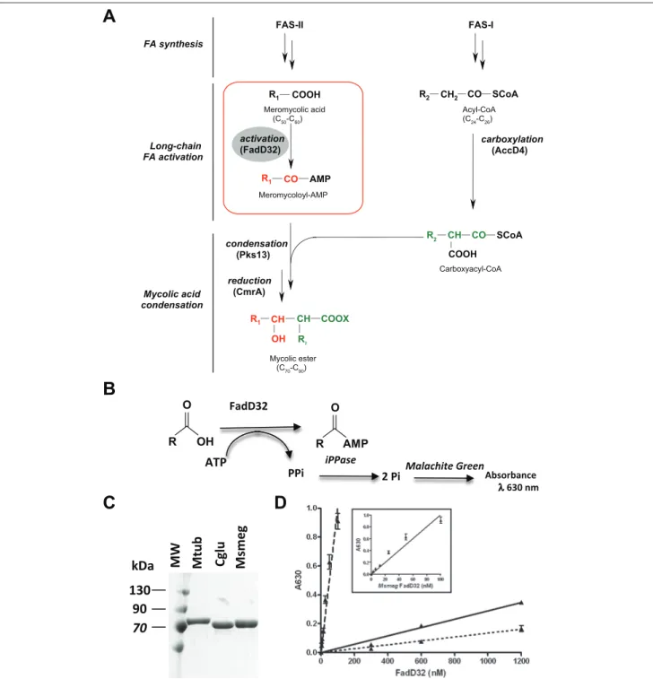
of mycolic acids involves the intervention of fatty acid synthases (FAS): FAS-I for de novo synthesis and FAS-II for elongation of fatty acids (FAs). The condensation (by Pks13) between two activated fatty acids produces, after reduction (by the ketoacyl reductase CmrA), the mature mycolic ester with the characteristic mycolic motif (alpha-branch, beta-hydroxyl). Both condensation substrates are activated: The acyl-CoA (C24–C26) is carboxylated by the acyl-CoA carboxylase complex (AccD4-containing complex), yielding the alpha-branch (R2) of mycolic acids. The very long-chain fatty acid (meromycolic acid, C50–C60) is activated as acyl-AMP by FadD32 (FAAL32), at the origin of the meromycolic chain (R1) of mycolic acids. R1 and R2 represent long hydrocarbon chains, and X is an unknown acceptor. (B) A schematic representation of FAAL reaction catalyzed by FadD32 and detection of inorganic phosphate (Pi) using the Pyrophosphatase-coupled reaction. PPi is pyrophosphate. (C) Purity of the different FadD32 orthologues after affinity and gel filtration chromatography purification. Protein fractions were analyzed on 10% sodium dodecyl sulfate polyacrylamide gel electrophoresis (SDS-PAGE). (D) FAAL activity of FadD32 orthologues: Mycobacterium tuberculosis (dotted line), Corynebacterium glutamicum (solid line), Mycobacterium smegmatis (dashed line), and inset. Assays were conducted in Tris-HCl (pH 8) using 200 µM lauric acid (C12) and 2 mM adenosine triphosphate (ATP) as substrates. Serial dilutions of enzymes were performed in Tris-HCl (pH 8). Means and standard errors of the mean (SEM) of four replicates are shown. Lines were obtained by linear regression, and data are representative of at least three independent experiments.
Healthcare, Little Chalfont, Buckinghamshire, UK), and FadD32 protein was eluted using 150 mM imidazole in 50 mM HEPES (pH 7.5), 500 mM NaCl, and 0.2 mM AEBSF. The fractions containing FadD32 protein were pooled and concentrated using Vivaspin 2 (10 000 Da molecular weight [MW] cutoff; Sartorius, Goettingen, Germany) and loaded onto a gel filtration column (HiLoad 16/60 Superdex 200; GE Healthcare) preequilibrated with 50 mM HEPES (pH 7.5), 500 mM NaCl, and 0.2 mM AEBSF. The purified pro-teins (as checked on sodium dodecyl sulfate polyacrylamide gel electrophoresis [SDS-PAGE] followed by a Coomassie blue staining) were then concentrated and stored at –20 °C in 50% glycerol.
Radiometric-TLC Enzyme Assays
The formation of the acyl-AMP product by FadD32 from fatty acid and adenosine triphosphate (ATP) in the presence of Mg2+ was monitored by radiolabeled-TLC (radio-TLC), as described previously.7,19 Briefly, the reaction mixtures were performed as described for spectrophotometric assays, in 30 µL in glass microtubes, and incubated for 80 min at RT. Reactions were then stopped by adding 5% final acetic acid. The reaction mixture was then spotted in duplicates on silica gel TLC plates (G60; Macherey-Nagel, Düren, Germany), with the radiolabeled fatty acid substrate being subsequently separated from the acyl-AMP product on the basis of their differential Rf (frontal ratio). Radiolabeled products were resolved in butan-1-ol/acetic acid/water (80:25:40, v/v/v) at RT and visualized by exposing the TLC to a Fujifilm (Tokyo, Japan) imaging plate prior to phos-phorimager detection (Typhoon Trio; GE Healthcare). The percentage of product was calculated after quantitation of both fatty acid and acyl-AMP using the ImageQuant soft-ware version 5.1 (Molecular Dynamics, Sunnyvale, CA).
Spectrophotometric Enzyme Assays
FadD32 enzyme catalyzes the formation of acyl-AMP using fatty acid and ATP as substrates (Fig. 1B). Pyrophosphate (PPi) released during this reaction was hydrolyzed using a Pyrophosphatase coupled reaction, and the resulting inor-ganic phosphate (Pi) was measured using a colorimetric assay. Addition of Malachite Green reagent to the reaction mix resulted in the formation of phosphomolybdate complex with an increase in absorbance at 630 nm (A630) (Fig. 1B). All assays were performed in quadruplicates in 384-well polystyrene microplates (Greiner Bio One, Courtaboeuf, France), using the PiColorLock Gold kit (Innova Biosciences, Cambridge, UK). Reactions were conducted at RT, in 30 µL of assay mix containing 50 mM HEPES (pH 7.5), 8 mM MgCl2, 0.01% Brij-58, 2 mM ATP, 20 µM lauric acid, 1 mM DTT (1,4-dithiothreitol), and 2 mU/mL Pyrophosphatase (Sigma-Aldrich). Reactions were initiated by the addition of
15 µL FadD32 enzyme diluted in 50 mM HEPES (pH 7.5) to 15 µL of 2× assay mix. After 80 min, reactions were stopped by adding 30 µL of cold reaction buffer and 15 µL of Malachite Green. The A630 was read after 5 min of incubation at RT in a µQuant counter (Bio-Tek, Winooski, VT). A reac-tion without enzyme (for protein dose curve experiments) or without substrate (for Km and Vmax determinations) was per-formed in each experiment and used as blank. To calculate the concentration of Pi, a calibration curve using known concentrations of Pi varying from 10 to 80 µM was per-formed in each experiment according to the manufacturer’s recommendations.
For apparent kinetic parameters (Km, Vmax, and kcat) determination, the initial velocity was measured (incuba-tion time of 40 min) as a func(incuba-tion of the studied substrate concentration at saturating concentrations of the nonvaried substrate. The kinetic parameters were determined when initial velocities were measured at a 90-min incubation time and found identical to those calculated at 40 min. The satu-ration curve was fit by nonlinear regression analysis using the following GraphPad Prism 4.02 (GraphPad Software, La Jolla, CA) equation: Vi = Vmax * [S]/(Km + [S]), where
Vmax is the maximal velocity, [S] is the substrate concentra-tion, and Km is the Michaelis-Menten constant. Kinetic parameters for fatty acids were determined at fixed concen-trations of ATP (2 mM) and by varying concenconcen-trations of the fatty acid (3.1–200 µM), whereas kinetic constants for ATP were determined at fixed concentrations of lauric acid (C12) (200 µM) and by varying concentrations of ATP (0.05–4 mM) (Suppl. Fig. S1). The catalytic constant (kcat) reflects the maximum rate of product formation and was calculated by dividing Vmax by the total enzyme concentra-tion kcat = Vmax/[ET]. The [ET] was 1.2 pmol in the 30-µL assay reaction, and Vmax was inferred from the Michaelis-Menten equation (described above and in Suppl. Fig. S1).
For inhibition studies, concentrations of ATP and lauric acid were adjusted to be above or around the Km (0.25 mM ATP and 20 µM lauric acid). The substrate analogue alkyl-adenylate AMPC12 (chemically synthesized as described previously19) was first diluted in DMSO, and the substrate mixture was applied to increasing concentrations of the compound. The reaction was started by adding the enzyme. Data were expressed as percentage of inhibition. Curve fit-ting and EC50 calculations were performed using sigmoidal dose response as the equation model (GraphPad Prism 4.02 software).
Thermostability of FadD32
The melting point (Tm) of FadD32 was measured by dif-ferential scanning fluorimetry (DSF) and was used both to characterize the enzyme thermal stability under various conditions (buffer, pH) and to identify ligands or inhibi-tors.21 A mix of enzyme (4 µM), SYPRO orange (5×)
(Invitrogen), buffer (100 mM), and NaCl (500 mM) was exposed to a temperature gradient from 25 to 80 °C with a 0.3 °C increment. All measurements were performed in triplicates in 96-well plates (Biorad, Marnes-la-Coquette, France). The thermal transition was monitored using an RTQPCR CFX96 Real-Time System (Biorad). Tm was given by the inflexion point of the curve RFU (relative fluorescence unit) = f(T). Before fluorimetry experiments, the FadD32 samples were soaked in the presence of sub-strate analogue (AMPC12) or in buffer at a final concentra-tion of 40 µM. For the DSF experiments, the His-tag of the affinity-purified FadD32 was removed by thrombin cleav-age (Novcleav-agen). The protein solution (after 5-fold dilution to reduce the imidazole concentration down to 30 mM) was submitted to a 3-h cleavage reaction at RT with 0.28 U/mL of thrombin. The cleaved protein was then reloaded onto a HisTrap HP (1-mL) affinity column to eliminate the non-cleaved fraction. The flow-through protein-containing frac-tions were then pooled, concentrated, and purified by size exclusion chromatography onto a Superdex 200 Prep Grade column (GE Healthcare). Samples used for DSF experi-ments were stored at –80 °C without glycerol.
Biochemical High-Throughput Screening
The above-mentioned biochemical assay to measure FadD32 activity was adapted for high-throughput screening (HTS). All the chemicals and biochemicals were procured from Sigma-Aldrich unless otherwise stated. The MsmegFadD32 enzyme was prepared as described above. The 30-µL reactions containing 50 mM HEPES buffer (pH 7.7), 0.25 mM ATP, 8 mM MgCl2, 0.001% Brij-35P, 1 mM DTT, 0.1 U/mL Pyrophosphatase, 20 nM MsmegFadD32 enzyme, and 20 µM lauric acid were carried out in 384-well microplates (Corning Costar; Corning Life Sciences, Tewksbury, MA) at room temperature (25 °C) for 100 min. Then, 30 µL of modified Baykov’s Malachite Green reagent22 was added and mixed for color development. Assay additions and mix-ing were done usmix-ing the Apricot PP-384-M Liquid Handlmix-ing System (Apricot Designs, Covina, CA). The plates were further incubated at 25 °C for 20 min. At the end of this incubation time, the plates were read at a 635-nm wave-length in a Spectramax 384 Plus microplate reader (Molecular Devices, Sunnyvale, CA). The single-point primary screening and the 10-point concentration response (CR) studies were performed using this assay. The inhibi-tion was calculated against the following reacinhibi-tion controls: MIN (0% reaction control) = 1 µL of 0.5 M EDTA (16.7 mM final) and MAX (100% reaction control) = 1 µL of 100% DMSO (3.3% final). Since the enzymatic mechanism process for the two substrates is yet unknown, the concen-trations of ATP and lauric acid were adjusted to be above or around their respective Km to select moderate to weak inhibitors of both substrates.
In the primary screening, 10 177 compounds were screened in singlicates at 20 µM. These compounds were randomly selected from a large, chemically diverse library of the AstraZeneca Corporate Compound collection to check the feasibility of HTS. This collection was made by acquiring chemically diverse sets of compounds from vari-ous sources as well as by adding compounds synthesized during various drug projects within AstraZeneca.
Promiscuity Test
High protein effect (promiscuity) assays were done to ascertain high protein (MsmegFadD32) effect on the com-pound CR. Basically, the above-mentioned high-through-put biochemical assay was done for a given set of compounds simultaneously at 1× (20 nM) and 5× (100 nM) enzyme concentrations and with a 10-point CR similar to the assay described before, except that the reaction time was reduced to 20 min to maintain the reaction linearity with increased enzyme concentration.23
Thermofluor (T
mShift) Assay
The Thermofluor assay was performed as described before24 to ascertain binding of these compounds to MsmegFadD32 enzyme. Thermal stability measurements were recorded in 96-well PCR plates covered with optical film (Bio-Rad, Hercules, CA), using a real-time PCR thermal cycler (iQ5 from Bio-Rad). The environmentally sensitive dye, SYPRO Orange (Invitrogen), was used to monitor protein unfold-ing. Thermal unfolding was monitored at 0.5 °C intervals from 25 °C to 95 °C. The assay was performed at the opti-mal reaction buffer in a 50-µL volume (50 mM HEPES [pH 7.7], 8 mM MgCl2, 6.25× SYPRO Orange, and 0.15 mg/mL
MsmegFadD32). The final DMSO concentration was
main-tained at 4% (v/v). Under these conditions, ATP, non-hydrolysable ATP analogue adenosine 5′-[γ-thio] triphosphate (ATP-γ-S) (Sigma-Aldrich) and lauric acid were tested for binding to MsmegFadD32 at 1× and 5× concentrations of their respective Km values. All 32 com-pounds selected after hit evaluation were tested in this assay for binding to MsmegFadD32 at three concentrations (12.5, 25, and 50 µM) (Suppl. Table S1A). The 16 “non-promiscuous” compounds were assessed for the competi-tion with ATP-γ-S (Suppl. Table S1B).
Results and Discussion
Enhancement of the FadD32 Purification
Yield
Previously, we successfully expressed and purified the
MtubFadD32 (MtubFadD32), yet with a relatively low
MtubFadD32 expression and/or solubility, we reasoned that
expression host strains reported to allow stabilization of mRNA levels of recombinant proteins should allow improved protein expression levels and stability, whereas auto-induc-tion media should slow down the expression rate while decreasing handling of cultures. Accordingly, the
MtubFadD32 protein was expressed by culturing Star One
Shot (Invitrogen) BL21(DE3)/pET15b-MtubfadD32 in auto-induction media for 72 h at 20 °C. Using these culture condi-tions, the amount of MtubFadD32 protein recovered after purification using the reported protocols was more than 7 mg/L of culture, which represented at least a 10-fold increase in the protein yield compared with conditions where protein expression was performed in LB medium (5-h induction with 0.5 mM isopropyl-β-d-thiogalactopyranoside [IPTG] at 25 °C).19 The protein obtained after the two-step purification was homogeneous (Suppl. Fig. S2).
FadD32 Assay Development
The MtubFadD32 activity was previously characterized using a radiometric assay coupled to thin-layer chromatog-raphy (radio-TLC). The search for FadD32 inhibitors through screening of chemical libraries requires the design of suitable readouts compatible with low volumes and robotics. The FadD32 protein displays FAAL activity in vitro, catalyzing the formation of an acyl-adenylate (acyl-AMP) from a fatty acid and ATP (in the presence of Mg2+), with the release of PPi (Fig. 1B). We decided to couple the FadD32 reaction to inorganic Pyrophosphatase (iPPase), which cleaves the PPi into Pi, the latter being measurable with inorganic phosphate-interacting reagents such as Malachite Green, followed by spectrophotometric detection at 630 nm, as demonstrated for Pi-generating enzymes.22
The initial assays of FadD32 activity using the Pyrophosphatase-coupled reaction were performed in 384-well plates in conditions similar to those applied in the radio-TLC assay previously reported,19 except for the [1-14C]C
12 fatty acid substrate, which was replaced by cold lauric acid (C12) and the addition of Pyrophosphatase in the reaction mixture. The FadD32 reaction was followed at 630 nm, and a Pi standard curve allowed quantitation of the Pi released, thus enabling the pyrophosphate or acyl-AMP concentrations formed during the reaction to be deduced. To test the linearity of the product formation in these condi-tions, a protein dose-response curve (with increasing con-centrations) of MtubFadD32 in the reaction was performed. For protein concentrations up to 600 nM in the assay, the increase in A630 remained low, and at least 1.2 µM was needed to detect FAAL activity (Fig. 1D, dotted line).
Among the various parameters to be optimized to adapt the FadD32 assay to HTS are (1) the protein concentration, opti-mally in the nanomolar range, and (2) the substrate’s (fatty acid) chain length and concentration, which should be kept to a minimum given its hydrophobic nature and low solubility.
The relatively high concentrations of MtubFadD32 added in the reaction seemed not compatible with the adaptation of the assay to HTS. We therefore considered using FadD32 ortho-logues as surrogates to develop an HTS assay, knowing that the use of the M. smegmatis orthologue in the mycobacterial PimB protein biochemical and structural studies has been shown to be valuable.25
The FadD32 proteins being highly conserved in all mycolic acid–producing bacteria (grouped in the
Corynebacteriales suborder), two FadD32 orthologues from Msmeg and Cglu—sharing, respectively, 75% and 40%
sequence identity with the MtubFadD32—were selected as surrogate candidates for comparative biochemical and inhibi-tion studies. The respective genes were cloned in pET15b expression vectors with an N-terminal 6xHistidine tag for production in E. coli strains. The three FadD32 proteins were expressed and purified (Fig. 1C) according to improved pro-tocols of expression and purification mentioned above (see Materials and Methods). The protein expression rates and purification yields obtained for MsmegFadD32 and
CgluFadD32 were significantly higher than those of MtubFadD32 (Table 1). Indeed, the calculated volumes of
culture required for performing a significant number of assays clearly distinguished the MsmegFadD32 and
CgluFadD32 from the Mtub protein and indicated that the
former orthologues are by far more convenient for HTS pur-poses than the Mtub protein (Table 1).
When the thermal stability of the three FadD32 orthologues was investigated through determination of the melting tem-perature (Tm), MtubFadD32 was the least stable protein, with a
Tm of 37 °C, whereas MsmegFadD32 and CgluFadD32 were much more thermally stable in our experimental conditions, with a Tm of 46 °C and 59 °C, respectively.
The activities of the MtubFadD32, MsmegFadD32, and
CgluFadD32 proteins were then measured in parallel in a
protein dose-response experiment, and their abilities to release Pi in the FadD32-Pyrophosphatase coupled assay were compared (Fig. 1D). A linear relationship between velocity and enzyme concentration was observed through-out the enzyme concentrations tested. The specific activity of CgluFadD32 was only twice that of MtubFadD32 (Table 1). In sharp contrast, the dose-response curve for the
MsmegFadD32 enzyme showed a linear response down to a
few nanomolar of protein in the assay (Fig. 1D, inset). Indeed, the specific activity of MsmegFadD32 was approxi-mately 75 times higher than that of the MtubFadD32 enzyme (Table 1). The CgluFadD32 displayed high ther-mal stability and purification yield, yet its low specific activity is not suitable for performing inhibition and HTS assays. Furthermore, when combining the purification yield, the amount of protein required in the assay, and enzyme activity for the three FadD32 orthologues (Table 1), the
Msmeg protein displayed the lowest volume of culture
nec-essary to perform a 100-microplate (384-well plates) assay, thus permitting the achievement of an HTS campaign with
a unique batch of protein. The higher sequence identity of
MsmegFadD32 with the Mtub enzyme, combined with its
favorable biophysical properties and high specific activity, made the MsmegFadD32 enzyme the most promising sur-rogate for conducting the assay development and optimiza-tion toward the HTS of inhibitors.
Assay Optimization Using FadD32 from M.
smegmatis
To gain insight into the optimal assay conditions (i.e., reaction volume and composition), several parameters were investi-gated using MsmegFadD32. A reaction volume of 30 µL was used to reduce the amount of protein in the assay while keep-ing the most favorable optical performance. To keep the benefit of using larger volumes at the reading step, which allowed reducing reading errors due to meniscus curvature and shorter path length,26 the reaction mixture was diluted 1:1 (v/v) postincubation with basal buffer before taking a readout. Indeed, higher ratios of signal/background (S/B of 4.2 vs. 2.6) and signal/noise (S/N of 97 vs. 23) compared with no buffer dilution were achieved (Suppl. Fig. S3A).
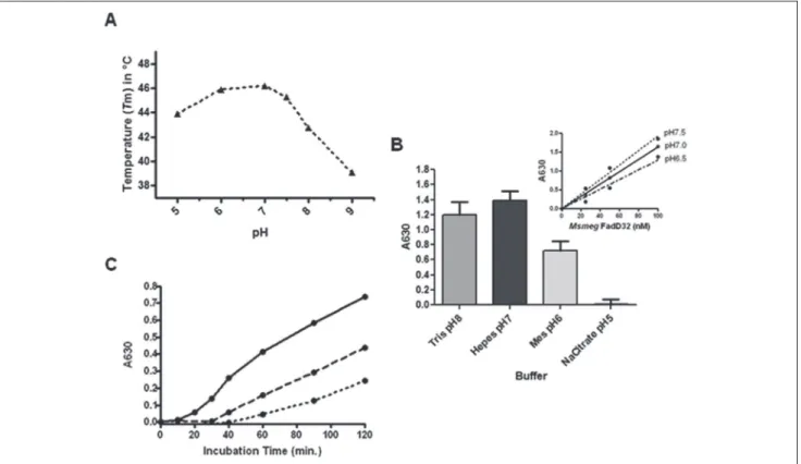
The critical parameters affecting the enzyme activity were the reaction buffer and pH. These were first investi-gated by inspecting the protein thermal stability using DSF, a widely used technique for screening optimal conditions for biological macromolecules.21 When thermal stability of the MsmegFadD32 protein was inspected at different buffer and pH conditions (from 5–8), a higher Tm was observed at neutral pH, indicating an increased stability of the protein at this pH (Fig. 2A). Investigation of FAAL activity of
MsmegFadD32 under the same conditions revealed that the
use of acidic buffer (pH 5) in the reaction mixture did not allow detection of FAAL activity, even at a higher protein concentra-tion in the assay (Fig. 2B). The highest protein activity was observed at neutral pH using HEPES buffer, with the pH 7.5 being the most suitable for further enzymatic assays (Fig. 2B, insert), in agreement with the thermal stability data.
A time course experiment of MsmegFadD32 activity was performed in the presence of three enzyme concentrations to determine the time window for reaction linearity. The
reaction rate was linear between 40 and 120 min, ensuring initial velocity (V0) conditions at all enzyme concentrations tested (Fig. 2C).
The low solubility of the fatty acids in the reaction mix-ture was a critical issue for the availability of the substrate in the FAAL reaction. Accordingly, the effects of detergents on the kinetic parameters of MsmegFadD32 (for lauric acid) were examined in the absence or presence of different agents. The presence of detergent in the assay significantly improved the reaction rate, with Brij (58 and 35) exhibiting the most pronounced effect (Suppl. Fig. S3B).
Since most of the library compounds are solubilized in DMSO, the effect of DMSO on MsmegFadD32 activity was investigated, and concentrations up to 7.5% did not appear to affect the activity of the enzyme (data not shown).
Enzymatic Characterization of M. smegmatis
FadD32
The choice of the fatty acid substrate and determination of kinetic parameters for both substrates (ATP and fatty acid) of FadD32 are critical for the determination of the conditions to set for inhibition and screening experiments. The apparent Km, Vmax, and kcat were determined for ATP and for fatty acids of various chain lengths (Table 2). The myristic acid (C14) dis-played the highest catalytic efficacy (kcat/Km), in agreement with the FadD32 substrate preference for long-chain fatty acids.19 For further inhibition and HTS experiments, lauric acid (C12) was chosen since it combines the advantage of hav-ing a relatively short aliphatic chain with a high catalytic effi-cacy. In addition, the Km value for C12 permits including a concentration of the substrate in the assay close to the Km (20 µM), which would allow the detection of all types (competi-tive, uncompeti(competi-tive, and noncompetitive) of inhibitors with a sufficient absorbance change. The lower Km of the C14 could be a disadvantage if one is looking for competitive inhibitors, and the assay would require several-fold the Km of the sub-strate to generate sufficient signal. The Km of ATP measured in the assay conditions was 0.08 mM.
We evaluated the correlation between the release of Pi using the PiColorLock Gold (Innova Biosciences)
Table 1. Properties of the Selected FadD32 Orthologues from Mycobacterium Species and Corynebacterium glutamicum
FadD32 Protein Identity, % Purification Yield, mg/L Specific Activity (200 µM Lauric Acid), nmol·min–1·mg–1 Volume of Culture for a 100-Microplate Assay, L
M. tuberculosis 100 7 32 ± 2 14
M. smegmatis 75 45 2443 ± 99 0.08
C. glutamicum 40 50 71 ± 2 1
Identity scores with the M. tuberculosis protein were computed from protein sequence alignment performed using the BLAST program (NCBI, Bethesda, MD). The protein purification yields are calculated per liter of culture expressing the protein of interest. The specific activity of each FadD32 was deduced from the linear regression protein dose-response curve (Figure 2B) (protein ranging from 1200 to 300 nM for MtubFadD32 and CgluFadD32 and from 100 to 3.125 nM for MsmegFadD32). The volume of culture required for performing a 100-microplate assay (384-well plates) was calculated based on addition of 1200 nM of MtubFadD32, 40 nM of MsmegFadD32, and 600 nM of CgluFadD32 protein per reaction.
detection and the formation of acyl-AMP determined by radio-TLC (Fig. 3). In the same conditions (except for the radiolabeled substrate), a protein dose-response curve experiment was performed, and the product formation was analyzed with either the radio-TLC assay (Fig. 3A) or the Pyrophosphatase-coupled FadD32 assay. The good corre-lation of the amount of product formed using both assays
validated the PiColorLock as a reliable and quantitative assay for measuring FadD32 activity (Fig. 3B). Thus, the optimization of the assay conditions and its adaptation to HTS afforded a robust and time-saving assay, which also represents a powerful tool for studying the biochemical properties of this FAAL and others (determination of enzyme kinetic parameters, inhibition studies) and makes
Figure 2. Assay optimizations using the FadD32 from Mycobacterium smegmatis. (A) Melting temperature (Tm) measurements of
MsmegFadD32 as a function of pH. The differential scanning fluorimetry measurements for the protein at the indicated pH are displayed. (B) Optimization of buffer and pH: Assays were conducted during 80 min in different buffers using 100 nM of the MsmegFadD32 enzyme. Inset: Fatty acyl-AMP ligase (FAAL) activity of MsmegFadD32 at different pH. Serial dilutions of enzymes were performed in the indicated HEPES buffer (dashed line: pH 6.5; dotted line: pH 7.5; solid line: pH 7.0) using 200 µM lauric acid and 2 mM adenosine triphosphate (ATP) as substrates. Means and standard errors of the mean (SEM) of four replicates are shown. Lines were obtained by linear regression. (C) Time course of MsmegFadD32 activity. Assays were conducted at room temperature in HEPES pH 7.5 using 20 µM lauric acid and 0.2 mM ATP using 80 nM (solid line), 40 nM (dashed line), or 20 nM (dotted line) of enzyme.
Table 2. Apparent Kinetic Parameters of MsmegFadD32
Substrate Km, µM Vmax, pmol·min–1 k
cat, min –1 k cat/Km, min –1 µM–1 ATP 76.0 ± 6.7 16.8 ± 0.4 14.0 ± 0.3 0.18 C10 259.0 ± 57.9 19 ± 2.8 16.1 ± 2.4 0.06 C12 25.9 ± 1.6 13.5 ± 0.3 11.2 ± 0.2 0.43 C14 5.2 ± 0.5 5.0 ± 0.1 4.2 ± 0.1 0.81
Initial velocities of the FadD32 reaction were assayed spectrophotometrically using the coupled assay. Reactions were conducted at room temperature during 40 min in HEPES pH 7.5 as described in Materials and Methods. The steady-state apparent parameters Km (Michaelis constant), Vmax (maximum velocity), kcat (catalytic constant), and kcat/Km (catalytic efficacy) were determined using nonlinear regression to fit the data to the Michaelis-Menten equation. ATP, adenosine triphosphate.
the assay an attractive option for the identification of FAAL inhibitors.
Binding of a Substrate Analogue and
Inhibition of FadD32 Activity
The shift in the thermal stability of MsmegFadD32 in the pres-ence of a substrate analogue (alkyl-adenylate, AMPC12), previously reported to inhibit the MtubFadD32 enzyme activ-ity,19 was investigated using T
m measurements. The addition of AMPC12 to the protein induced a significant (6 °C) ther-mal shift (∆Tm) compared with the protein alone at pH 7.5 (Fig. 4A). This shift indicated that the inhibitor binds to the protein, and this interaction very likely stabilizes
MsmegFadD32. The effect of AMPC12 on the protein activity
was tested and, expectedly, a dose-dependent inhibition of the enzyme activity was observed in the presence of increasing concentrations of the alkyl-AMP (Fig. 4A), with a calculated EC50 (half-maximal inhibitory concentration) of 1.4 µM.
As the use of the Tm shift appeared to be an interesting tool for evaluating the potential binding properties of com-pounds, as well documented in the literature,21 it was thus employed in the screening of the limited library in the
MsmegFadD32 HTS.
Screening Cascade and Compound Progression
The biochemical assay was validated for HTS by testing its reproducibility. An elaborate assay validation was per-formed before the throughput screening to assess the day-to-day assay variability (data not shown). A set of 200 compounds was screened on different occasions, and the calculated Z′ value27 was 0.7 (data not shown), thus allow-ing initiation of the screenallow-ing campaign. For the primary screening, 10 177 compounds were screened in singli-cates (at 20 µM) and, based on the 33% inhibition cutoff and stringent statistical criteria (hits outside mean + 2.5 interquartile range [IQR]*), 412 potential inhibitors were selected (Suppl. Fig. S4). These compounds were tested for the 10-point (100 µM down to 0.2 µM) CR assay, and those showing CR and EC50 values less than 100 µM were progressed further. Of 412 compounds, 200 were short-listed: 98 compounds with good CRs (with Hill slopes between 0.5 and 2.0) and 102 with acceptable CRs (with a relaxed criterion for Hill slopes between 0.5 and 3). These 200 CR-confirmed hits were then classified into chemical clusters. Only 32 of these compounds were fur-ther selected based on rigorous hit evaluation criteria,
such as chemical properties, ligand efficiency (cutoff value = 0.3), chemical tractability, and other drug- like properties.† The 32 compounds were then tested in
Figure 3. Fatty acyl-AMP ligase (FAAL) activity of MsmegFadD32
after assay optimizations. (A) Radio–thin-layer chromatography (TLC) analysis of the [1-14C]C12-AMP synthesized using increasing concentrations of the MsmegFadD32 enzyme. (B) A protein dose-response curve of MsmegFadD32 showing the formation of enzyme product using either inorganic phosphate (Pi) released in the PiColorLock-based assay (Innova Biosciences, Cambridge, UK) (open squares) or acyl-AMP formed in the radio-TLC assay (black triangles). Assays were performed at room temperature during 80 min in HEPES pH 7.5 using 40 µM lauric acid and 2 mM adenosine triphosphate (ATP) as substrates. Means and standard errors of the mean (SEM) of four replicates (PiColorLock assay) or duplicates (radio-TLC) are shown. Lines were obtained by linear regression, and data are representative of three independent experiments.
*Compound inhibition data sets follow “normal (Gaussian) distribution,” wherein the spread of the middle-half of the data is called interquartile range or IQR. Statistically, this spread is 4σ/3, where σ is the standard deviation. Statistically significant inhibition data points usually reside beyond the Median + 2 * IQR range as “outliers.” These outliers deserve attention as potential inhibitors.†If the compound is identified as a hit, it should satisfy the following hit evaluation criteria: (1) chemical nature: should have drug-like properties with adequate lipophilicity, ligand efficiency, and so on; (2) chemical tractability: should have multiple modification points to be able to make several analogues to form a chemical series in order to explore the most desirable derivative; and (3) pharmacophores: should not have known toxic motifs.
parallel in the promiscuity test (high protein effect) and the Thermofluor (Tm shift) assays.
In the promiscuity test, CR studies were done simultane-ously at 1× and 5× protein concentrations to ascertain an undesirable protein effect or nonspecific inhibition. A com-pound is defined as “promiscuous” (i.e., exhibits undesir-able protein effect) when it shows a 2-fold or more increase in its EC50 value at higher (5× or higher) protein concentra-tions. The 2-fold shift criterion for determining the promis-cuity was used since, for nonpromiscuous compounds, even a large change in enzyme concentration (2×–10×) is not expected to lead to changes in the EC50 as the inhibitor’s concentration is in far more excess than that of the protein,
and unless the inhibitor binds nonspecifically, the EC50s remain within a 2-fold change.23,28,29 Sixteen compounds were found to pass this test (i.e., these compounds did not show a high protein effect) (Suppl. Fig. S4).
The Thermofluor assay was done for the same 32 com-pounds to ascertain a significant dose-dependent shift in
MsmegFadD32 Tm as a result of binding of the compounds to the enzyme. Five of these compounds showed dose-dependent binding to the enzyme (Suppl. Table S1A) and did not show a nonspecific high protein effect and thus were “nonpromiscu-ous” in the promiscuity test (data not shown). Together, these data suggest that these compounds bind to the enzyme specifi-cally. To substantiate these results, competition studies were done, where binding of all 16 nonpromiscuous compounds to
MsmegFadD32 was checked in the presence of ATP-γ-S in the
Thermofluor assay. Convincingly enough, the same five com-pounds indicated dose-dependent binding, preferentially to the free form of the enzyme (with ∆Tm1 > ∆Tm2), suggesting that they are competitive inhibitors of MsmegFadD32 and that they competed with the ATP analogue for binding to the enzyme (Suppl. Table S1B). Binding of any of these compounds to the enzyme in the presence of the second substrate, lauric acid, however, could not be demonstrated because compounds did not show significant Tm shifts in the presence of C12. The latter alone or in the presence of ATP-γ-S did not show significant Tm shifts upon binding to MsmegFadD32 (data not shown). It is not uncommon that some substrates do not show significant Tm shifts upon binding to enzymes (in-house data) and that their binding to the enzyme can be shown with other biophysical assays such as 1D nuclear magnetic resonance spectroscopy.30 These five hits represent five chemical classes as shown in
Figure 4B, thus exhibiting (1) dose-dependent enzyme
inhibi-tion, (2) no protein effect (nonpromiscuous inhibitors), and (3) binding to MsmegFadD32 in the Thermofluor assay.
Thus, this screening campaign demonstrated that the essential FadD32 enzyme is a “druggable” target. This HTS was performed on the M. smegmatis FadD32 protein using a 10 000–compound limited library and has validated the opti-mized HTS assay, leading to the selection of five compounds as inhibitors of this enzyme with a high hit rate (4%). These are true inhibitors, as shown by binding as well as competi-tion studies. This proof of concept being made, the perspec-tive of this work will be to apply the same strategy using an HTS campaign with an extended library to select more com-pounds with potentially lower EC50. The selected molecules will be the starting point for testing the corresponding chemi-cal classes through “backscreening” on the M. tuberculosis enzyme. The efficacy of the most potent molecules will be tested on M. tuberculosis growth and checked on sensitive and resistant isolates. The application of high-throughput chemical screening for the discovery of small molecules that could inhibit FadD32 activity has significant potential in the development of novel anti-TB drugs.
Figure 4. Inhibition of MsmegFadD32 by the substrate analogue
AMPC12 and screening for inhibitors of its activity. (A) Inhibition curve of MsmegFadD32 activity by AMPC12. The chemical structure of adenosine 5′-alkylphosphate (alkyl-AMP) is displayed; R = C12 for AMPC12. Increasing concentrations of the compound were added to the assay, and FadD32 activity was measured in HEPES pH 7.5 in the presence of 20 µM lauric acid (C12) and 0.2 mM adenosine triphosphate (ATP) as substrates. Reactions were conducted at room temperature during 80 min and were done in quadruplicates. (B) Chemical classes, EC50, and Tm shift measurements of the five selected compounds. $Competes with ATP-γ-S in a dose-dependent manner for binding to the enzyme. For details, refer to Supplementary Table S1B.
Acknowledgments
We are grateful to Dr. Christian Chalut (IPBS, Toulouse) for provid-ing the pET15b-MtubfadD32 clone. Special thanks to Prashanti Madhavapeddi (AstraZeneca Infection iMed) for her inputs on the Thermofluor assay. We would also like to thank Adam Shapiro and Suresh Solapure (AstraZeneca Infection iMed) for critically review-ing the manuscript. The DSF equipment used in this study is part of the Integrated Screening Platform of Toulouse (PICT, IBiSA).
Declaration of Conflicting Interests
The authors declared no potential conflicts of interest with respect to the research, authorship, and/or publication of this article.
Funding
The authors disclosed receipt of the following financial support for the research, authorship, and/or publication of this article: This work was supported by the Centre National de la Recherche Scientifique (France), the MYCA contract from the Région Midi-Pyrénées and FEDER Funds from the European Community that funded the post-doctoral fellowships of S.G., and the European Community New Medicines for Tuberculosis (grant LSHP-CT-2005-018923).
References
1. World Health Organization. Multidrug and Extensively Drug-Resistant TB (M/XDR-TB): 2010 Global Report on Surveillance and Response. 2010. http://www.who.int/tb/pub-lications/2010/978924599191/en/index.html
2. Ma, Z.; Lienhardt, C.; McIlleron, H.; Nunn, A. J.; Wang, X. Global Tuberculosis Drug Development Pipeline: The Need and the Reality. Lancet 2010, 375, 2100–2109.
3. Marrakchi, H.; Bardou, F.; Laneelle, M.; Daffé, M. A. Comprehen-sive Overview of Mycolic Acid Structure and Biosynthesis. In The Mycobacterial Cell Envelope; Daffé M., Reyrat, J. M., Eds.; ASM Press: Washington, DC, 2008; pp 41–62.
4. Gokhale, R. S.; Saxena, P.; Chopra, T.; Mohanty, D. Versa-tile Polyketide Enzymatic Machinery for the Biosynthesis of Complex Mycobacterial Lipids. Nat. Prod. Rep. 2007, 24, 267–277.
5. Zuber, B.; Chami, M.; Houssin, C.; Dubochet, J.; Griffiths, G.; Daffe, M. Direct Visualization of the Outer Membrane of Mycobacteria and Corynebacteria in Their Native State. J. Bacteriol. 2008, 190, 5672–5680.
6. Sani, M.; Houben, E. N.; Geurtsen, J.; Pierson, J.; de Punder, K.; van Zon, M.; Wever, B.; Piersma, S. R.; Jimenez, C. R.; Daffe, M.; et al. Direct Visualization by Cryo-EM of the Mycobacterial Capsular Layer: A Labile Structure Containing ESX-1–Secreted Proteins. PLoS Pathog. 2010, 6, e1000794. 7. Trivedi, O. A.; Arora, P.; Sridharan, V.; Tickoo, R.; Mohanty, D.;
Gokhale, R. S. Enzymic Activation and Transfer of Fatty Acids as Acyl-Adenylates in Mycobacteria. Nature 2004, 428, 441–445. 8. Duckworth, B. P.; Nelson, K. M.; Aldrich, C. C.
Adenylat-ing Enzymes in Mycobacterium tuberculosis as Drug Targets. Curr. Topics Med. Chem. 2012, 12, 766–796.
9. Ferreras, J. A.; Stirrett, K. L.; Lu, X.; Ryu, J. S.; Soll, C. E.; Tan, D. S.; Quadri, L. E. Mycobacterial Phenolic Glycolipid Virulence Factor Biosynthesis: Mechanism and Small-Mol-ecule Inhibition of Polyketide Chain Initiation. Chem. Biol.
2008, 15, 51–61.
10. Simeone, R.; Leger, M.; Constant, P.; Malaga, W.; Marrak-chi, H.; Daffe, M.; Guilhot, C.; Chalut, C. Delineation of the Roles of FadD22, FadD26 and FadD29 in the Biosynthesis of Phthiocerol Dimycocerosates and Related Compounds in Mycobacterium tuberculosis. FEBS J. 2010, 277, 2715–2725. 11. Portevin, D.; de Sousa-D’Auria, C.; Montrozier, H.; Houssin, C.;
Stella, A.; Laneelle, M. A.; Bardou, F.; Guilhot, C.; Daffe, M. The AMP Ligase FadD32 and AccD4-Containing Acyl-CoA Carboxylase Are Required for the Synthesis of Mycolic Acids and Essential for Mycobacterial Growth: Identification of the Carboxylation Product and Determination of the Acyl-CoA Carboxylase Components. J. Biol. Chem. 2005, 280, 8862–8874. 12. Gavalda, S.; Leger, M.; van der Rest, B.; Stella, A.; Bardou,
F.; Montrozier, H.; Chalut, C.; Burlet-Schiltz, O.; Marrakchi, H.; Daffe, M.; et al. The Pks13/FadD32 Crosstalk for the Bio-synthesis of Mycolic Acids in Mycobacterium tuberculosis. J. Biol. Chem. 2009, 284, 19255–19264.
13. Portevin, D.; De Sousa-D’Auria, C.; Houssin, C.; Grimaldi, C.; Chami, M.; Daffe, M.; Guilhot, C. A Polyketide Synthase Catalyzes the Last Condensation Step of Mycolic Acid Bio-synthesis in Mycobacteria and Related Organisms. Proc. Natl. Acad. Sci. U. S. A. 2004, 101, 314–319.
14. Gande, R.; Gibson, K. J.; Brown, A. K.; Krumbach, K.; Dover, L. G.; Sahm, H.; Shioyama, S.; Oikawa, T.; Besra, G. S.; Eggeling, L. Acyl-CoA Carboxylases (accD2 and accD3), Together with a Unique Polyketide Synthase (Cg-pks), Are Key to Mycolic Acid Biosynthesis in Corynebacterianeae such as Corynebacterium glutamicum and Mycobacterium tuberculosis. J. Biol. Chem. 2004, 279, 44847–44857. 15. Boldrin, F.; Casonato, S.; Dainese, E.; Sala, C.; Dhar, N.; Palu,
G.; Riccardi, G.; Cole, S. T.; Manganelli, R. Development of a Repressible Mycobacterial Promoter System Based on Two Transcriptional Repressors. Nucleic Acids Res. 2010, 38, e134. 16. Forti, F.; Crosta, A.; Ghisotti, D. Pristinamycin-Inducible
Gene Regulation in Mycobacteria. J. Biotechnol. 2009, 140, 270–277.
17. Cortes, M.; Singh, A. K.; Reyrat, J. M.; Gaillard, J. L.; Nassif, X.; Herrmann, J. L. Conditional Gene Expression in Myco-bacterium abscessus. PLoS ONE 2011, 6, e29306.
18. Carroll, P.; Faray-Kele, M. C.; Parish, T. Identifying Vulnerable Pathways in Mycobacterium tuberculosis by Using a Knock-down Approach. Appl. Environ. Microbiol. 2011, 77, 5040–5043. 19. Leger, M.; Gavalda, S.; Guillet, V.; van der Rest, B.; Slama,
N.; Montrozier, H.; Mourey, L.; Quemard, A.; Daffe, M.; Mar-rakchi, H. The Dual Function of the Mycobacterium tubercu-losis FadD32 Required for Mycolic Acid Biosynthesis. Chem. Biol. 2009, 16, 510–519.
20. Studier, F. W. Protein Production by Auto-induction in High Den-sity Shaking Cultures. Protein Expr. Purif. 2005, 41, 207–234.
21. Pantoliano, M. W.; Petrella, E. C.; Kwasnoski, J. D.; Lobanov, V. S.; Myslik, J.; Graf, E.; Carver, T.; Asel, E.; Springer, B. A.; Lane, P.; et al. High-Density Miniaturized Thermal Shift Assays as a General Strategy for Drug Discovery. J. Biomol. Screen. 2001, 6, 429–440.
22. Baykov, A. A.; Evtushenko, O. A.; Avaeva, S. M. A Malachite Green Procedure for Orthophosphate Determination and Its Use in Alkaline Phosphatase-Based Enzyme Immunoassay. Anal. Biochem. 1988, 171, 266–270.
23. Feng, B. Y.; Shoichet, B. K. A Detergent-Based Assay for the Detection of Promiscuous Inhibitors. Nat. Protoc. 2006, 1, 550–553.
24. Venkatraman, J.; Bhat, J.; Solapure, S. M.; Sandesh, J.; Sarkar, D.; Aishwarya, S.; Mukherjee, K.; Datta, S.; Malolanarasim-han, K.; Bandodkar, B.; et al. Screening, Identification, and Characterization of Mechanistically Diverse Inhibitors of the Mycobacterium tuberculosis Enzyme, Pantothenate Kinase (CoaA). J. Biomol. Screen. 2012, 17, 293–302.
25. Guerin, M. E.; Kaur, D.; Somashekar, B. S.; Gibbs, S.; Gest, P.; Chatterjee, D.; Brennan, P. J.; Jackson, M. New Insights
into the Early Steps of Phosphatidylinositol Mannoside Bio-synthesis in Mycobacteria: PimB′ is an essential enzyme of Mycobacterium smegmatis. J. Biol. Chem. 2009, 284, 25687– 25696.
26. Harrison, R. O.; Hammock, B. D. Location Dependent Biases in Automatic 96-Well Microplate Readers. J. Assoc. Off. Anal. Chem. 1988, 71, 981–987.
27. Zhang, J. H.; Chung, T. D.; Oldenburg, K. R. A Simple Statistical Parameter for Use in Evaluation and Validation of High Through-put Screening Assays. J. Biomol. Screen. 1999, 4, 67–73. 28. McGovern, S. L.; Caselli, E.; Grigorieff, N.; Shoichet, B. K.
A Common Mechanism Underlying Promiscuous Inhibitors from Virtual and High-Throughput Screening. J. Med. Chem.
2002, 45, 1712–1722.
29. Shoichet, B. K. Interpreting Steep Dose-Response Curves in Early Inhibitor Discovery. J. Med. Chem. 2006, 49, 7274–7277. 30. Meyer, B.; Peters, T. NMR Spectroscopy Techniques for
Screening and Identifying Ligand Binding to Protein Recep-tors. Angew Chem. Int. Ed. Engl. 2003, 42, 864–890.
A
B
Suppl. Fig. S1: Michaelis-Menten curves for the determination of the appatent K
mand V
maxfor lauric acid (C
12) and ATP. Data were obtained using the PiColorLock-based assay.
Reactions were conducted at RT during 40 min in HEPES pH7.5 buffer using increasing
concentration of lauric acid with 2mM ATP (A) or increasing concentration of ATP with 200
µM of lauric acid (B). The K
mand V
maxwere determined using non-linear regression to fit
the data to the Michaelis-Menten equation.
MtubFadD32 following either expression in BL21(DE3)/purification in Tris pH8 (A) or
expression in E.coli BL21 Star™ (DE3) One Shot/purification in HEPES pH7.5 (B). Upper
panels display fractions from the first step affinity chromatography and lower panels show
fractions from second-step S-200 gel filtration chromatography, as analyzed by 12%
SDS-PAGE.
130 100 70 55Affinity
Gel filtration
66,2 97,4 45 66,2 97,4 45 kDa MW 66,2 97,4 45 kDa MW kDa MW kDa MWOptimization of the reaction volume. Assays were conducted during 100 min at RT in
Tris-HCl pH8 using 2 mM ATP, 200µM lauric acid in the absence (grey) or in the presence of 400
nM enzyme (black). The reaction volumes were either 20 µL of substrate mixture/20 µL of
enzyme (20:20) or 15 µL substrate mix/15 µL enzyme with addition of 30 µL of cold buffer
(4°C) after incubation (15:15:30). (B) Effect of detergents on MsmegFadD32 activity: assays
were conducted at room temperature during 80 min in HEPES buffer at pH7.5 using 40 nM
enzyme and 25 µM lauric acid (C
12) in the absence or in the presence of Brij
®58 (0.01%),
Brij
®35P (0.01%) or NP-40 (0.1%). Means and standard errors of the mean (SEM) of four
replicates are shown and data are representative of at least three independent experiments.
screening of a library of 10,177 compounds and the strategy for “Hit” selection and evaluation
leading to selection of five compounds, non-promiscuous and binders.
32 Compounds 33% inhibition cut-off Compounds < 100 µM with CR 16Non-promiscuous 5 Binders Tm Shift Assay Protein Effect 5 Compounds Non-promiscuous and binders
10177 compounds Primary Screen at 20 µM
412 Actives
Concentration Response (CR) Screening
200 compounds