Antiviral Polymeric Drugs and Surface Coatings by
Alyssa Maxine Larson B.S. in Biochemistry
University of California, Santa Barbara, 2008 Submitted to the Department of Chemistry
in Partial Fulfillment of the Requirements of the Degree of Doctor of Philosophy in Biological Chemistry
at the
MASSACHUSETTS INSTITUTE OF TECHNOLOGY June 2013
0 2013 Massachusetts Institute of All rights reserved
Technology
ARCnpAs
MASSACHUSETTS INS E OF TECHNOLOGYJUL
OI 2013
UBRARIES
Signature of Author: - - F-
of-I Certified by: Firmenich Professor of Department of Chemistry April 26, 2013 Accepted by: Robert W. Field Haslam and Dewey Professor of Chemistry Chairman, Departmental Committee on Graduate Students Alexander M. Klibanov Chemistry and Bioengineering
Thesis Supervisor -41 1 ,1
-This Doctoral Thesis has been examined by a committee of the Department of Chemistry as follows:
\) IJohn
M. EssigmannWilliam R. and Betsy P. Leitch Professor of Chemistry and Biological Engineering Thesis Committee Chair
Alexander M. Klibanov Firmenich Professor of Chemistry and Bioengineering
Thesis Supervisor
& Elizabeth M. Nolan
Pfizer Laubach Career Development Assistant Professor of Chemistry Thesis Committee Member
Antiviral Polymeric Drugs and Surface Coatings by
Alyssa Maxine Larson
Submitted to the Department of Chemistry on April 26, 2013, in Partial Fulfillment of the Requirements for the Degree of Doctor of Philosophy in Biological Chemistry Abstract
Viruses are a major cause of human morbidity and mortality in the world. New effective approaches to stop their spread are paramount. Herein, two approaches toward this goal are explored: (i) developing multivalent therapeutics (multiple copies of an antiviral agent covalently attached to a polymeric chain) with superior potency against their viral targets, and (ii) creating antiviral surface coatings that detoxify aqueous solutions containing various viruses on contact.
By harnessing the power of multivalency we endeavored to improve the potency of influenza inhibitors, as well as resurrect the potency of two FDA-approved influenza inhibitors for which widespread drug resistance now exists. In the former direction of research, we attached multiple copies of bicyclic naphthoquinone-like monomeric inhibitors to polymeric chains. When tested against the Wuhan strain of influenza, these multivalent conjugates were up to 240-fold more potent inhibitors than their monomeric predecessors. However, this improvement in potency was strain-dependent, as two other serotypically-different influenza strains were not
inhibited nearly as well by multivalent inhibitors.
This strategy was also employed to restore inhibition for the adamantane class of influenza inhibitors against drug-resistant strains. The chemical modifications to the adamantane scaffold necessary for polymer attachment imposed deleterious steric constraints which resulted in poorer inhibitory effect. Even despite these drawbacks, however, the drug-polymer conjugates were up to 30-fold more potent against drug-resistant strains than their monomeric counterparts. These efforts made strides toward the ultimate goal of recovery of influenza virus inhibition for the adamantanes.
To diminish transmission of viral infections, we explored the action of antimicrobial PEI-based (PEI = polyethylenimine) hydrophobic polycations against both enveloped and non-enveloped viruses. When solutions containing herpes simplex viruses (both 1 and 2) were brought in contact with NN-dodecyl,methyl-PEI coated on either polyethylene slides or latex condoms, they could be disinfected by up to 6-logs of viral titers. Our hydrophobic polycation also could be formulated into a suspension to disinfect herpes simplex virus-containing solutions, suggesting potential utility in a therapeutic modality.
We also investigated whether these findings were applicable to non-enveloped viruses, namely poliovirus and rotavirus. Aqueous solutions containing them indeed could be drastically disinfected by our hydrophobic polycation-coated slides; subsequent mechanistic studies suggested that this disinfection was due to adsorption of the viruses onto the coated surfaces from solution.
Thesis Supervisor: Dr. Alexander M. Klibanov
Acknowledgements
This PhD is the result of the support and love from many very special people, all of whom have contributed to my intellectual and personal growth for the past 5 years. I am grateful to my advisor, Alex Klibanov, who has taught me much more than how to be a scientist. Thank you for your "tough love" which helped me to develop "thick skin" as well as your endless jokes, stories, and anecdotes. Although you were not completely successful in "de-honeying" my voice, I believe you have made strides, and for that you should be proud. I am also appreciative to my thesis chair, Professor John Essigmann, for his intellectual guidance and help in getting into MIT by providing valuable feedback on my admissions essay. It was a joy to TA for you and I always cherished our several hour-long yearly meetings. Thank you to Professors Liz Nolan and Jianzhu Chen for their help in my research progress.
I am eternally grateful to my family and friends for keeping me sane these past 5 years. To my parents- thank you for your mental (and sometimes financial) support, for making many trips to the east coast, and for always being so excited about my research, even when I wasn't. To Lia- I am so glad we have been able to grow closer and enjoy many quick weekend trips since 2008 with good food, good wine, and excellent company. I can't wait to take you out once I get my first paycheck. To Pat- thank you for your love, support, and constant reminders that there is life outside buildings 56 and 78. Thanks for being my beeb. To Steph- you are my MIT
chemistry soul mate. If we hadn't become friends I am nearly certain I wouldn't have made it through. Thank you for all the amazing times at Tommy Doyles, the hundreds of Magners we've consumed together, and for being such an amazing friend since August of 2008. You are my (hunter) hero! To Jeremy and Emily- Thank you both for your fantastic friendships. Jeremy- our multi-weekly Starbucks trips were a pivotal part of my coping mechanism for stress at MIT. Working with you on all of our non-lab related endeavors was a delight. I am so glad that we saw each other in the free stuff like at SidPac! Emily- I have adored our girls' nights, shopping trips to Marshalls, and our many Harpoon Brewery excursions. I am so glad that Jeremy shared you with us! To all my other friends at MIT and elsewhere- thanks for the fun times at the Muddy, Boston visits, phone calls, emails, etc. I truly cherish you all.
Thank you to all Klibanov and Chen lab members (past and present) for your help with science, career advice, and for your friendships. To Jen- thank you for all the lunch dates and advice you gave me my first two years. It was great working with you and your professionalism and poise were inspirational to me. To Alisha- thank you for teaching me how to run chemical reactions and to perform plaque assays, both of which were pivotal to my research.
I am also grateful for the guidance I received from my GW@MIT mentor Lynne Wilson, who has been an invaluable resource for me as I transition from student to scientist.
Thank you to all of the Professors, students, admins, technicians, collaborators, classmates, and student group leaders who I have had the privilege to work with over the past 5
years. It has been a great honor. MIT is a very special place, and although at times it can be overwhelming and stressful, I am glad that I had the love and support to make it through. It has provided me with opportunities that no other institution could and it enabled me to have a very unique PhD experience that I am both proud of and thankful for.
Table of Contents
Abstract...3
A cknow ledge ents...4
T able of C ontents... 5
L ist of F igures...7
L ist o f T ab les...8
L ist of A bbreviations...9
Chapter 1. Background, significance, and thesis overview A . Introduction ... 12
B . R eferen ces...17
Chapter 2. Conjugating drug candidates to polymeric chains does not necessarily enhance anti-influenza activity A . Introduction ... 22
B. Results and Discussion...23
C. Materials and Methods... 37
D . R eferences... 42
Chapter 3. Conjugation to polymeric chains of influenza drugs targeting M2 ion channels partially restores their inhibition of drug-resistant mutants A . Introduction ... 48
B. Results and Discussion... 49
C. Materials and Methods... 61
D . R eferences...66
Chapter 4. Decreasing herpes simplex viral infectivity in solution by surface-immobilized and suspended N,N-dodecyl,methyl-polyethylenimine A . Introduction ... 70
B. Results and Discussion...71
C. Materials and Methods...78
D . R eferen ces...84
Chapter 5. Hydrophobic polycationic coatings disinfect poliovirus and rotavirus solutions A . Introduction ... 89
B. Results and Discussion...90
C. Materials and Methods...94
D . R eferences...98
Chapter 6. Biocidal packaging for pharmaceuticals, foods, and other perishables A . Introduction ... 10 1 B. Active Biocide-releasing Packaging ... 103
C. Active Packaging Materials that Do Not Release a Biocide...111
Appendix
A. Selected NMR spectra from Chapter 2...132
B. Selected NMR spectra from Chapter 3...137
C. Selected NMR spectra from Chapter 4...152
List of Figures
Figure 1-1 Cartoon of influenza A virus 13
Figure 1-2 Structure of DMPEI 15
Figure 2-1 Chemical structures of 5-hydroxynaphthalene-1,4-dione and its analogs 23 Figure 2-2 Chemical structures of previously reported anti-hemagglutinin agents 24 Figure 2-3 Chemical structures of anti-influenza inhibitors attached to polymers 26 Figure 2-4 Docking results of hemagglutinin inhibitors on two representative influenza 34
strain hemagglutinins
Figure 2-5 A cartoon depicting a multivalent vs. a monovalent interaction of an inhibitor 36 with the influenza A virus
Figure 3-1 Chemical structures of the two FDA-approved adamantane-class M2 ion 48 channel influenza A inhibitors
Figure 3-2 Synthetic route to generate amantadine-linker-azide for subsequent covalent 49 attachment to poly-L-glutamate
Figure 3-3 Synthetic route to generate rimantadine-linker-azide compounds for 50 subsequent covalent attachment to polymers
Figure 3-4 Chemical structures of poly-L-glutamate derivatized with amantadine or 51
rimantadine at various degrees of loading
Figure 3-5 Chemical structures of poly-L-glutamate derivatized with rimantadine and 58 various hydrophobic moieties
Figure 3-6 Chemical Structures of poly-L-glutamine, poly(acrylic acid Na salt), and 59 carboxymethylcellulose Na salt derivatized with rimantadine
Figure 4-1 Reduction of viral titers of HSV-1 and -2 when incubated the presence of a 71 DMPEI-coated polyethylene slide or a plain one
Figure 4-2 Reduction of viral titer of HSV- 1 when incubated with PMPEI or PEI 72 Figure 4-3 Reduction of viral titer of HSV-1 when incubated in the presence of a 74
DMPEI suspension
Figure 4-4 Reduction of viral titers of HSV-1 and -2 when incubated in PBS thickened 75 with 1.5% HEC in which various concentrations of DMPEI were suspended
Figure 4-5 Reduction of viral titers of HSV-1 and -2 when incubated in the presence of 77 piece of latex condom coated with DMPEI or a plain one
Figure 5-1 Reduction of viral titer of poliovirus when incubated the presence of a 90 DMPEI-coated polyethylene slide, HMPEI-coated glass slide or plain slides
Figure 5-2 The time course of reduction in viral titer against rotavirus from a 93 polyethylene slide coated with DMPEI or a plain one
Figure 6-1 Schematic depiction of various forms of biocidal active packaging 102 Figure 6-2 Schematic representation of the derivatization of polytetrafluoroethylene 112
with the antibiotic ampicillin
Figure 6-3 Live-dead analysis of S. aureus cells after exposure to an underivatized 115
amino-glass slide or of that covalently derivatized with HMPEI
Figure 6-4 Scanning electron microscopy images of S. aureus and E. coli cells after 117 interaction with either plain silicon wafers or DMPEI-coated ones
Figure 6-5 Scanning electron microscopy images of influenza viral particles incubated 118 with either plain silicon wafers or DMPEI-coated ones
Figure 6-6 Schematic mechanism of inactivation of influenza virus by DMPEI-coated 119 surfaces
List of Tables
Table 2-1 IC50 values for both small molecule influenza inhibitors and their polymer- 27
attached derivatives against the Wuhan strain of influenza A virus
Table 2-2 IC5 0 values for both small molecule influenza inhibitors and their polymer- 30
attached derivatives against the PR8 and turkey strains of influenza A virus.
Table 2-3 IC50 values for 5-hydroxy-2-methyl-1,4-naphthalenedione, that attached to 31 poly-L-glutamate, and that with a linker against three influenza A virus
strains
Table 2-4 IC50 values for 5-hydroxy-2-methyl-1,4-naphthalenedione conjugated to 32
polymers of varying degrees of backbone charge against the Wuhan and turkey strains of influenza A virus.
Table 3-1 The IC50 values for both monomeric amantadine and its poly-L-glutamate 54
conjugates against the Wuhan, PR8, and WSN strains of influenza A virus
Table 3-2 The IC5 0 values for monomeric rimantadine, 3-(1-aminoethyl)adamantan- 56
1-ol, as well as for the latter's various polymer conjugates against the Wuhan, PR8, and WSN strains of influenza A virus
Table 4-1 Antiviral activity against HSV-1 and toxicity toward Vero cells of DMPEI 75 suspensions
Table 5-1 The recovery of infectious poliovirus by washing uncoated and DMPEI- 92 coated polyethylene slides with the detergents cetyltrimethylammonium
Abbreviations
Standard 1-letter codes are used for amino acids ATCC BCS CC50 CMC CTAC DCC DDTMAC DIC DMPEI DMEM DMAP DMF DPTC EMEM FBS FDA Fmoc-Cl HA HxNy HBTU HEC HMPEI HSV HSV-1 HSV-2 IC5 0 IgG MDCK. MES MW MTS NHS NMR PBS PEI PFU PMPEI PR8 PTFE SAR THF Turkey
American type culture collection bovine calf serum
50% cell cytotoxicity concentration carboxymethylcellulose cetyltrimethylammonium chloride N,N'-dicyclohexylcarbodiimide dodecyltrimethylammonium chloride N,N'-diisopropylcarbodiimide N,N-dodecyl,methyl-polyethylenimine Dulbecco's modified Eagle medium 4-dimethylaminopyridine
dimethylformamide
di(2-pyridyl)thionocarbonate
Eagle's minimum essential medium fetal bovine serum
Food and Drug Administration fluorenylmethyloxycarbonyl chloride influenza hemagglutinin protein
hemagglutinin(serotype#)neuraminidase(serotype#)
O-(benzotriazol-1-yl)-N,N,N',N'-tetramethyluronium hexafluorophosphate hydroxyethyl-cellulose
N,N-hexyl,methyl-polyethylenimine herpes simplex virus
herpes simplex virus-1 KOS strain herpes simplex virus-2 186syn*-1 strain half-maximal inhibitory concentration human immunoglobulin G
Madin Darby canine kidney cells 2-(N-morpholino)ethanesulfonic acid molecular weight
[3-(4,5-dimethylthiazol-2-yl)-5-(3-carboxymethoxyphenyl)-2-(4-sulfophenyl)-2H-tetrazolium
N-hydroxysuccinimide nuclear magnetic resonance phosphate-buffered saline polyethylenimine
plaque forming units
per-methylated polyethylenimine influenza strain A/PR/8/34 (human) polytetrafluoroethylene
structure-activity relationship tetrahydrofuran
Wuhan influenza strain A/Wuhan/359/95 (human)
WSN influenza strain A/WSN/33 (laboratory adapted, human) X31 influenza strain A/X-31 (laboratory adapted, human)
CHAPTER 1
A. Introduction
Viruses are ubiquitous in the environment and our lives. One cannot go more than a few days without seeing a news story about some viral strain, new or old, wreaking havoc on humans who encounter it. Because of their stealth nature, many viruses evade the immune system of their host and cause chronic illnesses or sometimes deadly acute infections.1 Even for those viruses against which we are fortunate enough to have therapies, it is generally only a matter of time before resistance develops. 2-6 Vaccines, arguably one of the most important medical
breakthroughs of the 2 0th century, can provide protection against a variety of viral pathogens; however, even with their overwhelming success against some viruses,7'8 there are many others,
for example, HIV or herpes simplex viruses, for which effective vaccines have yet to be developed.9~11 Additionally, viruses like influenza mutate so often that new vaccines must be administered each year to combat them."'2 Furthermore, these vaccines are developed based on
educated guesses as to what epitopes will be present on the following year's circulating influenza strains;1,13,14 unfortunately, these guesses are not always accurate.
Transmission of many viruses from person to person often occurs through contaminated surfaces, also known as fomites.5 In this case, an infected individual might cough or sneeze onto
a surface after which a healthy person will touch it and potentially contract the virus. Other viruses, such as herpes simplex viruses and HIV, can be transmitted through sexual intercourse.16 The limitations of our current arsenal against viral infections necessitate additional, novel methods to combat viruses, either through more potent and efficacious therapeutics or by reducing their spread.
In the Klibanov lab, we are investigating two alternative and distinct approaches utilizing polymeric materials to tackle these issues. The first involves exploring the effects of multivalent
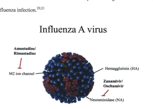
inhibitors against influenza viruses. Influenza A virus (Figure 1-1) is a spherical, enveloped virus, with a segmented RNA genome.17 The virus is decorated with many copies of three
surface proteins. The first, hemagglutinin (HA), is a trimeric protein that is responsible for recognizing terminal sialic acid moieties on host cells and binding to them to initiate infection.'7
The Neuraminidase (NA) protein is involved in the final step of viral infection that allows progeny virions to escape the infected cell so that they may infect new cells.'7 Neuraminidase is
the target of two of the four FDA-approved small molecule influenza inhibitors, namely Zanamivir and Oseltamivir.'8 The last surface protein, the M2 ion channel, is paramount in viral
uncoating; that is, the release of influenza's segmented viral RNA genome into the cell during infection.'7 It is the target of the other two FDA-approved small molecule influenza inhibitors,
amantadine and rimantadine.19 Unfortunately, due to the occurrence of a variety of single-point mutations in the M2 ion channel, nearly all circulating influenza strains have developed resistance to amantadine and rimantadine. Therefore they are no longer recommended for use for
treatment of influenza infection.2 0 2 1
Influenza A virus
Amantadine/ RimantadineI
Hemagglutinin (HA) M2 ion channel Zanamlvir/ Oseltamivir euraminidase (NA)Figure 1-1. Cartoon depicting influenza A virus. Three surface proteins are shown. The hemagglutinin protein depicted in blue helps initiate infection while the neuraminidase protein in depicted in pink allows
for progeny viral escape. The M2 ion channel depicted in purple aids in viral uncoating. Adapted from cdc.gov.
Previously, it has been shown by us and others that attachment of multiple copies of influenza surface protein ligands or inhibitors to flexible polymeric chains vastly improves their potency compared to that of their monomeric parents.'8,2 2
-2 6 This improvement in potency is due to multivalency, whereby multiple simultaneous interactions between polymer-drug conjugates and receptors on the viral surface result in an enhanced binding avidity.2 4 For the FDA-approved
neuraminidase inhibitor zanamivir, we have recently discovered that this improvement in potency can also be attributed to an additional drug mechanism that is unique to the drug-polymer conjugate.2 3 Importantly, the zanamivir-polymer conjugates are effective at potently
inhibiting not only native influenza strains, but also zanamivir-resistant ones. This latter action is attributed to multivalency and likely the aforementioned alternative mechanism.18,2 3
In Chapter 2 of this thesis, I will discuss a quest for multivalent inhibition by literature-predicted influenza inhibitors which target the hemagglutinin protein. Using the plaque reduction assay, relatively simple bicyclic quinone molecules, as well as multiple copies thereof covalently attached to a long polyglutamate-based polymeric chain, were examined as new inhibitors of various naturally-occurring strains of influenza A virus. The polymer-conjugated inhibitors were found to have a far greater potency (for some as high as two orders of magnitude when a long spacer arm was employed) than their corresponding parent molecules against the human Wuhan influenza strain. However, such polymeric inhibitors failed to exhibit higher potency compared to their small-molecule predecessors against the human PR8 and avian turkey influenza strains. These observations, further explored by means of molecular modeling, reveal the previously unrecognized unpredictability of the benefits of multivalency, possibly because of poor accessibility of the viral targets to polymeric agents.
Chapter 3 also addresses multivalent influenza inhibitors; however, instead of attempting to improve new influenza inhibitors (as in Chapter 2) we endeavored to "resurrect" the inhibitory potency of the two FDA-approved adamantane-class M2 ion channel influenza inhibitors. As mentioned above, these drugs are no longer recommended for use to treat influenza infection because of widespread resistance to them. By attaching multiple copies of amantadine or rimantadine to polymeric chains we explored whether it was possible to recover their potency in inhibiting drug-resistant influenza viruses as previously seen in zanamivir examples. Depending on loading densities, as well as the nature of the drug, the polymer, and the spacer arm, polymer-conjugated drugs were up to 30-fold more potent inhibitors of drug-resistant strains than their monomeric parents. Although a full recovery of the inhibitory action against drug-resistant strains was not achieved, this study may be a step toward salvaging anti-influenza drugs that are no longer effective.
Though therapeutics are an excellent line of defense toward viral infection, it also would be valuable to eliminate the transmission of viruses before they
cause disease in their host. By developing antimicrobial +
surface coatings, we have investigated reducing the
transmission of bacteria and viruses from contaminated Figure 1-2. Chemical structure of NN-dodecyl,methyl-polyethylenimine
surfaces. Previously, our lab has developed (DMPEI).
antimicrobial PEI-based (PEI = polyethylenimine) surface coatings which are lethal to Staphylococcus aureus, Escherichia coli, Candida albicans, and influenza.' ,27-31 In Chapter 4 of this thesis, I discuss my studies characterizing our most efficacious antimicrobial coating, NN-dodecyl,methyl-polyethylenimine (DMPEI) (Figure 1-2), against the previously untested herpes
simplex virus (HSV). In this chapter, surface-immobilized and suspended modalities of DMPEI are explored for their ability to reduce viral infectivity in aqueous solutions containing herpes simplex viruses (HSVs) 1 and 2. In our experiments, DMPEI coated on either polyethylene slides or male latex condoms dramatically decreased infectivity in solutions containing 1 or HSV-2. Moreover, DMPEI suspended in aqueous solution markedly reduced the infectious titer of the HSVs. These results suggest potential uses of DMPEI for both prophylaxis (in the form of coated condoms) and treatment (as a topical suspension) for HSV infections.
Earlier work with our antimicrobially coated surfaces (whether covalently or non-covalently) involved bacteria, fungi, and enveloped viruses (namely, influenza and HSVs). 1,16,28,3'In Chapter 5, we explored whether our PEI-based hydrophobic polycations were
active against the non-enveloped poliovirus and rotavirus. We discovered that covalently derivatizing glass surfaces with branched NN-hexyl,methyl-PEI (HMPEI) or physically depositing ("painting") linear DMPEI onto polyethylene surfaces enables the resultant coated materials to quickly and efficiently disinfect aqueous solutions containing poliovirus and rotavirus. Subsequent experiments revealed that washing these poliovirus-exposed DMPEI-coated surfaces with a detergent could recover the viruses in their infectious form. Therefore, HMPEI and DMPEI can disinfect solutions containing poliovirus and rotavirus by adsorption of viral particles.
In addition to the spread of disease via fomites as described above, many consumer goods must be protected from bacterial and fungal colonization to ensure their integrity and safety. By making these items' packaging biocidal, the interior environment can be preserved from microbial spoilage without altering the products themselves. In Chapter 6, we briefly review this concept, referred to as "active packaging", and discuss existing methods for constructing
active packaging systems, highlighting the work done in this regard in our lab. The methods described are based on either packaging materials that release biocides or those that are themselves intrinsically biocidal (or biostatic), with numerous variations within each category. B. References
1. Knipe DM, Howley PM. 2007. Fields' Virology. Philadelphia Wolters Kluwer Health/Lippincott Williams & Wilkins.
2. Gubareva LV. 2004. Molecular mechanisms of influenza virus resistance to neuraminidase inhibitors. Virus Res. 103(1):199-203.
3. Pielak RM, Schnell JR, Chou JJ. 2009. Mechanism of drug inhibition and drug resistance of influenza A M2 channel. Proc Natl Acad Sci. 106(18):7379-7384.
4. Piret J, Boivin G. 2011. Resistance of herpes simplex viruses to nucleoside analogues: mechanisms, prevalence, and management. Antimicrob Agents Ch. 55(2):459-472.
5. De Clercq E. 2006. Antiviral agents active against influenza A viruses. Nat Rev Drug Disc. 5(12):1015-1025.
6. Hayden FG, Pavia AT. 2006. Antiviral management of seasonal and pandemic influenza. J Infect Dis. 194(Supplement 2):S1 19-S126.
7. De Quadros CA, Andrus JK, Olive JM, de Macedo CG, Henderson DA. 1992. Polio eradication from the Western Hemisphere. Annu Rev Publ Health. 13(1):239-252.
8. Orenstein WA, Papania MJ, Wharton ME. 2004. Measles elimination in the United States. J Infect Dis. 189(Supplement 1):S1-S3.
9. Barouch DH. 2008. Challenges in the development of an HIV-1 vaccine. Nature 455(7213):613-619.
10. Johnston MI, Fauci AS. 2008. An HIV vaccine-challenges and prospects. N Eng J Med. 359(9):888-890.
11. Raje ni J, Iburmanovi V. 2006. Developments in herpes simplex virus vaccines: old problems and new challenges. Folia Microbiol. 51(2):67-85.
12. Perez-Tirse J, Gross PA. 1992. Review of cost-benefit analyses of influenza vaccine. Pharmacoeconmics. 2(3):198-206.
13. Leroux-Roels I, Leroux-Roels G. 2009. Current status and progress of prepandemic and pandemic influenza vaccine development. Expert Rev Vaccines. 8(4):401-423.
14. Smith NM, Bresee JS, Shay DK, Uyeki TM, Cox NJ, Strikas RA. 2006. Prevention and control of influenza: recommendations of the Advisory Committee on Immunization Practices (ACIP). Morbid Mortal Week Rep. 55(RR-10): 1.
15. Klibanov AM. 2007. Permanently microbicidal materials coatings. J Mat Chem.
17(24):2479-2482.
16. Larson AM, Oh H, Knipe DM, Klibanov AM. 2013. Decreasing herpes simplex viral infectivity in solution by surface-immobilized and suspended
NN-dodecyl,methyl-polyethylenimine. Pharm Res. 30(1):25-3 1.
17. Lamb RA, Krug RM. 2007. Orthomyoxoviridae: The viruses and their replication. In: Fields' Virology. Eds., Knipe DM, Howley PM. 5th ed. Philadelphia: Wolters Kluwer Health/ Lippincott Williams & Wilkins.
18. Weight AK, Haldar J, Alvarez de Cienfuegos L, Gubareva LV, Tumpey TM, Chen J, Klibanov AM. 2011. Attaching zanamivir to a polymer markedly enhances its activity against drug-resistant strains of influenza A virus. J Pharm Sci. 100(3):831-835.
19. Kozakov D, Chuang G-Y, Beglov D, Vajda S 2010. Where does amantadine bind to the influenza virus M2 proton channel? Trends Biochem Sci. 35(9):471-475.
20. Bright RA, Shay DK, Shu B, Cox NJ, Klimov Al 2006. Adamantane resistance among influenza A viruses isolated early during the 2005-2006 influenza season in the United States. JAMA. 295(8):891-894.
21. Bright RA, Medina M-j, Xu X, Perez-Oronoz G, Wallis TR, Davis XM, Povinelli L, Cox NJ, Klimov Al 2005. Incidence of adamantane resistance among influenza A (H3N2) viruses isolated worldwide from 1994 to 2005: a cause for concern. Lancet 366(9492):1175-1181. 22. Haldar J, Alvarez de Cienfuegos L, Tumpey TM, Gubareva LV, Chen J, Klibanov AM. 2010. Bifunctional polymeric inhibitors of human influenza A viruses. Pharm Res. 27(2):259-263.
23. Lee CM, Weight AK, Haldar J, Wang L, Klibanov AM, Chen J. 2012. Polymer-attached zanamivir inhibits synergistically both early and late stages of influenza virus infection. Proc Natl Acad Sci. 109(50):20385-20390.
24. Mammen M, Choi S-K, Whitesides GM. 1998. Polyvalent interactions in biological systems: implications for design and use of multivalent ligands and inhibitors. Angew Chemie Int Ed. 37(20):2754-2794.
25. Sigal GB, Mammen M, Dahmann G, Whitesides GM. 1996. Polyacrylamides bearing pendant a-sialoside groups strongly inhibit agglutination of erythrocytes by influenza virus: the strong inhibition reflects enhanced binding through cooperative polyvalent interactions. J Am Chem Soc. 118(16):3789-3800.
26. Choi S-K, Mammen M, Whitesides GM. 1997. Generation and in situ evaluation of libraries of poly (acrylic acid) presenting sialosides as side chains as polyvalent inhibitors of influenza-mediated hemagglutination. J Am Chem Soc. 119(18):4103-4111.
27. Park D, Wang J, Klibanov AM. 2006. One-step, painting-like coating procedures to make surfaces highly and permanently bactericidal. Biotechnol Prog. 22(2):584-589.
28. Haldar J, An D, Alvarez de Cienfuegos L, Chen J, Klibanov AM. 2006. Polymeric coatings that inactivate both influenza virus and pathogenic bacteria. Proc Natl Acad Sci. 103(47):17667-17671.
29. Haldar J, Chen J, Tumpey TM, Gubareva LV, Klibanov AM. 2008. Hydrophobic
polycationic coatings inactivate wild-type and zanamivir- and/or oseltamivir-resistant human and avian influenza viruses. Biotechnol Lett. 30(3):475-479.
30. Haldar J, Weight AK, Klibanov AM 2007. Preparation, application and testing of permanent antibacterial and antiviral coatings. Nat Protoc. 2(10):2412-2417.
31. Hsu BB, Wong SY, Hammond PT, Chen J, Klibanov AM. 2011. Mechanism of
inactivation of influenza viruses by immobilized hydrophobic polycations. Proc Natl Acad Sci. 108(1):61-66.
CHAPTER 2
Conjugating drug candidates to polymeric chains does not necessarily enhance anti-influenza activity
The work presented in this chapter was published with kind permission from Wiley Periodicals, Inc
in the following manuscript and is reproduced (Copyright C 2012):
Larson AM, Wang H, Cao Y, Jiang T, Chen J, Klibanov AM. 2012. Conjugating drug candidates to polymeric chains does not necessarily enhance anti-influenza activity. J Pharm Sci. 101(10):3896-905.
A. Introduction
Influenza A virus is highly transmissible and kills over 250,000 people worldwide each year. In the United States alone, some 20% of the population contracts the virus annually, leading to countless missed days of work and school and tens of billions of dollars in associated costs. 1,2 Two of the FDA-approved drugs for the treatment of influenza infections, oseltamivir
(TamifluTM) and zanamivir (RelenzaTM), have fallen short of expectations due to their mediocre activity in reducing the symptoms and duration of the infection, as well as emerging resistance in clinical isolates.3-5 Thus new and more effective anti-influenza therapeutic agents are greatly needed.
One proposed strategy for generating more potent inhibitors of influenza is to utilize the benefits of multivalency.2,4,6-1 1
Conjugating multiple copies of influenza inhibitors to a flexible polymeric chain has been shown to result in multivalent interactions between the polymer-attached inhibitors and the viral surface receptor proteins.2,4,6-1 1 These enhanced interactions, in
turn, lead to a much stronger binding compared to that of the small-molecule parents stemming from favorable entropic factors; additionally, water-swollen polymeric chains may sterically hinder physical contacts between the virus and the target cell.6
The foregoing benefits of multivalency for binding to influenza virus have been demonstrated for viral surface proteins with the natural ligand of hemagglutinin, N-acetylneuraminic (sialic) acid, and with the neuraminidase inhibitor zanamivir. 2,4,6-11 However, both of these compounds are structurally complex, requiring many-step syntheses to become amenable to attachment to polymeric chains in order to investigate the effect of multivalency.2,4
They are also difficult to modify selectively and thus not optimal for structure-activity relationship (SAR) studies. In the present work, we instead have employed simple organic
molecules'2 (Figure 2-1) with anti-influenza properties to investigate the SAR of multivalency.
In particular, we have assessed whether the aforementioned potential benefits of multivalency invariably translate into greater anti-influenza activity.
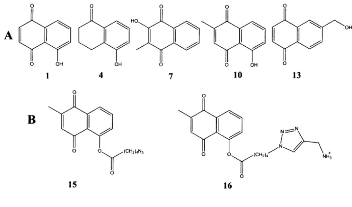
Figure 2-1. Chemical structures of (A) 5-hydroxynaphthalene-1,4-dione (1) and its analogs 4, 7, 10, and 13 used in this study; (B) modified 10 with an azide-terminated spacer arm (15) for use in conjugating to propargylamine-derivatized poly-L-glutamate and 10 derivatized with a spacer arm (16) for investigation
of the dependence of IC5 0 on the presence of the spacer arm by itself with no polymer.
0
A
I 4 7 10 13B
(CH2)4N3 0 15 16B. Results and Discussion
Since the inherent structural complexities of zanamivir and sialic acid do not allow for easy manipulations, for our SAR studies we chose to investigate whether there were simpler molecules also possessing anti-influenza activity. In the scholarly study by Bodian et al.,12
that bind to the hemagglutinin protein on influenza A viral strain X31 (H3N2) and stabilize the protein in its native conformation. This stabilization was proposed to hinder the conformational changes necessary for cell-viral membrane fusion, which is an essential step in the influenza infection cycle.1 2
Specifically, the binding site explored was a region approximately half-way between the most outer tip of the hemagglutinin protein and the viral membrane near the fusion peptide.12 These docking studies resulted in a group of benzoquinones and hydroquinones as
potential ligands to bind to hemagglutinin and inhibit the aforementioned conformational change. 12 HO HO 0 COOH HO 0 AcHN OO (CH 2)16CH3 H HO H
A
0 (CH 2)16CH3 F 0 O H2N OH SH H N H ClN NH N 0 N S N 0 F F F F NN 0 FD
I
EF
NFigure 2-2. Chemical structures of anti-hemagglutinin agents previously described in the literature: (A) Neu5Ac3aF-DSPE; 14 (B) N-(2,8-dimethyl-3-oxo- 1 -thia-4-azaspiro[4.5]decan-4-yl)-6-methylimidazo [2,1 -b]thiazole-5-carboxamide; 15 (C)
4-amino-5-chloro-2-hydroxy-N-((2S,6R,9aR)-6-methyloctahydro-1H-quinolizin-2-yl)benzamide; 16 (D) N-(3-cyanophenyl)-N-methyl-2-phenylcyclohexanecarboxamide; " (E)
methyl-0-methyl-7-ketopodocarpate; 18 (F) 3-fluoro-N-(2-(piperidin- 1
-yl)ethyl)-5-(trifluoromethyl)benzamide, 19 It is worth noting that 1 is a more potent anti-influenza inhibitor than
compound F (IC50 of 315 pM),'9 on par with compounds A, B, and C (IC50's of 5.6, 3-23, and 3-8 pLM,
One of the foregoing compounds, 5-hydroxynaphthalene-1,4-dione (1), was selected as a starting point for our studies. Using the plaque reduction assay method to test 1 for putative anti-influenza activity against the Wuhan strain of the anti-influenza A virus, we indeed found it to be a moderate inhibitor with an IC50 value of 4.7 ± 1.5 pM (Table 2-1, 1st entry). While other
unnatural anti-hemagglutinin inhibitors exist (Figure 2-2),14-19 all are structurally more complex than 1. Therefore, we decided to continue our studies herein with compound 1.
In light of the previous studies with polymer-attached zanamivir and sialic acid,2,4,6-11 we
next tested whether covalent conjugation of multiple copies of 1 to a polymeric chain would increase anti-influenza potency. To this end, 1 was attached to the physiologically benign and biodegradable polymer poly-L-glutamate at -10% loading (i.e., with approximately one tenth of all monomeric units being derivatized with the inhibitor). The resultant polymeric inhibitor 2a (Figure 2-3) exhibited an over 10-fold better IC50 value compared to its monomeric counterpart 1
(Table 2-1, 2nd entry), presumably due to the phenomenon of multivalency. Interestingly, lowering the degree of loading of the inhibitor on the polymer from 10% to 5% (to yield 2b) failed to improve the antiviral potency (Table 2-1), suggesting that the polymer-conjugated ligand molecules do not interfere with each other's ability to inhibit the virus at these loadings.
One can readily envisage how steric constraints imposed by the polymeric chain may hinder the ability of the inhibitor to bind to its viral receptor, thus masking the true power of multivalency. This hypothesis was verified by inserting a nine-atom spacer arm between the polymer and 1, resulting in compound 3. As seen in Table 2-1, this insertion indeed dramatically improved the IC50 value: 20-fold over 2a and some 240-fold over 1.
To examine the generality of these findings, several structural analogs of 1, not previously identified as anti-influenza inhibitors,12 were tested, along with their synthesized
O Na
A
B
2a: R = 1, x:y = 10:90 2b: R = 1, x:y = 5:95 5a: R = 4, x:y = 10:90 5b: R = 4, x:y = 4:96 8: R = 7, x:y = 10:90 11: R = 10, x:y = 4:96 14: R = 13, x:y = 10:90 0 o Nai 0 H N N NN N N 3: R = 1, x:y = 11:89 C ) 6: R = 4, x:y = 6:94 R 9: R = 7, x:y = 5:95 o 12: R = 10, x:y = 12:88 NaC
JH 17: R = 10, x:y = 4:96 18: R = 10, x:y:z = 10:50:40Figure 2-3. Chemical structures of anti-influenza inhibitors attached to poly-L-glutamate (A) with no spacer arm; (B) via a nine-atom spacer arm intended to reduce the putative steric hindrances imposed by the polymeric chains; (C) with the polymeric backbone of varying degrees of electrostatic charge.
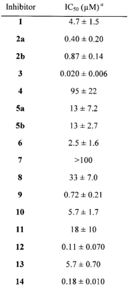
Table 2-1. IC50 values for both small molecules and their polymer-attached derivatives against the Wuhan strain of influenza A virus.
Inhibitor IC50 (tM) a
1 4.7 ±1.5
2a 2b
" The plaque reduction assay experiments were run at least in triplicate; the calculated mean and standard
deviation values are presented in the table. The IC50 values are expressed based on the concentration of
the small-molecule inhibitor. The IC50 value of bare poly-L-glutamate was found to exceed 1 mM and
thus should not appreciably contribute to the extent of inhibition.
poly-L-glutamate conjugates, against the Wuhan influenza strain. 5-Hydroxy-3,4-dihydronaphthalene-1(2H)-one (4) and 2-hydroxy-3-methylnaphthalene-1,4-dione (7) both were found to be inhibitors, albeit much weaker ones than 1. Conjugating them to the polymer directly, i.e., with no spacer arm (to produce 5a or 5b depending on the degree of loading and 8,
0.40 ± 0.20 0.87 ± 0.14 0.020 ±0.006 95 ± 22 13 ± 7.2 13 ± 2.7 2.5 ± 1.6 >100 33 7.0 0.72 0.21 5.7 1.7 18 10 0.11 0.070 5.7 0.70 0.18 0.010 3 4 5a 5b 6 7 8 9 10 11 12 13 14
respectively) led to marked (7 and >3-fold, respectively) improvements in the antiviral potency (Table 2-1). Moreover, as seen in Table 2-1, when the nine-atom spacer arm was inserted to further distance the ligand from the polymeric chain (to form 6 and 9, respectively), the inhibitory potency in both cases rose another several-fold to reach the overall improvement compared to those of the parents 4 and 7 of 38-fold and >140-fold, respectively.
The same general trend of a dramatically enhanced inhibitory potency upon attachment to the polymeric chain via the spacer arm was observed with yet another analog of 1 tested, namely 5-hydroxy-2-methyl-1,4-naphthalenedione (10). This compound, which differs from 1 only by a methyl substituent in the benzoquinone portion of the molecule, exhibited an IC50 value
comparable to that of 1; converting it to 12 produced a 52-fold jump in inhibitory activity (Table 2-1). Interestingly, however, in the case of 10 attached to the polymer with no spacer arm (11), no increase (and, in fact, a sizeable decline) in the potency was observed (Table 2-1), illustrating how subtle the SAR is.
The poly-L-glutamate conjugate of 13 (in the absence of a nine-atom linker), 14, exhibited a greater than 30-fold improvement over its monomeric counterpart (Table 2-1) demonstrating that attachment through the 6C position in the naphthoquinone moiety does not have a deleterious effect on the inhibitor improvement; in fact, it generated the most potent non-linker conjugated inhibitor.
In addition to a striking improvement in the inhibitory potency of the ligands upon conjugating them to poly-L-glutamate via the long spacer arm, their toxicity also diminished. In particular, both for 2a, 2b, and 3 vs. 1 and for 11 and 12 vs. 10 the cellular toxicity was at least an order of magnitude lower. For example, when compound 1 was used in the infection phase of the plaque reduction assay, cells that were incubated with the concentrations of inhibitor greater
than, or equal to, 30 pM displayed obvious fatal demise. In contrast, cells incubated with even greater than 300 pM concentrations of polymer-attached inhibitors 2a, 2b, and 3 were healthy and seemingly unaffected by the presence of inhibitor. Presumably, sequestering the toxic small molecules (50% cell cytotoxicity concentration, CC50, of -30 pIM for 112) to the polymer
prevents them from traversing the cellular membrane and exerting deleterious effects within the cell.
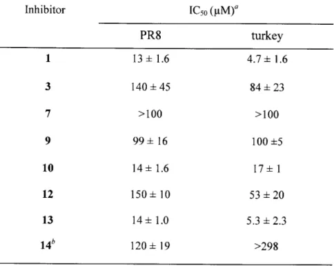
To determine to what extent the foregoing findings apply to other influenza A viruses, we selected two additional strains, namely the avian turkey and the human PR8 strains. Both strains have different serotypes of the hemagglutinin and neuraminidase proteins compared to Wuhan's, leading to subtle differences in the amino acid composition and structure of these proteins.20 Using the plaque reduction assay, we tested the monomeric inhibitors 1, 7, 10, and 13, as well as those attached to poly-L-glutamate either via the nine-atom spacer arm (compounds 3, 6, and 12) or directly (14). As in the case of the Wuhan strain, 1, 10, and 13 were substantial inhibitors of the viruses, whereas 7 was not (Table 2-2), suggesting similarities in the receptors' binding sites of all three strains. And yet, in stark contrast to the observations made with the Wuhan strain (Table 2-1), conjugation to the polymeric chains even via a long spacer arm not only failed to result in a significant improvement of the anti-influenza potency but, in the case of 1 and 10, actually made it markedly worse, i.e., increased the IC50 values (Table 2-2). For 14, conjugation of the inhibitor to the polymer though the C6 position in the naphthoquinone also caused a substantial deterioration in potency for both turkey and PR8 strains suggesting that the site of the linker's attachment is not entirely responsible for the reduction in inhibition.
We hypothesized that perhaps the addition of the linker group itself imposed new steric hindrances for binding of the inhibitor to the receptor sites on the turkey and PR8, but not
Table 2-2. IC5 0 values for both small molecules and their polymer-attached derivatives against
the PR8 and turkey strains of influenza A virus.
Inhibitor IC50 (pIM)a PR8 turkey 1 13 1.6 4.7 ±1.6 3 140 45 84 23 7 >100 >100 9 99 16 100 5 10 14 1.6 17 1 12 150 10 53 20 13 14 ±1.0 5.3 2.3 14' 120 19 >298
a The plaque reduction assay experiments were run at least in triplicate; the calculated mean and standard
deviation values are presented in the table. The IC50 values are expressed based on the concentration of
the small-molecule inhibitor. The IC5 0 values for compounds 4 and 6 exceeded 170 gM for both viral
strains. Thus they were not included in this table because no definitive conclusions concerning the effect of attachment to the polymer can be made.
bAll polymer conjugates characterized in this table contain the nine-atom spacer arm, except for 14 which
comprises 13 conjugated directly to poly-L-glutamate.
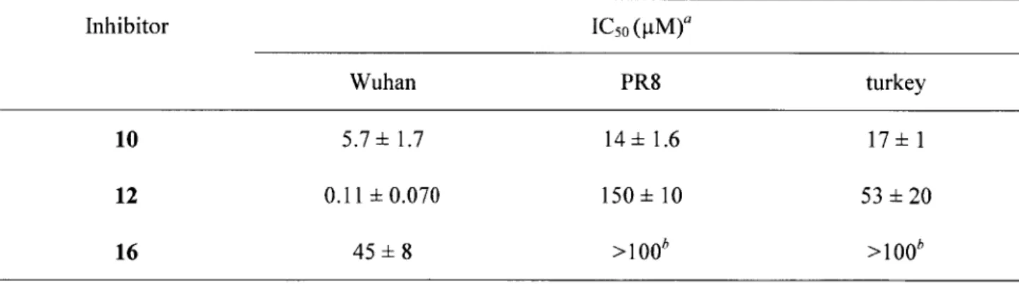
the Wuhan, strains of the virus. This would inevitably lead to a diminished ability of the inhibitor to bind to its receptor when subsequently attached to poly-L-glutamate. To test this hypothesis, we focused on the compound 10 group of inhibitors and synthesized compound 16 containing the same chemical structure as the inhibitor plus the spacer arm portion of 12 but in the absence of
polymer. As seen in Table 2-3, 16 indeed exhibited a drastically poorer anti-influenza inhibition against all three viruses compared to the parent compound 10.
Thus the deteriorated inhibition potency upon attachment of the nine-atom spacer arm indeed may account for at least some of the inferior inhibition observed for compounds 3, 9, and 12 compared to their monomeric precursors 1, 7, and 10, respectively, against the turkey and PR8 strains. However, in order for 12 to exhibit the multivalency benefits over 16 of greater than 100-fold (as it did with Wuhan strain, Table 2-3), the ICso value of 16 would have to be at least 104 pM for the PR8 and turkey strains. The results of the plaque reduction assay suggest that this is not the case though: a reduction in plaque numbers for both of these strains upon incubation with 90 tM 16 indicated that the ICso was close to 100 ptM (data not shown). The exact IC5 0 for
16 was not measurable however, due to cytotoxicity of the compound.
Table 2-3. IC50 values for compounds 10, 12, and 16 against three influenza A virus strains
Inhibitor IC5 0 (pM)a
Wuhan PR8 turkey
10 5.7 ±1.7 14 ±1.6 17 1
12 0.11 ±0.070 150 ±10 53 20
16 45± 8 >100 >100
a The plaque reduction assay experiments were run at least in triplicate; the calculated mean and standard
deviation values are presented in the table. The IC50 values are expressed based on the concentration of the small-molecule inhibitor.
b Although the IC5o values for the turkey and PR8 strains could not be conclusively determined because
the 50% cell cytotoxicity concentration (CC5o) for 16 only slightly exceeded 100 pM, there was a visible reduction in plaques upon incubation with 90 pM of 16.
Next we investigated the role of the polymer's charge in antiviral activity. To this end, two new polymer conjugates with compound 10 attached via the nine-atom spacer arm (17 and 18) were prepared and tested against the Wuhan and turkey strains. Compound 17, containing a neutral poly-L-glutamine backbone, showed modest improvements for the turkey strain over its negatively charged analog 12 but failed to exhibit a great enhancement over the monomer 10 (Table 2-4). Conversely, for the Wuhan strain, 17 demonstrated an almost 50-fold improvement over the monomer, similar to that afforded by compound 12 (Table 2-4). Likewise, compound 18 (containing a partly negatively-charged backbone) displayed no great enhancement for the turkey strain while exhibiting an almost 40-fold improvement over 10 for the Wuhan strain. These results indicate that the charge on the polymeric chain cannot account for the striking differences
observed between the turkey and Wuhan strains for monomeric vs. polymer-attached inhibitors.
Table 2-4. Comparison of IC50 values for 5-hydroxy-2-methyl-1,4-naphthalenedione (12)
conjugated to polymers of varying degrees of backbone charge against the Wuhan and turkey strains of influenza A virus.
Inhibitor IC50 (ptM)a
Wuhan turkey
12 0.11 ±0.070 53 ±20
17 0.12 0.061 7.4 ±3.9
18 0.15 ±0.035 35 ±9.0
'The plaque reduction assay experiments were run at least in triplicate; the calculated mean and standard
deviation values are presented in the table. The IC50 values are expressed based on the concentration of
To determine whether the difference in improvements for polymer-attached conjugates over monomeric inhibitors between the Wuhan and turkey strains was an artifact of our biological assay, i.e., whether the inhibitor was brought into contact with the virus at the inappropriate time during infection, we performed an additional plaque assay with 10 and 12 present not only during the pre-incubation and binding (as is conventionally done) but also with the inhibitor in the agar overlay. This modality of the plaque reduction assay exposes cells and viruses to the inhibitor for the entirety of the first and subsequent infection phases leaving the inhibitor available for all steps in the viral cycle and not just for binding or endocytosis. In this experimental mode, the inhibitory effect of 10 and 12 for the Wuhan strain changed only modestly: IC50's of 2.4 ± 0.39 pM and 0.061 ± 0.045 pM, respectively, for incubation of
inhibitor in the agar overlay vs. 5.7 ± 1.7 ptM and 0.11 ± 0.070 [tM for our conventional experiment. For the turkey strain, the inhibition by the monomer (10) improved more (IC5 0 of 17
± 1.0 pM when not included in the agar and 2.9 ± 0.49 piM when included in the agar) but the polymer-attached inhibitor's (12's) IC5 0 exceeded 5 pM; these observations confirm that the lack
of improvement for polymer-attached inhibitors against the turkey strain was not an artifact of our biological assay.
We then hypothesized that the preferred binding sites of the monomeric vs. the polymer-attached inhibitors might be distinct between the strains, thus being responsible for their vastly different inhibitory properties. To explore this possibility, we ran in silico docking experiments to determine the preferred binding sites for the monomeric inhibitor 10 and the linker-attached inhibitor 16 for the PR8 strain and a Wuhan surrogate strain, X31 (both Wuhan and X31 are H3N2). Note that the X-31 strain has a 87% sequence identity for the HA1 strand of the hemagglutinin molecule compared to the Wuhan strain (the HA2 Wuhan sequence is
unavailable)2 1 2 2
(for comparison, the PR8's HA1 strand has just a 33% sequence identity23). As seen in Figure 2-4A, for the X-31 strain both inhibitors bind in the same region of the hemagglutinin protein; this region coincides with the crystallographically determined binding site for tert-butyl hydroquinone (another fusion inhibitor described by Bodian et al. 12,13). In stark
contrast, for the PR8 strain, inhibitors 10 and 16 bind in vastly distinct locations on the hemagglutinin protein, with as much as 40 A separating them (Fig. 2-4B). Note that 10 binds to a similar location on the PR8 strain's hemagglutinin as both 10 and 16 in the X-3 1's protein;
A
~~,JA .16 10
161
10
Figure 2-4. The docking results for inhibitors 10 and 16 (both black) on the hemagglutinin protein (grey) of the X-31 strain (a surrogate used to represent the Wuhan strain) (A, left panel) and of the PR8 strain (B, right panel) of influenza A virus. As seen in A, the binding sites on the protein for the two inhibitors overlap in the former case. In contrast, the distance between the binding sites for the two inhibitors in B is some 40 A indicating that the molecules bind to the protein in vastly distinct locations. The images, generated in Pymol, depict only 10 and 16 docked on one hemagglutinin monomer for each viral strain.
however, 16 on the PR8 strain's hemagglutinin binds in a region that is closer to the viral envelope, thereby possibly hindering accessibility for the polymer-attached inhibitor.
One has to wonder why there are benefits of multivalency for all the compounds tested attached via a long spacer arm to a poly-L-glutamate for one strain of influenza, but not for the other two (Tables 2-1 and 2-2). It is unlikely that the observed differences in inhibition among viral strains are due to dissimilar spacing of hemagglutinin molecules along the viral surface among different strains. There are -400 hemagglutinin molecules on a given viral particle of roughly the same size regardless of the influenza virus strain. Since the mechanism of viral particle formation is the same for different strains, the spacing among hemagglutinin molecules on average should be similar as well.2 0 The binding of the first polymer-attached inhibitor
molecule to hemagglutinin is also unlikely to prevent binding of all the others. Although the binding of the first inhibitor molecule indeed might make it sterically impossible for a second, nearby counterpart to bind to hemagglutinin, that cannot be the case for more distant polymer-attached inhibitor molecules. Since our (-10% derivatized) polymeric chain contains some 50
randomly distributed inhibitor molecules, most of them should be sufficiently remote from the first one bound to interact with another hemagglutinin molecule.
The three influenza strains undoubtedly have some differences in the binding site for these inhibitors;2 0 in the case of the turkey and PR8 strains, these differences might interplay
with the polymer and/or linker deleteriously to weaken the binding of the conjugated inhibitor to the virus. Additionally, the topology of the viral surface might introduce accessibility problems for the inhibitors once they are attached to the bulky, hydrated polymeric chain. In previous studies investigating the effect of multivalency on anti-influenza inhibitors, the receptor of the multivalent ligand was proximal to the solvent-exposed outer edge of the surface protein.2,4,6-11
Although the exact location of binding of 1 and its analogs to hemagglutinins in the strains studied herein is unknown, our docking studies (Fig. 2-4) predict that for 10 and 16 the site is located approximately half-way down the protein for the X-31 strain and close to the viral envelope for the linker-attached inhibitor (16) on the PR8 strain.12 Perhaps while a small molecule readily accesses regions down the stock of the protein, conjugation to a bulky polymeric chain hampers the access below the dense canopy of proteins on the viral surface.
The concept of multivalency stipulates that several simultaneous interactions of ligands and receptors should result in a stronger, multipoint inhibitor-virus binding and hence give rise to
A
B
0
Figure 2-5. A cartoon depicting a multivalent vs. a monovalent interaction of an inhibitor with the virus. The top panel (A) depicts a polymer-attached inhibitor (polymer is black line, inhibitors are open circles) interacting with a viral envelope containing the receptors of the inhibitor (grey part-circle is the viral envelope, white open half-circles are receptors, dotted lines indicate a binding interaction). Attaching multiple copies of the inhibitor to a polymeric chain can result in a multivalent and hence much stronger interaction with the viral receptors. The bottom panel (B) depicts a monovalent interaction of parent inhibitor molecules with a viral receptor. In this case, the individual inhibitor molecules act independently of each other in binding to their receptors and hence do not benefit from entropically enhanced binding (i.e., from multivalency).
6
more potent inhibitors (Figure 2-5). Our results herein suggest, however, that this is indeed the case only if attaching an inhibitor to a polymeric chain does not impose negative spatial constraints not outweighed by the inherent benefits of multivalency. Since it is unknown in advance whether this will be the case, our data illustrate that the superiority of multivalent inhibitors of a virus compared to their monovalent predecessors cannot be automatically assumed.
Acknowledgements
I am thankful to Hongmei Wang for synthesizing compound 13, to Yang Cao for performing the docking simulations, and to all coauthors for help in editing the manuscript that resulted in this chapter.
C. Materials and Methods Materials
All small-molecule inhibitors except for 13, poly-L-glutamate Na salt (50 -100 kDa), solvents, and reagents were purchased from Sigma-Aldrich Chemical Co. (St. Louis, MO) and used without further purification. Dialysis membranes (3,500 kDa molecular weight cutoff) were from Spectrum Labs (Rancho Dominguez, CA) and PD-10 desalting columns from GE Healthcare (Buckinghamshire, U.K.).
Syntheses
Synthesis of 6-(hydroxymethyl)naphthalene-1,4-dione (13) was carried out as described by Antonini et al.2 4
Synthesis of 2a, 2b, 5a, 5b, 8, 11, and 14: Conjugation was carried out via a Steglich esterification with minor deviations from a reported procedure.2 5 Specifically, poly-L-glutamate
Na salt was converted to poly(L-glutamic acid) by dissolution in double-distilled (dd) H20,
lowering the pH to 1, and washing with 0.10 M HCl to remove free salts before an overnight lyophilization. Lyophilized poly(L-glutamic acid) (20 mg, 0.16 mmol) was dissolved in 0.80 mL of dry dimethylformamide (DMF), followed by the addition of N,N'-dicyclohexylcarbodiimide (DCC) (4.3 mg (0.021 mmol) in 0.30 mL of DMF for a ~10% derivatization), an anti-influenza agent (0.050 mmol in 0.20 mL of DMF), pyridine (10 pL, 0.12 mmol), and a catalytic quantity of 4-dimethylaminopyridine (DMAP) in 0.40 mL of DMF with vigorous stirring. The solution was stirred overnight at room temperature. The polymer was then isolated by precipitation in chloroform, washed with fresh chloroform to remove the unreacted anti-influenza agent, converted to the Na salt by dissolution in phosphate buffered saline (PBS), and dialyzed against ddH20 in a 3,500-Da MW cutoff dialysis membrane for 24 hr to remove free salts and other
impurities. TLC using a 3:1 (v/v) ethyl acetate:hexanes mixture confirmed purity and demonstrated the disappearance of the anti-influenza agent's spot (for example, Rf 0.68 for 10). Percent conjugation of the anti-influenza agent was calculated by means of 'H NMR in D20
using a Bruker 600 MHz instrument by comparing the ratio of the integration of a polymer peak with that of an anti-influenza agent's peak. In the case of a ~5% derivatization of the polymer with anti-influenza agent, 2.0 mg/0.010 mmol of DCC was added to the reaction mixture (2b, 5b, and 11). The NMR spectrum of 2a appears in appendix A.
1H NMR (D20) 6 (600 MHz) - for 2a and 2b: 1.6-2.0 (2H polymer, d, CH2), 2.1-2.5
(2H polymer, s, CH2), 4.0-4.4 (1H polymer, s, CH), 6.8-8.0 (5H, aromatics); for 5a and 5b:
1.8-2.1 (2H polymer, d, CH2), 2.1 (2H, m, cyclohexyl CH2), 2.2-2.4 (2H polymer, s, CH2), 2.6 (2H,
m, cyclohexyl CH2), 2.8 (2H, m, cyclohexyl CH2), 4.2-4.4 (1H polymer, s, CH), 7.3-7.9 (3H,
![Figure 2-2. Chemical structures of anti-hemagglutinin agents previously described in the literature: (A) Neu5Ac3aF-DSPE; 14 (B) N-(2,8-dimethyl-3-oxo- 1 -thia-4-azaspiro[4.5]decan-4-yl)-6-methylimidazo [2,1](https://thumb-eu.123doks.com/thumbv2/123doknet/14021827.457441/24.918.101.771.400.891/chemical-structures-hemagglutinin-previously-described-literature-dimethyl-methylimidazo.webp)