Full Terms & Conditions of access and use can be found at
https://www.tandfonline.com/action/journalInformation?journalCode=kgmi20
Gut Microbes
ISSN: (Print) (Online) Journal homepage: https://www.tandfonline.com/loi/kgmi20
Exosomes transfer miRNAs from cell-to-cell to
inhibit autophagy during infection with Crohn’s
disease-associated adherent-invasive E. coli
Anaïs Larabi, Guillaume Dalmasso, Julien Delmas, Nicolas Barnich & Hang Thi Thu Nguyen
To cite this article: Anaïs Larabi, Guillaume Dalmasso, Julien Delmas, Nicolas Barnich & Hang Thi Thu Nguyen (2020) Exosomes transfer miRNAs from cell-to-cell to inhibit autophagy during infection with Crohn’s disease-associated adherent-invasive E.�coli, Gut Microbes, 11:6, 1677-1694, DOI: 10.1080/19490976.2020.1771985
To link to this article: https://doi.org/10.1080/19490976.2020.1771985
View supplementary material Published online: 25 Jun 2020.
Submit your article to this journal Article views: 885
View related articles View Crossmark data
RESEARCH PAPER
Exosomes transfer miRNAs from cell-to-cell to inhibit autophagy during infection
with Crohn’s disease-associated adherent-invasive E. coli
Anaïs Larabia, Guillaume Dalmassoa, Julien Delmasa,b, Nicolas Barnicha, and Hang Thi Thu Nguyena
aM2iSH, UMR 1071 Inserm, Université Clermont Auvergne, Clermont-Ferrand, France; bService de Bactériologie, Centre Hospitalier Universitaire
(CHU) Gabriel Montpied, Clermont-Ferrand, France ABSTRACT
Adherent-invasive E. coli (AIEC), which abnormally colonize the intestinal mucosa of Crohn’s disease (CD) patients, are able to adhere to and invade intestinal epithelial cells (IECs), survive and replicate within macrophages and induce a pro-inflammatory response. AIEC infection of IECs induces secretion of exosomes that increase AIEC replication in exosome-receiving IECs and macrophages. Here, we investigated the mechanism underlying the increased AIEC replication in cells receiving exosomes from AIEC-infected cells. Exosomes released by uninfected human intestinal epithelial T84 cells (Exo-uninfected) or by T84 cells infected with the clinical AIEC LF82 strain (Exo-LF82), the nonpathogenic E. coli K12 strain (Exo-K12) or the commensal E. coli HS strain (Exo-HS) were purified and used to stimulate T84 cells. Stimulation of T84 cells with Exo-LF82 inhibited autophagy compared with Exo-uninfected, Exo-K12 and Exo-HS. qRT-PCR analysis revealed increased levels of miR-30c and miR-130a in Exo-LF82 compared to Exo-uninfected, Exo-K12 and Exo-HS. These miRNAs were transferred via exosomes to recipient cells, in which they targeted and inhibited ATG5 and ATG16L1 expression and thereby autophagy response, thus favoring AIEC intracellular replica-tion. Inhibition of these miRNAs in exosome-donor cells infected with AIEC LF82 abolished the increase in miR-30c and miR-130a levels in the released Exo-LF82 and in Exo-LF82-receiving cells, thus suppressing the inhibitory effect of Exo-LF82 on ATG5 and ATG16L1 expression and on autophagy-mediated AIEC clearance in Exo-LF82-receiving cells. Our study shows that upon AIEC infection, IECs secrete exosomes that can transfer specific miRNAs to recipient IECs, inhibiting autophagy-mediated clearance of intracellular AIEC.
ARTICLE HISTORY
Received 16 October 2019 Revised 14 April 2020 Accepted 14 May 2020
KEYWORDS
Crohn’s disease; AIEC; exosomes; miRNA; autophagy
Introduction
Crohn’s disease (CD) is an inflammatory bowel disease (IBD) with a multifactorial etiology, invol-ving a complex interaction between environmen-tal, genetic and microbial factors.1,2 An intestinal dysbiosis has been reported in CD patients, char-acterized by a decrease in the number of beneficial bacteria such as members of the Firmicutes phy-lum and an increase in potentially harmful bac-teria such as those from the Enterobacbac-teriaceae family. Our group and others have reported a high prevalence of pathogenic adherent- invasive Escherichia coli (AIEC) strains in the ileal mucosa of CD patients.3–6 The CD- associated AIEC strains have been shown to adhere to and to invade intestinal epithelial cells (IECs),7–9 to survive and replicate within macrophages10,11 with an increase in the macro-phages from CD patients compared to those from
healthy controls,12 to promote pro-inflammatory cytokine production, and to colonize the gut and induce intestinal inflammation in genetically sus-ceptible mouse models.13,14
Autophagy, a lysosomal degradation process, has emerged as a key player in the maintenance of intest-inal homeostasis and gut ecology, the protection against microbes and the control of appropriate intestinal immune responses.15 Efforts have been made to reveal the mechanisms by which dysfunc-tional autophagy predisposes to CD development.16 Among those, a defect in autophagy has been shown to result in impaired clearance of pathogenic bacteria including CD-associated AIEC, which is associated with aberrant immune responses.8,9,11,13,17-19 Reversely, we reported that AIEC can limit their elimination by the autophagy machinery of the host by increasing the level of microRNAs (miRNA, miR) 30c and 130a to suppress the CONTACT Hang Thi Thu Nguyen hang.nguyen@uca.fr M2iSH, UMR 1071 Inserm, Université Clermont Auvergne, Clermont-Ferrand 63001, France
Supplemental data for this article can be accessed on the publisher’s website. GUT MICROBES
2020, VOL. 11, NO. 6, 1677–1694
https://doi.org/10.1080/19490976.2020.1771985
expression of ATG (Autophagy-related) 5 and ATG16L1, respectively, two key players of autophagy
induction.8 This consequently favors the intestinal
colonization of AIEC and increases AIEC-induced inflammation in vitro and in vivo.8 Given that miR- 30c and miR-130a are upregulated in CD,8 dysfunc-tional autophagy might not be only caused by CD- associated risk variants in autophagy-related genes, but also by the epigenetic factors that negatively regulate expression of autophagy-related genes.
Recently, our group highlighted a previously uncovered function of exosomes, small extracel-lular vesicles with a diameter of 30 to 100 nm,20 in cell-to-cell communication during AIEC infection.21 Exosomes are released from a broad range of cell types and are found in most bodily fluids.20 Exosomes play a role in cell-to-cell communication by transferring lipids, proteins and genetic material such as mRNAs and miRNAs from a donor cell to a recipient cell.20 Moreover, the exosomal content is functional when being transferred to recipient cells, in which the exosomal mRNAs are translated into proteins22,23 and exosomal miRNAs are able to silence target genes.24–26 Thus, exosomes have been implicated in many physiopathological functions, such as signaling, immunity and infection.20 In the context of AIEC infection, we reported that AIEC infection promotes the secretion of exosomes by infected cells, which are in turn uptaken by naïve cells, leading to increased pro-inflammatory response and impaired clearance of intracellular AIEC.21 The mechanisms underlying the effects of exosomes in recipient cells, however, remain unknown.
Since a functional autophagy is required to restrain the intracellular replication of AIEC,19 we hypothesized that the increased AIEC replication in the cells that receive exosomes released from AIEC- infected IECs is due to impaired autophagy. Thus, in the current study, we aimed at investigating whether miR-30c and miR-130a, which were pre-viously shown to be upregulated in AIEC-infected cells,8 are packaged in exosomes and transferred to recipient cells to inhibit ATG5 and ATG16L1 expression, limiting autophagy-mediated clearance of intracellular AIEC.
Results
Exosomes released from AIEC LF82-infected IECs inhibit autophagy in recipient IECs
We previously reported that exosomes secreted by AIEC-infected human intestinal epithelial T84 cells increase intracellular replication of AIEC and inflammation in exosome-receiving cells.21 Since a functional autophagy is required to restrain the intracellular replication of AIEC,19 we hypothesized that exosomes released from AIEC-infected cells may inhibit autophagy in recipient cells, thus leading to abnormal AIEC replication. To confirm this, we purified exosomes secreted by uninfected T84 cells (designated as Exo-uninfected) or by T84 cells infected with the AIEC LF82 reference strain (designated as Exo-LF82) or the non-pathogenic E. coli K12 strain (designated as Exo-K12) or the commensal E. coli HS strain (designated as Exo-HS) for 12 h as previously described.21 Western blot analysis showed that the purified exosomes contained the exosomal marker CD63 and CD9 but did not carry the negative marker GRP94, which was detected only in T84 cell lysate (Figure 1a). T84 cells were then stimulated with the purified exo-somes, and western blot analysis for the shift of LC3-I (microtubule-associated protein 1 light chain 3; the free cytosolic form) to LC3-II (the form conjugated to phagophore and
autophago-some), an indication of autophagy induction,27
was performed. Upon stimulation with Exo- LF82, T84 cells exhibited decreased LC3-II level, suggesting impaired autophagy, compared with cells receiving Exo-uninfected, Exo-K12 or Exo-HS (Figure 1b,c). Inhibition of autophagy by Exo-LF82 was confirmed by the concomitant increase in p62/SQSTM1 (sequestosome 1), a receptor protein degraded by functional
autophagy27 (Figure 1b,c). Furthermore, Exo-
LF82 also inhibited LC3-II level and p62 degra-dation compared to Exo-uninfected, Exo-K12 or Exo-HS in T84 cells infected with the AIEC
LF82 strain (Figure 1d,e). These results suggest
that exosomes derived from AIEC-infected cells may inhibit autophagy in exosome-receiving cells at basal level or following AIEC infection.
Figure 1. AIEC LF82-infected IECs release exosomes that can inhibit autophagy in recipient cells. Exosomes were purified from the culture supernatant of uninfected T84 cells (Exo-uninfected) or T84 cells infected with the AIEC LF82 strain (Exo-LF82), the E. coli K12 MG1655 strain (Exo-K12) or the commensal E. coli HS strain (Exo-HS) for 12 h. (a) Western blot analysis of the exosomal markers CD63 and CD9, and the negative marker GRP94 using 30 μg of exosomal protein lysate or T84 protein lysate. (b-e) T84 cells were stimulated for 8 h with Exo-uninfected, Exo-LF82, Exo-K12 or Exo-HS (80 μg of exosome/mL of culture medium) and then uninfected (b, c) or infected with LF82 for 4 h (d, e). Representative western blot analysis (b, d) and quantification of LC3-II/β-actin band intensity (c, e) are shown. Data are representative of three independent experiments and are presented as means ± SEM. Statistical analysis was performed using the one-way ANOVA test followed by a Bonferroni posttest. *P < 0.05; **P ≤ 0.01; ***P ≤ 0.001.
MiR-30c and miR-130a levels are increased in the exosomes released from AIEC LF82-infected IECs and in exosome-receiving IECs
Our previous study demonstrated that upon AIEC infection, the levels of miR-30c and miR- 130a are increased in host IECs, thus inhibiting expression of ATG5 and ATG16L1, respectively, and subsequently impairing autophagy.8 As pre-viously observed, qRT-PCR analysis showed increased levels of miR-30c and miR-130a in T84 cells in response to AIEC LF82 infection compared to uninfected IECs or IECs infected with the K12 MG1655 or E. coli HS strains (Figure 2a). We hypothesized that in response to AIEC infection, miR-30c and miR-130a were packaged in exosomes and transferred to recipi-ent cells, in which they inhibit ATG5 and ATG16L1 expression and suppress autophagy. To confirm this, exosomes secreted from unin-fected T84 cells or T84 cells inunin-fected with the AIEC LF82, or K12 MG1655 or E. coli HS strain were purified, and the levels of miR-30c and miR-130a in these exosomes were analyzed by
qRT-PCR. As shown in Figure 2b, the levels of
miR-30c and miR-130a were significantly increased in Exo-LF82 compared to Exo- uninfected, Exo-K12 or Exo-HS.
Furthermore, T84 cells stimulated with Exo- LF82 exhibited increased miR-30c and miR-130a levels compared with those receiving Exo-
uninfected, Exo-K12 or Exo-HS (Figure 2c). This
suggests that these miRNAs were transferred from exosomes to recipient cells.
MiR-30 c and miR-130a are efficiently transferred via exosomes from donor cells to recipient cells, where they target and inhibit ATG5 and ATG16L1 expression
To demonstrate that miR-30c and miR-130a are directly transferred from cell-to-cell via exosomes, we transfected T84 cells with Alexa Fluor 555- labeled-miR-30c or Alexa Fluor 555-labeled-miR -130a, then extracted the released exosomes. Naïve T84 cells were then incubated with the purified
exosomes. Confocal microscopic analysis clearly showed the localization of Alexa Fluor 555- labeled miRNAs inside exosome-receiving cells (Figure 3a,b), indicating that these miRNAs are successfully transferred from donor cells to reci-pient cells via exosomes.
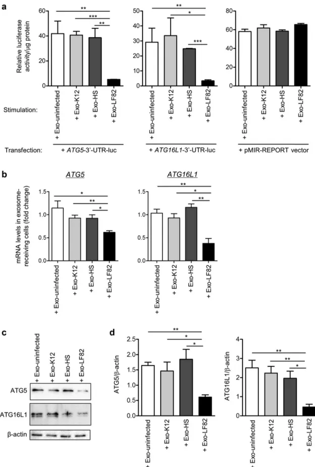
We next examined whether miR-30c and miR- 130a transferred from exosomes can regulate ATG5 and ATG16L1 expression in recipient cells. Naïve T84 cells were transfected with a construct in which the 3ʹ-untranslated region (UTR) of ATG5 or ATG16L1 mRNA was cloned downstream a luciferase-coding sequence in the pMIR-
REPORTTM luciferase vector (designated as ATG5-
3ʹ-UTR-luc or ATG16L1-3ʹ-UTR-luc construct, respectively). Transfected cells were then stimulated with Exo-uninfected, Exo-K12, Exo-HS or Exo- LF82. Exo-LF82 significantly reduced luciferase activity in recipient cells transfected with ATG5-3ʹ- UTR-luc or ATG16L1-3ʹ-UTR-luc construct com-pared to Exo-uninfected, Exo-K12 or Exo-HS, indi-cating that the exosomal miR-30c and miR-130a bind to the 3ʹ-UTRs of ATG5 and ATG16L1 mRNAs, respectively (Figure 4a). However, in cells transfected with the empty luciferase reporter, luci-ferase activity was not reduced upon stimulation with Exo-LF82 compared to Exo-uninfected, Exo- K12 or Exo-HS, indicating the specificity of binding between the exosomal miR-30c or miR-130a and the 3ʹ-UTR of ATG5 or ATG16L1 mRNA, respectively (Figure 4a). Importantly, T84 cells receiving Exo- LF82 exhibited significantly reduced levels of ATG5 and ATG16L1 mRNAs compared to those receiving Exo-uninfected, Exo-K12 or Exo-HS, as analyzed by
qRT-PCR (Figure 4b). Furthermore, western blot
analysis revealed a significant reduction of ATG5 and ATG16L1 protein expression in cells receiving Exo-LF82 compared to that in cells receiving Exo-
uninfected, Exo-K12 or Exo-HS (Figure 4c).
Together, these results show that upon AIEC infection, host cells can release exosomes that transfer miR-30c and miR-130a to recipient cells to inhibit expression of their target genes ATG5 and
ATG16L1 at both mRNA and protein levels, by
directly targeting the 3ʹ-UTRs of ATG5 and
Figure 2. MiR-30c and miR-130a levels are increased in the exosomes released from AIEC LF82-infected IECs and in exosome-receiving IECs. (a, b) Exosomes were purified from uninfected T84 cells (Exo-uninfected) or T84 cells infected with the AIEC LF82 strain (Exo-LF82), the E. coli K12 MG1655 strain (Exo-K12) or the E. coli HS strain (Exo-HS) for 12 h. The levels of miR-30c and miR-130a in exosome-donor cells (a) and in the purified exosomes (b) were analyzed by qRT-PCR. (c) T84 cells were stimulated with the purified exosomes (80 μg of exosome/mL of culture medium), and the levels of miR-30c and miR-130a in these cells were analyzed by qRT-PCR. Data are representative of three independent experiments and are presented as means ± SEM. Statistical analysis was performed using the one- way ANOVA test followed by a Bonferroni posttest. *P < 0.05; **P ≤ 0.01; ***P ≤ 0.001.
Figure 3. MiR-30c and miR-130a are efficiently transferred via exosomes from donor to recipient cells. T84 cells were transfected with 50 nM of Alexa Fluor 555-labeled-miR-30c (a) or Alexa Fluor 555-labeled-miR-130a (b) for 24 h, and uninfected or infected with the AIEC LF82, the E. coli K12 or the E. coli HS strain with a multiplicity of infection (MOI) of 10 for 12 h. Exosomes released from these cells were extracted and designated as Exo-uninfected, Exo-LF82, Exo-K12 and Exo-HS, respectively. Naïve T84 cells were incubated with the purified exosomes (80 μg of exosome/mL of culture medium) for 8 h. Confocal microscopic analysis of T84 cells that received exosomes from cells transfected with miR-30c (a) or miR-130a (b) labeled with Alexa Fluor 555 (red). Actin cytoskeleton was stained with FITC- phalloidin (green). Nuclei were stained with DAPI (blue). Scale bars = 5 µm.
Figure 4. MiR-30c and miR-130a transferred from exosomes inhibit ATG5 and ATG16L1 expression in exosome-receiving IECs. Exosomes were purified from uninfected T84 cells (Exo-uninfected) or T84 cells infected with the AIEC LF82 (Exo-LF82), the E. coli K12 MG1655 (Exo-K12) or the HS strain (Exo-HS) for 12 h. (a) T84 cells were transfected with ATG5-3ʹ-UTR-luc or ATG16L1-3ʹ-UTR-luc construct, in which the 3ʹ-UTR of ATG5 or ATG16L1 mRNA was cloned downstream a luciferase-coding sequence in the pMIR-REPORT luciferase vector, or with the empty vector. Sixteen hours after transfection, the cells were incubated with Exo-uninfected, Exo-K12,
Inhibition of miR-30 c and miR-130a in AIEC LF82- infected cells abolishes the increase in miR-30c and miR-130a levels in the released exosomes and in exosome-receiving cells
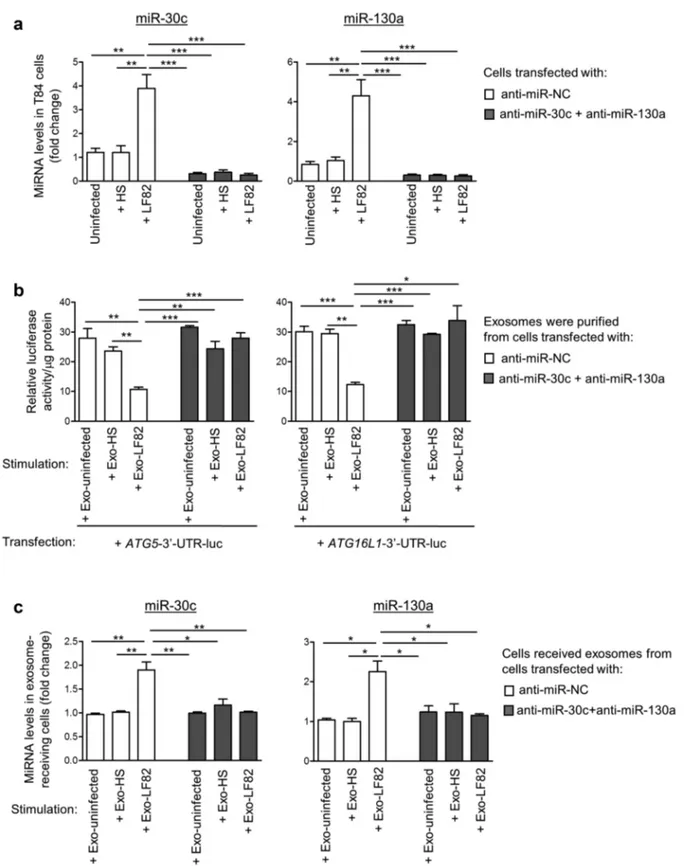
To further demonstrate that the inhibitory effect of Exo-LF82 on autophagy in exosome-receiving cells is dependent on miR-30c and miR-130a, we inhib-ited the levels of these miRNAs using their anti-senses, designated as anti-miR-30c and anti-miR -130a, in T84 donor cells uninfected or infected with LF82 or E. coli HS strain, and examined the effect of exosomes released by these cells on autop-hagy in recipient cells. As shown in Figure 5a, in T84 cells transfected with the anti-miR negative control (anti-miR-NC), LF82 infection led to an increase in miR-30c and miR-130a levels compared to E. coli HS infection or uninfected condition. However, in cells transfected with anti-miR-30c and anti-miR-130a, this increase was abolished (Figure 5a).
Furthermore, naïve T84 cells were transfected with ATG5-3ʹ-UTR-luc or ATG16L1-3ʹ-UTR-luc construct and then incubated with Exo- uninfected, Exo-HS or Exo-LF82 released from
cells transfected with anti-miR-NC or
a combination of anti-miR-30c and anti-miR -130a. In cells receiving the exosomes from anti- miR-NC-transfected cells, reduced luciferase activ-ity was observed upon incubation with Exo-LF82
compared to Exo-uninfected or Exo-HS (Figure
5b). This result, which was in agreement with the
data in Figure 4a, indicates that increased levels of
miR-30c and miR-130a in Exo-LF82 derived from anti-miR-NC-transfected cells were transferred to exosome-receiving cells, where they bound to the target 3ʹ-UTRs of ATG5 and ATG16L1 mRNAs. However, in cells receiving the exosomes secreted by cells transfected with anti-miR-30c and anti-miR -130a, no significant difference in luciferase activity upon incubation with Exo-uninfected, Exo-HS or
Exo-LF82 was observed (Figure 5b). This suggests
that the inhibition of miR-30c and miR-130a in exosome-donor cells abolishes the increase in the
levels of these miRNAs in cells receiving Exo-LF82
versus Exo-uninfected or Exo-HS. As expected,
miR-30c and miR-130a were detected at very low levels in Exo-uninfected, Exo-HS and Exo-LF82 derived from cells transfected with anti-miR-30c and anti-miR-130a with no significant difference (data not shown). Importantly, Exo-LF82 derived from cells transfected with anti-miR-NC can increase miR-30c and miR-130a levels in recipient cells compared to Exo-uninfected or Exo-HS, and this effect was not observed for Exo-LF82 derived from cells transfected with anti-miR-30c and anti-
miR-130a (Figure 5c).
Together, these data show that inhibition of miR-30c and miR-130a in AIEC LF82-infected donor cells abolishes the increase in miR-30c and miR-130a levels in the released exosomes and in exosome-receiving cells.
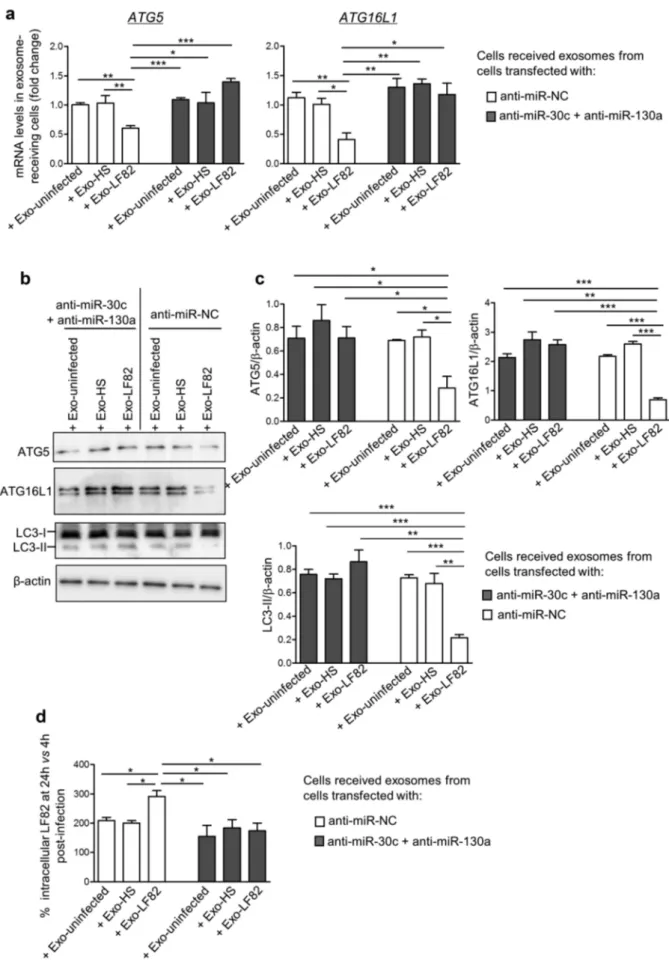
Inhibition of miR-30c and miR-130a in AIEC-infected cells abolishes the inhibitory effect of exosomes released from these cells on autophagy-mediated AIEC clearance in recipient cells
Exo-LF82 released from anti-miR-NC-transfected cells, compared to Exo-uninfected and Exo-HS, triggered the decreased expression of ATG5 and ATG16L1 at both mRNA and protein levels (Figure 6a–c) and inhibited autophagy response,
as shown by decreased LC3-II level (Figure 6b,c)
in recipient cells. This subsequently led to increased LF82 intracellular replication, presented as
percen-tage of LF82 colony-forming units
(CFU) determined at 24 h versus 4 h post-
infection, defined as 100% (Figure 6d). The CFU
of LF82 determined at 4 h post-infection in the cells that received Exo-LF82, Exo-uninfected and Exo- HS were not different (data not shown). Transfection of donor cells with anti-miR-30c and anti-miR-130a abolished the inhibitory effects of Exo-LF82 on ATG5 and ATG16L1 expression (Figure 6a–c) and on autophagy response
(Figure 6b,c) in recipient cells. This resulted in an
Exo-HS or Exo-LF82 (80 μg of exosome/mL of culture medium) for 8 h. Luciferase activity was measured and normalized to the protein concentration of cell lysate. (b-d) T84 cells were stimulated with exosomes as in afor 8 h, and ATG5 and ATG16L1 expression at mRNA and protein levels were analyzed by qRT-PCR (b) and western blot (c), respectively. (d) Quantification of band intensity in c. Data are representative of three independent experiments and are presented as means ± SEM. Statistical analysis was performed using the one- way ANOVA test followed by a Bonferroni posttest. *P < 0.05; **P ≤ 0.01; ***P ≤ 0.001.
Figure 5. Inhibition of miR-30c and miR-130a in AIEC-infected T84 cells abolishes the increase in miR-30c and miR-130a levels in exosomes and in exosome-receiving cells. (a) T84 cells were transfected with vehicle or with 50 nM of anti-miR-negative control (anti- miR-NC) or with a combination of anti-miR-30c and anti-miR-130a. One day post-transfection, the cells were uninfected or infected with the AIEC LF82 or the commensal E. coli HS strain for 12 h. The levels of miR-30c and miR-130a were analyzed by qRT-PCR. (b) Naïve T84 cells were transfected with the ATG5-3ʹ-UTR-luc or ATG16L1-3ʹ-UTR-luc construct, in which the 3ʹ-UTR of ATG5 or ATG16L1 mRNA was cloned downstream a luciferase-coding sequence in the pMIR-REPORT luciferase vector. Sixteen hours after transfection, the cells were stimulated with the exosomes purified from the culture supernatant of the cells in a(designated as Exo-uninfected, Exo-LF82 and Exo-HS) for 8 h (80 μg of exosome/mL of culture medium). Luciferase activity was measured at 1 day post-transfection and was normalized to the protein concentration of cell lysate. (c) Naïve T84 cells were stimulated with the purified exosomes as in b for 8 h, and miR-30c and miR-130a levels in the exosome-receiving cells were analyzed by qRT-PCR. Data are representative of three independent experiments and are presented as means ± SEM. Statistical analysis was performed using the one-way ANOVA test followed by a Bonferroni posttest. *P < 0.05; **P ≤ 0.01; ***P ≤ 0.001.
Figure 6. Inhibition of miR-30c and miR-130a in AIEC-infected T84 cells abolishes the inhibitory effect of exosomes on autophagy- mediated AIEC clearance in recipient cells. T84 cells were transfected with vehicle or with 50 nM of anti-miR-negative control (anti- miR-NC) or a combination of anti-miR-30c and anti-miR-130a. One day post-transfection, the cells were uninfected or infected with the AIEC LF82 or the commensal E. coli HS strain for 12 h, and exosomes were purified from the culture supernatant (designated as
efficient clearance of intracellular LF82 in cells receiving Exo-LF82 compared to Exo-uninfected or Exo-HS since similar levels of LF82 intracellular
replication in these cells were observed (Figure 6d).
Together, these data show that during AIEC infection, the inhibitory effect of exosomes on autophagy in recipient cells is mediated by miR- 30c and miR-130a, which are transferred from donor cells to recipient cells via exosomes.
Discussion
In recent years, significant progresses have been made in elucidating the mechanistic network underlying the role of exosomes in antibacterial defense, immunity and pathogenesis of several human diseases.20 In the current study, we showed that the exosomal shuttle can transfer specific miRNAs from cells to cells during CD-associated AIEC infection, which leads to impaired autop-hagy-mediated clearance of intracellular AIEC.
Exosomes have been emerged as an important intermediator participating in cellular crosstalk during pathogen infection by delivering their content com-posed of proteins, lipids, and RNAs. It has been shown that exosomes can directly transfer pathogen-related molecules from cells to cells, thereby impacting the infection progress.28 For example, cells intoxicated with the lethal toxin virulence factor, a toxin secreted by Bacillus anthracis, secrete exosomes transporting this toxin to naïve cells.29 Similarly, the cytotoxin- associated gene A (CagA), a major virulence factor secreted by Helicobacter pylori, was found in exosomes secreted by gastric epithelial cells inducibly expressing the cagA gene as well as in exosomes derived from the serum of patients carrying cagA-positive Helicobacter pylori.30 Furthermore, exosomes isolated from the serum of patients exhibiting a chronic gastritis and have H. pylori infection can induce expression of the soluble IL-6 receptor, which increases IL-1α expres-sion, in gastric epithelial cells.31 It has been reported
that Mycobacterium tuberculosis-infected macro-phages release extracellular vesicles including exo-somes that can deliver pathogen-associated molecular patterns (PAMPs), such as mycobacterial proteins, lipids, and nucleic acids, to naïve macro-phages, activating or inhibiting immune responses.23,32-35 Moreover, exosomes from Mycobacterium tuberculosis-infected macrophages or from the serum of Mycobacterium tuberculosis- infected mice can activate endothelial cells, suggesting a role for these exosomes in promoting leukocyte adhesion and cell migration as well as inflammation upon Mycobacterium tuberculosis infection.35 However, the exosomal components responsible for these effects remain to be identified. Regarding the exosomal miRNAs, increasing evidence has high-lighted the effect of pathogen infection on the miRNA composition of host cell-derived exosomes, which may have an impact on exosome-receiving cells and host immune responses, thus inhibiting the infec-tion or, on the contrary, promoting the immune escape of the pathogen.28 In the context of viral infec-tion, exosomes secreted by epithelial cells infected with Newcastle disease virus, a zoonotic virus affecting all species of birds, exhibit increased levels of miR- 1273f, miR-1184 and miR-198.24 These exosomal miRNAs inhibit interferon pathway in exosome- receiving cells, thus promoting viral infection.24 Similarly, infection of human HT-29 colon cancer cells with enterovirus 71 induces the secretion of exo-somes that can transfer miR-146a to recipient cells, in which this miRNA inhibits expression of IRAK1 (interleukin-1 receptor-associated kinase 1), TRAF6 (tumor necrosis factor receptor-associated factor 6) and STAT1 (signal transducer and activator of tran-scription 1), thus suppressing IFN response and facilitating enterovirus 71 infection.36 Conversely, exosomes derived from bronchoalveolar lavage fluid of mice infected with influenza virus dis-play an increased level of miR-483-3p, which exacer-bates pro-inflammatory and anti-viral responses in Exo-uninfected, Exo-LF82 and Exo-HS, respectively). Naïve T84 cells were stimulated with the purified exosomes for 8 h (80 μg exosome/mL of culture medium). (a) ATG5 and ATG16L1 mRNA expression levels in exosome-receiving cells were analyzed by qRT-PCR. Western blot analysis of ATG5, ATG16L1 and LC3 expression (b) and band intensity quantification (c). (d) Exosome-receiving cells were infected with the AIEC LF82 strain, and the number of intracellular LF82 bacteria was determined using gentamicin protection assay. The results are presented as percentage of CFU of LF82 determined at 24 h versus 4 h post-infection, defined as 100%. Data are representative of three independent experiments and are presented as means ± SEM. Statistical analysis was performed using the one- way ANOVA test followed by a Bonferroni posttest. *P < 0.05; **P ≤ 0.01; ***P ≤ 0.001.
recipient lung epithelial cells in vitro.37 In the context of bacterial infection, it was shown that upon lipopo-lysaccharide (LPS) exposure, miR-155 and miR-146a from murine bone marrow-derived dendritic cells (BMDC) are released into exosomes and are efficiently transferred to recipient BMDC.25 Upon being upta-ken, these miRNAs mediate target gene repression to modulate inflammatory gene expression and repro-gram cellular response to endotoxins.25 Similarly, exo-somes derived from the serum of a sepsis-related lung injury mouse model established by intraperitoneal injection of LPS are selectively enriched in miR- 155.38 In vitro, these exosomes stimulate NF-κB acti-vation, induce production of TNF-α and IL-6, and favor macrophage proliferation. In vivo, the intrave-nous injection of these exosomes to naïve mice increased the number of pro-inflammatory M1 macrophages in the lungs and induce lung inflammation.38 MiR-155 level was also found increased in exosomes derived from Helicobacter pylori-infected macrophages, and this exosomal miRNA regulates inflammatory responses in exo-some-receiving macrophages to limit H. pylori repli-cation and prevent the gastritis caused by H. pylori infection.39 Likewise, upon infection of macrophages with Mycobacterium bovis Bacillus Calmette-Guerin (BCG), the miRNA content of macrophage-derived exosomes is altered.40 In silico analysis suggested that the differentially expressed exosomal miRNAs may subvert host metabolic pathway to enable BCG survi-val within infected macrophages.40
Evidence has also suggested that extracellular vesicles, which include exosomes, seem to play a role in IBD. The extracellular vesicles purified from the intestinal lumen of IBD patients convey a higher amount of IL-6, IL-8, IL-10 and TNF-α compared to those from healthy subjects, and the levels of these pro-inflammatory mediators were positively correlated with CD severity score.41 The extracellular vesicles isolated from the lumen of IBD patients also activate IECs in vitro, leading to increased IL-8 secretion.41 Furthermore, these extracellular vesicles, as well as extracellular vesicle- treated IECs, induce the migration of a greater number of macrophages than untreated IECs.41 A higher amount of human proteins associated with oxidative antimicrobial activity was also shown in extracellular vesicles (including exo-somes, microvesicles and bacterial outer membrane
vesicles) collected from the interface between intestinal mucosa and intestinal lumen of IBD patients compared to those collected from control subjects, and was correlated with an alteration of microbial functions in IBD patients.42
In the context of CD-associated AIEC infection, we recently reported that exosomes are new mediators of host-AIEC interaction with their capacity to trigger a pro-inflammatory response and promote AIEC intracellular replication in exosome-receiving cells.21 However, the mechanisms behind the functional effects of exosomes on recipient cells remain unknown. Previous works of our group have shown that autophagy is a key mechanism of host defense to limit the intracellular replication of AIEC,9,11,19 and that the CD-associated risk variants in the autophagy- related genes ATG16L1 and IRGM lead to defective autophagy and impaired elimination of intracellular AIEC.17,19 Conversely, AIEC is able to counteract autophagy defense by increasing the level of miR-30c and miR-130a to suppress the expression of the autop-hagy-related proteins ATG5 and ATG16L1 in host cells, thus favoring AIEC colonization and promoting AIEC-induced inflammation.8 In the present study, we identified the mechanism by which exosomes derived from AIEC-infected cells modulate AIEC intracellular replication in recipient cells. We showed that exosomes secreted by AIEC LF82-infected human intestinal epithelial T84 cells exhibited an increased level of miR-30c and miR-130a compared with exo-somes secreted by uninfected cells or cells infected with nonpathogenic E. coli strains. These miRNAs were efficiently transferred via exosomes from donor to recipient T84 cells, in which they directly targeted the 3ʹ-UTRs of ATG5 and ATG16L1 mRNAs, thus inhibiting ATG5 and ATG16L1 expression at both mRNA and protein levels. This consequently resulted in impaired autophagy-mediated clearance of AIEC. Along with other studies, our results showed that bacterial infection-induced miRNA transfer within exosomes plays a role in cell-to-cell communication via modulating innate immune responses, thus favor-ing pathogen survival.
To further confirm that the observed effects are specifically dependent of miR-30c and miR-130a, we inhibited the levels of these miRNAs in exo-some-donor cells and the functional consequences of this inhibition were examined. We showed that inhibition of miR-30c and miR-130a in exosome-
donor cells before AIEC LF82 infection abolished the increase in their levels in the secreted exosomes and in exosome-receiving cells. Consequently, the inhibitory effect of the exosomes secreted from LF82-infected cells on autophagy in exosome- receiving cells was not observed; thus, autophagy was sustained and LF82 intracellular replication was well controlled. Collectively, these results indi-cated that the exosomal miR-30 c and miR-130a, increased in response to AIEC infection, are suffi-cient to impair autophagy in exosome-receiving cells, thus leading to increased AIEC intracellular replication. Moreover, these results highlighted for the first time a role for exosomal miRNAs in mod-ulating autophagy in exosome-receiving cells, and reinforce the importance of autophagy, miRNAs and exosomes in CD pathogenesis.
In conclusion, together with our previous study showing that AIEC can subvert host autophagy response by upregulating the levels of miR-30c and miR-130a to replicate inside host cells, we propose that AIEC dysregulate host miRNAs to inhibit autophagy, and this can be amplified from cell to cell via the exosomal shuttle, thus favoring
AIEC colonization (Figure 7). This importantly
suggests that the loss of tightly regulated autop-hagy, caused by miRNA dysregulation, in handling AIEC, which might contribute to the pathogenesis of CD, could happen at local and distant sites in
a host. This should be kept in mind while develop-ing future personalized strategies to treat CD patients with abnormal AIEC colonization.
Materials and methods Bacterial strains
The AIEC LF82 reference strain isolated from a chronic ileal lesion of a CD patient,7 the nonpatho-genic E. coli K-12 MG1655 strain and the commensal E. coli O9:H4 strain (E. coli HS)43 were used. Bacteria were grown in Luria-Bertani (LB) broth (Pronadisa) overnight at 37°C without shaking.
Cell culture
Human intestinal epithelial T84 cells (ATCC CCL-248TM) were maintained in an atmosphere containing 5% CO2 at 37°C in a Dulbecco’s
Modified Eagle Medium/Nutrient F-12 HAM culture medium (Gibco) supplemented with 10% fetal bovine serum (FBS, Dutscher), 1% L-glutamine (Gibco), 1% minimal essential medium vitamins 100X free from L-Glutamine (Dutscher), and 1% antibiotic and antimycotic solution (10,000 U of penicillin, 10 mg of streptomycin, and 0.025 mg of amphotericin B per mL; GE Healthcare HyClone).
Figure 7. Model for the role of exosomes in transferring miR-30c and miR-130a from cell-to-cell to inhibit autophagy during infection with CD-associated AIEC. AIEC upregulates miR-30c and miR-130a in host cells to inhibit autophagy and this can be amplified from cells to cells via the exosomal shuttle, which transfers these miRNAs to adjacent cells. This consequently leads to impaired autophagy- mediated AIEC clearance, favoring AIEC colonization.
Bacterial infection of human T84 cells and determination of intracellular bacterial number using gentamicin protection assay
For exosome extraction, T84 cells were seeded on 150 cm2 cell culture flask (Falcon) and infected with a multiplicity of infection (MOI) of 10. T84 cells were maintained in contact with bacteria for 3 h in culture medium without antibiotics (infection medium). Then, cells were washed with phosphate- buffered saline (PBS; Gibco) and incubated for 9 h with culture medium containing 10% exosome- depleted FBS (System Biosciences, Mountain View, CA) instead of regular FBS to avoid coex-traction of bovine exosomes and 100 µg/mL of gentamicin (Euromedex) to remove extracellular bacteria.
For infection of exosome-receiving cells, T84 cells were seeded on 12-well or 24-well plates (Falcon), stimulated with exosomes (80 μg of exosomes per 1 ml of culture medium) for 8 h and infected with a MOI of 10. Cells were maintained in contact with bacteria for 3 h in infection medium and were washed with PBS. Cells were then incubated with infection med-ium containing 100 µg/mL of gentamicin for the indicated time.
Invasion assay was performed as described previously.8 Briefly, cells were lysed using 1% Triton X-100 (Euromedex) in deionized water. Serial dilutions of samples were made and pla-ted onto LB agar plate, and the number of colony-forming units (CFU) was counted to determine the number of intracellular bacteria.
Isolation of exosomes from cell culture supernatant Twelve hours after infection, the supernatant of infected cells was collected, and exosomes were extracted using the ExoQuickTM Exosome Precipitation Solution (System Biosciences) follow-ing the manufacturer’s protocol. Briefly, superna-tant was centrifuged at 3,000 g for 15 min to eliminate cell debris. Then, supernatant was passed through a Mustang E membrane 0.2-µm pore size filters (Pall Corporation) to remove bacterial LPS and microvesicles larger than exosomes. ExoQuickTM was added to the supernatant with a volume ratio of 1:5, the suspension was mixed
by inverting the tubes and incubated at 4°C over-night. Finally, the suspension was centrifuged at 1,500 g for 30 min to pellet the exosomes.
Protein extraction and western blot analysis
T84 cells or purified exosomes were lysed in radio- immunoprecipitation assay (RIPA) lysis buffer con-taining 150 mM NaCl, 0.5% sodium deoxycholate, 50 mM Tris-HCl pH 8, 0.1% sodium dodecyl sul-fate, 0.1% Nonidet P-40, and supplemented with protease inhibitors (Roche). Proteins were quanti-fied using Bio-Rad DCTM protein assay kit accord-ing to the manufacturer’s instructions (Bio-Rad).
Cell and exosome lysates were separated on sodium dodecyl sulfate polyacrylamide gels, trans-ferred to nitrocellulose membranes, blocked in 5% bovine serum albumin (BSA) in PBS containing 0.1% Tween-20 and then probed with appropriate primary antibodies: anti-CD63 (EXOAB-CD63A-1, System Biosciences), anti-CD9 (EXOAB-CD9A-1, System Biosciences), anti-GRP94 (sc-11402, Santa Cruz), anti-ATG16L1 (#8089, Cell Signaling Technology), anti-ATG5 (#12994, Cell Signaling Technology), anti-LC3 (L7543, Sigma), anti-p62 /SQSTM1 (sc-28359, Santa Cruz) and anti-β-actin (#4970, Cell Signaling Technology). After washes, membranes were incubated with the appropriate horseradish peroxidase (HRP)-conjugated second-ary antibodies (Cell Signaling Technology and System Biosciences), and blots were revealed using the Clarity Western enhanced chemiluminescence detection kit (Bio-Rad) and the ChemiDocTM XRS System (BioRad).
In vitro transfection and luciferase assay
Anti-miR negative control #1 and the antisenses of miR-30c and miR-130a were purchased from Ambion. T84 cells were transfected with 50 nM of antisenses of miRNAs using Lipofectamine 2000 (Invitrogen) and Opti-MEM I reduced serum med-ium (Invitrogen) 24 h before being infected with AIEC LF82, E. coli K12 MG1655 or E. coli HS strain for 12 h. Exosomes were purified from the culture supernatant of these cells and designated as Exo- uninfected, Exo-LF82, Exo-K12 and Exo-HS, respectively.
The 3′-untranslated regions (UTRs) of human
ATG5 and ATG16L1 mRNAs were cloned into the
pMIR-REPORTTM Luciferase vector (Ambion) to
generate ATG5-3ʹ-UTR-luciferase or ATG16L1-3ʹ- UTR-luciferase construct, respectively, as previously described.8 For luciferase assay, T84 cells were seeded on 48-well plate. Twenty-four hours later, cells were transfected with 750 ng of ATG5-3ʹ-UTR- luciferase or ATG16L1-3ʹ-UTR-luciferase construct or of the pMIR-REPORTTM Luciferase vector, and 16 h after transfection, the cells were stimulated with 80 μg of exosomes per 1 ml of culture medium for 8 h. Firefly luciferase activity was measured using the dual-luciferase reporter assay system (Promega) and a Luminoskan Ascent luminometer (Thermo Electron Corp.). Relative luciferase activity was nor-malized to μg of protein, determined from cell lysate using Bio-Rad DCTM protein assay kit according to the manufacturer’s instructions (BioRad).
RNA extraction, cDNA synthesis and quantitative RT-PCR (qRT-PCR) analysis
Total RNAs were extracted from purified exosomes or cultured cells using the miRNeasy mini kit (Qiagen) according to the manufacturer’s instruc-tions. For exosomal samples, 25 fmol of each spike- in controls Caenorhabditis elegans cel-mir-39-3p, cel-mir-54-3p and cel-mir-238-3p (mirVana™ miRNA mimic, Life Technologies) were added into the lysis solution per extraction.
Total RNAs were reversely transcribed using the miRNA 1st strand cDNA synthesis kit (Agilent) to quantify mature miRNA levels or the PrimeScript RT reagent kit (Takara) to quantify mRNA expression levels. qRT-PCR was performed using SsoAdvanced™ Universal SYBR® Green Supermix (Bio-Rad) on a CFX96 Touch™ Real-Time PCR detection system (Bio-Rad) using specific primers listed in Table S1. For the quantification of mature miRNA levels, the universal reverse primer provided in the miRNA 1st strand cDNA synthesis kit was used with the specific forward primers indicated in Table S1. Spike-in con-trols cel-mir-39-3p, cel-mir-54-3p and cel-mir-238-3p or U6 were used as internal controls for quantification of miRNAs in exosomes or in cells, respectively. Human 18S was used as an internal control for mRNA quantification in cells. Fold-induction was
calculated using the comparative threshold cycle num-ber (Ct) method as follows: ∆∆Ct = (Cttarget mRNA/ miRNA – Ctinternal control)condition of interest – (Cttarget mRNA/miRNA – Ctinternal control)control condition, and the
final data were derived from 2−∆∆CT. When spike-in controls cel-mir-39-3p, cel-mir-54-3p and cel-mir- 238-3p were added into samples and used as internal controls, the average of Ct of the three miRNAs was used as Ctinternal control.
Fluorescent microscopy
T84 cells were transfected with AlexaFluor555-labeled -miR-30c or AlexaFluor555-labeled-miR-130a (Ambion) for 24 h, and uninfected or infected with the AIEC LF82 strain or the E. coli K12 or E. coli HS strain with a MOI of 10 for 12 h. The supernatants of these cells were collected and exosomes were purified. T84 cells seeded on coverslips were stimulated with the purified exosomes (80 μg exosome/mL of culture medium) for 8 h. Cells were then fixed with 4% formaldehyde, permeabilized with 0.1% Triton X-100 for 10 min, saturated with PBS containing 0.025% Triton X-100, 3% BSA and 5% FBS for 1 h. Actin cytoskeleton was stained with FITC-phalloidin (A12379, Life technologies), and nuclei were stained with 2-(4-Amidinophenyl)-6-indolecarbamidine dihydrochloride (DAPI; Sigma) for 45 min at room temperature. Coverslips were mounted in EUKITT® medium (O. Kindler). Images were taken using Zeiss LSM 800 with Airyscan confocal microscope.
Statistical analysis
Values are expressed as means ± standard error of the mean (SEM). Statistical analyses between sev-eral groups were performed using ANOVA fol-lowed by a Bonferroni posttest (Kruskal–Wallis if not parametric) with GraphPad Prism version 7 software (GraphPad Software, La Jolla, CA). A P value less than 0.05 was considered statistically significant. *P < 0.05; **P ≤ 0.01; ***P ≤ 0.001. Acknowledgments
We thank the platform CLIC (CLermont-ferrand Imagerie Confocale, Université Clermont Auvergne) for assistance with the confocal microscopy.
Disclosure of Potential Conflicts of Interest No potential conflicts of interest were disclosed.
Funding
This work was supported by the Ministère de la Recherche et de la Technologie, Inserm (UMR1071) and INRAE (USC 2018), the Agence Nationale de la Recherche of the French government through the program “Investissements d’Avenir” (16-IDEX-0001 CAP 20-25) (to Hang Nguyen) and the European Union FP7 People Marie Curie International Incoming Fellowship (to Hang Nguyen).
References
1. Palmela C, Chevarin C, Xu Z, Torres J, Sevrin G, Hirten R, Barnich N, Ng SC, Colombel J-F. Adherent- invasive Escherichia coli in inflammatory bowel disease. Gut. 2018;67:574–587.
2. Carrière J, Darfeuille-Michaud A, Nguyen HTT. Infectious etiopathogenesis of Crohn’s disease. World J Gastroenterol. 2014;20:12102–12117. doi:10.3748/wjg. v20.i34.12102.
3. O’Brien CL, Bringer M-A, Holt KE, Gordon DM, Dubois AL, Barnich N, Darfeuille-Michaud A, Pavli P. Comparative genomics of Crohn’s disease- associated adherent-invasive Escherichia coli. Gut.
2017;66:1382–1389.
4. Martinez-Medina M, Aldeguer X, Lopez-Siles M, González-Huix F, López-Oliu C, Dahbi G, Blanco JE, Blanco J, Garcia-Gil JL, Darfeuille-Michaud A. Molecular diversity of Escherichia coli in the human gut: new ecological evidence supporting the role of adherent-invasive E.coli (AIEC) in Crohn’s disease. Inflamm Bowel Dis. 2009;15:872–882. doi:10.1002/ ibd.20860.
5. Darfeuille-Michaud A, Boudeau J, Bulois P, Neut C, Glasser A-L, Barnich N, Bringer M-A, Swidsinski A, Beaugerie L, Colombel J-F. High prevalence of adher-ent-invasive Escherichia coli associated with ileal mucosa in Crohn’s disease. Gastroenterology.
2004;127:412–421. doi:10.1053/j.gastro.2004.04.061. 6. Martin HM, Campbell BJ, Hart CA, Mpofu C, Nayar M,
Singh R, Englyst H, Williams HF, Rhodes JM. Enhanced Escherichia coli adherence and invasion in Crohn’s dis-ease and colon cancer. Gastroenterology.
2004;127:80–93. doi:10.1053/j.gastro.2004.03.054. 7. Darfeuille-Michaud A, Neut C, Barnich N, Lederman E,
Di Martino P, Desreumaux P, Gambiez L, Joly B, Cortot A, Colombel J-F. Presence of adherent Escherichia coli strains in ileal mucosa of patients with Crohn’s disease. Gastroenterology. 1998;115:1405–1413. 8. Nguyen HTT, Dalmasso G, Müller S, Carrière J,
Seibold F, Darfeuille-Michaud A. Crohn’s disease- associated adherent-invasive Escherichia coli modulate
levels of microRNAs in intestinal epithelial cells to reduce autophagy. Gastroenterology. 2014;146:508–519. doi:10.1053/j.gastro.2013.10.021.
9. Bretin A, Carrière J, Dalmasso G, Bergougnoux A, B’chir W, Maurin AC, Müller S, Seibold F, Barnich N, Bruhat A, et al. Activation of the EIF2AK4-EIF2A /eIF2α-ATF4 pathway triggers autophagy response to Crohn disease-associated adherent-invasive Escherichia coli infection. Autophagy. 2016;12:770–783. doi:10.1080/15548627.2016.1156823.
10. Glasser AL, Boudeau J, Barnich N, Perruchot MH, Colombel JF, Darfeuille-Michaud A. Adherent invasive Escherichia coli strains from patients with Crohn’s disease survive and replicate within macrophages without indu-cing host cell death. Infect Immun. 2001;69:5529–5537. doi:10.1128/IAI.69.9.5529-5537.2001.
11. Lapaquette P, Bringer M-A, Darfeuille-Michaud A. Defects in autophagy favour adherent-invasive Escherichia coli persistence within macrophages leading to increased pro-inflammatory response. Cell Microbiol. 2012;14:791–807. doi:10.1111/j.1462- 5822.2012.01768.x.
12. Vazeille E, Buisson A, Bringer MA, Goutte M, Ouchchane L, Hugot JP, de Vallée A, Barnich N, Bommelaer G, Darfeuille-Michaud A. Monocyte- derived macrophages from Crohn’s disease patients are impaired in the ability to control intracellular adher-ent-invasive Escherichia coli and exhibit disordered cytokine secretion profile. J Crohns Colitis.
2015;9:410–420. doi:10.1093/ecco-jcc/jjv053.
13. Bretin A, Lucas C, Larabi A, Dalmasso G, Billard E, Barnich N, Bonnet R, Nguyen HTT. AIEC infection triggers modification of gut microbiota composition in genetically predisposed mice, contributing to intestinal inflammation. Sci Rep. 2018;8:12301. doi:10.1038/ s41598-018-30055-y.
14. Carvalho FA, Barnich N, Sivignon A, Darcha C, Chan CHF, Stanners CP, Darfeuille-Michaud A. Crohn’s disease adherent-invasive Escherichia coli colo-nize and induce strong gut inflammation in transgenic mice expressing human CEACAM. J Exp Med.
2009;206:2179–2189. doi:10.1084/jem.20090741. 15. Larabi A, Barnich N, Nguyen HTT. New insights into
the interplay between autophagy, gut microbiota and inflammatory responses in IBD. Autophagy. 2019:1–14. 16. Nguyen HTT, Lapaquette P, Bringer M-A D-MA.
Autophagy and Crohn’s disease. J Innate Immun.
2013;5:434–443. doi:10.1159/000345129.
17. Brest P, Lapaquette P, Souidi M, Lebrigand K, Cesaro A, Vouret-Craviari V, Mari B, Barbry P, Mosnier J-F, Hébuterne X, et al. A synonymous variant in IRGM alters a binding site for miR-196 and causes deregula-tion of IRGM-dependent xenophagy in Crohn’s disease. Nat Genet. 2011;43:242–245. doi:10.1038/ng.762. 18. Dalmasso G, Nguyen HTT, Faïs T, Massier S, Barnich N,
Delmas J, Bonnet R. Crohn’s disease-associated adherent- invasive Escherichia coli manipulate host autophagy by
impairing SUMOylation. Cells. 2019;8:35. doi:10.3390/ cells8010035.
19. Lapaquette P, Glasser A-L, Huett A, Xavier RJ, Darfeuille-Michaud A. Crohn’s disease-associated adherent-invasive E.coli are selectively favoured by impaired autophagy to replicate intracellularly. Cell Microbiol. 2010;12:99–113. doi:10.1111/j.1462- 5822.2009.01381.x.
20. Carrière J, Barnich N, Nguyen HTT. Exosomes: From Functions in Host-Pathogen Interactions and Immunity to Diagnostic and Therapeutic Opportunities. In: Nilius B., de Tombe P., Gudermann T., Jahn R., Lill R., Petersen O. (eds) Reviews of Physiology, Biochemistry and Pharmacology, Vol. 172. Cham: Springer; 2016. ISBN 978-3-319-49901-7. Online ISBN 978-3-319- 49902-4. https://doi-org.proxy.insermbiblio.inist.fr/ 10.1007/112_2016_7
21. Carrière J, Bretin A, Darfeuille-Michaud A, Barnich N, Nguyen HTT. Exosomes released from cells infected with Crohn’s disease–associated adherent-invasive Escherichia coli activate host innate immune responses and enhance bacterial intracellular replication. Inflamm Bowel Dis. 2016;22:516–528.
22. Valadi H, Ekström K, Bossios A, Sjöstrand M, Lee JJ, Lötvall JO. Exosome-mediated transfer of mRNAs and microRNAs is a novel mechanism of genetic exchange between cells. Nat Cell Biol. 2007;9:654–659. doi:10.1038/ncb1596.
23. Singh PP, Li L, Schorey JS. Exosomal RNA from Mycobacterium tuberculosis-infected cells is functional in recipient macrophages. Traffic. 2015;16:555–571. doi:10.1111/tra.12278.
24. Zhou C, Tan L, Sun Y, Qiu X, Liao Y, Song C, Liu W, Nair V, Ding C. Exosomes carry microRNAs into neighboring cells to promote diffusive infection of new-castle disease virus. Viruses. 2019;11:527. doi:10.3390/ v11060527.
25. Alexander M, Hu R, Runtsch MC, Kagele DA, Mosbruger TL, Tolmachova T, Seabra MC, Round JL, Ward DM, O’Connell RM. Exosome-delivered microRNAs modulate the inflammatory response to endotoxin. Nat Commun. 2015;6. doi:10.1038/ ncomms8321.
26. Montecalvo A, Larregina AT, Shufesky WJ, Stolz DB, Sullivan MLG, Karlsson JM, Baty CJ, Gibson GA, Erdos G, Wang Z, et al. Mechanism of transfer of func-tional microRNAs between mouse dendritic cells via exosomes. Blood. 2012;119:756–766. doi:10.1182/ blood-2011-02-338004.
27. Klionsky DJ, Abdelmohsen K, Abe A, Abedin MJ, Abeliovich H, Arozena AA, Adachi H, Adams CM, Adams PD, Adeli K, et al. Guidelines for the use and interpretation of assays for monitoring autophagy (3rd edition). Autophagy. 2016;12:1–222.
28. Zhang W, Jiang X, Bao J, Wang Y, Liu H, Tang L. Exosomes in pathogen infections: a bridge to deliver molecules and link functions. Front Immunol.
2018;9:1–12. doi:10.3389/fimmu.2018.00001.
29. Abrami L, Brandi L, Moayeri M, Brown MJ, Krantz BA, Leppla SH. van der Goot FG. Hijacking multivesicular bodies enables long-term and exosome-mediated long-distance action of anthrax toxin. Cell Rep.
2013;5:986–996. doi:10.1016/j.celrep.2013.10.019. 30. Shimoda A, Ueda K, Nishiumi S, Murata-Kamiya N,
Mukai S, Sawada S, Azuma T, Hatakeyama M, Akiyoshi K. Exosomes as nanocarriers for systemic delivery of the Helicobacter pylori virulence factor CagA. Sci Rep. 2016;6:18346. doi:10.1038/srep18346. 31. Chen Y, Wang X, Yu Y, Xiao Y, Huang J, Yao Z,
Chen X, Zhou T, Li P, Xu C. Serum exosomes of chronic gastritis patients infected with Helicobacter pylori med-iate IL-1α expression via IL-6 trans-signalling in gastric epithelial cells. Clin Exp Immunol. 2018;194:339–349. doi:10.1111/cei.13200.
32. Cheng Y, Schorey JS. Extracellular vesicles deliver Mycobacterium RNA to promote host immunity and bacterial killing. EMBO Rep. 2019;20:e46613. doi:10.15252/embr.201846613.
33. Walters SB, Kieckbusch J, Nagalingam G, Swain A, Latham SL, Grau GER, Britton WJ, Combes V, Saunders BM. Microparticles from mycobacteria-infected macrophages promote inflammation and cellular migration. J Immunol. 2013;190:669–677. doi:10.4049/ jimmunol.1201856.
34. Hare NJ, Chan B, Chan E, Kaufman KL, Britton WJ, Saunders BM. Microparticles released from Mycobacterium tuberculosis-infected human macro-phages contain increased levels of the type I interferon inducible proteins including ISG15. Proteomics.
2015;15:3020–3029. doi:10.1002/pmic.201400610. 35. Li L, Cheng Y, Emrich S, Schorey J. Activation of
endothe-lial cells by extracellular vesicles derived from Mycobacterium tuberculosis infected macrophages or mice. PLoS One. 2018;13:e0198337. doi:10.1371/journal. pone.0198337.
36. Fu Y, Zhang L, Zhang F, Tang T, Zhou Q, Feng C, Jin Y, Wu Z. Exosome-mediated miR-146a transfer sup-presses type I interferon response and facilitates EV71 infection. PLoS Pathog. 2017;13:e1006611. doi:10.1371/ journal.ppat.1006611.
37. Maemura T, Fukuyama S, Sugita Y, Lopes TJS, Nakao T, Noda T, Kawaoka Y. Lung-derived exosomal miR-483- 3p regulates the innate immune response to Influenza virus infection. J Infect Dis. 2018;217:1372–1382. doi:10.1093/infdis/jiy035.
38. Jiang K, Yang J, Guo S, Zhao G, Wu H, Deng G. Peripheral circulating exosome-mediated delivery of miR-155 as a novel mechanism for acute lung
inflammation. Mol Ther. 2019;27:1–14. doi:10.1016/j. ymthe.2019.07.003.
39. Wang J, Deng Z, Wang Z, Wu J, Gu T, Jiang Y, Li G. MicroRNA-155 in exosomes secreted from Helicobacter pylori infection macrophages immunomodulates inflam-matory response. Am J Transl Res. 2016;8:3700–3709. 40. Alipoor SD, Mortaz E, Tabarsi P, Farnia P, Mirsaeidi M,
Garssen J, Movassaghi M, Adcock IM. Bovis bacillus calmette–guerin (BCG) infection induces exosomal miRNA release by human macrophages. J Transl Med.
2017;15:105. doi:10.1186/s12967-017-1205-9.
41. Mitsuhashi S, Feldbrügge L, Csizmadia E, Mitsuhashi M, Robson SC, Moss AC. Luminal extracel-lular vesicles (EVs) in inflammatory bowel disease (IBD) exhibit proinflammatory effects on epithelial
cells and macrophages. Inflamm Bowel Dis.
2016;22:1587–1595. doi:10.1097/MIB.000000000000 0840.
42. Zhang X, Deeke SA, Ning Z, Starr AE, Butcher J, Li J, Mayne J, Cheng K, Liao B, Li L, et al. Metaproteomics reveals associations between microbiome and intestinal extracellular vesicle proteins in pediatric inflammatory bowel disease. Nat Commun. 2018;9:2873. doi:10.1038/ s41467-018-05357-4.
43. Rasko DA, Rosovitz MJ, Myers GSA, MongodinEF, Fricke WF, Gajer P, Crabtree J, Sebaihia M, Thomson NR, Chaudhuri R, et al. The pangenome structure of Escherichia coli: comparative genomic analysis of e. coli commensal and pathogenic isolates. J Bacteriol. 2008;190:6881–6893. doi:10.1128/JB.00619-08.