Protection Against Chronic Pyelonephritis in Rats by Suppression of
Acute Suppuration: Effect of Colchicine and Neutropenia
Jacques Bille and Michel P. Glauser From the Division des Maladies Infectieuses, Department de Medicine, Centre Hospitalier Universitaire Vaudois, Lausanne, Switzerland Previous experiments in rats have suggested that renal scarring after acute, obstructive
pyelonephritis due toEscherichiacoliresults from parenchymal damage due to acute in-flammation and suppuration. To assess the role of acute infiltration by polymorphonu-clear leukocytes (PMNLs) in the pathogenesis of chronic pyelonephritis (CPN), rats were either treated with colchicine to depress leukcoyte motility or rendered neutropen-ic with a single dose of cyclophosphamide. Colchneutropen-icine given during acute pyelonephritis reduced kidney inflammation and protected against CPN two months later. Similarly, neutropenia reduced acute inflammation and protected against chronic parenchymal destruction and scarring. Protection against renal scarring in both colchicine-treated and neutropenic rats occurred despite higher renal bacterial counts during acute pyelonephritis. These experiments provide further evidence that CPN (renal scarring) results from kidney damage that occurs during early acute obstructive pyelonephritis. This damage appears to result from infiltration of the kidney by PMNLs rather than direct damage from bacterial infection.
The mechanisms that lead to the development of chronic pyelonephritis (CPN) after acute renal in-fection are not well defined. Persistent inin-fection does not seem to playa critical role in the develop-ment of renal scarring, because kidneys from pa-tients with CPN are often sterile at autopsy [1, 2]. In experimental pyelonephritis, viable bacteria are not required for progression of the lesions [3-5].
Furthermore, bacterial antigens persisting in the late phase of human and experimental pyelone-phritis [2, 6, 7] have been shown not to trigger significant kidney destruction [6, 7]. Two other immune mechanisms have been proposed. Either alterations in antigenicity of kidney tissue [8] or common antigens which are shared by Escherichia coli and renal tissue [9] have been postulated to
Received for publication July 20, 1981, and in revised form March 3, 1982.
Portions of this work appeared as an abstract (no. 532) in the Proceedings of the 21st Interscience Conference on Antimicro-bial Agents and Chemotherapy and in Clinical Research
28:364A, 1980.
This work was supported in part by grant no. 3.814-0.79 from the Swiss Foundation for Scientific Research.
We thank Dr. J. A. McCutchan for assistance in preparation of the manuscript and S. Bovey for secretarial assistance.
Please address requests for reprints to Dr. Michel P. Glauser, Division des Maladies Infectieuses, Department de Medicine, Centre Hospitalier Universitaire Vaudois, 1011 Lausanne, Switzerland.
220
stimulate a chronic, destructive immune response. However, the demonstration of antibodies to kidney tissue in CPN has been unsuccessful so far, both in humans [10] and in experimental animals [11]. Thymus-dependent autoimmune phenomena have been shown not to play a significant role in the development of CPN in rats [5, 12]. In fact, athymic, nude mice challenge iv with Streptococ-cus faecalis seem to develop more intense kidney infection and scarring than their normal litter-mates [13].
To investigate the mechanisms that lead to CPN, we have used a model of ascending obstruc-tive pyelonephritis due to E. coli that resembles se-vere human kidney infection because the disease is acquired by the retrograde route in the presence of an obstruction [14]. Previous experiments in which rats were treated with antibiotics starting at different times during acute pyelonephritis have suggested that the acute inflammatory process plays a major role in the development of kidney scars leading to CPN and that infection seems less important [5]. In those studies, however, the di-minished inflammation by early treatment might have been due to bacterial inhibition by antibio-tics. During inflammatory processes, polymor-phonuclear leukocytes (PMNLs) can damage tissue by exocytosis of lysosomal enzymes. Therefore, we gave rats with acute pyelonephritis
either colchicine, thus impairing PMNL move-ment, or cyclophosphamide, thus inducing severe neutropenia.
Materials and Methods
Induction ofpyelonephritis. Retrograde pyelo-nephritis was induced in male Wistar rats weighing 200-250 g (Madorin, Fiillingsdorf, Switzer-land) [14]. In brief, 103E. colitype 06 organisms
(Williams strain) [14] were slowly infused into the bladder and a ligature was loosely tied around the left ureter; 18 hr later the ligature was removed. This procedure produces partial obstruction followed by severe unilateral pyelonephritis. Three days after this operation, the pyelonephritic kidneys are greatly enlarged and display numerous abcesses over the cortex [14]. Two months after the operation, the kidneys have extended scars with severe destruction of kidney parenchyma [14].
Experimental design and killing ofrats. In this model of pyelonephritis, inflammation, as mea-sured by the ratio of the weight of the left pyelone-phritic kidney to that of the right normal kidney, reaches a peak three days after the operation and bacterial inoculation [5]. Colchicine-treated, neutropenic, and control rats were killed at this time in order to evaluate the effect of colchicine and neutropenia on acute pyelonephritis. The kidneys were examined for gross evidence of acute pyelonephritis after three days or for scars of CPN after two months. The distribution of abscesses or scars on the surface of the kidney was graded by dividing the kidney into eight equal parts and counting the number of parts where there was at least one abscess or scar. A distribu-tion index was calculated from the sum of the number of parts having abscesses or scars [14]. The kidneys were weighed and cut in half; one half was homogenized in 0.85070 NaCI and serially di-luted before plating onto MacConkey's agar (Dif-co Laboratories, Detroit) for (Dif-colony (Dif-counts ex-pressed in log cfu. The other half of each kidney was processed for histologic examination.
Severity ofpyelonephritis. Kidney weight pro-vides the best quantitative measure of the severity of pyelonephritis - it increases in proportion to suppuration during acute obstructive pyelone-phritis, whereas it decreases in proportion to the
destruction of kidney tissue during CPN [15]. To minimize the effects of variation of kidney weights among animals, the ratio of the left kidney weight to the right kidney weight (the L: R weight ratio) was used [5]. This is especially important when dealing with long-term experiments, wherein the total body weight may vary greatly among animals. The L: R weight ratio also expresses the compensatory hypertrophy of the right kidney during CPN, which is proportional to the destruc-tion and shrinkage of the left kidney [15].
Administration of colchicine. Colchicine pow-der (Fluka, Buochs, Switzerland) was diluted in sterile water to a concentration of 1 mg/ml. Colchicine was administered ip daily to the rats in a dosage of 0.4 mg/kg of body weight per day for three consecutive days. The first injection was given one day before the operation. Control rats were given one injection of 0.1 ml of 0.85% NaCI ip daily for three consecutive days.
Administration of cyclophosphamide and in-duction of neutropenia. Cyclophosphamide pow-der (Endoxan; Asta Werke, Bielefeld, Fepow-deral Re-public of Germany) was diluted in sterile water to a concentration of 25 mg/ml. Cyclophosphamide was given ip to the rats four days before the opera-tion in a dose of 100 mg/kg of body weight. Con-trol rats were given 1.0 ml of 0.85% NaCI ip. Leu-kocyte counts of the tail blood were made with a hemacytometer.
Statistical evaluation. The number of cfu/g of kidney, the L: R weight ratio of the kidneys, and the surface area of the kidney occupied by ab-scesses were compared by Student's unpaired r-test.
Results
Effect of colchicine treatment on pyelonephri-tis. Colchicine treatment was associated with early mortality in the rats receiving the drug. Dur-ing the first three days after the operation, seven (14%) of 49 colchicine-treated rats died, four in the acute and three in the chronic pyelonephritis experiments. No deaths occurred in the control rats. The severity of the disease in the dead ani-mals as measured by the L : R weight ratio, the dis-tribution of macroscopic lesions, and the bacterial counts were not different from those observed in
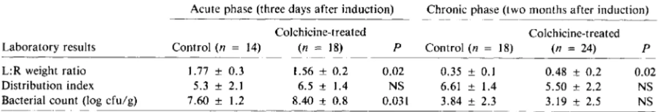
Table 1. Effect of treatment with colchicine on the severity of experimentally induced pyelonephritis in rats.
Laboratory results L:R weight ratio Distribution index Bacterial count (log cfu/g)
Acute phase (three days after induction) Chronic phase (two months after induction)
Colchicine-treated Colchicine-treated
Control(n = 14) (n = 18) P Control(n = 18) (n = 24) P
1.77 ± 0.3 1.56 ± 0.2 0.02 0.35 ± 0.1 0.48 ± 0.2 0.02
5.3 ± 2.1 6.5 ± 1.4 NS 6.61 ± 1.4 5.50 ± 2.2 NS
7.60 ± 1.2 8.40 ± 0.8 0.031 3.84 ± 2.3 3.19 ± 2.5 NS
NOTE. Colchicine (0.4 mg/kg of body weight per day) was administered ip to rats for three days beginning one day before pye-lonephritis was induced in the left kidney by operation [14]. Control rats were given one ip injection of 0.1 ml of0.85010NaCI per day for three days. Three days after the operation, the ratio of the weight of the left kidney to the weight of the right kidney (L:R weight ratio) was calculated to measure the intensity of the inflammation of the left kidney. Two months later, the L:R weight ratio was calculated to express the destruction of tissue in the left kidney. The surface of the left kidney was divided into eight equal sec-tions and the distribution index was defined as the number of secsec-tions with one or more abscesses or scars. Infection was expressed as the log of the number of cfu ofEscherichia colitype 06/g of left kidney homogenate. Data are means ± SD. NS= not significant.
colchicine-treated rats that were killed at the same time.
Table 1 compares pyelonephritis in colchicine-treated rats and control rats. When expressed by the L: R weight ratio, acute inflammation was slightly but significantly reduced in colchicine-treated rats. The distribution of lesions on the sur-face of the left kidney in the colchicine-treated rats was not different from that in the control rats, but the gross abscesses were smaller. In contrast to the reduction of L: R weight ratio, the bacterial counts in the kidneys were significantly higher in colchicine-treated rats than in the control rats.
Table 1 also shows the long-term effect of col-chicine treatment. There was significant protec-tion of the colchicine-treated rats against the parenchymal destruction of the pyelonephritic kidney compared with the control animals. De-spite a similar distribution of scars at the surface of the kidney, scars were smaller and less
numer-ous in colchicine-treated rats. Bacterial counts were similar in both groups of rats after two months.
Effect of neutropenia on pyelonephritis.
Ad-ministration of cyclophosphamide four days be-fore the operation resulted in severe leukopenia and neutropenia at the time of the operation and bacterial inoculation. The mean ± SD leukocyte count in the peripheral blood was reduced from 9,300 ± 2,800 cells/rum" in the control rats to 1,010 ± 360 cells/rnrn" on the day of the opera-tion, to 820 ± 380 cells/rum" the following day (coinciding with the end of the ureteral obstruc-tion), and to 1,780 ± 930 cells/rum" two days after the operation. No circulating PMNLs were found on the first two days after the operation.Cyclophosphamide treatment was accompanied by22010 mortality (five of 25 rats) during the first three days after the operation. No deaths occurred in the control rats. Pyelonephritis in the dead ani-mals as measured by the L: R weight ratio of the Table 2. Effect of treatment with cyclophosphamide on the severity of experimentally induced pyelonephritis in rats.
Laboratory results L:R weight ratio Distribution index Bacterial count (log cfu/g)
Acute phase (three days after induction) Chronic phase (two months after induction)
Cyclophosphamide-
Cyclophosphamide-Control(n = 21) treated(n =9) P Control(n = 24) treated(n = 10) P
2.03 ± 0.3 1.63 ± 0.3 0.0023 0.31 ± 0.15 0.50 ± 0.2 0.0085
5.5 ± 2.1 6.05 ± 1.6 NS 6.64 ± 2.0 6.96 ± 1.4 NS
8.29 ± 1.0 9.45 ± 1.1 0.086 3.43 ± 2.0 4.26 ± 2.3 NS
NOTE. Cyclophosphamide (100 mg/kg of body weight) was administered ip to rats four days before pyelonephritis was in-duced in the left kidney by operation [14]. Control rats were given 1.0 ml of 0.85% NaCI ip at the same time. Three days after the operation, the ratio of the weight of the left kidney to the weight of the right kidney (L:R weight ratio) was calculated to measure the intensity of the inflammation of the left kidney. Two months later, the L:R weight ratio was calculated to express the destruction of tissue in the left kidney. The surface of the left kidney was divided into eight equal sections, and the distribution index was defined as the number of sections with one or more abscesses or scars. Infection was expressed as the log of the number of cfu ofEscherichia colitype 06/g of left kidney homogenate. Data are means ± SD. NS = not significant.
kidneys, the distribution of macroscopic lesions, and the bacterial counts was not different from that in surviving neutropenic rats killed at the same time. Table 2 compares pyelonephritis in neutropenic and control rats. When expressed by the L: R weight ratio, inflammation during the acute phase of the disease was reduced in the cyclophosphamide-treated rats compared with that in control rats. In contrast, the bacterial counts were significantly higher.
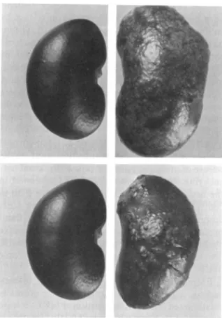
As in the colchicine experiments, the distribu-tion of abscesses over the renal cortical surface was similar in neutropenic and control rats, but the size and the number of abscesses was markedly reduced. This striking difference is shown in figure 1.
After two months of infection, cyclophospha-mide-treated rats displayed a significant reduction in parenchymal destruction and scarring, despite a slightly higher number of residual bacteria. Fur-thermore, although the distribution of scars was similar to that in control rats, the scars were smaller and fewer.
Microscopic examinations of sections of acutely infected kidneys showed dramatic differences be-tween neutropenic rats and control rats. Control rats had many large abscesses containing innumer-able PMNLs. Single abscesses extended from the
medulla through the cortex and invariably
distended the overlying capsule. Lesions in neutropenic animals which looked macroscopical-ly like abscesses, appeared microscopicalmacroscopical-ly to con-tain masses of bacteria within distended tubules, but contained only a few PMNLs. These lesions were smaller and seldom distended the overlying capsule. Thus, lesions in acute pyelonephritis in neutropenic rats were both fewer and quite differ-ent in microscopic appearance from those in con-trol rats. These histologic differences probably ac-counted for the differences in kidney weight.
Despite differences in kidney weight during the acute phase, the histology of the scars examined two months after the operation was similar in cy-clophosphamide-treated and control rats.
Discussion
Previous studies of the pathogenesis of CPN after acute, ascending, obstructive pyelonephritis in the rat [5] have suggested that acute inflammation and suppuration determined the severity of residual
Figure 1. Control (left) and acutely pyelonephritic
(right) kidneys from (top) untreated and (bottom) neu-tropenic rats.Cyclophosphamide was given to rats four days before the induction of pyelonephritis to induce severe leukopenia and neutropenia.
renal scarring. If the peak of inflammation was prevented by early antibiotic therapy, very little parenchymal destruction occurred. Even if low doses of antibiotic sufficient to suppress acute in-flammation but not to sterilize the kidney were given, little scarring developed despite persistent infection throughout the course of the disease. Moreover, if antibiotic treatment was started after the peak inflammatory process had fully devel-oped, severe renal destruction and CPN evolved in the absence of bacteria. These two observations suggest that the bacteria themselves act only to in-cite the acute inflammatory response and there-after play an insignificant role in the development of CPN. Similar observations stressing the need for early antibiotic treatment to prevent renal scars have been recently made by other in-vestigators [16].
[5], the dramatic protection against CPN might have been due to decreased endotoxin release by antibiotic suppression of bacterial growth. The in-trarenal injection of dogs with lipid A, the toxic component of lipopolysaccharide endotoxins of gram-negative bacteria, produces an inflamma-tory response that is related to the dose of lipid A [17]. In the present report, colchicine treatment and cyclophosphamide-induced neutropenia di-minished the peak of inflammation during acute pyelonephritis, but renal bacterial counts were higher throughout the course of acute-to-chronic pyelonephritis. Ifbacterial infection itself or the release of endotoxin directly injures the renal par-enchyma, more severe CPN should be found in the neutropenic or colchicine-treated rats than in control rats. Instead, we found less severe CPN despite higher bacterial counts. Decreased inflam-mation during the acute phase of suppurative pyelonephritis, not depressed bacterial growth, protected against kidney destruction in the chronic phase of the disease.
PMNLs are a crucial element in acute inflam-mation. Under various stimuli that are normally encountered during inflammation, PMNLs release substances that injure tissues. [18, 19]. Studies of acute myocardial infarction in rats have shown that tissue damage is directly related to PMNL in-filtration and is reduced by measures which inhibit exudation [20]. In gouty arthritis, inflammation is mainly mediated by the release of inflammatory substances from PMNLs actively phagocytosing urate crystals [21]. Colchicine has been used for centuries to treat this condition. Its anti-inflam-matory effect has been shown to be due to inter-ference with leukocyte locomotion and chemo-taxis and to diminished lysosomal degranulation [22]. In experimental pyogenic infection [23], col-chicine has been shown to delay the delivery of PMNLs to the site of infection and thus reduce inflammation. However, infection was more severe in these experiments, at least in part because of the delay in delivery of leukocytes. Col-chicine acted similarly in acute pyelonephritis in rats in the present study. The inflammatory pro-cesses as measured by the L: R weight ratios of the kidneys were reduced after colchicine treatment, whereas bacterial counts increased. This resulted in reduced parenchymal destruction in the chronic phase, suggesting that the toxic and tissue-destruc-tive processes during acute inflammation could be
in part due to infiltration of leukocytes that was more important than infection in the pathogenesis of renal scars.
Whereas colchicine treatment provided indirect support for a crucial role of PMNLs in the patho-genesis of renal scarring, experiments in rats ren-dered neutropenic during acute pyelonephritis provided more direct evidence. It has been shown in another model of acute pyelonephritis that complement depletion by means of cobra venom factor results in diminished exudation of PMNLs in the renal parenchyma and preserves the mor-phology of the kidney [24]. More recently, Shima-mura [25] observed a direct relationship between infiltration of PMNLs and renal tissue damage during the first two days of renal infection in neutropenic rats. In our neutropenic rats with acute pyelonephritis, infection in the kidneys was as widespread as in the control rats but was less severe as judged by smaller abscess size, lower kid-ney weight, and much less infiltration of PMNLs. In addition, renal scarring two months later was diminished.
Because treatments with colchicine and cyclo-phosphamide were accompanied by some mortal-ity, we explored the possibility that both drugs could influence food and water intake. We won-dered if the reduced inflammation found during acute pyelonephritis could have been due to dehy-dration resulting from diminished water intake. This was not the case, because pyelonephritic rats housed in metabolic cages and given the same amount of food and water as taken by cyclophos-phamide-treated rats had as severe pyelonephritis as control rats fed as much as they wanted.
Experiments by Miller et al. [26] using cyclo-phosphamide to treat pyelonephritis in rats illus-trates the role of early infiltration by PMNLs in the pathogenesis of renal scars. In these experi-ments, cyclophosphamide was administered two and four days after initiating renal infection. The resulting neutropenia, which was delayed in rela-tion to the peak of inflammarela-tion, did not prevent renal scars. The contrast between our results and those with delayed induction of neutropenia sup-port the view that initial infiltration by PMNLs produces the renal injury that ultimately results in CPN.
Cyclophosphamide might prevent renal scars by suppression of humoral or cellular immune mech-anisms responsible for renal parenchymal
dam-age. Indeed, next to its immediate leukopenic ef-fect, cyclophosphamide has a profound effect on both T and B lymphocytes [27]. However, exten-sive investigation has not shown a role for humor-al immunity in the pathogenesis of CPN [2, 5-7,
10, 11]. In the present report, E. coli agglutinin
levels measured two months after infection were similar in cyclophosphamide-treated and in con-trol pyelonephritic rats (mean ± SD 10g2 serum titer, 19.8 ± 1.8 and 20.2 ± 2.4, respectively). Thus, it seems unlikely that parenchymal damage was prevented by diminished antibody produc-tion. Cellular immune mechanisms have been shown not to be responsible for kidney damage during renal infection [12, 28]. Furthermore, in our model, rats thymectomized at birth showed no dif-ference in the extent of CPN from that observed in sham-thymectomized rats [5]. If cyclophospha-mide given just before infection in our experi-ments had acted by preventing a cellular immune mechanism, the administration of the drug just after initiation of infection as done by Miller et al. [26] should have also diminished renal scars. Be-cause this was not the case, it reinforces our inter-pretation that PMNLs, rather than cellular or hu-moral immunity, mediate tissue damage during the inflammatory processes resulting from infec-tion in the kidney.
References
1. Angell, M. E., ReIman, A. S., Robbins, S. L. "Active" chronic pyelonephritis without evidence of bacterial in-fection. N. Engl. J Med. 2781303-1308, 1968. 2. Aoki, S., Imamura, S., Aoki, M., McCabe, W. R.
"Abac-terial" and bacterial pyelonephritis: immunofluorescent localization of bacterial antigen. N. Engl. J. Med. 281: 1375-1382, 1969.
3. Kalmanson, G. M., Sommers, S.c.,Guze, L. B. Pyelone-phritis. VII. Experimental ascending infection with pro-gression of lesions in the absence of bacteria. Archives of Pathology 80:509-516, 1965.
4. Glassock, R. J., Kalmanson, G. M., Guze, L. B. Pyelone-phritis. XVIII. Effect of treatment on the pathology of enterococcal pyelonephritis in the rat. Am. J. Pathol. 76:49-60, 1974.
5. Glauser, M. P., Lyons, J. M., Braude, A. I. Prevention of chronic experimental pyelonephritis by suppression of acute suppuration. J. Clin. Invest. 61:403-407, 1978. 6. Sanford, J. P., Hunter, B. W., Donaldson, P.
Localiza-tion and fate ofEscherichia coli in hematogenous
pyelo-nephritis. J. Exp. Med. 116:285-294, 1962.
7. Cotran, R. S. RetrogradeProteus pyelonephritis in rats:
localization of antigen and antibody in treated sterile
pyelonephritic kidneys. J. Exp. Med. 117:813-821, 1963.
8. Kovats, T. G. The role of endotoxin in autoimmune pro-cesses. Naturwissenschaften 48:572-573, 1961. 9. Holmgren, J., Hanson, L.A.,Holm, S. E., Kaijser, B. An
antigenic relationship between kidney and certain Es-cherichia coli strains. Int. Arch. Allergy Appl.
Im-munol. 41:463-474, 1971.
10. Kalmanson, G. M., Guze, L. B. Pyelonephritis. An at-tempt to demonstrate anti-kidney antibody in the sera of patients with chronic bacteriuria. Am. J. Med. Sci. 246:532-536, 1963.
11. Miller, T. E., Smith, J. W., Lehmann, J. W., Sanford, J. P. Autoimmunity in chronic experimental pyelo-nephritis. J. Infect. Dis. 122:191-195, 1970.
12. Coles, G. A., Chick, S., Hopkins, M., Ling, R., Radford, N. J. The role of the T cell in experimental pyelo-nephritis. Clin. Exp. Immunol. 16:629-636, 1974. 13. Pitchon, H. E., Kalmanson, G. M., Glassock, R. J.,
Ishida, K., Guze, L. B. Experimental enterococcal pyelonephritis in athymic (nude) mice [abstract no.
1070]. In Proceedings of the 19th Interscience
Confer-ence on Antimicrobial Agents and Chemotherapy. American Society for Microbiology, Washington, D.C., 1980.
14. Brooks, S. J. D., Lyons, J. M., Braude, A. I. Immuni-zation against retrograde pyelonephritis. I. Production of an experimental model of severe ascending Escheri-chia coli pyelonephritis without bacteremia in rats. Am.
J. Pathol. 74:345-358, 1974.
15. Brooks, S. J. D., Lyons, J. M., Braude, A. I. Immuniza-tion against retrograde pyelonephritis. III. VaccinaImmuniza-tion against chronic pyelonephritis due toEscherichia coli. J.
Infect. Dis. 136:633-639, 1977.
16. Miller, T., Phillips, S. Pyelonephritis: the relationship be-tween infection, renal scarring, and antimicrobial thera-py. Kidney Int. 19:654-662, 1981.
17. Westenfelder, M., Galanos, C. Experimental lipid A-in-duced nephritis in the dog. A possible role of lipid A in the pathogenesis of abacterial chronic pyelonephritis. Infection 2:174-177,1974.
18. Weissmann, G., Smolen, J. E., Korchak, H. M. Release of inflammatory mediators from stimulated neutro-phils. N. Engl. J. Med. 303:27-34, 1980.
19. Babior, B. M. Oxygen-dependent microbial killing by phagocytes [in two parts]. N. Engl. J. Med. 298:659-668,721-725, 1978.
20. Maclean, D., Fishbein, M. c., Braunwald, E., Maroko, P. R. Long-term preservation of ischemic myocardium after experimental coronary artery occlusion. J. Clin. Invest. 61:541-551,1978.
21. Seegmiller, J. E., Howell, R. R., Malawista, S. E. The in-flammatory reaction to sodium urate: its possible rela-tionship to the genesis of acute gouty arthritis. J.A.M.A. 180:469-475, 1962.
22. Malawista, S. E. Colchicine: a common mechanism for its anti-inflammatory and anti-mitotic effects. Arthritis Rheum. 11:191-197, 1968.
23. Malawista, S. E., Andriole, V. T. Colchicine: anti-in-flammatory effect of low doses in a sensitive bacterial system. J. Lab. Clin. Med. 72:933-942, 1968.
24. Sullivan, M. J., Harvey, R. A., Shimamura, T. The ef-fects of cobra venom factor, an inhibitor of the comple-ment system, on the sequence of morphological events in the rat kidney in experimental pyelonephritis. YaleJ. BioI. Med. 50:267-273, 1977.
25. Shimamura, T. Mechanisms of renal tissue destruction in an experimental acute pyelonephritis. Exp. Mol. PathoI. 34:34-42, 1981.
26. Miller, T. E., Burnham, S., North, J. D. K.
1mmuno-logical enhancement in the pathogenesis of pyelo-nephritis. Clin. Exp. 1mmunol. 24:336-345, 1976. 27. Turk, J. L., Parker, D. The effect of cyclophosphamide
on the immune response.J. Immunopharmacol. 1: 127-137, 1979.
28. Miller, T., Burnham, S., Simpson, G. Selective deficiency of thymus-derived lymphocytes in experimental pyelo-nephritis. Kidney Int. 8:88-97, 1975.