347
CONCISE COMMUNICATION
Effects of Clinically Used Antioxidants in Experimental Pneumococcal
Meningitis
Marc Auer,1Luz-Andrea Pfister,1David Leppert,2 Martin G. Ta¨uber,1and Stephen L. Leib1
1Institute for Medical Microbiology, University of Bern, Bern, and2Department of Research and Neurology, University Hospitals, Basel, Switzerland Reactive oxygen intermediates mediate brain injury in bacterial meningitis. Several
anti-oxidant drugs are clinically available, including N-acetylcysteine (NAC), deferoxamine (DFO), and trylizad-mesylate (TLM). The present study evaluated whether these antioxidants are beneficial in a model of pneumococcal meningitis. Eleven-day-old rats were infected intracis-ternally with Streptococcus pneumoniae and randomized to intraperitoneal treatment every 8 h with NAC (200 mg/kg), DFO (100 mg/kg), TLM (10 mg/kg), or saline (250 mL). TLM-treated animals showed a significantly reduced mortality compared with controls (P!.03). Meningitis led to extensive cortical injury at225 2.2 h after infection (median, 14.6% of cortex; range, 0–61.1%). Injury was significantly (P!.01) reduced to 1.1% (range, 0–34.6%) by NAC, to 2.3% (range, 0–19.6%) by DFO, and to 0.2% (range, 0–36.9%) by TLM (the difference was not significant among the 3 groups). None of the drugs reduced hippocampal injury. Thus, several clinically used antioxidants reduced cortical injury in experimental pneu-mococcal meningitis.
Reactive oxygen intermediates (ROIs), a family of molecules derived from the partial reduction of molecular oxygen, have been implicated as mediators of neuronal damage in bacterial meningitis [1–3]. ROIs are released by the host from stimulated granulocytes, macrophages, and microglia, and Streptococcus pneumoniae has been shown to utilize pyruvate oxidase to pro-duce ROIs. Because of their free radical nature (i.e., the pres-ence of an unpaired electron) and loss of spin restriction, ROIs react avidly with organic molecules and are cytotoxic by oxi-dizing membrane phospholipids, proteins, nucleic acids, and nucleotides.
A number of factors make the central nervous system par-ticularly susceptible to oxidative damage. The brain receives ∼20% of the cardiac blood output and thus has a high oxygen tension, and brain cell membranes are rich in polyunsaturated fatty acids that can be oxidized by ROIs. Brain tissue has
rel-Received 11 February 2000; revised 3 April 2000; electronically published 6 July 2000.
Presented in part: 39th Interscience Conference on Antimicrobial Agents and Chemotherapy, San Francisco, 26–29 September 1999 (abstract 1779). The animal studies were approved by the Animal Care and Experimen-tation Committee of the Kanton of Bern, Switzerland, and followed Na-tional Institutes of Health guidelines for the performance of animal experiments.
Financial support: Swiss National Science Foundation (NRP 4038-52841); NIH (NS-32553, NS-34028).
Reprints or correspondence: Dr. Stephen L. Leib, Institute for Medical Microbiology, University of Bern, Friedbu¨hlstr. 51, 3010 Bern, Switzerland ([email protected]).
The Journal of Infectious Diseases 2000; 182:347–50
q 2000 by the Infectious Diseases Society of America. All rights reserved.
0022-1899/2000/18201-0049$02.00
atively low levels of endogenous antioxidant defense systems. In previous studies based upon a model of bacterial meningitis caused by group B streptococci, we localized the generation of ROIs to cells of the subarachnoid and ventricular inflammation and to the cerebral vasculature, the site of blood-brain barrier breakdown [1].
In an infant rat model of meningitis due to group B strep-tococci, the spin-trapping agent a-phenyl-tert-butyl nitrone sig-nificantly attenuated neuronal damage [1]. Other antioxidants attenuated a number of early events associated with bacterial meningitis, such as early increases in cerebral blood flow, in-tracranial pressure, brain water content, cerebrospinal fluid (CSF) white blood cell count, and pial arteriolar vasodilatation [4–6].
For the present study, we used an established rat model of experimental neonatal meningitis due to S. pneumoniae, which leads to neuronal damage [7, 8]. The model shows features identical to those described in neonates and infants with neo-natal meningitis. Two distinct forms of neuronal injury have been documented: extensive cortical injury and apoptotic injury to the dentate gyrus of the hippocampus [1].
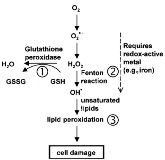
In this study, we examined the effect of 3 agents that interfere with ROI production at different stages (figure 1). N-acetyl-cysteine (NAC), which increases endogenous antioxidant de-fenses (glutathione), is clinically used for paracetamol intoxi-cation [4, 9]. Deferoxamine (DFO), an iron-chelating agent, decreases the concentration of oxidative radicals by inhibiting the catalyzation of radical production by iron [6, 10]. Trylizad-mesylate (TLM), which protects cell membranes from peroxi-dation, is therapeutically used to prevent brain injury in patients
348 Auer et al. JID 2000;182 (July)
Figure 1. Formation of reactive oxygen species and antioxidant mechanisms in biologic systems. Superoxide (O22), formed by a one-electron reduction of molecular O2, is converted to H2O2by dismutation and further to OH by the Cu1/Fe21-catalyzed Fenton reaction. The resultant free radical damage to lipid (peroxidation), proteins, and DNA leads to various forms of cell injury. The major antioxidant enzymes are superoxide dismutase, catalase, and glutathione peroxi-dase. GSH, reduced glutathione; GSSG, oxidized glutathione. As num-bered in the figure, the antioxidants used for therapy were (1) N-acetylcysteine (NAC), which increases the endogenous antioxidant glutathione; (2) deferoxamine (DFO), which decreases the concentra-tion of oxidative radicals by neutralizing iron; and (3) trylizad-mesylate (TLM), which inhibits lipid peroxidation.
with subarachnoid-space bleeding [11]. In the present study, we studied the 3 drugs to explore whether these clinically available antioxidants should be tested in clinical trials as adjunctive therapy for bacterial meningitis.
Material and Methods
Model of meningitis. Nursing Sprague-Dawley rats were in-fected on postnatal day 11 by direct intracisternal injection, with a 32-gauge needle, of 10 mL of a saline solution containing S.
pneumoniae (serogroup 3), as described elsewhere [7, 8]. The
in-oculum size was log10 6.55 0.35 cfu/mL. Eighteen hours after
infection, the rats were weighed and assessed clinically for their ambulatory activity and their ability to right themselves. The oc-currence of spontaneous seizures was documented during the fol-lowing 2 h. CSF (1–30 mL) was obtained by puncture of the cisterna magna. To document meningitis, 10 mL of CSF was cultured quan-titatively. Starting 18 h after infection, the animals were treated subcutaneously with 100 mg/kg ceftriaxone. Those surviving 24 h of infection were killed with an overdose of pentobarbital (100 mg/ kg intraperitoneally). Animals who died unobserved were excluded from the histopathologic evaluation.
Therapeutics. Three molecular classes of antioxidant agents were used: NAC (Fluimucil, 20% infusion solution; Inpharzam,
Cadempino, Switzerland), DFO (Sigma, Division of Fluka Chemie, Buchs, Switzerland), and TLM (Freedox, 150-mg sterile solution; Pharmacia and Upjohn, Du¨bendorf, Switzerland). Therapy was begun at the time of infection and was given every 8 h. The NAC group (n = 24) received 200 mg/kg, the DFO group (n = 25) re-ceived 100 mg/kg, and the TLM group (n = 25) received 10 mg/kg. As a control group, 30 rats received saline. The doses used in this study correspond to doses from published studies using rat models of experimental meningitis for NAC [4], a rat model of pneumonia for DFO [10], and a rat model of the “shaken-baby syndrome” for TLM [11].
Histopathology. For histopathologic examination, killed ani-mals were perfused with 4% paraformaldehyde in PBS, and 12 coronal brain sections per animal were evaluated for neuronal in-jury to the cortex and hippocampus, as described elsewhere [7, 8]. The area of cortical brain damage was expressed as the mean value per animal of the percentage of the total cortex in each of the 12 sections. Apoptotic death of neurons in the dentate granule cell layer of the hippocampus was scored, and an averaged score per animal was calculated from all evaluated sections. All histopath-ologic evaluations were done by an investigator blinded to the clinical, microbiologic, and treatment data of the respective animal.
Measurement of tumor necrosis factor–a (TNF-a). The con-centration of TNF-a in the CSF samples was measured by ELISA (Cytoscreen Rat Tumor Necrosis Factor–Alpha Ultra Sensitive; BioSource International, Camarillo, CA) [7]. CSF samples (10 mL) from infected animals of each study group (NAC,n = 13; DFO, ; and TLM, ) and from controls ( ) were sampled
n = 18 n = 15 n = 15
18 h after infection and immediately centrifuged, and the super-natant was frozen at2807C until analyzed, following the manu-facturer’s instructions.
Statistics. Continuous data were presented asmean5 SD, cat-egorical data as median and range. Data not normally distributed were analyzed by the Kruskal-Wallis test, followed by the Mann-Whitney test for pairwise comparison. Proportions between groups were analyzed using Fisher’s exact test. Survival curves were an-alyzed by Kaplan-Meier analysis for survival data.
Results
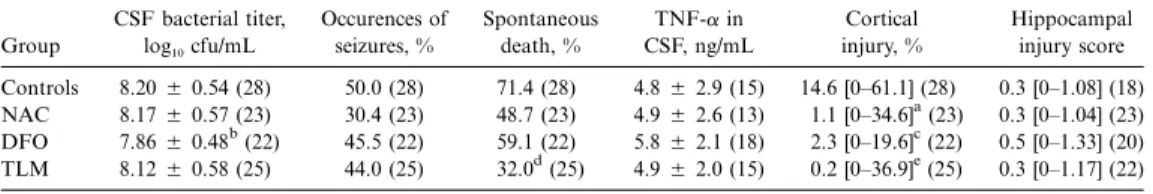
Effect of antioxidants on clinical parameters of meningitis. All infected animals had meningitis, as evidenced by lethargy or obtundation and by positive bacterial CSF titers at 18 h after infection. Treatment with NAC and TLM showed no ef-fect on CSF bacterial titers (table 1). Animals in the DFO treatment group had lower CSF bacterial titers than animals in all other groups, which was likely a consequence of growth inhibition by iron limitation in the CSF. Treatment with anti-oxidants did not significantly affect the occurrence of seizures (table 1). However, compared with controls, TLM-treated an-imals showed a significant (P!.03) attenuation of spontaneous mortality, while treatment with NAC and DFO had no signif-icant beneficial effect (table 1).
Effect of antioxidants on CSF concentration of TNF-a. The CSF concentrations of TNF-a were measured as an indicator of inflammation. There was no significant difference (P1.05)
JID 2000;182 (July) Antioxidants in Pneumococcal Meningitis 349
Table 1. Effect of antioxidant treatment on clinical parameters of meningitis.
Group CSF bacterial titer, log10cfu/mL Occurences of seizures, % Spontaneous death, % TNF-a in CSF, ng/mL Cortical injury, % Hippocampal injury score Controls 8.205 0.54 (28) 50.0 (28) 71.4 (28) 4.85 2.9 (15) 14.6 [0–61.1] (28) 0.3 [0–1.08] (18) NAC 8.175 0.57 (23) 30.4 (23) 48.7 (23) 4.95 2.6 (13) 1.1 [0–34.6]a(23) 0.3 [0–1.04] (23) DFO 7.865 0.48b(22) 45.5 (22) 59.1 (22) 5.85 2.1 (18) 2.3 [0–19.6]c(22) 0.5 [0–1.33] (20) TLM 8.125 0.58 (25) 44.0 (25) 32.0d(25) 4.95 2.0 (15) 0.2 [0–36.9]e(25) 0.3 [0–1.17] (22) NOTE. CSF, cerebrospinal fluid; TNF-a, tumor necrosis factor–a; NAC, N-acetlycysteine; DFO, deferoxamine; TLM, trylizad-mesylate. CSF bacterial titer and TNF-a in CSF are presented asmean5 SD; cortical injury and hippocampal injury scores are presented as median [range]. Numbers inparentheses = n.
a vs. controls. P!.02 b vs. controls. P!.03 c vs. controls. P!.01 d vs. controls. P!.03 e vs. controls. P!.01
between any of the experimental groups at 18 h after infection (table 1).
Effect of antioxidants on neuronal injury. Two distinct forms of neuronal injury were identified in this experimental model of bacterial meningitis. The neurons of the cortex showed mor-phologic signs that were compatible with necrosis, such as marked swelling of the cell body and loss of cellular demar-cation and cytoarchitecture [1]. The injury patterns in the hip-pocampus were compatible with apoptosis. The occurrence of apoptosis in the dentate gyrus in this model was confirmed in a previous study by positive-terminal deoxynucleotidyl trans-ferase–mediated dUTP nick end labeling (TUNEL) of frag-mented DNA in the dentate gyrus but not in the cortex [1].
All 3 antioxidants decreased the extent of cortical injury. The decrease varied from a median of 14.6% (range, 0–61.1%) in 28 control rats to 1.1% (range, 0–34.6%) in 23 NAC-treated rats (P!.02), to 2.3% (range, 0–19.6%) in 22 DFO-treated an-imals (P!.01), and to 0.2% (range, 0–36.9%) in 25 rats treated with TLM (P!.01). There were no significant differences in neuroprotective efficacy among the 3 antioxidant therapies (ta-ble 1).
In contrast, antioxidant treatment had no effect on the extent of apoptosis in the hippocampus. The median injury score was 0.3 (range, 0–1.04) for the NAC group (n = 23), 0.5 (range, 0–1.33) for the DFO group (n = 20), 0.3 (range, 0–1.17) for the TLM group (n = 22), and 0.3 (range, 0–1.08) for the control group (n = 18; table 1).
Discussion
In the present study, we used an infant rat model to evaluate the effect of clinically used antioxidants on the clinical and neuropathological outcomes during pneumococcal meningitis. We demonstrated that 3 classes of antioxidants (NAC, DFO, and TLM) that interfere with the harmful effects of ROIs at different levels exert a neuroprotective effect in the cortex (figure 1). Despite this beneficial effect, we found no reduction of the inflammatory response (i.e., CSF concentration of TNF-a) or
of hippocampal apoptosis as a result of treatment with any of the 3 compounds.
We tested the effect of NAC because it has been used as an antioxidant in a variety of experimental and clinical conditions [4, 9]. At present, NAC is known to have 2 modes of action: direct scavenging of free radicals and augmentation of intra-cellular levels of glutathione [4, 9]. NAC has proven efficacy as an antioxidant in humans and animals with various diseases, including experimental bacterial meningitis, in which it signif-icantly attenuated the increase in brain water content, intra-cranial pressure, and CSF pleocytosis 24 h after infection [4]. The results of the present study expand these findings by show-ing a beneficial effect of the drug on cortical injury in neonatal experimental meningitis. The basis for this beneficial effect is not completely clear. We have previously documented that ROIs are generated at the level of the cerebral vasculature and that cerebral blood flow improved upon treatment with the radical scavenger a-phenyl-tert-butyl nitrone [1]. Thus, in keep-ing with the evidence that ischemia contributes to cortical injury in this model, the neuroprotection of NAC in the cortex might be mediated by preventing disturbances of cerebral blood flow [3, 4, 8].
An important factor that contributes to the central nervous system’s susceptibility to oxidative damage is its rich content of iron. Extensive experimental support exists for the early oc-currence and pathophysiologic importance of oxygen radical formation and cell membrane lipid peroxidation in the injured nervous system. The radical-initiated peroxidation of neuronal, glial, and vascular cell membranes and myelin is catalyzed by free iron released from hemoglobin, transferrin, and ferritin by lowered tissue pH and oxygen radicals. Thus, a disruption of the normal iron homeostasis during bacterial meningitis may lead to an increase in oxygen free radical generation and con-secutive tissue damage.
DFO was used to evaluate the role of iron in our model of meningitis. Compounds such as DFO are excellent chelating agents of free Fe31and are currently being used clinically to treat iron-overload patients. In our study, DFO attenuated cor-tical injury. One possible mechanism for this beneficial effect
350 Auer et al. JID 2000;182 (July)
is through inhibition of the Cu1/Fe21-catalyzed Fenton reac-tion. This would reduce the generation of hydroxyl radicals as well as that of other reactive products, thus leading to reduced oxidative damage in the brain [2]. However, more studies are needed to delineate the exact mechanisms by which DFO exerts its protective role against ischemia-induced cortical injury in this model.
The 21-aminosteroid TLM has been shown to be a potent inhibitor of lipid peroxidation and to reduce traumatic and ischemic damage in a number of experimental systems [12]. TLM has been shown to decrease the posttraumatic increase in blood-brain barrier permeability observed after head injury in rats [12, 13]. As shown by in vitro and in vivo studies, the efficacy depends on a combination of mechanisms: (1) The molecules of TLM insert within the lipid bilayer of the cell membrane, scavenging toxic lipid-peroxyl radicals and reducing the production of highly reactive hydroxyl radicals [12]; (2) the insertion of TLM into the cell membrane decreases the membrane phospholipid fluidity and thereby mitigates the pro-gression of lipid peroxidation as it spreads across the membrane [12]; and (3) TLM increases the concentration of vitamin E, an important endogenous lipophilic antioxidant [14].
Administration of 21-aminosteroids in experimental endo-toxemia, sepsis, and meningitis showed improvement of sys-temic and hemodynamic parameters, attenuation of inflam-mation, preservation of tissue oxygenation, and protection of endothelial cell integrity [5, 14]. In our model, animals become invariably bacteremic in the course of the disease and are thus prone to sepsis, which likely contributes to mortality [7]. TLM, in contrast to the other antioxidants examined, significantly attenuated the spontaneous mortality in this study, in keeping with the known benefits of the drug in sepsis models.
None of the 3 antioxidants studied here attenuated CSF in-flammation, as measured by the concentration of TNF-a in CSF. Previous studies found that neurons in the dentate granule cells of the hippocampus undergo apoptosis during experimen-tal bacterial meningitis, independent of ischemic damage. This process is mediated in part by TNF-a [15]. The lack of an effect of the antioxidant drugs on CSF concentrations of TNF-a is congruent with the lack of neuroprotection in the hippocampus. In summary, using a model of bacterial meningitis caused by S. pneumoniae, we demonstrated a beneficial effect of treatment with the antioxidants NAC, DFO, and TLM on cortical injury, whereas the drugs were not efficacious in preventing hippo-campal injury. Thus, while antioxidant strategies are clearly promising for the adjunctive therapy of bacterial meningitis, an
ideal drug should act at the level of both the cortex and the hippocampus.
Acknowledgments
We thank Dr. Stephan Christen for helpful discussion and Oliver Schu¨tz and Philipp Joss for technical assistance.
References
1. Leib SL, Kim YS, Chow LL, Sheldon RA, Ta¨uber MG. Reactive oxygen intermediates contribute to necrotic and apoptotic neuronal injury in an infant rat model of bacterial meningitis due to group B streptococci. J Clin Invest 1996; 98:2632–9.
2. Koedel U, Pfister HW. Oxidative stress in bacterial meningitis. Brain Pathol
1999; 9:57–67.
3. Koedel U, Pfister HW. Superoxide production by primary rat cerebral en-dothelial cells in response to pneumococci. J Neuroimmunol 1999; 96: 190–200.
4. Koedel U, Pfister HW. Protective effect of the antioxidant N-acetyl-L-cysteine in pneumococcal meningitis in the rat. Neurosci Lett 1997; 225:33–6. 5. Lorenzl S, Koedel U, Frei K, Bernatowicz A, Fontana A, Pfister HW.
Pro-tective effect of a 21-aminosteroid during experimental pneumococcal meningitis. J Infect Dis 1995; 172:113–8.
6. Pfister HW, Koedel U, Lorenzl S, Tomasz A. Antioxidants attenuate micro-vascular changes in the early phase of experimental pneumococcal men-ingitis in rats. Stroke 1992; 23:1798–804.
7. Leib SL, Leppert D, Clements J, Ta¨uber MG. Matrix metalloproteinases contribute to brain damage in experimental pneumococcal meningitis. Infect Immun 2000; 68:615–20.
8. Pfister LA, Tureen JH, Shaw S, et al. Endothelin inhibition improves cerebral blood flow and is neuroprotective in pneumococcal meningitis. Ann Neu-rol 2000; 47:329–35.
9. Allameh A, Vansoun EY, Zarghi A. Role of glutathione conjugation in pro-tection of weanling rat liver against acetaminophen-induced hepatotox-icity. Mech Aging Dev 1997; 95:71–9.
10. Merali S, Chin K, Del Angel L, Grady RW, Armstrong M, Clarkson AB Jr. Clinically achievable plasma deferoxamine concentrations are therapeutic in a rat model of Pneumocystis carinii pneumonia. Antimicrob Agents Chemother 1995; 39:2023–6.
11. Smith SL, Hall ED. Tirilazad widens the therapeutic window for riluzole-induced attenuation of progressive cortical degeneration in an infant rat model of the shaken baby syndrome. J Neurotrauma 1998; 15:707–19. 12. Hall ED, Andrus PK, Smith SL, et al. Neuroprotective efficacy of
micro-vascularly-localized versus brain-penetrating antioxidants. Acta Neuro-chir Suppl (Wien) 1996; 66:107–13.
13. Smith SL, Scherch HM, Hall ED. Protective effects of tirilazad mesylate and metabolite U-89678 against blood-brain barrier damage after subarach-noid hemorrhage and lipid peroxidative neuronal injury. J Neurosurg
1996; 84:229–33.
14. Spapen H, Zhang H, Vincent JL. Potential therapeutic value of lazaroids in endotoxemia and other forms of sepsis. Shock 1997; 8:321–7. 15. Bogdan I, Leib SL, Bergeron M, Chow L, Ta¨uber MG. Tumor necrosis
factor-alpha contributes to apoptosis in hippocampal neurons during experi-mental group B streptococcal meningitis. J Infect Dis 1997; 176:693–7.