RESEARCH PAPER
Physiological characterization of Mg deficiency in
Arabidopsis thaliana
Christian Hermans1,2,*,†and Nathalie Verbruggen1
1
Laboratoire de Physiologie et de Ge´ne´tique Mole´culaire des Plantes, Universite´ Libre de Bruxelles, Bd du Triomphe CP 242, 1050 Brussels, Belgium
2
Bioenergetics Laboratory, University of Geneva, Ch des Embrouchis, Geneva 1254, Switzerland
Received 25 January 2005; Accepted 20 April 2005
Abstract
Although the symptoms of magnesium deficiency are well documented in plants, the primary physiological effects of low Mg availability remain largely unknown. This paper describes the physiological responses of Mg starvation in Arabidopsis thaliana. Growth character-istics, Mg and sugar concentration, and photochemical performance were measured at regular intervals during the induction of Mg deficiency. These data show that Mg deficiency increased the sugar concentration and altered sucrose export from young source leaves be-fore any noticeable effect on photosynthetic activity was seen. The decline in photosynthetic activity might be elicited by increased leaf sugar concentrations. Transcript levels of Cab2 (encoding a chlorophyll a/b protein) were lower in Mg-deficient plants before any obvious decrease in the chlorophyll concentration. These transcriptional data suggest that the reduction of chlorophyll is a response to sugar levels, rather than a lack of Mg atoms for chelating chlorophyll.
Key words: Arabidopsis, Cab2, chloropyll a fast fluorescence transient, magnesium deficiency, photosynthesis, sugars.
Introduction
It has long been known that magnesium availability to plants imposes limits on photosynthesis (Evans and Sorger, 1966; Mengel and Kirkby, 1987; Marschner, 1995). Mg is essential for chloroplasts, being the central element in chlorophyll (Beale, 1999). It participates in thylakoid membrane organization and grana stacking (Guller and
Krucka, 1993; Lu et al., 1995; Kaftan et al., 2002), acts as a cofactor and allosteric activator of enzymes involved in CO2fixation (Marschner, 1995), and is involved in energy
transfer via adenosine triphosphate (Igamberdiev and Kleczkowski, 2001, 2003) and pH control (Wu et al., 1991). Accordingly, a number of studies have decribed the effect of Mg deficiency on photochemical reactions or CO2
fixation (Terry and Ulrich, 1974; Cao and Tibbits, 1992; Sun and Payn, 1999; Laing et al., 2000; Sun et al., 2001). However, some limitations may arise from these experi-mental strategies because it is likely that Mg deficiency affects metabolic processes other than photosynthesis at an earlier stage. Recent studies in Beta vulgaris (Hermans et al., 2004, 2005) and in Vicia faba (Hariadi and Shabala, 2004a, b) showed that neither chlorophyll concentration nor plant biomass were effective for diagnosis of Mg deficiency in the early stages. In fact, Mg analysis remains the most accurate tool to diagnose Mg deficiency (Hariadi and Shabala, 2004a). Nonetheless, sucrose accumulation in source leaves is found to be an early symptom of Mg deficiency (Fischer and Bremer, 1993; Cakmak et al., 1994a, b; Mehne-Jakobs, 1995; Fischer et al., 1998; Hermans et al., 2004, 2005) and occurs before any notice-able effect on photosynthetic activity (Cakmak et al., 1994b; Hermans et al., 2004).
Arabidopsis thaliana was used as a case study to characterize the development of Mg deficiency symptoms and early physiological responses. Growth characteristics, Mg and sugar concentrations, as well as photochemical performance were measured at regular interval during the induction of Mg deficiency. Mg deficiency increased sugar concentration and apparently altered sucrose export from young source leaves before any effect on photo-synthetic activity. The accumulation of sugars in leaves
* Present address: Biology Department, Colorado State University, Fort Collins, CO 80523, USA.
y
To whom correspondence should be addressed in Brussels. Fax: +32 2 6505412. E-mail: chermans@ulb.ac.be
doi:10.1093/jxb/eri215 Advance Access publication 27 June, 2005
ª The Author [2005]. Published by Oxford University Press [on behalf of the Society for Experimental Biology]. All rights reserved.
may result in the down-regulation of photosynthetic genes and, consequently, account for the decline in chlorophyll concentration and photosynthetic activity. In addition, this work provides the first physiological data in Arabidopsis that can form a basis for further genetic analysis, as, to our knowledge, none of the recent transcriptomic analysis for essential nutrient metabolism in that species has so far been related to magnesium.
Materials and methods Plant material and culture
Seeds of Arabidopis thaliana (L.) Heynh Columbia and C24 ecotypes were germinated in peat-based compost. Upon transfer to hydroponics systems, roots of the plantlets were rinsed in distilled water and immediately placed on polystyrene tiles covering the containers (capacity of 4.0 l) filled with mineral solution (see Fig. 1A and B for the experimental design). C24 ecotype plantlets were grown in soil for 2 weeks and in nutrient solution for three more weeks in a day regime of 12 h light (100 lE mÿ2sÿ1)/12 h darkness, before the beginning of the treatment (Fig. 1A). Columbia ecotype plantlets were grown in soil for 2 weeks and in nutrient solution for 1 week in
12 h light (150 lE mÿ2sÿ1)/12 h darkness (Fig. 1E), or for 1 month in a short-day regime of 8 h light (150 lE mÿ2sÿ1)/16 h dark (Fig. 1B). The concentrations of macronutrients in mM were 1.0 Ca(NO3)2,
1.0 MgSO4, 0.88 K2SO4, and 0.25 KH2PO4, and the concentrations
of micronutrients in lM were 20 FeEDTA, 10 NaCl, 10 H3BO3, 1.0
ZnSO4, 1.0 MnSO4, 0.10 CuSO4, and 0.01 (NH4)6Mo7O24. The pH
of the solution was adjusted to 5.860.1 with 1 M KOH. At the beginning of the treatment, half of the plants were fed with a Mg-free nutrient solution, which was the same as above, except that 1.0 mM MgSO4 was replaced by 1.0 mM Na2SO4, in order to maintain
a constant anion/cation balance and to avoid sulphur deficiency. Nutrient solutions were replaced every 4 d. The growth conditions in the culture room were: temperature of 2262 8C and relative humidity of 6565%.
Chemical analysis
0.50 g dried crushed material powder was ashed in a muffle furnace at 450 8C. Ashes were digested with 7 N nitric acid and the filtrate assayed for Mg by atomic absorption spectrometry, at the 285.2 nm wavelength (Perkin Elmer AAS 3110).
Photosynthesis measurements
The behaviour of photosystem II was assessed using Chl a fast fluorescence kinetics (direct induction with 660 nm exposure), recorded with the Plant Efficiency Analyser fluorometer (Hansatech
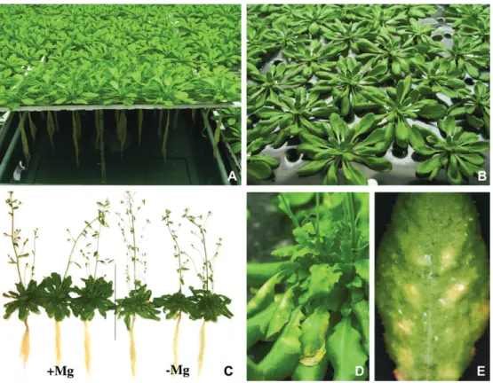
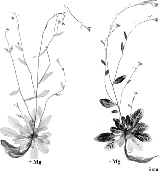
Fig. 1. Hydroponics culture and visual symptoms of Mg deficiency observed in Arabidopsis thaliana. (A) Illustration of the hydroponics culture of A. thaliana C24. Plants, grown for 2 weeks in soil and 3 weeks in nutrient solution, had 20 expanded leaves and were initially vegetative before the Mg-deficiency treatment. Photoperiod during the treatment duration was 12 h light (100 lE mÿ2sÿ1)/12 h darkness. (B) Hydroponics culture of A. thaliana Columbia plants (2 weeks growth in soil and 4 weeks growth in nutrient solution), which had 20–25 expanded leaves. A short-day regime of 8 h light (150 lE mÿ2sÿ1)/16 h darkness was imposed. The polystyrene tiles, on which the shoots were positioned, were covered with a black plastic film to limit growth of algae. (C) Representative control (left) and Mg-deficient (right) A. thaliana C24 reproductive plants after 16 d of treatment. Mg was removed from the nutrient solution at day 0. (D) Chlorotic Mg-deficiency symptoms observed in a reproductive rosette of A. thaliana C24 after 20 d of treatment. Mg was omitted at day 0. (E) Interveinal chlorosis and necrosis in a leaf of A. thaliana Columbia after 16 d of treatment. Mg was removed from the nutrient solution at day 0, when A. thaliana Columbia vegetative plants (2 week’s growth in soil and 1 week’s growth in nutrient solution) had over 15 expanded leaves. A day regime of 12 h light (150 lE mÿ2sÿ1)/12 h darkness was imposed.
Instruments, UK). The OJIP transients (fluorescence levels Fo
(50 ls), F300ls, FJ (2 ms), FI(30 ms), and FM (tFMAX)) (Strasser
et al., 1995) were analysed according to the JIP-test procedure, as previously described by Hermans et al. (2003).
Carbohydrate analysis
The frozen tissues were ground in a mortar and extracted three times with ethanol (80% v/v) at 85 8C. Extraction was assumed to be complete because a fourth ethanol extraction of the starch pellet did not contain detectable amounts of glucose. Soluble sugars were determined using a spectrophotometric enzymatic assay (Lambda 14 spectrophotometer, Perkin Elmer), through the NAD+reduction at
340 nm, following a procedure slightly modified from Sokolov et al. (1998). For starch determination, the pellet was autoclaved and digested with amylase and amyloglucosidase. The glucose released before and after digestion was measured with the same spectropho-tometric assay. All enzymes used in this assay were purchased from Roche Diagnostics (Germany).
Iodine staining
Starch was visualized by iodine staining. Leaves bleached in boiling ethanol (80% v/v), were stained with a Lugol solution (2 mM I2,
6 mM KI) and briefly destained with distilled water. RNA isolation and northern-blot analysis
Total RNAs were isolated as described in Ausubel et al. (1998). Total RNA was size-fractioned by electrophoresis in a MOPS– formaldehyde–1% agarose gel and blotted onto Hybond-N nylon membranes (Amersham Pharmacia Biotech, UK). A 0.24–9.5 kb RNA Ladder (Invitrogen Life Technologies) was used to determine the size of transcripts. Hybridizations were done with DNA probes labelled with 32P-dCTP, using the Mega prime labelling system (Amersham Pharmacia Biotech).
The membranes were probed with a PCR fragment (between nucleotides 17 and 165) of the Arabidopsis chlorophyll a/b binding protein gene 2 (Cab2, Leutwiler et al., 1986). Equal loading of membranes was assessed by hybridization with 18S rRNA from Arabidopsis thaliana.
Hybridizations were carried out at 65 8C in a buffer containing 53 SSC (13 SSC is 150 mM NaCl and 15 mM sodium citrate, pH 7.0) and 100 lg mlÿ1denatured salmon sperm. After 16 h hybridiz-ation, the blots were washed twice for 20 min at 55 8C in 23 SSC/ 0.1% SDS and once for 20 min in 0.23 SSC/0.1% SDS. Hybridiz-ation signals were quantified with the phosphor imager Storm 860 (Molecular Dynamics).
14
[C]-sucrose uptake
The upper epidermis of the leaf was abraded with carborundum (0.060 mm). [U-14C]-sucrose (9.25 kBq per leaf) was applied as liquid droplets (10 ll) containing 10 mM sucrose and 20 mM MES buffer (pH=5.5). [U-14C]-sucrose was purchased from Amersham Pharmacia Biotech. Small portions of tissues were bleached as described in Grusak et al. (1990), and their radioactivity was counted by liquid scintillation spectroscopy (Tri-carB 1900TR Liquid Scintillation Analyser, Packard Instruments).
Results and discussion
Effect of Mg deficiency on growth, biomass partitioning, and mineral concentration
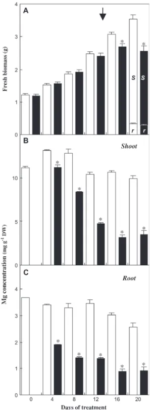
The impact of Mg deficiency was studied in Arabidopsis plants grown hydroponically (Fig. 1A). Slight interveinal Fig. 2. Effect of Mg deficiency on biomass allocation, Mg content in
Arabidopsis thaliana C24 (plants described in Fig. 1A, C, D). (A) Fresh biomass evolution of individual plants during the treatment. At the end of the treatment (day 20), biomass allocation between root (r) and shoot (s) was measured. Mean of 39–40 values 6SE. (B) Mg concentration in shoots during the treatment. Mean of three independent mineralizations of a powdered sample of five shoots 6SE. C, Mg concentration in roots during the treatment. Mean of three independent mineralizations of a powdered sample of five roots 6SE. White bars: control plants; black bars: Mg-deficient plants. Asterisks indicate significant difference between treatments at P=0.05 level or less. The arrow indicates the appearance of chlorosis.
chlorosis appeared on the uppermost fully expanded leaves of the rosettes 15 d after the removal of Mg from the nutrient solution, and intensified into necrotic spots. By day 16 of treatment, the individual fresh biomass of Mg-deficient plants had decreased by more than one-tenth compared with control plants (Figs 1C, 2A). At day 20 (Fig. 1D), corres-ponding to the end of the treatment, the fresh biomass of the shoot and the root were reduced by one-third and one-tenth, respectively (Fig. 2A), resulting in a decrease of the shoot-to-root fresh biomass ratio of more than one-fifth, relative to the control. Through all the Mg-deficiency treatments (up to 3 weeks) carried out on Arabidopsis, the root biomass never markedly decreased (Fig. 1C). These observations are consistent with previous reports on sugar beet (Hermans et al., 2004, 2005), but differ from other studies in which a prominent decrease of the root biomass relative to the shoot has been seen, as in bean plantlets (Fischer and Bremer, 1993; Cakmak et al., 1994a) or spinach plants (Fischer et al., 1998). Direct comparisons are difficult to make because studies were performed with different plant species at different growth stages. However, the bean plants studied by Cakmak et al. (1994a, b), had a much lower number of source leaves when deficiency was induced, compared with these experiments. Therefore, Mg limitation
might have affected leaf expansion of all leaves and shoot biomass, which could have readily restricted assimilate flux to the root as well.
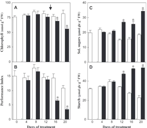
The Mg concentration in roots and shoots decreased after 4 d of treatment and the decrease was more abrupt in roots (Fig. 2B, C). At day 20, the Mg concentration in roots and shoots of Mg-deficient plants were 0.9 and 3.5 mg gÿ1DW, respectively, representing approximately one-third of the control values (Fig. 2B, C). Mg concentration in the upper-most fully expanded blades fell to 1.3 compared with 11.6 mg gÿ1 DW in control blades (data not shown), a value below 2 mg gÿ1 DW, which is considered as a general threshold value for the occurrence of Mg-deficiency symp-toms in leaves (Mengel and Kirkby, 1987). Very little is known about the mechanisms of Mg acquisition and re-partition between plant organs (Gardner, 2003). Shaul et al. (1999) and Shaul (2002) suggested that a Mg2+/H+antiporter called AtMHX1 (magnesium-proton exchanger) could de-termine the distribution of absorbed Mg2+ ions between the tonoplast and the cytosol of xylem parenchyma cells in the root vascular cylinder of Arabidopsis. AtMHX1 could act in a buffering capacity, by sequestering Mg2+ in the vacuole, in excess of that which can be loaded into the xylem (Shaul et al., 1999). It is proposed here that, under Fig. 3. Effect of Mg deficiency on chlorophyll concentration, photochemical performance, and sugar levels in Arabidopsis thaliana C24 (plants described in Fig. 1A, C, D). (A) Total chlorophyll concentration in uppermost expanded leaves. Mean of five acetone extractions 6SE. (B) Photochemical Performance Index of photosystem II (Hermans et al., 2003) in the uppermost expanded leaves. (C, D) Soluble sugars (sum of glucose, fructose, and sucrose) and starch concentration, expressed as lmol equivalent glucose gÿ1FW, in the uppermost expanded leaves. Samples were collected at the end of the photoperiod during the treatment. Mean of three replicate samples 6SE. White bars: control plants; black bars: Mg-deficient plants. Asterisks indicate significant difference between treatments at P=0.05 level or less. The arrow indicates the appearance of chlorosis.
deficiency conditions, the vacuolar Mg2+ storage pool in roots may readily be used to meet the demand in the shoots. However, Mg remobilization from the root can only pre-vent a drastic drop in shoot Mg concentrations for a limited period following Mg-withdrawal. (Fig. 2B, C).
Effect of Mg deficiency on photosynthetic apparatus and sugar levels
In order to establish the hierarchy of physiological respon-ses induced by Mg deficiency, the chlorophyll concentra-tion, the photochemical performance of photosystem II, and the sugar concentration in the uppermost fully expanded leaves of the rosettes were measured. The total chlorophyll concentration in Mg-deprived leaves first remained con-stant, despite the decrease of Mg concentration in shoots (Fig. 2B), but eventually decreased significantly (P <0.05) by one-tenth of the control value at day 16 and by one-third at day 20 (Fig. 3A). Nonetheless, it is likely that the chlorophyll concentration did not decrease as a direct
response to the deficiency, given that Mg deficiency was reported to increase the relative proportion of the Mg structural pool associated with chlorophyll (Mengel and Kirkby, 1987).
The detection of the Mg nutritional stress was also assessed using estimates of the Performance Index (PI ), characterizing overall photochemical processes in photo-system II (Hermans et al., 2003). The PI values started to decrease from day 16 in Mg-deficient leaves (Fig. 3B), along with the loss of chlorophyll (Fig. 3A) and the appearance of chlorosis (Fig. 1C). The total pool of sugars (sum of glucose, fructose, and sucrose) and starch increased in plants as an early response to Mg deficiency (Fig. 3C, D), before any significant decrease of the total chlorophyll concentration or of the PI (Fig. 3A, B). A change in leaf texture was noted as an early response to Mg deficiency, being obvious by day 12. Glucose first accumulated to more than twice the control value at day 12, and eventually sucrose by more than 2-fold at the end of the treatment (data not shown). Fig. 4. Iodine staining of starch in Arabidopsis thaliana Columbia exposed to Mg deficiency (treatment described in Fig. 1E). Mg-deficient and control plants were stained with iodine solution to reveal the presence of starch at the end of the dark period at day 14 of treatment, two days before the appearance of chlorotic symptoms (Fig. 1E).
To visualize the differences in starch distribution fol-lowing a dark period, iodine staining of whole Arabidopsis plants was performed. Starch was more abundant in the interveinal zones of the upper expanded leaves of the rosette (Fig. 4). The observed pattern of starch accumula-tion between the veins preceded the appearance of chlorotic symptoms (Fig. 1E).
A model of sucrose partitioning during Mg deficiency: differential distribution of sucrose depending on the position of source leaves
Increased sugar concentrations were found in the uppermost fully developed leaves of the rosette (Fig. 3C, D). These observations suggest that sucrose synthesis and utilization were out of balance, or that Mg deficiency somehow interfered with sucrose export from the source leaf or both simultaneously. However, it remains puzzling how Mg deficiency could affect sucrose partitioning. According to Cakmak et al. (1994b), in bean plants, the high suscepti-bility of sucrose partitioning to Mg delivery may be related to the carrier-mediated uptake of sucrose in the conducting complex of the phloem. This process requires MgATP as the substrate for H+-pumps (Delrot and Bonnemain, 1981; Bush, 1989). Conversely, Fischer et al. (1998) postulated that a congestion of metabolites in source leaves of spinach plants arises from a limited consumption in sink tissues, resulting from an inhibition of growth by low Mg supply.
To investigate the origins of sugar accumulation,14 [C]-sucrose movement was tracked in Arabidopsis plants (Grusak et al., 1990). The partitioning of radioactive molecules between sink organs depended on the position of the source leaves (Fig. 5). After distribution, the largest fraction was found in roots when the lowest source leaves of the rosette were labelled, and Mg deficiency did not affect the translocation to the roots or other sink organs (Fig. 5A). When the uppermost fully expanded leaves were labelled (Fig. 5B), the largest fraction of radioactivity was distributed to the floral stems of control plants. In Mg-deficient plants, more than one-tenth of the total labelling was not translocated and stayed in the upper expanded leaves (P <0.05), and less than half of the labelling was found in the floral stems compared with controls (Fig. 5B). The fact that as much 14[C]-sucrose molecules were detected in Mg-deficient sink organs as in the control ones, when radioactivity was applied to the lower mature leaves (Fig. 5A), tended to indicate that sucrose export from these leaves and sink metabolism were apparently not restricted in Mg-deficient plants. On the contrary, when14[C]-sucrose was applied to the uppermost fully expanded leaves, less radioactive material was detected in the sink organs (Fig. 5B). The 50% increase in sucrose accumulation in those leaves compared with control ones cannot account for the 300% reduction in marked molecules exported to the flower stems (Fig. 5B). This supports the idea of reduced sucrose export. These leaves, in which deficiency symptoms first
appeared (Fig. 1D), were also the ones with the lowest Mg level (1.3 mg Mg gÿ1DW at day 20 of treatment), below the critical Mg level (Mengel and Kirkby, 1987). Sub-sequently, the overall aerial biomass was decreased com-pared with control plants, while roots kept on growing normally. With the ongoing development of the deficiency, intermediate and lower leaves also started to accumulate higher sucrose and starch amount too.
The effect of Mg deficiency on photosynthetic gene expression: a likely effect of sugars
Sugar-derived signals regulate the expression of genes involved in general metabolism, but also in photosynthesis (Sheen, 1990; Oswald et al., 2001). It is possible, therefore, Fig. 5. Effect of Mg deficiency on14[C]-sucrose transport in Arabid-opsis thaliana C24 (plants described in Fig. 1A, C, D). Plants labelled apoplasmically after deposition of14[C]-sucrose molecules on the lowest leaf numbered 10–15 in order of appearance (A) and on the uppermost expanded leaf numbered 20–25 (B). 10 ll droplets containing 14 [C]-sucrose (9.25 kBq) were applied on abraded zones of leaves after 12 d of Mg deficiency treatment. Partitioning of labelled sucrose molecules between donor leaves and sink organs (root, floral stems, and youngest immature leaves) as a percentage of the total radioactivity measured in the whole plant. Mean 6SE of five replicates. Asterisk indicates significant difference between treatments at P=0.05 level or less.
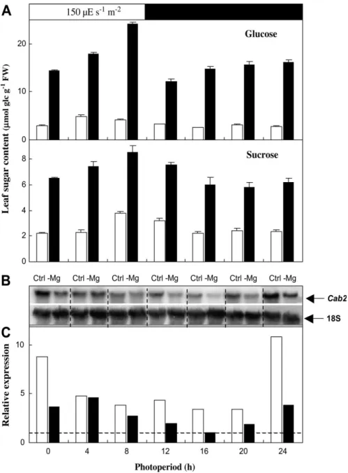
that many of the prospective changes in the photosynthetic apparatus that occur in shoots of Mg-deficient plants are effected through transcriptional changes elicited by in-creased leaf sugar concentrations. In view of this, northern analysis of Cab2, which encodes a chlorophyll a/b binding protein in photosystem II (Oswald et al., 2001) was performed. The diurnal transcripts profile of Cab2, together with sugar concentrations were analysed in the uppermost fully expanded leaves of A. thaliana Columbia rosettes
during day 11 of treatment. Glucose, fructose (data not shown), and sucrose concentrations gradually increased upon illumination (Fig. 6A) and showed a similar diurnal profile to that previously observed in Arabidopsis by Zeeman and Rees (1999), and Gibon et al. (2004). However, sugar levels were two or three times greater in Mg-deficient than in control leaves. Cab2 transcripts in control plants were more abundant during the first h of the light period (Fig. 6B, C) and decreased thereafter. A similar
Fig. 6. Effect of Mg deficiency on the diurnal accumulation of sugars and on Cab2 regulation in Arabidopsis thaliana Columbia exposed to Mg deficiency (plants described in Fig. 1B). (A) Glucose and sucrose concentrations, expressed as lmol equivalent glucose gÿ1FW, in the uppermost expanded leaves (typically numbered 20–25 from the oldest to the most recent) of rosettes during the photoperiod of day 11. Means of three extractions of a pooled powder of five plants 6SE. (B) Northern blot analysis of Cab2 expression on day 11. Fifteen lg of total RNA were loaded in each lane. (C) Quantified signals of Cab2 normalized to the 18S loading control. The signal of Mg-deficient sample at 16 h was defined as 1 (dashed line). The experiment was repeated twice with similar observations. White columns: control plants, black columns: Mg-deficient plants. Physiological parameters on day 11 of treatment were in control and Mg-deficient plants as followed: root fresh biomass: 0.4960.02 g and 0.5460.02 g, shoot biomass: 3.0560.09 g and 2.2060.07 g (n=35); total chlorophyll concentration: 22366.4 and 20069.2 nmol gÿ1FW (n=5).
profile was observed in Mg-deficient plants, however, the transcript levels were lower in Mg-deprived plants, prior to a marked decrease in the total chlorophyll concentra-tion (see legend to Fig. 6).
A negative correlation has widely been reported between sugar concentration and chlorophyll concentration, photo-synthetic activity, and photophoto-synthetic genes expression (Foyer, 1988; Sheen, 1990; Scha¨fer et al., 1992; Oswald et al., 2001). Oswald et al. (2001) proposed a model where photosynthetic electron transport and cytosolic sugar status could give rise to a signal that regulates Cab2 transcription in cell suspension culture of Arabidopsis thaliana. Here, with Mg deficiency, high sugar levels in source leaves possibly repress Cab2 expression and, to some extent, could be responsible for the decrease in chlorophyll con-centration. Indeed, decreased Cab2 transcript abundance broadly reflects a lower chlorophyll concentration in the presence of exogenous sugar (Oswald et al., 2001; Martin et al., 2002). Therefore, we suggest that the chlorophyll breakdown upon Mg deficiency involves a depressive effect of sugars on Cab2 expression, rather than a lack of Mg atoms for chelating chlorophyll molecules. In the same way, it was previously shown that high sugar and low nitrate supplies jointly caused a reduction in Cab trans-cripts abundance (Martin et al., 2002).
In conclusion, these data support the hypothesis that Mg deficiency in Arabidopsis affects sucrose export from source leaves (starting with the youngest ones), rather than sink growth and that sugar accumulation in leaves has a negative effect on photosynthetic gene transcription and photosynthetic activity. A proper understanding of Mg deficiency in plants will require future intensive studies linking physiological and biochemical processes with sugar signalling and sugar regulation of genes involved in carbohydrate metabolism, long-distance transport, and partitioning.
Acknowledgements
C Hermans received grant-aided support from the Fonds pour la Formation a` la Recherche dans l’Industrie et l’Agriculture (Belgium). This research was supported by grant from the Interuni-versity Attraction Pole Programme: Belgian Science Policy (Project V/13). We thank Professor RJ Strasser (Bioenergetics Laboratory, University of Geneva, Switzerland) for his support in chlorophyll fluorescence measurement and for his supervision over many years. Professor Elaine Tobbin (Department of Molecular, Cell and De-velopmental Biology, UCLA, Los Angeles, USA) is thanked for the Cab2 clone and Dr GN Johnson (Biological School, The University of Manchester, UK) for proofreading. We are also grateful to Professor J-P Dehaye for access to the Scintillator.
References
Ausubel FM, Brent R, Kingston RE, Moore D, Seidman JG, Smith JA, Struhl K (eds).1998. Current protocols in molecular biology. New York, USA: Greene Publishing Associates and Wiley-Interscience.
Beale SI.1999. Enzymes of chlorophyll biosynthesis. Photosynthesis Research 60, 43–73.
Bush DR.1989. Proton-coupled sucrose transport in plasmalemma vesicles isolated from sugar beet (Beta vulgaris L. cv. Great Western) leaves. Plant Physiology 89, 1318–1323.
Cakmak I, Hengeler C, Marschner H.1994a. Partitioning of shoot and root dry matter and carbohydrates in bean plants suffering from phosphorus, potassium and magnesium deficiency. Journal of Experimental Botany 45, 1245–1250.
Cakmak I, Hengeler C, Marschner H.1994b. Changes in phloem export of sucrose in leaves in response to phosphorus, potassium and magnesium deficiency in bean plants. Journal of Experimental Botany 45, 1251–1257.
Cao W, Tibbits TW.1992. Growth, carbon dioxide exchange and mineral accumulation in potatoes grown at different magnesium concentrations. Journal of Plant Nutrition 15, 1359–1371. Delrot S, Bonnemain JL.1981. Involvement of protons as substrate
for the sucrose carrier during phloem loading in Vicia faba leaves. Plant Physiology 67, 560–564.
Evans HJ, Sorger GJ. 1966. Role of mineral elements with emphasis on the univalent cations. Annual Review of Plant Physiology 17, 47–76.
Fischer ES, Bremer E.1993. Influence of magnesium deficiency on rates of leaf expansion, starch and sucrose accumulation and net assimilation in Phaseolus vulgaris. Physiologia Plantarum 89, 271–276.
Fischer ES, Lohaus G, Heineke D, Heldt HW.1998. Magnesium deficiency results in accumulation of carbohydrates and amino acids in source and sink leaves of spinach. Physiologia Plantarum 102,16–20.
Foyer CH. 1988. Feedback inhibition of photosynthesis through source–sink regulation in leaves. Plant Physiology and Biochem-istry 26, 483–492.
Gardner RC. 2003. Genes for magnesium transport. Current Opinion in Plant Biology 6, 263–267.
Gibon Y, Bla¨sing OE, Palacios-Rojas N, Pankovic D, Hendriks JHM, Fisahn J, Ho¨hne M, Gu¨nther M, Stitt M. 2004. Adjustment of diurnal starch turnover to short days: de-pletion of sugar during the night leads to a temporary inhibition of carbohydrate utilization, accumulation of sugars and post-translational activation of ADP-glucose pyrophosphorylase in the following light period. The Plant Journal 39, 847–862.
Grusak MA, Delrot S, Ntsika G.1990. Short-term effect of heat-girdles on source leaves of Vicia faba: Analysis of phloem loading and carbon partitioning parameters. Journal of Experimental Botany 41, 1371–1377.
Guller L, Krucka M. 1993. Ultrastructure of grape-vine (Vitis vinifera) chloroplasts under Mg- and Fe-deficiency. Photo-synthetica 29, 417–425.
Hariadi Y, Shabala S.2004a. Screening broad beans (Vicia faba) for magnesium deficiency. I. Growth characteristics, visual de-ficiency symptoms and plant nutrition status. Functional Plant Biology 31, 529–537.
Hariadi Y, Shabala S.2004b. Screening broad beans (Vicia faba) for magnesium deficiency. II. Photosynthetic performance and leaf bioelectrical responses. Growth characteristics, visual de-ficiency symptoms and plant nutrition status. Functional Plant Biology 31, 539–549.
Hermans C, Bourgis F, Faucher M, Delrot S, Strasser RJ, Verbruggen N.2005. Magnesium deficiency in sugar beet alters sugar partitioning and phloem loading in young mature leaves. Planta 220, 541–549.
Hermans C, Johnson GN, Strasser RJ, Verbruggen N. 2004. Physiological characterization of magnesium deficiency in sugar
beet: acclimation to low magnesium differentially affects photo-systems I and II. Planta 220, 344–355.
Hermans C, Smeyers M, Rodriguez RM, Eyletters M, Strasser RJ, Delhaye JP. 2003. Quality assessment of urban trees: a comparative study of physiological characterization, airborne imaging and on-site fluorescence monitoring by the OJIP-test. Journal of Plant Physiology 160, 81–90.
Igamberdiev AU, Kleczkowski LA.2001. Implications of adenylate kinase-governed equilibrium of adenylates on contents of free magnesium in plant cells and compartments. Biochemical Journal 360,225–231.
Igamberdiev AU, Kleczkowski LA. 2003. Membrane potential, adenylate levels and Mg2+are interconnected via adenylate kinase equilibrium in plant cells. Biochimica et Biophysica Acta 1607, 111–119.
Kaftan D, Brumfeld V, Nevo R, Scherz A, Reich Z.2002. From chloroplasts to photosystems: in situ scanning force microscopy on intact thylakoid membranes. EMBO Journal 21, 6246–6253. Laing W, Greer D, Sun O, Beets P, Lowe A, Payn T. 2000.
Pysiological impacts of Mg deficiency in Pinus radiata: growth and photosynthesis. New Phytologist 146, 47–57.
Leutwiler LS, Meyerowitz EM, Tobin EM.1986. Structure and expression of three light-harvesting chlorophyll a/b binding protein genes in Arabidopsis thaliana. Nucleic Acids Research 14,4051–4064.
Lu YK, Chen YR, Yang CM. 1995. Influence of Fe- and Mg-deficiency on the thylakoid membranes of a chlorophyll-deficient ch5 mutant of Arabidopsis thaliana. Botanical Bulletin of Acade-mia Sinica 36, 175–179.
Marschner H. 1995. Mineral nutrition of higher plants. Berlin: Springer-Verlag.
Martin T, Oswald O, Graham IA. 2002. Arabidopsis seedling growth, storage lipid mobilization, and photosynthetic gene expression are regulated by carbon:nitrogen availability. Plant Physiology 128, 472–481.
Mehne-Jakobs B.1995. The influence of magnesium-deficiency on carbohydrate concentrations in Norway spruce (Picea abies) needles. Tree Physiology 15, 577–584.
Mengel K, Kirkby EA.1987. Principles of plant nutrition, 4th edn. Worblaufen-Bern, Switzerland: International Potash Institute, 481–492.
Oswald O, Martin T, Dominy PJ, Graham IA.2001. Plastid redox state and sugars: interactive regulators of nuclear-encoded photo-synthetic gene expression. Proceedings of the National Academy of Sciences, USA 98, 2047–2052.
Scha¨fer C, Simper H, Hofmann B. 1992. Glucose feeding results in coordinated changes in chlorophyll content, ribulose-1,5-bisphosphate carboxylase-oxygenase activity and photosyn-thetic potential in photoautotrophic suspension cultured cells of Chenopodium rubrum. Plant, Cell and Environment 15, 343–350.
Shaul O, Hilgemann DW, Aldmeida-Engler J, Van Montagu M, Inze´, Galili G. 1999. Cloning and characterization of a novel Mg2+/H+exchanger. EMBO Journal 18, 3973–3980.
Shaul O.2002. Magnesium transport and function in plants: the tip of the iceberg. Biometals 15, 309–323.
Sheen J.1990. Metabolic repression of transcription in higher plants. The Plant Cell 2, 1027–1038.
Sokolov LN, De´jardin A, Kleczkowski LA.1998. Sugars and light/ dark exposure trigger differential regulation of ADP-glucose pyrophosphorylase genes in Arabidopsis thaliana (thale cress). Biochemical Journal 336, 681–687.
Strasser RJ, Srivastava A, Govindjee. 1995. Polyphasic chloro-phyll a fluorescence transient in plants and cyanobacteria. Photo-chemistry and Photobiology 61, 32–42.
Sun OJ, Gielen GJHP, Tattersall Smith RSC, Thorn AJ.2001. Growth, Mg nutrition and photosynthetic activity of Pinus radiata: evidence that NaCl addition counteracts the impact of low Mg supply. Trees 15, 335–340.
Sun OJ, Payn TW.1999. Magnesium nutrition and photosynthesis in Pinus radiata: clonal variation and influence of potassium. Tree Physiology 19, 535–540.
Terry N, Ulrich A.1974. Effects of magnesium deficiency on the photosynthesis and respiration of leaves of sugar beet. Plant Physiology 54, 379–381.
Wu W, Peters J, Berkowitz GA.1991. Surface charge-mediated effects of Mg2+ on K+ flux across the chloroplast envelope are associated with regulation of stromal pH and photosynthesis. Plant Physiology 97, 580–587.
Zeeman SC, Rees T.1999. Changes in carbohydrates metabolism and assimilate export in starch-excess mutants of Arabidopsis. Plant, Cell and Environment 22, 1445–1453.
![Fig. 5. Effect of Mg deficiency on 14 [C]-sucrose transport in Arabid- Arabid-opsis thaliana C24 (plants described in Fig](https://thumb-eu.123doks.com/thumbv2/123doknet/14904411.655290/6.918.496.816.86.627/effect-deficiency-sucrose-transport-arabid-arabid-thaliana-described.webp)