. . . .
. . . .
RIP3, a kinase promoting necroptotic cell death,
mediates adverse remodelling after myocardial
infarction
Mark Luedde
1†, Matthias Lutz
1†, Natalie Carter
1†, Justyna Sosna
2, Christoph Jacoby
3,
Mihael Vucur
4, Je´re´mie Gautheron
4, Christoph Roderburg
4, Nadine Borg
3,
Florian Reisinger
5, Hans-Joerg Hippe
1, Andreas Linkermann
6, Monika J. Wolf
7,
Stefan Rose-John
8, Renate Lu¨llmann-Rauch
9, Dieter Adam
2, Ulrich Flo¨gel
3,
Mathias Heikenwalder
6, Tom Luedde
4, and Norbert Frey
1,10*
1
Department of Internal Medicine III: Cardiology and Angiology, University of Kiel, Arnold-Heller-Straße 3, Haus 6, 24105 Kiel, Germany;2
Institute of Immunology, University of Kiel, Kiel, Germany;3
Department of Molecular Cardiology, University of Duesseldorf, Duesseldorf, Germany;4
Department of Internal Medicine III, University Hospital, Aachen, Germany;5 Institute of Virology, Technical University of Munich, Munich, Germany;6
Department of Nephrology, University of Kiel, Kiel, Germany;7
Institute of Physiology, University of Zurich and Zurich Center for Integrative Human Physiology, Zurich, Switzerland;8
Department of Biochemistry, University of Kiel, Kiel, Germany;9
Department of Anatomy, University of Kiel, Kiel, Germany; and10 DZHK (German Centre for Cardiovascular Research), Partner Site Hamburg/Kiel/Lu¨beck, Kiel, Germany
Received 1 April 2014; revised 27 May 2014; accepted 30 May 2014; online publish-ahead-of-print 11 June 2014 Time for primary review: 14 days
Aims Programmed necrosis (necroptosis) represents a newly identified mechanism of cell death combining features of both apoptosis and necrosis. Like apoptosis, necroptosis is tightly regulated by distinct signalling pathways. A key regulatory role in programmed necrosis has been attributed to interactions of the receptor-interacting protein kinases, RIP1 and RIP3. However, the specific functional role of RIP3-dependent signalling and necroptosis in the heart is unknown. The aims of this study were thus to assess the significance of necroptosis and RIP3 in the context of myocardial ischaemia.
Methods and results
Immunoblots revealed strong expression of RIP3 in murine hearts, indicating potential functional significance of this protein in the myocardium. Consistent with a role in promoting necroptosis, adenoviral overexpression of RIP3 in neo-natal rat cardiomyocytes and stimulation with TNF-a induced the formation of a complex of RIP1 and RIP3. Moreover, RIP3 overexpression was sufficient to induce necroptosis of cardiomyocytes. In vivo, cardiac expression of RIP3 was up-regulated upon myocardial infarction (MI). Conversely, mice deficient for RIP3 (RIP32/2) showed a significantly better ejection fraction (45 + 3.6 vs. 32 + 4.4%, P , 0.05) and less hypertrophy in magnetic resonance imaging studies 30 days after experimental infarction due to left anterior descending coronary artery ligation. This was accompanied by a dimin-ished inflammatory response of infarcted hearts and decreased generation of reactive oxygen species.
Conclusion Here, we show that RIP3-dependent necroptosis modulates post-ischaemic adverse remodelling in a mouse model of MI.
This novel signalling pathway may thus be an attractive target for future therapies that aim to limit the adverse conse-quences of myocardial ischaemia.
-Keywords Programmed necrosis † Receptor interacting protein 3 † Myocardial infarction † Remodelling † Inflammation
1. Introduction
Acute myocardial infarction is one of the leading causes of death world-wide.1Despite considerable progress, contemporary therapy can only partially address the central problem that leads from acute ischaemia
and infarction to chronic heart failure: a major loss of cardiomyocytes with subsequent remodelling and contractile dysfunction.
Apoptosis, which is mediated by death receptors like the TNF recep-tor2or fas receptor/CD95,3has been considered a possible target for novel therapies,4as this process is tightly regulated by specific signalling
†Mar.L., Mat.L., and N.C. contributed equally to this work.
*Corresponding author. Tel:+49 431/597 1441; fax: +49 431/597 1470, Email: norbert.frey@uksh.de, freynorbert@hotmail.com
Published on behalf of the European Society of Cardiology. All rights reserved.&The Author 2014. For permissions please email: journals.permissions@oup.com.
Cardiovascular Research (2014) 103, 206–216 doi:10.1093/cvr/cvu146
pathways and could thus potentially be inhibited. For example, TNF-a has been shown to be up-regulated during myocardial infarction (MI) and to enhance apoptosis in this clinical condition.5 However, the overall rate of apoptotic cells in the infarcted region was ,1% in this study and recent theories question a significant role of apoptosis in post-ischaemic remodelling.6,7 Recently, a novel mechanism called ‘pro-grammed necrosis’ or necroptosis has been suggested as another im-portant mediator of cell death in the heart.6Similar to apoptotic cell death, this process is tightly regulated by distinct molecules, but leads to the typical morphological signs of necrosis such as defects of membrane integrity and inflammation, thus combining features of both mechan-isms.6,8–11The signalling pathways that activate programmed necrosis are not fully understood. However, in vitro studies could demonstrate that TNF-a-dependent formation of a complex between receptor-interacting protein 1 (RIP1) and another kinase, receptor-receptor-interacting protein 3 (RIP3) is an essential step for inducing programmed necro-sis.8,9,12In this process, RIP3 appears to play a key role, controlling the phosphorylation of RIP1, a necessary step in programmed necrosis.8,12
An important role of RIP3-mediated necroptosis has been demon-strated during viral infection of the liver and in tissue damage due to in-flammatory bowel disease.13In the heart, inhibition of RIP1 by the small molecule necrostatin-1 leads to a reduction of infarct size, implying func-tional importance of necroptosis also in myocardial ischaemia.14–16In contrast, the functional significance of RIP3 in the heart is still unknown. Here, we demonstrate that RIP3 is expressed in the heart and RIP3 is activated during MI. Moreover, mice lacking RIP3 reveal improved cardiac performance, accompanied by a decreased inflamma-tory response and generation of reactive oxygen species (ROS).
2. Methods
2.1 In vitro experiments
For the preparation of neonatal rat ventricular cardiomyocytes (NRVCMs), 1- to 2-day-old Wistar rats were sacrificed by decapitation and hearts were immediately removed for digestion with collagenase II (Worthington) at 378C, as previously described.17Cells were infected with the indicated multi-plicity of infection under serum-free conditions 24 h after plating and subsequently cultured supplemented with 2% fetal calf serum for 48 h. After-wards, cells were analysed by flow cytometric analysis, immunoblots, MTT cell viability assays, real-time PCR, immunohistology, or electron micros-copy (see Supplementary material online for detailed description of applied methods).
2.2 In vivo studies
RIP3-deficient (RIP32/2) mice,18kindly provided by Genentech, Inc., South San Francisco, CA, USA, and littermate WT mice (C57/BL6N-background) were studied at 10 – 12 weeks of age. Mice were subjected to permanent left anterior descending coronary artery (LAD) ligation and subsequently assessed for left ventricular (LV) morphology and function. Furthermore, transgenic and WT mouse hearts were analysed by (immuno-)histology and electron microscopy as well as for the content of ROS. For LAD ligation as well as heart removal, mice received 0.1 mg Temgesic/kg body weight by subcutaneous injection for analgesia and were anaesthetized by inhalation of isoflurane (3 – 5%). For postoperative analgesia, 0.1 mg Temgesic/kg body weight was applied once daily for 5 days post-surgery. All studies were approved by the animal ethics committee of Schleswig-Holstein, Germany. Magnetic resonance imaging (MRI) experiments were performed as described elsewhere.19Detailed methods are available in Supplementary material online.
2.3 Statistical analysis
All results were expressed as mean + SEM. We performed statistical ana-lysis using one- or two-way ANOVA followed by Student – Newman – Keuls post hoc tests or t-tests as appropriate. A P-value of ,0.05 was consid-ered statistically significant.
3. Results
3.1 RIP3 is expressed in cardiomyocytes and
co-localizes with mitochondria
In order to unravel a potential function of RIP3 in the myocardium, we first assessed its expression in the heart. While cardiac expression of RIP3 (55 kDa) was lower compared with spleen and lung, it was signifi-cantly higher than in brain and kidney (Figure1A and B). Of note, the shifted bands above from kDa in liver and kidney might be due to post-translational modification of RIP3. RIP3 was also expressed in isolated NRVCMs (Figure1C). Since it has been suggested that expression of RIP3 correlates with the functional relevance of programmed necrosis in certain cell types,9we hypothesized that RIP3 may also play a signifi-cant role in the heart. Adenoviral overexpression of RIP3 led to a shift in its molecular weight, indicating activation of RIP3.20 As a negative control, this shifted band could be inhibited by RIP3-specific synthetic miRNA (Figure1C). As a positive control, we stimulated L929 mouse fibroblasts that are particularly prone to programmed necrosis,9,20 with TNF-a and Z-Val-Ala-Asp-FMK (zVAD) caspase inhibitor. After 3 h of stimulation, immunoblots from these cells revealed the shifted band at the same molecular weight as activated RIP3 in NRVCMs (Figure 1D), suggesting that the molecular weight shift of RIP3 in NRVCMs is indeed due to activation of RIP3. Analyses of NRVCMs by confocal laser scanning immunohistochemistry confirmed endogenous RIP3 expression in NRVCMs and showed perinuclear clustering of RIP3 (Figure1D). In this regard, it has recently been reported that mitochon-dria reveal perinuclear clustering during programmed cell death.21 Inter-estingly, co-staining of NRVCMs with a RIP3 antibody and ‘Mitotracker’ revealed close co-localization of RIP3 and mitochondria in these cells (Figure1E – G). AdV RIP3 induced increased phosphorylation activity in NRVCMs (Figure 2A). In contrast, overexpression of a kinase-dead RIP3 mutant did not induce increased phosphorylation of RIP3, demon-strating that adenoviral overexpression of RIP3 promotes kinase activity of RIP3. As an additional indication of RIP3 activation, immunoblots revealed increased polyubiquitination by Lys63 (K63) residue linkage at the level of activated RIP3 (+151.9% vs. lacZ +24.9%, Figure2B and C ).
3.2 Overexpression of RIP3 induces the
formation of RIP1/RIP3 complex and
necrosis of cardiomyocytes
Cell culture experiments in human cell lines showed that association of RIP1 and RIP3 in response to TNF-a stimulation represents the crucial initial step in programmed necrosis.8,12To test whether TNF-dependent formation of RIP1/RIP3 complexes is also present in cardiomyocytes, we infected NRVCMs with an adenoviral vector encoding for rat RIP3 (NCBI Reference Sequence: NM_139342.1) or GFP (green fluorescent protein) as control. Forty-eight hours after infection, NRVCMs were pretreated with a 50 mM zVAD-fmk caspase inhibitor, since caspases have been shown to inhibit RIP3 – RIP1 complex formation in vitro,12 and stimulated with 100 ng/mL of recombinant TNF-a for different
time intervals. Immunoprecipitation with a RIP1 antibody followed by immunoblotting with a RIP3 antibody revealed rapid formation of RIP1/RIP3 complexes as early as 30 min after TNF stimulation with a maximum at 60 min (Figure3A). As a sign of increased turnover of
RIP1/RIP3 complexes in RIP3 overexpressing NRVCMs, RIP1 content was significantly decreased in these cells (287.6 + 2.7%, P , 0.001, n ¼ 3; Figure3B and C). Next, we assessed the consequences of RIP3 ac-tivation on cardiomyocyte survival. Of note, adenoviral overexpression Figure 1 RIP3 is expressed in cardiomyocytes. (A) Immunoblot revealing RIP3 expression in the mouse heart. GAPDH was used as loading control. RIP3 expression in the heart is lower compared with lung and spleen, higher than in brain and kidney (n ¼ 3, B). (C ) Immunoblot showing that RIP3 is expressed in NRVCMs. Adenoviral overexpression of RIP3 induced a shift of RIP3 in molecular weight (RIP3 OE). Both endogenous and weight-shifted RIP3 expression could be inhibited by two miRNA viruses: miRIP3-1 and miRIP3-2, but not by overexpression with miRNA control virus (micon), demonstrating the spe-cificity of the shifted RIP3-bar. (D) As a positive control, stimulation of L929 mouse fibroblast with TNF-a and zVAD induced a shift in the molecular weight of RIP3. (E) Confocal laser immunocytology: co-staining of RIP3 (stained red) and a-actinin in an NRVCM. In NRVCMs, RIP3 showed a perinuclear clustering (F, stained green), similar to mitochondria (G, stained red). (H ) Co-staining of mitochondria and RIP3. n ¼ 3 independent experiments. Con: unstimulated control; AdV RIP3: RIP3 adenovirus.
M. Luedde et al.
of RIP3 significantly increased the amount of propidium iodide (PI)-positive cells (68.9 + 1.2 vs. 26.8 + 1.3% in unstimulated control cells, P , 0.01, Figure 3D and E), as measured by FACS analysis. Notably, overexpression of a kinase-inactive RIP3 mutant (RIP3 K51A) did not alter the proportion of PI-positive cells, demonstrating that kinase activity of RIP3 is required for the induction of cell death of NRVCMs (see Supplementary material online, Figure S1). Combined 7AAD/annexinV staining revealed no difference in apoptotic cell rates between both groups (data not shown). Simultaneous TNF-a stimula-tion and caspase inhibistimula-tion by zVAD-FMK slightly decreased the RIP3-dependent cell death rate (Figure3E). However, neither TNF-a stimulation nor caspase inhibition alone affected RIP3-dependent cell death rate (see Supplementary material online, Figure S2), confirming that RIP3-induced cell death is not executed via the ‘canonical’ apoptotic caspase-dependent pathway. In line with the FACS analyses, adenoviral overexpression of RIP3 markedly reduced survival of NRVCMs (257 + 0.83% vs. control, P , 0.01, Figure3F), as measured by another cell via-bility assay (MTT assay) and significantly increased cell lysis, as measured by augmented troponin T release into the cell culture media (2143 + 337 pg/mL vs. 801 + 89 pg/mL, P , 0.01, Figure3G). In contrast, adeno-viral miRNA-based knockdown of RIP3 did not alter the basal cell death rate of cultured cardiomyocytes (data not shown). This effect might favour the concept that RIP3-dependent effects are confined to stress
conditions and are dispensable under basic conditions. Of note, miRNA-based knockdown of RIP1 (see Supplementary material online, Figure S2C ) only weakly inhibited RIP3-dependent cell death (see Supplemen-tary material online, Figure S2D), indicating that RIP1 activity is not required for RIP3-mediated cell death of cardiomyocytes. In conclusion, we demonstrate that RIP3 is sufficient for induction of programmed necrosis in cardiomyocytes.
3.3 RIP3 expression is induced in myocardial
infarction
To probe for a potential pathophysiological role of RIP3 in vivo, we exam-ined activation of RIP3-dependent signalling in a mouse model of MI. Interestingly, 24 h after permanent ligation of the LAD, significantly up-regulated RIP3 protein levels were detected in ischaemic mouse hearts (+70.69 + 14.36%, P , 0.05, Figure4A and B), consistent with ac-tivation of RIP3-dependent signalling pathways in response to myocardial ischaemia. Histological analyses revealed up-regulation of RIP3 especially in the peri-infarct zone of the LV (Figure3E), implying functional relevance of RIP3 in myocardial infarction.
3.4 RIP3 mediates inflammation and ROS
generation after myocardial infarction
To further examine the functional relevance of RIP3 in MI, we subjected RIP32/2mice18to permanent LAD ligation (n ¼ 22). This intervention did not result in differences with regard to the survival rate compared with WT controls (see Supplementary material online, Figure S3A). Next, we evaluated the effects of RIP3 deficiency on biochemical, histo-logical, and ultrastructural levels. Haematoxylin – eosin stainings of WT and RIP32/2hearts after MI are shown in Figure5A. To examine the role of apoptosis in RIP3-dependent remodelling, we measured caspase 3 cleavage in WT and RIP32/2hearts 24 h after LAD ligation. By immuno-histology and western blot, we did not detect alterations of caspase 3 cleavage in both RIP32/2hearts and WT controls (Figure5B – D). Fur-thermore, stainings for Cd31 revealed a comparable decrease of endo-thelial cells in the infarcted areas in both genotypes (Figure5E). Likewise, electron microscopy of both WT and RIP32/2 hearts revealed no obvious morphological differences between both groups (Figure5F). In contrast, 4 days after infarction we observed a marked attenuation of in-flammatory cell invasion into infarcted regions in RIP3-deficient hearts (Figure5B). Inflammation is a major consequence of myocardial ischae-mia19and —in contrast to apoptotic cell death—a pivotal concomitant effect of programmed necrosis in vivo.9,12We therefore systematically assessed whether the degree of inflammation was decreased in RIP32/2hearts after LAD ligation. Indeed, CD3 staining and quantifica-tion of T-cell invasion revealed a significantly lower number of inflamma-tory cells in RIP32/2 hearts (Figure 6A and B). The decreased inflammatory response of RIP32/2hearts was accompanied by an un-altered systemic level of leucocytes and T cells in RIP32/2mice com-pared with controls. Furthermore, we could not detect any morphological alterations of livers, lungs, kidneys, and spleens of RIP3 and WT mice 4 days after LAD ligation (see Supplementary material online, Figure S4). In addition to inflammation, generation of ROS has been presumed to be another key mediator of RIP3-induced organ damage.6,10We therefore examined RIP32/2hearts and WT controls for ROS content 24 h after LAD ligation. RIP32/2hearts revealed signifi-cantly decreased ROS generation vs. WT hearts (2.31 + 0.58 vs. 5.32 + 1.18, P , 0.05, Figure6C). In a reverse approach, we also assessed culti-vated NRVCMs overexpressing RIP3 for ROS contents by FACS analysis. Figure 2 Increased phosphorylation and ubiquitination activity ofRIP3 in neonatal rat cardiomyocytes. (A) Semi-quantitative phospho-gel staining showing increased phosphorylating activity in cells overex-pressing RIP3. Overexpression of a kinase-dead RIP3 mutant (AdRIP3 A51 K) led to no increase of phosphorylation activity (n ¼ 3). (B) Immunoblot revealing polyubiquitin chains formed by Lys63 (K63) residue linkage at the level of activated RIP3 in NRVCMs that were infected with a RIP3 adenovirus vs. control cells that were infected with a LacZ virus (AdVLacZ), demonstrating increased ubiqitination ac-tivity of activated RIP3 (+151.9% vs. lacZ +24.9%, n ¼ 3, C). NRVCMs overexpressing a kinase-dead RIP3 mutant (AdV RIP3 K51A)) served as negative control, and immunoblot against GAPDH served as loading control. *P , 0.01.
Interestingly, these cells showed an increased ROS content (+25 + 3.6% vs. LacZ-expressing cells, Figure6D), emphasizing the functional role of ROS generation as a potential primary effector of RIP3 in the heart. Taken together, ROS generation and inflammation could both function as mediators of RIP3-dependent myocardial damage due to is-chaemia. To further assess the role of altered cardiac energetics in RIP3-dependent remodelling, we used31P magnetic resonance spectroscopy. We found a higher PCr/ATP ratio of RIP2/2mouse hearts vs. WT con-trols; however, the difference did not reach statistical significance (see
Supplementary material online, Figure S5). Thus, likely additional mechanisms are also relevant in RIP3-dependent cardiac remodelling.
3.5 RIP3 mediates adverse remodelling after
myocardial infarction
To examine whether RIP3 impairs cardiac remodelling after MI, we assessed cardiac morphology and function of RIP32/2mice and WT controls under basal conditions, 24 h, and 30 days post-MI by cardiac Figure 3 RIP3 induces programmed necrosis of cardiomyocytes. (A) Adenoviral overexpression of RIP3 in NRVCMs stimulates the formation of a RIP1/ RIP3 complex, as shown by immunoprecipitation after stimulation with TNF-a and pre-incubation with the pan-caspase inhibitor zVAD-fmk. (B) Immuno-blot revealing that adenoviral overexpression of RIP3 leads to a significant decline in RIP1 expression (287.6 + 2.7%, P , 0.001, n ¼ 3, C ). (D) PI staining and FACS analysis show significantly increased cell death of NRVCMs overexpressing RIP3. zVAD-fmk and TNF-a stimulation did not significantly alter the proportion of PI-positive cells (E), n ¼ 4 independent experiments. (F ) Treatment of NRVCMs with zVAD-fmk and TNF-a did not significantly alter cell viability, whereas overexpression of RIP3 in NRVCMs markedly decreased cardiomyocyte survival (n ¼ 4). (G) Overexpression of RIP3 in NRVCMs lead to a significant increase of cell lysis, as measured by troponin T release into culture media (n ¼ 4). Con: unstimulated control; AdV GFP, AdV LacZ: control adenovirus; AdV RIP3: RIP3 adenovirus; AdV RIP3 K51A: RIP3 virus expressing a kinase-inactive RIP3 mutant; PI: propidium iodide; FL2H: fluorescence signal (high); FSC-H: forward scatter; *P , 0.05;†P , 0.01;‡P , 0.001.
M. Luedde et al.
MRI (Figure7and see Supplementary material online, Movies). Basic mor-phological and functional parameters were comparable between both groups (Table1). Noteworthy, gadolinium-enhanced MRI scans of the left ventricle 24 h after LAD ligation and histology-based quantification of fibrotic areas 30 days after LAD ligation showed no significant differ-ences in infarct size between both groups (Figure7B and D, and see Sup-plementary material online, Figure S6).
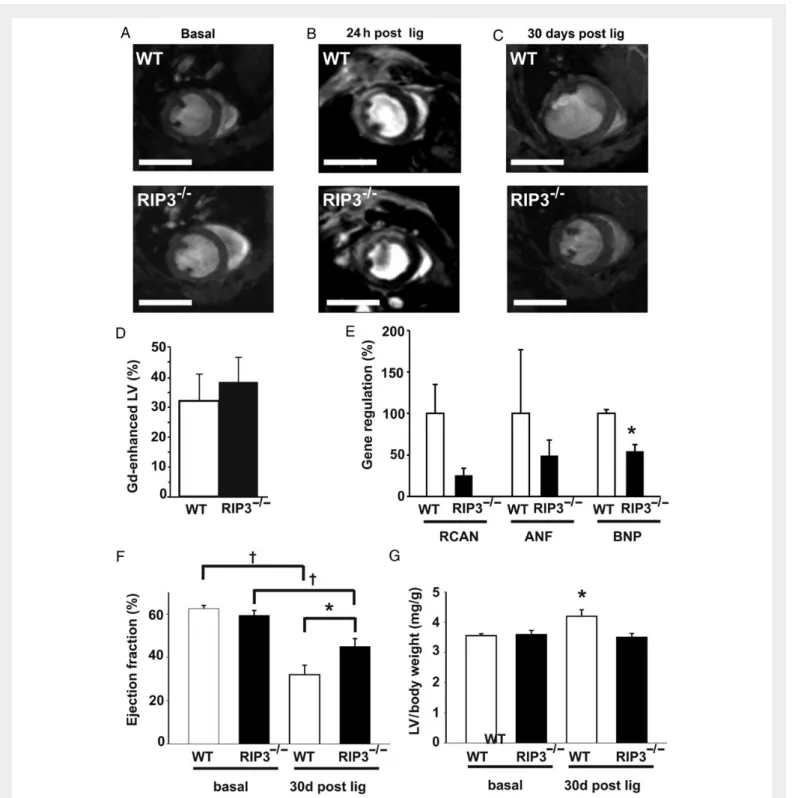
However, 30 days after LAD ligation, RIP32/2mice demonstrated a significantly better ejection fraction than WT controls (45 + 3.6%, n ¼ 11 vs. 32 + 4.4%, n ¼ 10, P , 0.05; Figure 7F). Consistently, RIP32/2hearts showed less hypertrophy than WT controls, as demon-strated by a decreased LV/body weight ratio (3.50 + 0.13 mg/g, n ¼ 11 vs. 4.20 + 0.21 mg/g, n ¼ 10, P , 0.05; Figure7G). Moreover, real-time PCR experiments revealed lower levels of B-type natriuretic peptide (BNP) expression in RIP3-deficient hearts (Figure7E) and serum tropo-nin T levels were significantly lower in RIP3-deficient mice (see Supple-mentary material online, Figure S3B), also consistent with improved myocardial remodelling of these mice post-infarction. Taken together,
these data indicate that RIP3 does not alter short-term infarct size, but promotes long-term adverse post-infarct remodelling in vivo.
4. Discussion
Programmed necrosis mediated by RIP3 has recently been identified as a novel mechanism of cell death with major functional importance in several organs, including liver,12 pancreas,9 and bowel.13 We here show that RIP3 is also expressed in the heart, forms a complex with RIP1 in cardiomyocytes, and can be activated upon TNF stimulation. Nevertheless, cardiomyocytes appear to be less prone to necroptosis compared with, e.g. Jurkat cells,12since caspase inhibition and TNF-a stimulation alone were not sufficient to induce significant cell death. In contrast, forced overexpression of RIP3 is a potent stimulus for necrop-tosis of NRVCMs, in line with the hypothesis that protective effects may act upstream of RIP3. For example, inhibition of the RIP1/RIP3 complex by caspase 8 has recently been suggested as one possible mechanism protecting cells from necroptosis.22,23The need of RIP3 overexpression Figure 4 RIP3 is up-regulated in ischaemic hearts. (A and B) Immunoblot demonstrating significant up-regulation of RIP3 24 h after permanent ligation of the LAD in mouse hearts (n ¼ 4). (C ) Representative immunohistological staining of RIP3 in a sham-operated WT mouse heart. (D) As a negative control, RIP32/2mouse hearts were not stained by the RIP3 antibody. (E) 24 h after LAD ligation, WT hearts revealed increased staining of RIP3 in the peri-infarct area (n ¼ 4).
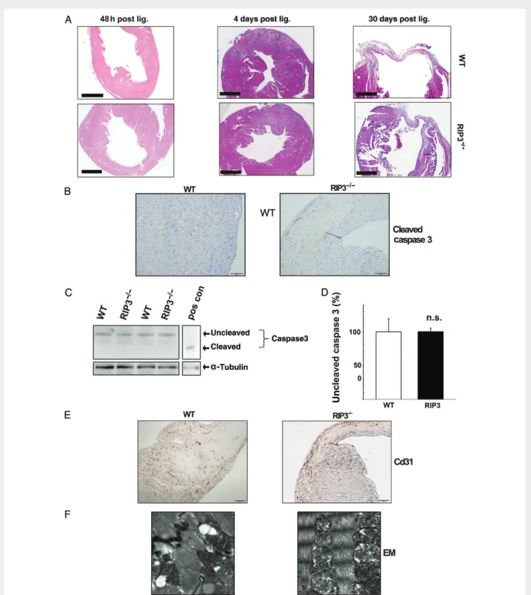
Figure 5 No altered apoptosis rate in infarcted RIP32/2hearts. (A) Representative haematoxylin – eosin stainings of WT and RIP32/2hearts 48 h, 4 days,
and 30 days after LAD ligation. Scale bars denote 1000 mM. (B) Immunohistological stainings of both WT and RIP32/2mouse hearts revealed discretely scattered cells that were positive for cleaved caspase 3. (C ) Immunoblot of WT and RIP32/2protein extracts 24 h post LAD ligation showing no significant
difference in caspase 3 cleavage in both groups. Liver extract from LPS-treated TAK1 knockout mice 15 was used as positive control (pos con) for caspase 3 cleavage. The content of uncleaved caspase 3 was unaltered between both groups (n ¼ 4) (D). (E) As a marker of angiogenesis, both WT and RIP32/2
hearts showed an attenuated staining for Cd31 in infarcted areas. (F ) Representative electron microscopy imaging of border zones of infarcted areas of WT and RIP32/2mouse hearts 24 h after LAD ligation, revealing amorphous matrix densities. n.s.: no statistically significant difference.
M. Luedde et al.
to detect the RIP1/RIP3 complex in NRVCMs may be due to a faster turnover of complexes, e.g. due to rapid RIP1 cleavage12in NRVCMs compared with Jurkat cells. Consistently, we demonstrated that RIP3 overexpression leads to a significant decrease of RIP1 protein content in NRVCMs. Another explanation might be based on low RIP3 expres-sion in the heart under basal conditions, which hampers detection of the RIP1/RIP3 complexes in NRVCMs. Up-regulation of RIP3 under stress conditions (Figure4A) implies that a functional relevance of RIP3 and pro-grammed necrosis in the heart is limited to stress situations like myocar-dial ischaemia. In line with this notion, RIP32/2mice show normal life span and fertility18as well as normal cardiac function under basal condi-tions. Moreover, miRNA-based knockdown of RIP3 in NRVCMs did not alter viability of NRVCMs compared with LacZ-infected control cells (data not shown) and knockdown of RIP3 did have no effect on TNF-a-stimulated NRVCMs (see Supplementary material online, Figure S2). In this context, complex formation of RIP1 and RIP3 and sub-sequent induction of programmed necrosis due to RIP3 overexpression may mimic stress situations, consistent with the occurrence of pro-grammed necrosis in cardiomyocytes.
The complex formation of RIP1 and RIP3 as early as 30 min after TNF-a stimulation of NRVCMs as well as up-regulation of RIP3 24 h after myocardial ischaemia imply that RIP3-dependent effects are exe-cuted in an early response to exogenous stress. Recent work by Oerle-mans et al.15demonstrates a reduction of infarct size due to chemical inhibition of RIP1 by Nec-1 24 h after induction of ischaemia. Consist-ently, we could demonstrate the ‘canonical’ pathway of TNF-induced complex formation of RIP1 and overexpressed RIP3 in NRVCMs. However, overexpression of RIP3 alone was sufficient to induce
programmed necrosis of NRVCMs without a need for additional stimu-lation by TNF-a (Figure3). In this regard, cardiomyocytes differ from other cells, e.g. L929 cells. In these cells, RIP3 overexpression requires additional TNF-a stimulation to induce programmed necrosis.24Two explanations for this interesting result are possible:
first, RIP3 overexpressed in NRVCMs is constitutively active, ‘bypass-ing’ the upstream TNF-related activation pathway. This explanation is supported by the fact that knockdown of RIP1 did not significantly inhibit RIP3-dependent cell death of cardiomyocytes either (see Supple-mentary material online, Figure S2C), demonstrating a RIP1-independent effect of RIP3 in cardiomyocytes.
Secondly, in the heart, additional yet unknown activation pathways of RIP3 might exist apart from TNF-a. In this regard, recent studies demon-strated that several different receptors and signalling pathways upstream from RIP3 may activate necroptosis.25The specific activation pathway of RIP3 seems to be cell type-dependent and also depending on the specific activating stress stimulus.25In respect to our model of myocardial is-chaemia, this explanation is supported by the fact that we did not find significant differences in the regulation of TNF-a-related genes between WT and RIP2/2mouse hearts (see Supplementary material online, Figure S7). Moreover, treatment of WT and RIP32/2 hearts with the TNF-a inhibitor, etanercept, did not effectively rescue WT mice after LAD ligation (data not shown). Thus, most likely other up-stream pathways apart from TNF mediate RIP3 activation in myocardial ischaemia.
In contrast to these early effects of RIP1 inhibition in the model of Oerlemans et al., infarct size of RIP32/2 mice is not altered in our model. At first sight, it is therefore surprising that RIP3 nevertheless Figure 6 RIP3 mediates inflammation and generation of ROS in myocardial infarction. (A) RIP32/2hearts revealed a significantly decreased invasion of
CD3-positive cells 4 days after LAD ligation vs. WT controls. (B) Scale bars denote 100 mM. (C ) RIP32/2hearts revealed significantly decreased levels of ROS vs. WT controls 24 h after LAD ligation (n ¼ 8). (D) In line with this ROS-generating effect of RIP3 in vivo, neonatal rat cardiomyocytes overexpressing RIP3 showed a moderately increased ROS production compared with NRVCMs expressing a control virus (LacZ, n ¼ 4). LV: left ventricular weight; Lig.: LAD ligation. *P , 0.05.
mediates long-term cardiac remodelling as well as contractile function. Yet, a significant difference between programmed necrosis and apop-tosis is the strong inflammatory response driven by necrosis.12 Consist-ently, we could demonstrate that the post-ischaemic inflammatory response of RIP32/2mice is markedly decreased compared with WT mice. Thus, apoptotic cell death was unlikely to account for the differ-ences between both groups. Moreover, we could not detect any
evident change in apoptotic cell death between both groups, as measured by caspase 3 cleavage. Since a role of inflammation in post-infarction remodelling has been demonstrated before,26 reduced inflammation may be one possible mechanism for the observed protective effects of RIP3 ablation on adverse remodelling in our study. This mechanism seems to be heart-specific, as the total count and proportion of T cells in the blood of RIP32/2mice was unaltered compared with WT mice. Figure 7 RIP3 mediates adverse left ventricular remodelling in response to myocardial ischaemia. (A) Cardiac MRI from WT and RIP32/2mice under basal conditions. (B) Gadolinium-enhanced MRI short-axis views of WT and RIP32/2hearts 24 h after LAD ligation, showing that infarct size is not altered between both groups, as quantified in (D) (n ¼ 4). (C ) MRI short-axis views 30 days after LAD ligation, scale bars: 5 mm. Thirty days after LAD ligation, RIP32/2hearts revealed a significant down-regulation of BNP vs. WT controls, a significantly better ejection fraction (EF) (E) and less hypertrophy (F ) than WT controls (n ¼ 12, *P , 0.05,†P , 0.01).
M. Luedde et al.
However, since our results are based on the constitutive knockout of RIP3, we cannot definitely rule out that RIP3 deficiency of leucocytes may have a cardiomyocyte-independent effect in this regard.
In addition to a decreased inflammatory response, we could demon-strate decreased generation of ROS in RIP32/2mice after LAD ligation (Figure6C). Generation of ROS has been considered a crucial mechanism of RIP3-induced cell damage.8,10In our model of permanent myocardial ischaemia, ROS might be a key mediator that ‘translates’ short-term effects of RIP3 signalling into long-term adverse ischaemic remodel-ling.27Oxidative stress has been demonstrated to activate prohyper-trophic signalling pathways like mitogen activated protein kinases28 and to cause a slippage of myofibrils and LV dilation,29 ultimately leading to decreased ventricular contractility. Thus, inhibition of ROS generation is likely to be causative for preserved contractility in the RIP3 knockout mouse. Of note, oxidative stress also induces inflamma-tory pathways in the heart;29thus, the relative contribution of these two mechanisms to RIP-mediated LV remodelling cannot be considered in-dependently. Mitochondria have been recognized to be a source of ex-cessive ROS generation in MI.28 The co-localization of RIP3 and mitochondria implies that mitochondrial ROS generation is crucial in RIP3-dependent myocardial remodelling. In line with this notion, several recent publications have identified the mitochondrial proteins such as mixed lineage kinase domain-like protein (MLKL) and phospho-glycerate mutase family member 5 (PGAM5) as downstream effectors of RIP3.20,30–33These results have even led to the consideration of the RIP1/RIP3 complex being a ‘mitochondrial attack complex’,20,30with dis-sipation of the mitochondrial transmembrane potential (DcM)
ultimate-ly leading to fragmentation of mitochondria under specific stress conditions32Indeed, we could detect a trend towards improved ener-getics of RIP32/2mouse hearts after LAD ligation in vivo. Moreover, the co-localization of RIP3 and mitochondria in cardiomyocytes is con-sistent with the functional importance of mitochondria in RIP3-mediated cardiac damage. However, RIP3 overexpression in cardiomyocytes did neither cause fragmentation of mitochondria nor significant alteration of the mitochondrial transmembrane potential (DcM) compared with
control cells (data not shown). As possible explanations for this interest-ing findinterest-ing, ROS might be generated by other sources, like endosomes,34 peroxisomes,35or infiltrating immune cells. In line with this assumption, a novel report by Tait et al.36generally challenges the functional role of
mitochondria in programmed necrosis, demonstrating that in some cells, mitochondria are not necessarily required in this process. It thus appears possible that RIP3 may affect regulatory functions on metabolism apart from mitochondrial damage, especially in an energy-dependent organ like the heart.
In conclusion, we here demonstrate that RIP3 exerts negative effects on post-ischaemic cardiac remodelling. In addition to the data of Oerle-mans et al. on RIP1 in ischaemia – reperfusion injury, these results imply a fundamental role of pathways mediating programmed necrosis in cardiac ischaemia. Because late cardiac remodelling with subsequent post-ischaemic heart failure is still a major clinical problem, these signalling pathways might represent attractive targets for future therapeutic inter-ventions.
5. Limitations of this study
The in vivo results presented in this study are based on a constitutive knockout of RIP3. Although we have not observed any morphological differences between transgenic and WT control mice in peripheral blood cells as well as other organs, we cannot definitely rule out that RIP3 deficiency of other cell types may have contributed to the observed effects in the heart. Moreover, our study does not provide a quantitative comparison of apoptotic vs. necroptotic cell death in the course of myo-cardial ischaemia. Thus, we cannot definitively conclude that necropto-sis is the dominant form of cell death in post-ischaemic myocardial damage. Nevertheless, we feel that the alterations of myocardial remod-elling in the RIP3 knockout mouse strongly argue for an important role of the necroptotic RIP3 signalling pathway in post-MI remodelling. Further studies are needed to finally compare the relative contribution of each form of cell death in this and other disease models.
Supplementary material
Supplementary material is available at Cardiovascular Research online.
Acknowledgements
The technical assistance of Vanessa Mangels, Daniel Kull, Gabriele Brunke, and Sebastian Cucuruz is gratefully acknowledged.
. . . . . . . . Table 1 Morphological and functional parameters of RIP32/2mice hearts and WT controls under basal conditions and 30 days post LAD ligation, assessed by MRI
WT RIP32/2
Basal 30 days post LAD ligation Basal 30 days post LAD ligation
Weight (g) 23.6 + 0.5 27.7 + 0.4 24.9 + 0.6 26.8 + 0.5
Heart rate (/min) 487 + 15.1 562 + 14.6 495 + 22.0 531 + 15.1
EDV (mL) 62 + 2.3 121 + 19.9 71 + 4.8 83 + 8.6
ESV (mL) 23 + 1.6 89 + 20.4 30 + 3.9 48 + 8.7
Stroke volume (mL) 39 + 1.3 32 + 1.8 42 + 1.6 35 + 1.4
Ejection fraction (%) 63 + 1.6 32 + 4.4 59 + 2.3 45 + 3.6*
Cardiac output (mL/min) 19 + 0.6 18 + 1.3 21 + 1.3 19 + 0.6
LV weight (mg) 89 + 4.0 116 + 6.0 84 + 2.3 94 + 4.4*
LV/body weight (mg/g) 3.6 + 0.1 4.2 + 0.2 3.6 + 0.1 3.5 + 0.1* WT, wild type; EDV, end-diastolic left ventricular volume; ESV, end-systolic left ventricular volume; LV, left ventricular.
Conflict of interest: none declared.
Funding
This work was supported by a grant of the Bundesministerium fu¨r Bildung und Forschung, Germany (Nationales Genomforschungsnetz: NGFNplus to N.F.), a grant of the Deutsche Stiftung Herzforschung (F12/12 to Mar.L., Mat.L., and M.H.), the European Research Council within the FP-7 (ERC-2007-Stg/208237-Luedde-Med3-Aachen to T.L.), the German Re-search Foundation (SFB/TRR57, P06 to T.L.; SFB 877, A1 to S.R.-J.; B2 to D.A.), and ERC-StG-2010 261317 grant and a Helmholtz-young investigator grant (to M.H.).
References
1. World Health Organization. The top 10 causes of death. Fact sheet N8 310, 2014. 2. Sugano M, Koyanagi M, Tsuchida K, Hata T, Makino N. In vivo gene transfer of soluble
TNF-alpha receptor 1 alleviates myocardial infarction. FASEB J 2002;16:1421 – 1422. 3. Wollert KC, Heineke J, Westermann J, Ludde M, Fiedler B, Zierhut W, Laurent D,
Bauer MK, Schulze-Osthoff K, Drexler H. The cardiac Fas (APO-1/CD95) Receptor/ Fas ligand system: relation to diastolic wall stress in volume-overload hypertrophy in vivo and activation of the transcription factor AP-1 in cardiac myocytes. Circulation 2000;101:1172 – 1178.
4. Bialik S, Geenen DL, Sasson IE, Cheng R, Horner JW, Evans SM, Lord EM, Koch CJ, Kitsis RN. Myocyte apoptosis during acute myocardial infarction in the mouse localizes to hypoxic regions but occurs independently of p53. J Clin Invest 1997;100:1363 – 1372. 5. Sun M, Dawood F, Wen WH, Chen M, Dixon I, Kirshenbaum LA, Liu PP. Excessive tumor necrosis factor activation after infarction contributes to susceptibility of myocardial rupture and left ventricular dysfunction. Circulation 2004;110:3221 – 3228.
6. Kung G, Konstantinidis K, Kitsis RN. Programmed necrosis, not apoptosis, in the heart. Circ Res 2011;108:1017 – 1036.
7. Dorn GW II. Apoptotic and non-apoptotic programmed cardiomyocyte death in ven-tricular remodelling. Cardiovasc Res 2009;81:465 – 473.
8. Declercq W, Vanden Berghe T, Vandenabeele P. RIP kinases at the crossroads of cell death and survival. Cell 2009;138:229 – 232.
9. He S, Wang L, Miao L, Wang T, Du F, Zhao L, Wang X. Receptor interacting protein kinase-3 determines cellular necrotic response to TNF-alpha. Cell 2009;137: 1100 – 1111.
10. Zhang DW, Shao J, Lin J, Zhang N, Lu BJ, Lin SC, Dong MQ, Han J. RIP3, an energy me-tabolism regulator that switches TNF-induced cell death from apoptosis to necrosis. Science 2009;325:332 – 336.
11. Christofferson DE, Yuan J. Necroptosis as an alternative form of programmed cell death. Curr Opin Cell Biol 2010;22:263 – 268.
12. Cho YS, Challa S, Moquin D, Genga R, Ray TD, Guildford M, Chan FK. Phosphorylation-driven assembly of the RIP1-RIP3 complex regulates programmed necrosis and virus-induced inflammation. Cell 2009;137:1112 – 1123.
13. Welz PS, Wullaert A, Vlantis K, Kondylis V, Fernandez-Majada V, Ermolaeva M, Kirsch P, Sterner-Kock A, van Loo G, Pasparakis M. FADD prevents RIP3-mediated epithelial cell necrosis and chronic intestinal inflammation. Nature 2011;477:330 – 334.
14. Lim SY, Davidson SM, Mocanu MM, Yellon DM, Smith CC. The cardioprotective effect of necrostatin requires the cyclophilin-D component of the mitochondrial permeability transition pore. Cardiovasc Drugs Ther 2007;21:467 – 469.
15. Oerlemans MI, Liu J, Arslan F, den Ouden K, van Middelaar BJ, Doevendans PA, Sluijter JP. Inhibition of RIP1-dependent necrosis prevents adverse cardiac remodeling after myo-cardial ischemia-reperfusion in vivo. Basic Res Cardiol 2012;107:270.
16. Smith CC, Davidson SM, Lim SY, Simpkin JC, Hothersall JS, Yellon DM. Necrostatin: a potentially novel cardioprotective agent. Cardiovasc Drugs Ther 2007;21:227 – 233.
17. Hippe HJ, Luedde M, Lutz S, Koehler H, Eschenhagen T, Frey N, Katus HA, Wieland T, Niroomand F. Regulation of cardiac cAMP synthesis and contractility by nucleoside di-phosphate kinase B/G protein beta gamma dimer complexes. Circ Res 2007;100: 1191 – 1199.
18. Newton K, Sun X, Dixit VM. Kinase RIP3 is dispensable for normal NF-kappa Bs, signaling by the B-cell and T-cell receptors, tumor necrosis factor receptor 1, and Toll-like recep-tors 2 and 4. Mol Cell Biol 2004;24:1464 – 1469.
19. Flogel U, Ding Z, Hardung H, Jander S, Reichmann G, Jacoby C, Schubert R, Schrader J. In vivo monitoring of inflammation after cardiac and cerebral ischemia by fluorine magnetic resonance imaging. Circulation 2008;118:140 – 148.
20. Wang Z, Jiang H, Chen S, Du F, Wang X. The mitochondrial phosphatase PGAM5 func-tions at the convergence point of multiple necrotic death pathways. Cell 2012;148: 228 – 243.
21. Galluzzi L, Kepp O, Kroemer G. Mitochondria: master regulators of danger signalling. Nat Rev Mol Cell Biol 2012;13:780 – 788.
22. Kaiser WJ, Upton JW, Long AB, Livingston-Rosanoff D, Daley-Bauer LP, Hakem R, Caspary T, Mocarski ES. RIP3 mediates the embryonic lethality of caspase-8-deficient mice. Nature 2011;471:368 – 372.
23. Oberst A, Dillon CP, Weinlich R, McCormick LL, Fitzgerald P, Pop C, Hakem R, Salvesen GS, Green DR. Catalytic activity of the caspase-8-FLIP(L) complex inhibits RIPK3-dependent necrosis. Nature 2011;471:363 – 367.
24. Vanlangenakker N, Bertrand MJ, Bogaert P, Vandenabeele P, Vanden Berghe T. TNF-induced necroptosis in L929 cells is tightly regulated by multiple TNFR1 complex I and II members. Cell Death Dis 2011;2:e230.
25. Han J, Zhong CQ, Zhang DW. Programmed necrosis: backup to and competitor with apoptosis in the immune system. Nat Immunol 2011;12:1143 – 1149.
26. Frantz S, Bauersachs J, Ertl G. Post-infarct remodelling: contribution of wound healing and inflammation. Cardiovasc Res 2009;81:474 – 481.
27. Whelan RS, Kaplinskiy V, Kitsis RN. Cell death in the pathogenesis of heart disease: mechanisms and significance. Annu Rev Physiol 2010;72:19 – 44.
28. Takimoto E, Kass DA. Role of oxidative stress in cardiac hypertrophy and remodeling. Hypertension 2007;49:241 – 248.
29. Hori M, Nishida K. Oxidative stress and left ventricular remodelling after myocardial in-farction. Cardiovasc Res 2009;81:457 – 464.
30. Chan FK, Baehrecke EH. RIP3 finds partners in crime. Cell 2012;148:17 – 18. 31. Sun L, Wang H, Wang Z, He S, Chen S, Liao D, Wang L, Yan J, Liu W, Lei X, Wang X. Mixed
lineage kinase domain-like protein mediates necrosis signaling downstream of RIP3 kinase. Cell 2012;148:213 – 227.
32. Vanden Berghe T, Vanlangenakker N, Parthoens E, Deckers W, Devos M, Festjens N, Guerin CJ, Brunk UT, Declercq W, Vandenabeele P. Necroptosis, necrosis and second-ary necrosis converge on similar cellular disintegration features. Cell Death Differ 2010; 17:922 – 930.
33. Vanlangenakker N, Vanden Berghe T, Bogaert P, Laukens B, Zobel K, Deshayes K, Vucic D, Fulda S, Vandenabeele P, Bertrand MJ. cIAP1 and TAK1 protect cells from TNF-induced necrosis by preventing RIP1/RIP3-dependent reactive oxygen species pro-duction. Cell Death Differ 2011;18:656 – 665.
34. Li Q, Spencer NY, Oakley FD, Buettner GR, Engelhardt JF. Endosomal Nox2 facilitates redox-dependent induction of NF-kappaB by TNF-alpha. Antioxid Redox Signal 2009; 11:1249 – 1263.
35. Fliegner D, Westermann D, Riad A, Schubert C, Becher E, Fielitz J, Tschope C, Regitz-Zagrosek V. Up-regulation of PPARgamma in myocardial infarction. Eur J Heart Fail 2008;10:30 – 38.
36. Tait SW, Oberst A, Quarato G, Milasta S, Haller M, Wang R, Karvela M, Ichim G, Yatim N, Albert ML, Kidd G, Wakefield R, Frase S, Krautwald S, Linkermann A, Green DR. Wide-spread mitochondrial depletion via mitophagy does not compromise necroptosis. Cell Rep 2013;5:878 – 885.
M. Luedde et al.