M A J O R A R T I C L E
Emergence of New Pandemic GII.4 Sydney
Norovirus Strain Correlates With Escape From
Herd Immunity
Kari Debbink,1Lisa C. Lindesmith,2Eric F. Donaldson,2Veronica Costantini,3Martina Beltramello,4Davide Corti,4,5 Jesica Swanstrom,2Antonio Lanzavecchia,4Jan Vinjé,3and Ralph S. Baric1,2
1
Department of Microbiology and Immunology and2Department of Epidemiology, University of North Carolina, Chapel Hill;3Division of Viral Diseases, Centers for Disease Control and Prevention, Atlanta, Georgia; and4Institute for Research in Biomedicine, and5Humabs Biomed SA, Bellinzona, Switzerland
Background. GII.4 noroviruses are a significant source of acute gastroenteritis worldwide, causing the majority of human norovirus outbreaks. Evolution of the GII.4 major capsid protein occurs rapidly, resulting in the emer-gence of new strains that produce successive waves of pandemic disease. A new pandemic isolate, GII.4 2012 Sydney, largely replaced previously circulating strains in late 2012. We compare the antigenic properties of GII.4 2012 Sydney with previously circulating strains.
Methods. To determine whether GII.4-2012 Sydney is antigenically different from recently circulating strains GII.4-2006 Minerva and GII.4-2009 New Orleans in previously identified blockade epitopes, we compared reactivity and blockade profiles of GII.4-2006, GII.4-2009, and GII.4-2012 virus-like particles in surrogate neutralization/ blockade assays using monoclonal antibodies and human polyclonal sera.
Results. Using monoclonal antibodies that map to known blockade epitopes in GII.4-2006 and GII.4-2009 and human outbreak polyclonal sera, we demonstrate either complete loss or significantly reduced reactivity and block-ade of GII.4.2012 compared to GII.4-2006 and GII.4-2009.
Conclusions. GII.4-2012 Sydney is antigenically different from GII.4-2006 Minerva and GII.4-2009 New Orleans in at least 2 key blockade epitopes. Viral evolution in key potential neutralization epitopes likely allowed GII.4-2012 to escape from human herd immunity and emerge as the new predominant strain.
Keywords. norovirus; GII.4 Sydney; virus-like particles; viral evolution; virus emergence.
Noroviruses are a leading cause of acute gastroenteritis, resulting in approximately 21 million infections in the United States annually [1]. The highly heterogeneous human noroviruses are divided into 2 principal gen-ogroups designated GI and GII, which are further sub-divided into 9 and 21 genotypes, respectively [2,3]. The GII.4 noroviruses are responsible for >70% of all noro-virus outbreaks [4]. Norovirus disease patterns in human populations include epidemic outbreaks of disease every 2–3 years, punctuated by the emergence
of an antigenically distinct GII.4 strain that appears to escape human herd immunity to the previous circulat-ing strain [5–8]. Thefirst major GII.4 norovirus pan-demic was associated with the GII.4-1997 US95/96 strain [9,10], which was replaced in 2002 with GII.4-2002 Farmington Hills [11]. GII.4-2002 was then re-placed in 2004 by the Hunter strain [12–14]. In 2006, GII.4-2006 Minerva replaced Hunter [13,15,16] and was the predominant strain until 2009 when it was gradually replaced with GII.4-2009 New Orleans [17]. In March 2012, a new strain, called GII.4-2012 Sydney, wasfirst described, and by November 2012, it had re-placed GII.4-2009 as the primary norovirus strain in most countries in the northern hemisphere [18,19].
Noroviruses are single-stranded, positive sense RNA viruses that belong to the family Caliciviridae. The nor-ovirus genome is approximately 7.5 kb and contains 3 open reading frames (ORFs). ORF1 encodes the non-structural proteins, ORF2 encodes the VP1 major Received 7 March 2013; accepted 14 May 2013; electronically published 1
August 2013.
Correspondence: Ralph Baric, PhD, 2107 McGavran-Greenberg, CB 7435, Chapel Hill, NC 27599-7435 (rbaric@email.unc.edu).
The Journal of Infectious Diseases 2013;208:1877–87
© The Author 2013. Published by Oxford University Press on behalf of the Infectious Diseases Society of America. All rights reserved. For Permissions, please e-mail: journals.permissions@oup.com.
capsid protein, and ORF3 encodes the VP2 minor capsid protein [20]. The P2 subdomain of VP1 contains potential neu-tralizing antibody epitopes and interacts with histoblood group antigens (HBGAs), which are a diverse family of carbohydrates and serve as putative receptors for norovirus entry [6,7,21–27]. HBGAs are differentially expressed in humans and differentially bound by norovirus strains [20,21,25,27,28].
There is currently no cell culture or small animal model available to study human noroviruses, slowing progress toward understanding the mechanisms of protective immunity, virus evolution, and the development of an effective vaccine. However, virus-like particles (VLPs), which are morphological-ly and antigenicalmorphological-ly comparable to native virions, serve as a virus surrogate, and, in conjunction with bioinformatics and surrogate neutralization assays, provide a good model to study questions regarding viral evolution and human immunity, which will ultimately inform vaccine design [6–8,29].
Using human and mouse monoclonal antibodies and time-ordered wild-type and chimeric GII.4 VLPs, we and others have previously shown that the emergence of these new pan-demic strains is often associated with alterations in GII.4 block-ade epitopes mapped to the surface of the major capsid protein P2 subdomain [5–8,30–33]. In this manuscript, we demon-strate that the emergence of GII.4-2012 Sydney is associated with changes in major blockade epitopes, especially epitopes A [6] and D [5], that lead to altered antigenicity as compared to GII.4-2009 New Orleans.
MATERIALS AND METHODS Virus-like Particles
A synthetically-derived outbreak GII.4-2012 ORF2 comple-mentary DNA was synthesized (BioBasic), and inserted into the Venezuelan equine encephalitis virus 3526 replicon vector (VRP-3526). For GII.4-2006 and GII.4-2009 VLPs, virus repli-con particles (VRPs) were produced and inoculated onto baby hamster kidney (BHK) cells as previously described [5]. For GII.4-2012, VLPs were expressed from Venezuelan equine en-cephalitis replicon vector RNA after electroporation of BHK cells and purified as described previously [6,8]. Structural in-tegrity of VLPs was confirmed by electron microscopy, enzyme immunoassay, and carbohydrate binding [5].
HBGA-VLP Binding Assay
HBGA binding assays were performed as previously described [6,
21]. In brief, synthetic HBGAs or pig gastric mucin type III (PGM; Sigma Chemicals) was diluted to 10 µg/mL and coated onto plates. Two micrograms/mL VLPs were added and detected by polyclonal rabbit sera and then antirabbit immunoglobulin G (IgG)–horseradish peroxidase (HRP) (GE Healthcare) followed by One-Step Ultra TMB EIA HRP substrate solution (Thermo-Fisher). Data are the average of 3 replicates with similar results
from at least 2 independent experiments. Positive reactivity is defined as a mean optical density (OD) >3 times the background binding signal.
Enzyme Immunoassays
Mouse and human monoclonal antibody (mAb) reactivity was determined by EIA, as reported [5,7]. In brief, 1 µg/mL VLP in phosphate-buffered saline was coated onto plates, followed by addition of 1 µg/mL purified IgG and then antimouse or anti-human IgG-HRP (GE Healthcare) followed by One-Step Ultra TMB EIA HRP substrate solution. Data are the average of 3 replicates from at least 2 independent experiments. Positive re-activity is defined as a mean OD 450 nm >0.2 after background subtraction [8].
VLP-Carbohydrate Ligand-Binding Antibody Blockade Assays PGM contains HBGAs α-1,2-fucose (H antigen) and α-1,4-fucose (Lewis antigen) [7,21,34] and has been validated as a substrate for norovirus VLP antibody-blockade assays [5, 7]. Blockade assays were performed as previously described [8]. VLPs bound to PGM or biotinylated B were detected by rabbit anti-GII.4 norovirus polyclonal sera. The percentage of control binding was defined as the VLP-ligand binding level in the presence of test antibody or sera compared to the binding level in the absence of antibody multiplied by 100 [7,35]. All anti-bodies were tested for blockade potential at 2-fold serial dilu-tions ranging from 0.004 to 2 µg/mL for mouse mAbs, from 0.008 to 16 µg/mL for human mAbs, or from 0.008% to 1% for human serum. Data shown are the average of at least 2 repli-cates and are representative of similar data from at least 2 inde-pendent trials. Sigmoidal dose response analysis was performed as previously described [8]. EC50values (the antibody or serum concentration required to block 50% of the VLP-carbohydrate binding) among VLPs were compared using 1-way analysis of var-iance with Dunnett posttest.P < .05 was considered significant. Hybridoma Production, IgG Purification, and GII.4 Outbreak Serum Samples
Mouse and human mAbs were produced and purified as de-scribed previously by our group [5,29]. De-identified human
con-valescent serum samples collected from 8 subjects infected with GII.4-2009, as previously described, were used in this study [8]. RESULTS
GII.4 noroviruses undergo genetic changes over time, which are associated with changes in antigenicity [29]. We previously identified 3 GII.4 blockade epitopes in VP1, designated A, D, and E, which are altered in GII.4-2012 and other contemporary strains and affect GII.4 norovirus antigenic profiles [5–8] (Figure1). In early strains, epitope A appears immunodomi-nant, accounting for 40%–55% of the total blockade response [7,9]; GII.4-2009 and GII.4-2012 share 4 common residues but
have 2 amino acid changes at P294 T and A368E. Epitope D shares 2 residues between GII.4-2009 and GII.4-2012, with dif-ferences at S393G and P396H. Epitope E has one differential residue between GII.4-2012 and GII.4-2009, a I413 T change. To test the impact of these sequence changes on antigenicity, we produced GII.4-2012 Sydney and chimeric VLPs. Overall, GII.2-2012 exhibited decreased EIA binding to all synthetic HBGAs tested compared to GII.4-2009 (Figure 2). Next, we compared EIA reactivity and blockade ability of mAbs and human outbreak sera between GII.4-2012 and previous pre-dominant strains GII.4-2009 and GII.4-2006.
GII.4-2006 Mouse Monoclonal EIA Cross-Reactivity and Blockade Response
We previously characterized a set of GII.4-2006 mouse mAbs, all of which exhibit strong EIA reactivity with GII.4-2006 and GII.4-2009 and are blockade antibodies that target overlapping residues across epitope A [6–8, 29]. To determine whether GII.4-2006 mouse mAbs are able to distinguish GII.4-2012 from previous GII.4 strains, we compared 5 GII.4-2006 (G2,
G3, G4, G6, G7) mouse mAbs for EIA reactivity with GII.4-2012, GII.4-2006, and GII.4-2009 VLPs. By EIA, GII.4-2012 did not bind with GII.4-2006-G3 and G4 mAbs (Figure3A),
whereas reactivity with GII.4-2006-G2, G6, and G7 mAbs was significantly reduced (P < .05) 1.8- to 4.5-fold compared with GII.4-2006 and GII.4-2009 (Figure3A).
Blockade is a more sensitive measure of antigenic variation and, unlike EIA, can measure potential functional differences in antigenicity that correlate with protective immunity in vivo [36,
37]. Three GII.4-2006 blockade mouse mAbs that map to epitope A demonstrated EIA reactivity with GII.4-2012 (GII.4-2006-G2, G6, and G7). To examine functionally relevant anti-genic differences between GII.4-2012 and previous strains, we performed surrogate neutralization blockade assays. 2006-G2, although able to block both 2006 and GII.4-2009 VLPs, lost the ability to block GII.4-2012 VLP interaction with HBGA (Figure3B). GII.4-2006-G6 and G7 mAbs were both
able to block GII.4-2012 VLP interaction with HBGA, but EC50 values were significantly reduced (3.9- to 6.3-fold) compared with GII.4-2006 and GII.4-2009 VLPs (Figure 3C and 3D). Of 5
Figure 1. GII.4 norovirus structure and genetic variability in blockade epitopes.A, Schematic representation of the norovirus genome. Open reading frame (ORF) 1 encodes the nonstructural proteins, ORF2 encodes the major capsid protein, and ORF3 encodes the minor capsid protein. The major capsid protein is divided into the shell and the P1 and P2 subdomains.B, Blockade epitopes for GII.4 noroviruses. A structural model of a GII.4 norovirus P2 dimer indicates the location of previously identified blockade epitopes A, D, and E. C, GII.4-2012 changes in evolving blockade epitopes. Amino acid sequences from GII.4-2006, GII.4-2009, and GII.4-2012 were aligned and changes occurring in epitopes A, D, and E are noted in the tables.D, Previously reported epitope binding specificity of GII.4-2006 and GII.4-2009 mouse monoclonal antibodies (mAbs) and GII.4 human mAbs.
GII.4-2006 blockade mouse mAbs that target epitope A, only 2 (G6, G7) retained limited EIA and blockade ability against GII.4-2012 VLPs, and both of these were significantly reduced. GII.4-2009 Mouse Monoclonal EIA Cross-Reactivity and Blockade Response
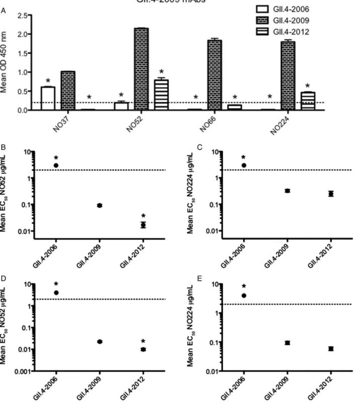
To further compare the impact of epitope A changes on GII.4-2012 antigenic structure and function, we evaluated GII.4-GII.4-2012 reactivity with 4 GII.4-2009 blockade mouse mAbs. Previous work demonstrated that mAbs NO37 and NO52 target epitope A, but the binding sites for mAbs NO66 and NO224 have not yet been mapped on the structure [8]. NO37 cross-reacts with GII.4-2006 and GII.4-2009 [8], but does not bind GII.4-2012 (Figure4A). The other NO antibodies tested react with
GII.4-2009 but not GII.4-2006 [8]. NO52 and NO224 binding to GII.4-2012 were significantly reduced (2.75- to 3.9-fold, respec-tively) compared to GII.4-2009. NO66 failed to react by EIA with GII.4-2012 under these treatment conditions (Figure4A).
In addition, we tested the GII.4-2009 mouse mAbs that bind GII.4-2012 in EIA (NO52 and NO224) for blockade potential against this strain. Both NO52 and NO224 were able to block GII.4-2012 (Figure 4B and 4C). Interestingly, NO52 blocked
GII.4-2012 significantly more efficiently than the homotypic GII.4-2009 VLP (Figure 4B), indicating that this mAb may
target a unique motif within epitope A compared with other epitope A–targeting antibodies. EC50blockade titers for GII.4-2012 and GII.4-2009 by NO224 were not significantly different
(Figure 4C). These results support our data from GII.4-2006
mAbs, demonstrating that antigenicity between GII.4-2012 and previously circulating strains is significantly different at epitope A. These results also confirm previous findings that epitope A mAbs likely engage overlapping, yet distinct epitope residues. Human Monoclonal Antibody EIA Cross-Reactivity and Blockade Response
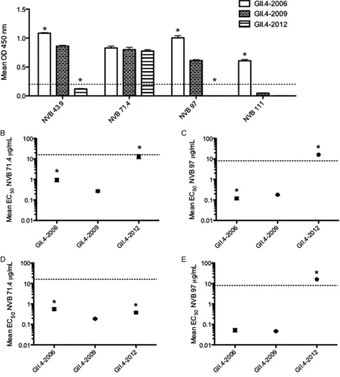
Although mouse mAbs provide an excellent tool to examine targeted, strain-specific norovirus antibody responses, it is pos-sible that the immune systems of mice and humans engage nor-ovirus epitopes differently. Therefore, we examined whether human mAb EIA reactivity and blockade responses were also able to differentiate between GII.4-2012 and ancestral strains. We previously characterized 4 blockade human mAbs, which recognize either GII.4-2006 or GII.4-2009, or both (NVB 43.9, NVB 71.4, NVB 97, NVB 111) [5]. Two of these mAbs map to epitope A (NVB 43.9, NVB 111) and another maps to epitope D (NVB 97) [5]. NVB 71.4 recognizes a conserved GII.4 block-ade epitope across time-ordered strains that has not yet been mapped onto the structure. Wefirst tested these GII.4 blockade human mAbs for EIA reactivity with GII.4-2012. Consistent with a previous report, NVB 43.9, NVB 71.4, and NVB 97 rec-ognize GII.4-2006 and GII.4-2009, whereas NVB 111 binds only GII.4-2006 [5]. When we examined reactivity of these human mAbs with GII.4-2012, we found that NVB 43.9 and NVB 111 (epitope A), and NVB 97 (epitope D) lost EIA Figure 2. GII.4-2012 histoblood group antigen (HBGA) binding. Virus-like particles (VLPs) representing GII.4-2006, GII.4-2009, and GII.4-2012 strains were assayed for their ability to bind synthetic biotinylated HBGAs or pig gastric mucin type III (PGM). The mean optical density (OD) 450 nm was calculat-ed and graphcalculat-ed. Error bars represent SEM. VLP reactivity is defined as a positive signal >3 times the background binding, indicated by the dashed line. Abbreviations: CHO, carbohydrate; Le, Lewis.
reactivity with GII.4-2012 (Figure 5A). NVB 71.4 retained
binding to GII.4-2012 similar to levels seen with GII.4-2006 and GII.4-2009 (Figure5A). These results corroborate the
im-portance of epitope A changes in antibody recognition of GII.4-2012. In addition, loss of binding to the epitope D mAb indicates that this epitope has also evolved in the GII.4-2012 strain.
Next, we determined whether human blockade mAbs could further distinguish between emergent GII.4-2012 and contem-porary GII.4-2009 strains. GII.4-2012 was only recognized by one human blockade mAb, NVB 71.4. This broadly
cross-reactive and blocking antibody is capable of preventing HBGA binding with GII.4 strain VLPs from GII.4-1987 through GII.4-2009 [5]. When tested for blockade response against GII.4-2012, NVB 71.4 weakly blocked this strain, with an EC50 signif-icantly higher than for GII.4-2006 and GII.4-2009 (Figure5B).
We also evaluated NVB 97 blockade activity against GII.4-2012 despite its inability to bind GII.4-2012 by single dilution EIA. NVB 97 targets epitope D, another previously identified GII.4 blockade epitope [5]. Corroborating EIA data, NVB 97 was not able to block GII.4-2012 VLP-binding ligand interaction by blockade assay (Figure5C). Overall, human mAb blockade data
Figure 3. GII.4-2006 mouse monoclonal antibody (mAb) enzyme immunoassay (EIA) reactivity and blockade response against GII.4-2012.A, Mouse mAbs (1 µg/mL) against GII.4-2006 (G2, G3, G4, G6, and G7) were assayed for ability to react with GII.4-2006, GII.4-2009, and GII.4-2012 virus-like particles (VLPs) by EIA. The mean optical density (OD) 450 nm was calculated and graphed. Error bars represent SEM. *Reactivity is significantly different from that of the homotypic GII.4-2006 VLP. Reactivity is defined as a positive signal >0.2 by EIA, represented by the dashed line. VLP reactivity below the dashed line is considered nonreactive.B–G, Mouse mAbs against GII.4-2006 (G2, G6, G7) were assayed for ability to block GII.4-2006, GII.4-2009, and GII.4-2012 VLP interaction with carbohydrate ligand pig gastric mucin type III (B–D) or biotinylated B (E–G). The mean percentage of control binding (percent of the VLP bound to carbohydrate ligand in the presence of an antibody compared to the amount of VLP bound with no antibody present) of each VLP wasfit with a sigmoidal curve, and the mean EC50(µg/mL) blockade titers (the antibody concentration at which 50% of VLP-PGM binding is blocked) for 2006,
GII.4-2009, and GII.4-2012 were calculated. Error bars represent 95% confidence intervals. *Mean EC50blockade titer for the test VLP is significantly different
from the mean EC50for GII.4-2006 (P < .05). Monoclonal antibodies that did not block a particular VLP were assigned an EC50of 4 µg/mL for statistical
anal-ysis and are shown on the graph by data points above the upper limit of detection (dashed line). Statistics for both EIA and blockade assays were calculat-ed by 1-way analysis of variance with Dunnett posttest.
Figure 4. GII.4-2009 mouse monoclonal antibody (mAb) enzyme immunoassay (EIA) reactivity and blockade response against GII.4-2012.A, Mouse mAbs (1 µg/mL) against GII.4-2009 (NO37, 52, 66, and 224) were assayed for ability to react with GII.4-2006, GII.4-2009, and GII.4-2012 VLPs by EIA. The mean optical density (OD) 450 nm was calculated and graphed. Error bars represent SEM. *Reactivity is significantly different from that of the homotypic GII.4-2009 virus-like particle (VLP). Reactivity is defined as a positive signal >0.2 by EIA, represented by the dashed line. VLP reactivity below the dashed line is considered nonreactive.B–E, Mouse mAbs against GII.4-2009 (NO 52, 224) were assayed for ability to block GII.4-2006, GII.4-2009, and GII.4-2012 VLP interaction with carbohydrate ligand pig gastric mucin type III (B and C) or biotinylated B (D–E). The mean percentage of control binding (percentage of the VLP bound to carbohydrate ligand in the presence of an antibody compared to the amount of VLP bound with no antibody present) of each VLP was fit with a sigmoidal curve, and the mean EC50(µg/mL) blockade titers (the antibody concentration at which 50% of VLP-PGM binding is blocked) for
GII.4-2006, GII.4-2009, and GII.4-2012 were calculated. Error bars represent 95% confidence intervals. *Mean EC50blockade titer for the test VLP is significantly
different from the mean EC50for GII.4-2009 (P < .05). Monoclonal antibodies that did not block a particular VLP were assigned an EC50of 4 µg/mL for
statis-tical analysis and are shown on the graph by data points above the upper limit of detection (dashed line). Statistics for both EIA and blockade assays were calculated by 1-way analysis of variance with Dunnett posttest.
Figure 5. GII.4 human monoclonal antibody (mAb) enzyme immunoassay (EIA) reactivity and blockade response against GII.4-2012.A, Human mAbs against GII.4 norovirus (1 µg/mL) were assayed for ability to react with GII.4-2006, GII.4-2009, and GII.4-2012 virus-like particles (VLPs) by EIA. The mean optical density (OD) 450 nm was calculated and graphed. Error bars represent SEM. *Reactivity is significantly different from that of the GII.4-2009 VLP. Re-activity is defined as a positive signal >0.2 by EIA, represented by the dashed line. VLP reactivity below the dashed line is considered nonreactive. B–E, Human mAbs against GII.4 noroviruses were assayed for ability to block GII.4-2006, GII.4-2009, and GII.4-2012 VLP interaction with carbohydrate ligand pig gastric mucin type III (B and C) or biotinylated B (D and E). The mean percentage of control binding (percentage of the VLP bound to carbohydrate ligand in the presence of an antibody compared to the amount of VLP bound with no antibody present) of each VLP wasfit with a sigmoidal curve, and the mean EC50(µg/mL) blockade titers (the antibody concentration at which 50% of VLP-PGM binding is blocked) for GII.4-2006, GII.4-2009, and GII.4-2012
were calculated. Error bars represent 95% confidence intervals. *Mean EC50blockade titer for the tested VLP is significantly different from the mean EC50
for GII.4-2009 (P < .05). Monoclonal antibodies that did not block a particular VLP at the highest mAb concentration tested were assigned an EC50of 2
times the upper limit tested in µg/mL for statistical analysis and are shown on the graph by data points above the upper limit of detection (dashed line). Statistics for both EIA and blockade assays were calculated by 1-way analysis of variance with Dunnett posttest.
are consistent with mouse mAb blockade data, demonstrating that GII.4-2012 is antigenically distinct in major blockade epi-topes from previously circulating contemporary strains GII.4-2006 and GII.4-2009.
Mapping of GII.4-2012 Amino Acids Involved in Epitope A Antigenic Differences
To more specifically map amino acids important for antigenic differences between GII.4-2009 and GII.4-2012, 3 epitope A chimeras between GII.4-2009 and GII.4-2012 were constructed, where residues from 2009 were inserted into the GII.4-2012 background (GII.4-2012.T294P, GII.4-2012.E368A, and GII.4-2012.09A). We performed blockade assays using epitope A targeting anti-bodies against these chimeras and compared them to parental strain blockade data. Overall, data show that GII.4-2012 residue 368E is important for the antigenic differences in epitope A between GII.4-2009 and GII.4-2012, as this residue impacted the blockade ability of all 8 epitope A–targeting mAbs tested (2006-G2, G3, G4, G6, G7, NO37, NO52, and NVB 43.9), and representative data are shown (Figure 6A). Residue 294 T was
also important, synergistically contributing with 368E to the difference in blockade response for 4 of 8 mAbs (2006-G3, NO37, NO52, NVB 43.9), and representative data are shown (Figure6B). Importantly, blockade profiles between GII.4-2009
and GII.4-2012.09A were different for some antibodies (Figure6A), indicating that there are other residues not
includ-ed in the defininclud-ed epitope A that contribute to antigenicity changes between GII.4-2009 and GII.4-2012.
GII.4-2009 Outbreak Human Sera Against GII.4-2012
Blockade results with mAbs demonstrated differences between GII.4-2009 and GII.4-2012 in epitopes A and D, but mAb data do not represent the total antibody response. To determine if the polyclonal antibody response is different between GII.4-2009 and GII.4-2012 and to what degree epitope A accounts for any differences, we tested the blockade activity of GII.4-2009 outbreak convalescent human sera from 8 individuals against GII.4-2009, GII.4-2012, and GII.4-2012.09A VLPs. EC50titers demonstrated that significantly more sera was necessary to block GII.4-2012 (0.066%) and GII.4-2012.09A (0.048%) com-pared to GII.4-2009 (0.021%) (Figure 7). This demonstrates that only approximately 30% of the blockade against GII.4-2009 is retained against GII.4-2012, and at least 11% of the de-creased response is due to changes in epitope A. Individually, it took significantly more sera to block GII.4-2012 compared to GII.4-2009 in 7 of 8 serum samples (Supplementary Figure 1A–
G) and significantly less sera to block in 1 of 8 serum samples (Supplementary Figure 1H). In 4 of 8 serum samples ( Supple-mentary Figure 1B, C, E, and H), epitope A was responsible for a significant change in blockade, accounting for between 21% and 100% of the change.
DISCUSSION
GII.4 noroviruses are the principal cause of epidemic norovirus gastroenteritis in human populations. The GII.4 genotype un-dergoes epochal evolution whereby a predominant circulating Figure 6. Monoclonal antibody (mAb) blockade of chimeric GII.4-2012 virus-like particles (VLPs). Mouse and human epitope A–targeting monoclonal an-tibodies against GII.4 noroviruses (2006-G2, G3, G4, G6, G7, NO37, NO52, and NVB 43.9) were assayed for ability to block GII.4-2012.T294P, GII.4-2012. E368A, and GII.4-2012.09A interaction with carbohydrate ligand, and graphs representative of 2 distinct patterns are shown. The mean percentage of control binding ( percent of the VLP bound to carbohydrate ligand in the presence of an antibody compared to the amount of VLP bound with no antibody present) of each VLP wasfit with a sigmoidal curve, and the mean EC50(µg/mL) blockade titers (the antibody concentration at which 50% of VLP-PGM
binding is blocked) for GII.4-2009, GII.4-2012 VLP, GII.4-2012.T294P, GII.4-2012.E368A, and GII.4-2012.09A were calculated. Error bars represent 95% con fi-dence intervals. *Mean EC50blockade titer for the tested VLP is significantly different from the mean EC50for GII.4-2009 (P < .05). Monoclonal antibodies
that did not block a particular VLP at the highest mAb concentration tested were assigned an EC50of 2 times the upper limit tested in µg/mL for statistical
analysis and are shown on the graph by data points above the upper limit of detection (dashed line). Statistics for both enzyme immunoassay and blockade assays were calculated by 1-way analysis of variance with Dunnett posttest.
strain is replaced by an emergent strain containing antigenic changes facilitated by alterations in the P2 subdomain of the norovirus capsid [5,21,30,31,33,38]. Increased evolution of GII.4 noroviruses over other genotypes has been correlated with mutation rate, antigenic space, and herd immunity [39]. These antigenic changes over time have also been shown to be specifically associated with GII.4 blockade epitopes [5–8]. These data support the hypothesis that emergence of new strains is driven by evolutionary escape from human herd im-munity [21,40].
To determine if antigenic differences exist between GII.4-2012 and the recent circulating ancestral strains GII.4-2006 and GII.4-2009, we compared reactivity and blockade capacity using time-ordered VLPs, mouse and human monoclonal anti-bodies, and GII.4-2009 human outbreak convalescent sera. Of 9 tested mAbs that bind GII.4-2006 and GII.4-2009 (G2, G3, G4, G6, G7, NO37, NVB 43.9, NVB 71.4, and NVB 97), only 4 retained the ability to react with GII.4-2012 (G2, G6, G7, and NVB 71.4), although at significantly lower levels compared with GII.4-2006 and GII.4-2009. Of the 3 mouse mAbs that reacted with GII.4-2009 and not GII.4-2006 (NO52, NO66, NO224), 2 of them were able to react with GII.4-2012 (NO52, NO224), albeit at significantly reduced levels. The majority of the tested mAbs target the A epitope, which has previously
been identified as the predominant GII.4-blockade epitope in earlier strains. Our data also support earlier data that suggests the presence of several overlapping epitopes within epitope A. Epitope A–targeting mAbs, except NO52, either lost block-ade activity or required significantly more sera to block GII.4-2012 ligand-binding interactions. Interestingly, whereas NO52 binding to GII.4-2012 is reduced compared to GII.4-2009, this mAb blocks GII.4-2012 more efficiently than GII.4-2009. Pos-sible explanations for this include that NO52 binding to GII.4-2009 and GII.4-2012 may result in slightly different antibody positioning that more efficiently blocks VLP-HBGA binding in GII.4-2012, NO52 may recognize a rare overlapping epitope that is only targeted by the immune systems of a small fraction of the human population, or NO52 may represent a novel mouse-specific epitope. In any event, differences among block-ade potential of epitope A mAbs highlight the need forfine res-olution mapping of these overlapping epitopes as well as the need for assays that measure the fraction of a polyclonal re-sponse against a unique monoclonal antibody epitope [41]. Our mAb data clearly support the hypothesis that major anti-genic differences exist in epitope A between GII.4-2012 and previously circulating strains, and that this epitope may repre-sent the major site for driving GII.4 escape from herd immuni-ty over the past 15 years [5,6,8,42].
Previous studies by our group and others support the hy-pothesis that human immune responses may select for muta-tions in the HBGA binding pocket, selecting for varying HBGA recognition patterns over time in GII.4 noroviruses [5,20,21,
25–27]. HBGA binding assays revealed similar, but reduced binding patterns as compared to contemporary GII.4-2006 and 2009 strains, indicating that more sophisticated measures of af-finity binding may be needed to untangle the subtle changes in HBGA interactions in GII.4-2012 Sydney. Supporting earlier findings, human mAb NVB 97 completely lost reactivity and blockade against GII.4-2012 D epitope. Epitope D minimally consists of varying residues 393–395, but likely includes other adjacent residues that may alter norovirus strain binding affini-ty and specificity to HBGAs as well as antibody binding and blockade. Modulation of several residues in close proximately to the HBGA binding pocket influence HBGA binding [6,21,
42–44], but most of these residues have not been evaluated for their roles in antibody binding. Overall, results from mAb binding and blockade assays demonstrate that recognition of epitopes A and D between GII.4-2009 and GII.4-2012 are very different, and most neutralizing antibodies generated against epitopes A and D during a GII.4-2009 New Orleans strain in-fection would probably not protect against the new GII.4-2012 Sydney strain.
Blockade data from human outbreak sera also showed sig-nificant reductions in the global antibody blockade response for GII.4-2012 compared with GII.4-2009. Importantly, a comparison of mean outbreak human sera EC50 titers from a
Figure 7. GII.4-2012 and GII.4-2012.09A blockade by GII.4-2009 out-break human sera. Human convalescent polyclonal sera from 8 individuals infected in 2010 with GII.4-2009 New Orleans were assayed for ability to block GII.4-2009, GII.4-2012, and GII.4-2012.09A virus-like particle (VLP) in-teraction with carbohydrate ligand. The mean percentage of control binding (percentage of the VLP bound to carbohydrate ligand in the pres-ence of sera compared to the amount of VLP bound with no sera present) of each VLP wasfit with a sigmoidal curve, and the mean EC50(% sera)
blockade titers (the serum concentration at which 50% of VLP-PGM binding is blocked) for GII.4-2009, GII.4-2012, and GII.4-2012.09A were calculated. Error bars represent 95% confidence intervals. *Mean EC50
blockade titer is significantly different from the mean EC50for GII.4-2009
(P < .05). Statistics were calculated by 1-way analysis of variance with Dunnett posttest.
GII.4-2009 New Orleans outbreak between GII.4-2009 and GII.4-2012 indicate that GII.4-2012 Sydney retained only ap-proximately 30% of the GII.4-2009 blockade response, and 2 changes in epitope A accounted for 11% of the change in block-ade response. On an individual level, epitope A changes signifi-cantly accounted for changes in blockade response in half of the serum samples. This suggests that individual responses to new GII.4 strains are highly varied, and the immune response to specific epitopes may be shaped by previous exposure history. As GII.4 noroviruses are mucosal pathogens with short incubation periods (<30 hours) and rapid clinical disease manifestations (approximately 48 hours), waning immunity may rapidly permit repeat acute infections in some but not all individuals, depending on the specificity and affinity of the global blockade specific re-sponse to different epitopes, perhaps at both an individual and population level. The frequency at which monoclonal antibodies are produced against a specific epitope may be a key predictor of short vs long-term immunity, with antibody responses to the most frequently targeted epitopes remaining the longest and se-lecting for the most antigenic variation. Given that the majority of blockade monoclonal antibodies that were isolated target epitope A, our data suggest that antibody responses against A would wane more slowly than antibody responses against more rarely targeted epitopes such as D, E, and the universal epitope. Interestingly, the ancestral blockade response against the universal neutralization site is weak against GII.4-2012, suggesting the emergence of muta-tions in this epitope as well. Overall, our data clearly demonstrate that 2012 Sydney is antigenically different from both GII.4-2006 and GII.4-2009 and support the hypothesis that emergence of GII.4 strains is the result of escape from herd immunity as they undergo evolution in major neutralizing epitopes.
One of the factors complicating vaccine design is the high degree of GII.4 antigenic variation over time. Because of this variation, the GII.4 component of a successful norovirus vaccine will likely need to be reformulated over time. Our work describes an important platform approach to identify GII.4 strains with pandemic potential and provides important in-sights into effective vaccine design. We demonstrate the importance of key reagents such as time-ordered VLPs and monoclonal antibodies that identify surface varying residues in-volved in escape from herd immunity. Tracking sequence and antigenic changes over time may reveal new patterns of evolu-tion, distinguish important overlapping epitopes, and identify the emergence of new blockade epitopes. By identifying impor-tant blockade epitopes for GII.4 noroviruses and tracking those particular sites in new strains as they emerge, vaccines could be reformulated quickly and tailored specifically to new epidemic and pandemic strains. We may also identify important thera-peutic antibodies targeting these epitopes in circulating strains, which would be valuable for treating long-term chronic infec-tions in immunosuppressed patients, as has been demonstrated for respiratory syncytial virus [45]. In particular, our data
suggest that epitopes A and D are major drivers of escape from herd immunity in contemporary strains, and screening new strains for changes in these and other potential neutralization epitopes may provide a quick and valuable method for effective vaccine design and reformulation.
Supplementary Data
Supplementary materialsare available atThe Journal of Infectious Diseases online (http://jid.oxfordjournals.org/). Supplementary materials consist of data provided by the author that are published to benefit the reader. The posted materials are not copyedited. The contents of all supplementary data are the sole responsibility of the authors. Questions or messages regarding errors should be addressed to the author.
Notes
Acknowledgments. We thank Victoria Madden and C. Robert Bagnell Jr of the Microscopy Services Laboratory, Department of Pathology and Laboratory Medicine, University of North Carolina–Chapel Hill (UNC-CH), for expert technical support. We also acknowledge the UNC-CH Genome Analysis Facility.
Financial support. This work was supported by the National Institutes of Health (NIH), National Institute of Allergy and Infectious Diseases (NIAID; grant AI056351), and by NIH institutional training grant T32-AI007419. K. D. is supported in part by a Career Guidance for Trainees grant from the Burroughs Wellcome Fund. R. S. B. received grant support for research and travel to meetings from the NIH, NIAID. E. F. D. was paid salary by the Food and Drug Administration and grant funding from the NIH. A. L. received grant support from the NIH. Salaries of L. C. L. and J. S. were paid by grant funds from the NIH, NIAID. Salary of K. D. was paid by grant funds from the NIH and Burroughs Wellcome Fund. D. C. was paid by grant funds from the NIH.
Disclaimer. The funders had no role in study design, data collection and analysis, decision to publish, or preparation of the manuscript. The findings and conclusions in this report are those of the author(s) and do not necessarily represent the official position of the Centers for Disease Control and Prevention (CDC). Mention of company names or products does not constitute endorsement by the CDC. This article did receive clear-ance through the appropriate channels at the CDC prior to submission.
Potential conflicts of interest. A. L. is on the board of Humabs Biomed SA. D. C. has received payment from Humabs Biomed. All other authors report no potential conflicts.
All authors have submitted the ICMJE Form for Disclosure of Potential Conflicts of Interest. Conflicts that the editors consider relevant to the content of the manuscript have been disclosed.
References
1. Scallan E, Hoekstra RM, Angulo FJ, et al. Foodborne illness acquired in the United States—major pathogens. Emerg Infect Dis 2011; 17:7–15. 2. Zheng DP, Ando T, Fankhauser RL, Beard RS, Glass RI, Monroe SS.
Norovirus classification and proposed strain nomenclature. Virology 2006; 346:312–23.
3. Updated norovirus outbreak management and disease prevention guidelines. MMWR Recomm Rep2011; 60:1–18.
4. Fankhauser RL, Monroe SS, Noel JS, et al. Epidemiologic and molecular trends of“Norwalk-like viruses” associated with outbreaks of gastroen-teritis in the United States. J Infect Dis2002; 186:1–7.
5. Lindesmith LC, Beltramello M, Donaldson EF, et al. Immunogenetic mechanisms driving norovirus GII.4 antigenic variation. PLoS Pathog 2012; 8:e1002705.
6. Debbink K, Donaldson EF, Lindesmith LC, Baric RS. Genetic mapping of a highly variable norovirus GII.4 blockade epitope: potential role in escape from human herd immunity. J Virol2012; 86:1214–26.
7. Lindesmith LC, Debbink K, Swanstrom J, et al. Monoclonal antibody-based antigenic mapping of norovirus GII.4-2002. J Virol2012; 86: 873–83.
8. Lindesmith LC, Costantini V, Swanstrom J, et al. Emergence of a noro-virus GII.4 strain correlates with changes in evolving blockade epitopes. J Virol2013; 87:2803–13.
9. Vinje J, Altena SA, Koopmans MP. The incidence and genetic variabili-ty of small round-structured viruses in outbreaks of gastroenteritis in the Netherlands. J Infect Dis1997; 176:1374–8.
10. Noel JS, Fankhauser RL, Ando T, Monroe SS, Glass RI. Identification of a distinct common strain of“Norwalk-like viruses” having a global dis-tribution. J Infect Dis1999; 179:1334–44.
11. Widdowson MA, Cramer EH, Hadley L, et al. Outbreaks of acute gas-troenteritis on cruise ships and on land: identification of a predominant circulating strain of norovirus—United States, 2002. J Infect Dis 2004; 190:27–36.
12. Bull RA, Tu ET, McIver CJ, Rawlinson WD, White PA. Emergence of a new norovirus genotype II.4 variant associated with global outbreaks of gastroenteritis. J Clin Microbiol2006; 44:327–33.
13. Kroneman A, Vennema H, Harris J, et al. Increase in norovirus activity reported in Europe. Euro Surveill2006; 11:E061214 1.
14. Phan TG, Kuroiwa T, Kaneshi K, et al. Changing distribution of norovi-rus genotypes and genetic analysis of recombinant GIIb among infants and children with diarrhea in Japan. J Med Virol2006; 78:971–8. 15. Siebenga J, Kroneman A, Vennema H, Duizer E, Koopmans M.
Food-borne viruses in Europe network report: the norovirus GII.4 2006b (for US named Minerva-like, for Japan Kobe034-like, for UK V6) variant now dominant in early seasonal surveillance. Euro Surveill2008; 13. 16. Centers for Disease Control and Prevention. Norovirus activity—
United States, 2006–2007. MMWR Morb Mortal Wkly Rep 2007; 56:842–6.
17. Vega E, Barclay L, Gregoricus N, Williams K, Lee D, Vinje J. Novel sur-veillance network for norovirus gastroenteritis outbreaks, United States. Emerg Infect Dis2011; 17:1389–95.
18. Centers for Disease Control and Prevention. Notes from the field: emergence of new norovirus strain GII.4 Sydney—United States, 2012. MMWR Morb Mortal Wkly Rep2013; 62:55.
19. van Beek J, Ambert-Balay K, Botteldoorn N, et al. Indications for worldwide increased norovirus activity associated with emergence of a new variant of genotype II.4, late 2012. Euro Surveill2013; 18. 20. Prasad BV, Hardy ME, Dokland T, Bella J, Rossmann MG, Estes MK.
X-ray crystallographic structure of the Norwalk virus capsid. Science 1999; 286:287–90.
21. Lindesmith LC, Donaldson EF, Lobue AD, et al. Mechanisms of GII.4 norovirus persistence in human populations. PLoS Med2008; 5:e31. 22. Chen R, Neill JD, Estes MK, Prasad BV. X-ray structure of a native
cali-civirus: structural insights into antigenic diversity and host specificity. Proc Natl Acad Sci U S A2006; 103:8048–53.
23. Cao S, Lou Z, Tan M, et al. Structural basis for the recognition of blood group trisaccharides by norovirus. J Virol2007; 81:5949–57.
24. Lochridge VP, Jutila KL, Graff JW, Hardy ME. Epitopes in the P2 domain of norovirus VP1 recognized by monoclonal antibodies that block cell interactions. J Gen Virol2005; 86:2799–806.
25. de Rougemont A, Ruvoen-Clouet N, Simon B, et al. Qualitative and quantitative analysis of the binding of GII.4 norovirus variants onto human blood group antigens. J Virol2011; 85:4057–70.
26. Choi JM, Hutson AM, Estes MK, Prasad BV. Atomic resolution struc-tural characterization of recognition of histo-blood group antigens by Norwalk virus. Proc Natl Acad Sci U S A2008; 105:9175–80.
27. Shanker S, Choi JM, Sankaran B, Atmar RL, Estes MK, Prasad BV. Structural analysis of histo-blood group antigen binding specificity in a norovirus GII.4 epidemic variant: implications for epochal evolution. J Virol2011; 85:8635–45.
28. Marionneau S, Ruvoen N, Le Moullac-Vaidye B, et al. Norwalk virus binds to histo-blood group antigens present on gastroduodenal epithe-lial cells of secretor individuals. Gastroenterology2002; 122:1967–77. 29. Lindesmith LC, Donaldson EF, Baric RS. Norovirus GII.4 strain
anti-genic variation. J Virol2011; 85:231–42.
30. Bok K, Abente EJ, Realpe-Quintero M, et al. Evolutionary dynamics of GII.4 noroviruses over a 34-year period. J Virol2009; 83:11890–901. 31. Bull RA, Eden JS, Rawlinson WD, White PA. Rapid evolution of
pan-demic noroviruses of the GII.4 lineage. PLoS Pathog2010; 6:e1000831. 32. Zakikhany K, Allen DJ, Brown D, Iturriza-Gomara M. Molecular
evo-lution of GII-4 norovirus strains. PLoS One2012; 7:e41625.
33. Allen DJ, Gray JJ, Gallimore CI, Xerry J, Iturriza-Gomara M. Analysis of amino acid variation in the P2 domain of the GII-4 norovirus VP1 protein reveals putative variant-specific epitopes. PLoS One 2008; 3: e1485.
34. Tian P, Yang D, Jiang X, et al. Specificity and kinetics of norovirus binding to magnetic bead-conjugated histo-blood group antigens. J Appl Microbiol2010; 109:1753–62.
35. Harrington PR, Lindesmith L, Yount B, Moe CL, Baric RS. Binding of Norwalk virus-like particles to ABH histo-blood group antigens is blocked by antisera from infected human volunteers or experimentally vaccinated mice. J Virol2002; 76:12335–43.
36. Nurminen K, Blazevic V, Huhti L, et al. Prevalence of norovirus GII-4 antibodies in Finnish children. J Med Virol2011; 83:525–31.
37. Reeck A, Kavanagh O, Estes MK, et al. Serological correlate of protection against norovirus-induced gastroenteritis. J Infect Dis2010; 202:1212–8. 38. Siebenga JJ, Vennema H, Renckens B, et al. Epochal evolution of
GGII.4 norovirus capsid proteins from 1995 to 2006. J Virol2007; 81:9932–41.
39. Bull RA, White PA. Mechanisms of GII.4 norovirus evolution. Trends Microbiol2011; 19:233–40.
40. Donaldson EF, Lindesmith LC, Lobue AD, Baric RS. Norovirus patho-genesis: mechanisms of persistence and immune evasion in human populations. Immunol Rev2008; 225:190–211.
41. Corti D, Suguitan AL Jr., Pinna D, et al. Heterosubtypic neutralizing antibodies are produced by individuals immunized with a seasonal in-fluenza vaccine. J Clin Invest 2010; 120:1663–73.
42. Allen DJ, Noad R, Samuel D, Gray JJ, Roy P, Iturriza-Gomara M. Char-acterisation of a GII-4 norovirus variant-specific surface-exposed site involved in antibody binding. Virol J2009; 6:150.
43. Tan M, Xia M, Cao S, et al. Elucidation of strain-specific interaction of a GII-4 norovirus with HBGA receptors by site-directed mutagenesis study. Virology2008; 379:324–34.
44. Yang Y, Xia M, Tan M, et al. Genetic and phenotypic characterization of GII-4 noroviruses that circulated during 1987 to 2008. J Virol2010; 84:9595–607.
45. Santos RP, Chao J, Nepo AG, et al. The use of intravenous palivizumab for treatment of persistent RSV infection in children with leukemia. Pe-diatrics2012; 130:e1695–9.