Ubiquinone-8 Stimulates Phagocytosis in Macrophages
by Modulation of the Kinetics of the Fc Receptor
L. H. Block, A. Georgopoulos, D. Biemesderfer,C. Herzog, M. Kashgarian, and V. Sitaramam
From the Department of Medicine, University of Basel, Basel, Switzerland; the Chemotherapy Clinic, University of Vienna, Vienna, Austria; the Department of Pathology, Yale University School of Medicine, New Haven, Connecticut; and the National Institute of Nutrition, Hyderabad, India
The effect of exogenous ubiquinone-8 (Q8) on IgG- and C3b-mediated phagocytosis of sensitized sheep red blood cells and of opsonizedStaphylococcus aureus by macrophages was studied by morphological and quantitative methods. Q8 stimulated the initial events of phagocytosis, that is, attachment and ingestion, in which occupancy of the Fe receptor by IgG was shown to be of critical significance. The kinetics of competitive inhibition of phagocytosis of opsonized bacteria by macrophages by using Fe fragments suggested the intimate role of the kinetics of the Fe receptor in the initial events of phagocytosis and, further, the modulation of the kinetics of the Fe receptor by Q8 as the basis of en-hanced phagocytosis by Q8.
Ubiquinones have a defined role in biologic oxida-tions at the level of mitochondria and plasma mem-branes. The mechanism of action of ubiquinones in enhancing nonspecific immune mechanisms that leads to complete protection against bacterial infec-tions (both gram negative and gram positive) in ex-perimental animals is not clearly defined. Thus far, major emphasis has been focused on cytostasis, cy-tolysis, and release of H2 0 2(which are mediated by
ubiquinone in phagocytes) as the basis of its bac-tericidal influence [1-4].
Phagocytosis is primarily a macrotransport phe-nomenon and occurs through discrete steps of at-tachment and internalization, a process finally cul-minating in specific bactericidal events, such as the generation of H202 [5]. The possibility of a
prebac-tericidallevel of action ofubiquinoneson the phago-cyte has been indicated for several reasons:(l) phago-cytosis is an energy-requiring process [6]; (2) addition of exogenous ubiquinone stimulates oxidation of NADPH by the plasma membrane [3];(3) in vivo bacterial clearance from the blood with concomi-tant sequestration within the liver and spleen was en-hanced by administration of ubiquinone-8 (Q8)[1]; and (4) Q8 also stimulated in vivo phagocytosis of opsonized sheep red blood cells by mouse peritoneal macrophages [7].
Received for publication 24 May 1985, and in revised form 21 August 1985.
Please address requests for reprints to Dr.L.H. Block, Depart-ment of Medicine, Kantonsspital, CH-4031 Basel, Switzerland.
209
A systematic evaluation of the role of Q8 in the initial events of attachment and ingestion is impor-tant for two reasons. Because these early events are also triggered by IgG and C3b, an analysis of such interactions would aid in an understanding of the phagocytic mechanisms, and a clear distinction be-tween internalization and bactericidal events would also aid in the elucidation of the pathophysiology of specific disease states wherein either or both of these events could be suppressed. In this paper we describe systematic morphological and biochemical studies that show that Q8 enhances both attachment and internalization steps of phagocytosis. The over-all effect of Q8 was to stimulate the rates of attach-ment and ingestion, a process that was also syner-gistic to the triggering action of IgG and C3b. We also report here unique kinetic evidence that exoge-nous Q8 modulates the apparent affinity of the Fe receptor to its specific ligands, namely, IgG and Fe fragments.
Materials and Methods
Preparation ofmononuclear phago cytes. Buffy
coat cells were isolated from individual units of 500 ml of fresh, human heparinized blood (in Ficoll-Hypaque gradients) obtained from healthy donors [8]. After two washes in HBSS (Microbiological As-sociates, Walkersville, Md) at room temperature, the cells were suspended in Eagle's MEM containing 24 mMHEPES (GIBCO, Grand Island, NY). The via-bility of the cells was determined by trypan-blue
ex-elusion and was found to be >96070. This fraction contained 17.5070 rnonocytes, 78.5070 lymphocytes, and 4070 granulocytes, as determined by differential cell counting on cell preparation stained with Turk's solution.
For separation of mononuclear phagocytes, cells were incubated in plastic petri dishes (35-mm di-ameter; Falcon Plastics, Oxnard, Calif) at a concen-tration of 1x 10⁸cells/ml per dish for 1 hr at 37 C
in a 5070 COz atmosphere. After this incubation period, the nonadhering cells (containing lympho-cytes and PMNLs) were decanted and the dishes gently rinsed with warm MEM. The cells from several dishes were pooled, washed (for 10 min at 400g and 4 C) with MEM, and quantitated by the nonspecific esterase method [9]. The mononuclear phagocyte content of this cell preparation was 87% ± 7.5070. The cell counts were done in triplicate with the aid of a Neubauer chamber. All assays were done within hours after preparation of the cells, which were stored at 0 C.
Purification ofQ8. Q8 was isolated from the cell
walls of Escherichia coli strain 01 by using high-pressure liquid chromatography, as previously de-scribed [5]. Lack of contamination of the compound with endotoxin was confirmed both by amoebocyte lysate assay [10] and by analysis for 2-keto-3-deoxyoctonate, with appropriate standards [11]. In addition, Q8 was found to be free of pyrogenicity over a wide range of doses (25-250 mg/kg) when ad-ministered to rabbits. The positive control was bac-terial endotoxin, purified fromProteus vulgaris,at a dose of 0.03 Ilg in sterile saline. In the pyrogen-testing assays, the mean body temperature of rab-bits was 37 C ± 0.1 C, which changed to 37.3 C ± 0.2 C with Q8 (n = 11;P
<
.05) and 37.8 C ± 1.2 C with bacterial endotoxin(n=
9;P<
.05), which caused biphasic fevers. Q8 was used only in a highly reduced state (90010-95070), which is critical for its activity.Procedures for preparation of radiolabeled bac-teria and sensitization of sheep red blood cells
(SRBCs). We inoculated 20 ml of trypticase-soy
broth withStaphylococcus aureusstrain 50 2A, and a (l4C]-L-amino acid mixture (50 IlCi; New England Nuclear, Boston, l\1ass) was added. The culture was incubated overnight at 37 C. Bacteria were then heat killed in boiling water for 30 min, centrifuged at 900 g and washed twice with modified HBSS (Micro-biological Associates). S.aureuswere resuspended in HBSS, and the number of bacteria added to the
suspensions was determined by Petroff-Hausser chamber.
For sensitization of SRBCs with antibody to SRBC, SRBCs (Cordis, Miami) were washed twice in a buffer containing 10 mM EDTA, 5 mM 5,5-diethylbarbituric acid, 140 mM NaCl, 55 mM glu-cose, and 0.1070 gelatin (pH 7.4); resuspended in the same buffer; and adjusted to a concentration of 2
x
108 cells/mI. Aliquots of 0.5 ml of thissuspen-sion were each mixed with an appropriate amount of 7 S or 19 S rabbit antibody to SRBC (Cordis), adjusted to a final volume of 0.7 ml, and incubated in polypropylene tubes (Falcon Plastics) for 30 min at 37 C followed by a second incubation of 30 min at 0 C.
For SRBC sensitization with C3b, the antibody-treated SRBCs (sensitized with IgG and IgM frac-tions of rabbit antibody to SRBC) were washed twice with a buffer containing 5 mM 5, 5-diethylbarbituric acid, 140 mM NaCI, 55 mM glucose, 0.1% gelatin,
0.15 mM
csci,
and 0.5 mM MgClz (pH 7.4) fol-lowed by sequential addition of purified C1 through C4 (Cordis), as described by Theofilopoulos et aI. [12]. To confirm the presence of antibody and com-plement on the cell surface, we exposed the antibody-and C3b-coated SRBCs to fluorescein-labeled an-tisera to IgG and human C3 and subsequently ex-amined them with a fluorescence microscope (Zeiss, Oberkochen, FRG).Experimental treatment of m acrophages and the
phagocytic assay. Q8 in dimethylformamide
(Sig-ma, St. Louis) was added to the cultures. The sol-vent did not exceed 1070 (vol/vol). Preliminary in vitro studies with Q8 indicated that the most pronounced effects occurred when Q8 was in the range between
10-4M and 10-5M. Therefore, experiments were
car-ried out with Q8 at a final concentration of 10-5M.
The corresponding controls contained 1% dimethyl-formamide alone, which did not cause measurable effects. All experimental materials were added in 100 III of MEM. Before performance of the phagocytic assays, macrophages were exposed to Q8 for 60 min at 37 C in an atmosphere of 5070 COz.
Studies on phagocytosis were performed by add-ing 100 III of 14C-labeled S. aureus(containing 2 x 10⁸ bacteria) to each monolayer culture (2 x 10⁷
cells/ml per dish) and incubating each culture for different time intervals; three plates were used for each time point. Incubation was stopped by pour-ing off the supernatant, and monolayers were washed four times with warm N-ethylmaleimide (1 mM),
which quenched further endocytic activity. To dis-tinguish between bound and ingested organisms, we
added lysostaphin, at a concentration of 10Ulml,
to kill extracellular bacteria [13]. After two washes with N-ethylmaleimide, the cell layers were allowed
to dry overnight; then, 1 ml of 0.2N NaOH was
added for digestion (2 hr at 37 C). Each suspension was neutralized with 0.4 ml of 3070 acetic acid, and l-rnl aliquots were removed for counting and added to 10ml of Quicksziuti 212® (Ziusser Analytic, U.K.). For protein measurements, 100-111 samples weretaken from each culture, and cell protein was determined according to the method of Lowry et al. [14]. Tak-ing into account the specific activity of the radioac-tivity (cpm per organism), we calculated the num-ber of bacteria phagocytized by the mononuclear phagocytes from the uptake of radioactivity. Phago-cytic activity was then expressed as the number of bacteria x 103
/ jJgof cell protein. Radioactivity was
counted in an Auto Gamma-Scintillation spectrom-eter (Packard Instrument, Downers Grove, Ill) with an efficiency of 50070. All assays were run in triplicate. During phagocytosis, the incubation mixtures con-tained 10070 either freshly prepared or complement-inactivated serum (complement-inactivated by incubation at 55 C for 30 min). The competition experiments with Fe fragments were performed in the presence of a sub-fraction of human gammaglobulin containing only IgG [15].
For assessment of erythrophagocytosis by human mononuclear phagocytes, freshly prepared SRBCs were sensitized as indicated above; washed twice with
a buffer containing 10 mM EDTA, 5 mM
5,5-diethylbarbituric acid, 140mM NaCI, 55mM
glu-cose, and 0.1070 gelatin (pH 7.4); and washed once with MEM. The mononuclear phagocyte:SRBC ra-tion during incubara-tions was 1:10. Erythrophagocy-tosis was allowed for various time intervals at 37 C
in a 5070 CO2 atmosphere and was stopped as
de-scribed above. The noninternalized SRBCs were lysed by using a 0.83070 ammonium chloride solu-tion. Reliability of the 0.83070 ammonium chloride solution to yield complete lysis of extracellular SRBCs was routinely confirmed by microscopic ex-amination. After one more washing step with MEM, the cover slips were removed, dried, and stained with Wright's stain. Phagocytosis was calculated by count-ing internalized SRBCs within 500 mononuclear phagocytes [16]. Mononuclear phagocytes were divided into four class intervals that contained zero, one to two, three to four, and five or more SRBCs
per cell. Results were expressed by calculating the
phagocytic index (PI) as follows: PI = Pi Xi/Pi,
where Pi equals the number of macrophages in the ith class interval and Xi, the average uptake of SRBCs per cell.
Scanning electron microscopy. Electron
micro-scopic studies on the action of Q8 were performed with the use of guinea pig peritoneal macrophages, prepared according to the method of Nathan et al. [17]. The collected macrophages were loaded into capillary tubes by using the method of David and David [18]. One end of the tube was sealed with wax. The capillary tube was then centrifuged for 4 min
at 800g and cut at the cell-liquid interphase. The
capillary tubes were transferred to migration cham-bers (Miles Laboratories, Elkhart, Ind), and the sealed ends were fixed on a wax spot. The open end was positioned on either a 4-mm diameter silicon disk or a 4-mm square piece of millipore filter. The macrophages migrated onto the silicon disks or the millipore filters in the presence or absence of Q8 for 18 hr at 37 C, after which the monolayers were ex-posed to the bacteria and SRBCs for different time intervals. The cells were then processed for scanning electron microscopy and transmission electron mi-croscopy, as described previously [7]. Observations represent the average of four independent experi-ments.
Results
Morphological studies of the effect of Q8 on the
uptake of
s.
aureus by guinea pig macrophages. Theeffect of any agent on a primary surface phenome-non such as phagocytosis should first be established in terms of specificity and duration of the response, to rule out the qualitative influence of changes in surface architecture, which is indeed quite complex for a macrophage. The attachment of organisms to a phagocyte was studied with an improved scanning electron microscopic technique recently developed by Biemesderfer et al. [7]; this technique permits a detailed examination for surface changes with little or no disruption of the monolayer. Addition of
op-sonized S. aureus, both to control and Q8-treated
macrophages, resulted in extensive ruffling of the plasma membrane as well as lamellipodia formation (figures 1 and 2), in conformity with an earlier re-port [19]. The only morphological difference noted was that the Q8-treated rnacrophages exhibited two-to fourfold increases in attachment of bacteria when
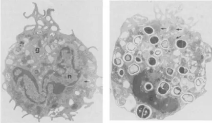
Figure 1. Scanning electron micrograph of control mac-rophages incubated with opsonized S. au reus (arrow)for 5 min. Note the extensive lamellipodia (L) formation (x1,464).
compared with controls. This was observed as early
as5-10min, with a parallel increase in the frequency
of engulfment of bacteria. Itshould be emphasized
that this difference was only quantitative; no qualita-tive differences could be observed on the macrophage surfaces by scanning electron microscopy.
Longer periods of incubation of both control and
Q8-treated macrophages (i.e.,4-6hr) resulted in a
number of ultrastructural changes common to both of the groups: the presence of a large number of bac-teria as well as reduction in the degree of ruffling, presumably due to sequestration of the plasma mem-brane within (figure 3, right), which was not seen in the early stages of phagocytosis (figure 3, left); dense clumping of the chromatin; less prominent Golgi apparatus when compared with the earlier stages of phagocytosis; large accumulation of poly-ribosomes and rough endoplasmic reticulum; and
Figure 2. Q8-treated macrophages incubated with S.aureusfor 5 min. Compare the number of attached particles(arrows)with those in figure 1. L, lamellipodia
(x1,464).
electron-dense particles containing patches of par-ticles (figures 3, right and 4). The transmission elec-tron microscopic picture at later intervals was con-sistent with the reported increase in RNA synthesis and a higher rate of delivery of mature ribosomal RNA to the cytoplasm in human monocytes
follow-ing phagocytosis [20]. These changes are likely to
be secondary to the early phagocytic events within the phagocytes, with only quantitative differences between control and Q8-treated cells that cannot be discerned by transmission electron microscopy. For instance, when macrophages were incubated with Q8 for various time intervals, a significant increase
(25070-40070 at 2 hr) in protein synthesis was noted when compared with controls (data not shown). The fact that the typical morphological and biochemi-cal changes that are characteristic for the "activated
macrophage" could not be observed before12-14hr
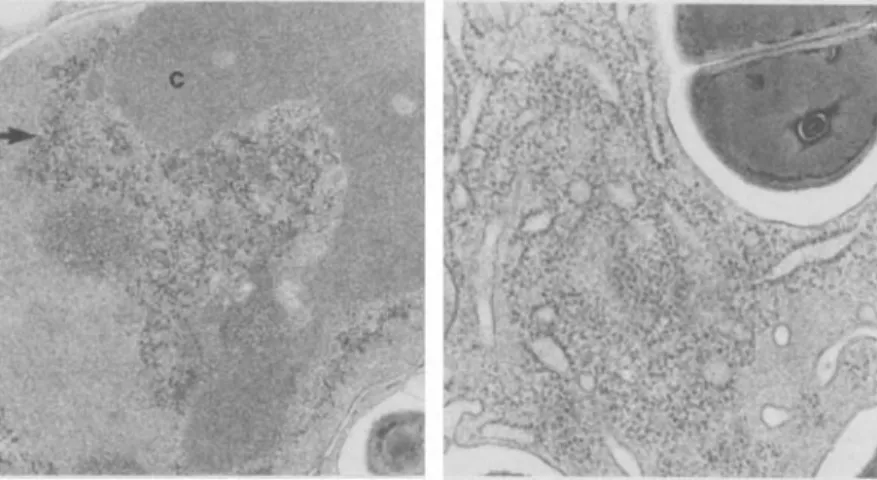
Figure 3. Left, transmission electron micrograph of Q8-treated macrophage incubated for 30 min with S. aureus.
Note Golgi apparatus (g), nucleus (n), and rough endoplasmic reticulum (ar-row; X5,330).Right, Q8-treated mac-rophage incubated for 5 hr with S.
aureus. Note the dense chromatin (c) in the nucleus, the large patches of polyribosomes (arrows) in the cyto-plasm, and the smooth plasma mem-brane(x5,330).
Figure 4. Left, a higher power elec-tron micrograph of the nucleus of a Q8-treated macrophage at a late stage of phagocytosis (4-6 hr). The clumped chromatin (c) and the patches of electron-dense particles (arrow) are evident (x 21,320). Right, this micro-graph demonstrates the accumulation of polyribosomes around the rough endoplasmic reticulum (x 30,340).
- - - _ . _ - _ . _... -Table 1. The effect of C3b and IgG and IgM fractions on attachment and ingestion of SRBCs by mononuclear phagocytes.
NOTE. SRBCs were sensitized with IgG (10 ng/108 SRBCs) or IgM (15~g/108SRBCs) in a total volume of 0.7 ml and then added to monolayers(lWIml per dish, preincubated for 1 hr with
and without Q8) for 60 min at 37 C in an atmosphere contain-ing 5010 CO2after which microscopic examination and
assess-ment of the phagocytic index were performed. Five hundred mononuclear phagocytes were scored for each sample: - = no attachment and ingestion,(+) = 0-1, + = 1-2, + + = 3-4, + + + = 5-6, + + + + = 7-8, + + + + + = ~8SRBCs at-tached to or ingested by mononuclear phagocytes. The data represent the mean of four independent experiments.
cytosis of S.aureusby acting as a trigger for endocy-tosis; (2) the absence of IgM receptors at the surface of the macrophage prevents its binding directly to the membrane in the absence of C3b; and(3)though Fe and C3b receptors apparently act independently of one another in the binding and phagocytosis of sensitized particles, the two receptors can act syner-gistically. Occupancy of the receptors seems to be quantitatively of major significance in the stimula-tion of phagocytosis by Q8. The attachment step per se seems to be of primary significance, since changes
++++ +++++ (+) + Ingestion ++ +++ Without Q8 With Q8 (10-5M) (+) ++++ +++++ (+) +++++ Attachment IgG fraction + + IgG fraction plus C3b + + + IgM fraction IgM fraction plus C3b + + SRBCs Without sensitized with Q8
of incubation indicates that the Q8 effect on pro-tein synthesis at 2 hr cannot be attributed to the "ac-tivated macrophage."
Because the enhanced attachment and ingestion of bacteria in Q8-treated cells could not be ascribed to either short-term or long-term ultrastructural changes, either at the surface or in the interior, these events were further investigated by detailed quan-titative methods.
Effect of Q8 on attachment and ingestion of
SRBCsbymacrophages. The electron microscopic
studies were initially elaborated at a semiquantita-tive level of light microscopy with regard to the in-fluence of Q8 on attachment and ingestion of sensi-tized SRBCs by macrophages. We did this in light of our earlier, similar observations based on Q8 treat-ment in vivo, wherein the mouse peritoneal macro-phages were shown to exhibit enhanced attachment to SRBCs following an in vivo administration of Q8 [7]. Both attachment and ingestion were individu-ally scored in each case. Itcan be seen (table 1) that Q8 per se was marginally effective; that IgG but not IgM stimulated both attachment and ingestion, al-though both of these immunoglobulin fractions were antibodies to SRBCs; that C3b stimulated attach-ment and ingestion in the presence of both IgG and IgM; and that Q8 potentiated the actions of the im-munoglobulins as well as the effect of C3b. The ef-fect of these agents on attachment consistently par-alleled that on ingestion, and IgG was the single most effective agent in initiating phagocytosis, since IgM, with or without Q8, was without significant effect unless C3 b was also present.
These results were consistent with the observations that(1)immunocomplexed IgG increases the
phago-Figure 5. Effect of Q8 on sensitization profiles for erythrophagocytosis by human mononuclear phagocytes following presensitization with IgM and IgO antibodies to SRBC and subsequent addition of IgO antibody to SRBC, C3b, and Q8. Mononuclear phagocytes were sen-sitized with IgM antibody to SRBC, as described in Materials and Methods. The data represent the mean of four independent experiments ± SO.
'0 20~30-4~~f-'9~0-~ MINUTES
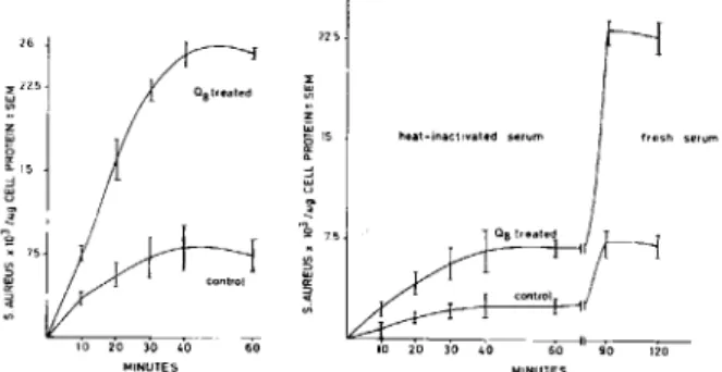
Figure 6. Left, the effect of Q8 on phagocytosis of 14C_ labeled S.aureus by human mononuclear phagocytes in the presence of fresh serum. The data represent the mean of four independent experiments ± SE. Right, the ef-fect of Q8 on phagocytosis of 14C-Iabeled S.aureus by human mononuclear phagocytes in the presence of heat-inactivated serum after reconstitution with fresh serum. the data represent the mean of four independent experi-ments ± SE.
tivity as well as the stimulatability of phagocytosis by Q8 (figure 6). These results indicated the paral-lelism between the phagocytosis of SRBCs and S. aureus in relation to the action of Q8.
Effect ofQ8on the inhibitory role ofFe fragments
on the phagocytosis ofS.aureus by macrophages. Fc
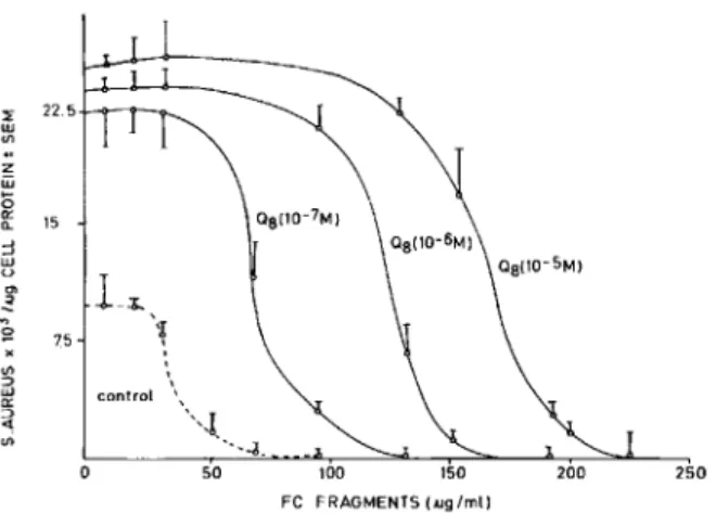
fragments are known to inhibit phagocytosis of op-sonized particles, presumably by competing for the Fereceptors. The enhanced phagocytosis by Q8 could be due to an increased rate of endocytosis with or without an increase in the number of Fe receptors on the surface of the macrophage. On the other hand, if the enhanced phagocytosis is mediated by an increased affinity for the Fe receptor as well, a change in the affinity of the Fc receptor may be dis-cerned in a competition experiment wherein phago-cytosis of opsonized bacteria is quantitated as a func-tion of Fe fragments in the incubafunc-tion medium. Data in figure 7 show that phagocytosis of opsonized bac-teria (at a constant concentration of IgG) can be inhibited by Fe fragments such that the 50010 inhibi-tory concentration(Iso)was rv35/Ag/ml (or approxi-mately two orders of magnitude higher concentra-tion of Fe fragments). Addiconcentra-tion of Q8 enhanced not only the magnitude of phagocytosis but also theIso of Fe fragments. In fact, stimulation of phagocyto-sis tended to near saturation at 10-7M Q8, whereas
the150proportionately increased to 10-sMQ8.These
results were consistent with the possibility that the addition of exogenous Q8 modified the kinetics of the Fe receptor, an occurrence facilitating the
trig-261 ;
rl '."""
~~
i.
75j con' rc , <t ' 10 20 30 40--- 60 MINUTES control 'C3b 600 450 300 IgG (ng/10 8 SRBC) 100 150in ingestion always paralleled changes in attachment. In subsequent studies, only ingestion was quantitated because measurement of attachment was less amena-ble to quantitation, particularly with bacteria.
Because receptor occupancy seemed to be critical to the magnitude of phagocytosis, we varied the IgG in the incubations and quantitated the ingestion of SRBCs as a function of IgG concentration in the medium. Stimulation of phagocytosis by IgG, alone or in the presence of C3b, exhibited near saturation at rv600 ng of IgG/108 SRBCs (figure 5). On the
other hand, the stimulation of phagocytosis by Q8 appeared to be saturated even at a concentration of rv350 ng of IgG/108SRBCs. These results suggested
that although C3b and IgG could be merely addi-tive, the presence of exogenous Q8 enhanced the ap-parent affinity of the Fc receptor to IgG.
Effect ofserum andQ8on bacterial ingestion by
m acrophages. Phagocytosis of bacteria was
mark-edly stimulated by Q8 in the presence of serum. This threefold stimulation was reflected not only in the magnitude but also in the rate of phagocytosis (fig-ure 6). Heat inactivation of the serum led to signifi-cant and proportionate reductions of the rate as well as the magnitude of phagocytosis in both control and Q8-treated macrophages. Treatment of macrophages with heat-inactivated serum did not affect the mac-rophages adversely, since restoration with fresh se-rum resulted in restoration of the full phagocytic
ac-a a '" lil 1000 3\ til W l -~ g 3000 < 1[ l r <
d
~ 2000 o ~Discussion
Figure 7. The effect of Q8 on the IgG-mediated Fe frag-ment inhibition of phagocytosis by human mononuclear phagocytes. The data represent the mean of five indepen-dent experiments ± SD.
gering effect of IgG on phagocytosis. The quantita-tive relation between IgG concentration and phago-cytosis in these macrophages supported a primarily kinetic interpretation.
appears to be primarily of quantitative (kinetic) rather than qualitative (structural) significance in triggering phagocytosis, an occurrence contributing to the specificity of the microdomain that ultimately constitutes the phagocytic vesicle. The results
ob-tained with SRBCs as well as with S.
aureus
gave aconsistent picture in this regard. The phagocytic events of attachment and ingestion essentially be-haved consistently as tandem-rate processes, their magnitude being related to the availability of ubi-quinones, i.e., the rate of respiration. That the stim-ulation of various functions in phagocytes by added ubiquinone does not seem to be a nonspecific "tick-ling" is evidenced in a previous report [4]. First, the stimulation of a respiratory burst is seen in disrupted cells that are no longer responsive to latex particles and phorbol myristate acetate. Second, when dis-rupted cells are pretreated with Nikkol detergent, the addition of NADPH and ubiquinone still results in oxygen consumption.
The experimental evidence suggests that the ki-netics of the Fe receptor, both in terms of the maxi-mal rate and apparent affinity of the receptor to IgG, varies with the exposure of the macrophages to ex-ogenous Q8. This implies that the enhanced oxida-tions at the plasma membrane could affect the
ki-netic properties of the Fereceptor protein vis-a-vis
the phagocytic process. Kinetic evidence related to such macrophenomena is generally suspect due to the inherent complexity of the system, although ki-netic interpretations have often yielded illuminating insights in cellular processes such as growth. Prece-dence for such kinetic eviPrece-dence has recently been reported, primarily in terms of the perturbations pro-duced in the bilayer as a consequence of respiration
[23-25]. (1) Respiration and density of the surface
charge were shown to induce variable porosity to os-molytes in the mitochondrial inner membrane (due to dynamic perturbations in the bilayer) that leads to enhanced requirement for isotonicity. (2) Hypo-osmotic activation of a variety of membrane-bound enzyme systems (transporters, etc.) was reported, which includes NADPH oxidase of the plasma mem-brane [26], besides oxidative phosphorylation in
mitochondria. (3) Respiration was shown to affect
the kinetics of {3-galactoside transport inE.coli and
dicarboxylate transport in mitochondrial inner mem-branes. In fact, these studies prompted us to exam-ine the possible alterations in the kexam-inetics of the Fe receptor. Indeed, addition of Q8 increased not only the maximal rate of phagocytosis, but also the
ap-250 200 50 100 150 Fe FRAGMENTS(.ug/mll
l-~
'~ control \"-1 --"-<1_ z ~ o g: 15 ::J ~ en ~ 75The results reported here offer direct evidence for the stimulatory influence of Q8 on the initial events of phagocytosis, namely, attachment and ingestion. The mechanism of action of Q8 does not appear to require any postulate other than its well-authenti-cated role in electron transport, even with regard to these initial events. Electron transport is diffusion controlled [21]. In a diffusion-coupled reaction that requires collision of membrane components, factors that affect the rate of lateral diffusion must, in turn, affect component interaction [22]. The major parameters in any collison-based reaction mecha-nism are the concentration of reactants and their diffusion coefficients. Since the diffusion coefficient of a component in the membrane relates to its bulk-iness, use of ubiquinones with a shorter chain length, such as Q7 or Q8 as compared with the native ubi-quinone QIO, would be consistent with the observed stimulation of respiration. This agrees with a report by Crawford and Schneider [4] who recently identi-fied the role of ubiquinones in microbicidal events. Thus, the shorter ubiquinones (e.g., Q5) were more potent in the stimulation of a respiratory burst than were the higher series analogues.