HAL Id: inserm-01904372
https://www.hal.inserm.fr/inserm-01904372
Submitted on 24 Oct 2018
HAL is a multi-disciplinary open access
archive for the deposit and dissemination of
sci-entific research documents, whether they are
pub-lished or not. The documents may come from
teaching and research institutions in France or
abroad, or from public or private research centers.
L’archive ouverte pluridisciplinaire HAL, est
destinée au dépôt et à la diffusion de documents
scientifiques de niveau recherche, publiés ou non,
émanant des établissements d’enseignement et de
recherche français ou étrangers, des laboratoires
publics ou privés.
monoclonal antibodies
Julien Saint-Pol, Martine Billard, Emmanuel Dornier, Etienne Eschenbrenner,
Lydia Danglot, Claude Boucheix, Stephanie Charrin, Eric Rubinstein
To cite this version:
Julien Saint-Pol, Martine Billard, Emmanuel Dornier, Etienne Eschenbrenner, Lydia Danglot, et al..
New insights into the tetraspanin Tspan5 using novel monoclonal antibodies. Journal of Biological
Chemistry, American Society for Biochemistry and Molecular Biology, 2017, 292 (23), pp.9551 - 9566.
�10.1074/jbc.M116.765669�. �inserm-01904372�
New insights into the tetraspanin Tspan5 using novel
monoclonal antibodies
Received for publication, October 31, 2016, and in revised form, March 29, 2017 Published, Papers in Press, April 20, 2017, DOI 10.1074/jbc.M116.765669 Julien Saint-Pol‡§1, Martine Billard‡§, Emmanuel Dornier§¶2,3, Etienne Eschenbrenner‡§3, Lydia Danglot储**, Claude Boucheix‡§, Stéphanie Charrin‡§, andX Eric Rubinstein‡§4
From‡Inserm, U935, F-94807 Villejuif, the§Universite´ Paris-Sud, Institut Andre´ Lwoff, F-94807 Villejuif,¶Inserm, U1004, F-94807 Villejuif , the储CNRS, UMR7592, Université Paris Diderot, Sorbonne Paris Cité, Institut Jacques Monod, F-75205 Paris, and **Inserm, ERL U950, 75205 Paris, France
Edited by Amanda J. Fosang
Tspan5 is a member of a subgroup of tetraspanins referred to as TspanC8. These tetraspanins directly interact with the met-alloprotease ADAM10, regulate its exit from the endoplasmic reticulum and subsequent trafficking, and differentially regu-late its ability to cleave various substrates and activate Notch signaling. The study of Tspan5 has been limited by the lack of good antibodies. This study provides new insights into Tspan5 using new monoclonal antibodies (mAbs), including two mAbs recognizing both Tspan5 and the highly similar tetraspanin Tspan17. Using these mAbs, we show that endogenous Tspan5 associates with ADAM10 in human cell lines and in mouse tis-sues where it is the most abundant, such as the brain, the lung, the kidney, or the intestine. We also uncover two TspanC8-spe-cific motifs in the large extracellular domain of Tspan5 that are important for ADAM10 interaction and exit from the endoplas-mic reticulum. One of the anti-Tspan5 mAbs does not recognize Tspan5 associated with ADAM10, providing a convenient way to measure the fraction of Tspan5 not associated with ADAM10. This fraction is minor in the cell lines tested, and it increases upon transfection of cells with TspanC8 tetraspanins such as Tspan15 or Tspan33 that inhibit Notch signaling. Finally, two antibodies inhibit ligand-induced Notch signaling, and this effect is stronger in cells depleted of the TspanC8 tetraspanin Tspan14, further indicating that Tspan5 and Tspan14 can com-pensate for each other in Notch signaling.
Tetraspanins form a family of proteins with four transmem-brane domains expressed by all metazoans. These proteins pos-sess a number of specific features, including conserved residues and a specific fold in the largest of the two extracellular domains (the large extracellular loop (LEL)5) that differentiate
them from other proteins with four transmembrane domains (1– 4). Identification of pathological mutations in humans and genetic approaches in the mouse or invertebrates have shown the importance of these molecules. In mammals, particular tet-raspanins have, for example, been shown to play a key role in sperm-egg fusion, vision, kidney function, immunity, or muscle regeneration (1– 4). A remarkable property of the most charac-terized tetraspanins is to associate at the cell surface with one another and with non-tetraspanin integral proteins to organize a network of interaction referred to as the “tetraspanin web” or tetraspanin-enriched microdomains. In this network, tetraspa-nins associate directly and specifically with a limited number of molecular partners that they connect to other tetraspanins. Well characterized primary complexes include the complexes formed by CD151 (Tspan24) and the laminin-binding integrins ␣31 and ␣61 as well as the complex formed by CD81 (Tspan28) and CD19, a co-stimulatory molecule of B lympho-cytes. In addition, CD81 shares with CD9 (Tspan29) two com-mon partners, CD9P-1/EWI-F and EWI-2, that are Ig domain proteins of unknown function (1– 4).
Tspan5 is a highly conserved tetraspanin; the human, mouse, and rat proteins are completely identical and share 91, 44, and 38% identity with the closest orthologs found in Danio rerio,
Drosophila melanogaster,and Caenorhabditis elegans, respec-tively (5– 8). Tspan5 is a member of a subgroup of tetraspanins that have 8 cysteines in the LEL (others have 6 or 4 cysteines) and are consequently referred to as TspanC8 (7–10). Mammals express six of these TspanC8 tetraspanins that share a common partner, the metalloprotease ADAM10, a member of the ADAM (A Disintegrin And Metalloprotease domain) family of metalloproteases (8, 10, 11). These membrane-anchored enzymes mediate a proteolytic cleavage of various transmem-brane proteins within their extracellular region, a process referred to as ectodomain shedding (12, 13). ADAM10 cleaves off the ectodomain of more than 40 transmembrane proteins, including cytokine and growth factor precursors, as well as adhesion proteins such as E- and N-cadherins (13). Notably, ADAM10-mediated cleavage of the amyloid precursor protein prevents the formation of the amyloid peptide A, a major component of amyloid plaques observed in Alzheimer’s disease (14). ADAM10 plays also an essential role in Notch signaling; Notch ectodomain cleavage by ADAM10 allows a second cleav-age by the␥-secretase complex that results in the release of the This work was supported in part by core funding from INSERM and by
speci-fic grants from the NRB-Vaincre le Cancer, Institut du Cancer et d’Immunogénétique, and the Institut National du Cancer. The authors declare that they have no conflicts of interest with the contents of this article. 1Recipient of a fellowship from the Institut National du Cancer.
2Recipient of a fellowship from the Association pour la Recherche sur le Cancer.
3Recipients of fellowships from the French Ministry of Research.
4To whom correspondence should be addressed: Inserm, U935, 14 Av. Paul Vaillant Couturier, F-94807, Villejuif, France. Tel.: 33 1 4559 5317, E-mail: eric.rubinstein@inserm.fr.
5The abbreviations used are: LEL, large extracellular loop; UP, uroplakin; ER, endoplasmic reticulum; PNGase, peptide:N-glycosidase; EndoH, endogly-cosidase H; BFA, brefeldin A.
ARTICLE
J. Biol. Chem. (2017) 292(23) 9551–9566
9551
at INSERM on June 12, 2017
http://www.jbc.org/
Notch intracellular domain and its translocation to the nucleus where it acts as a transcriptional cofactor (15–18).
TspanC8 tetraspanins regulate several aspects of ADAM10. They all regulate the exit of ADAM10 from the ER and target it either to late endosomes (Tspan10, 17) or the plasma mem-brane (Tspan5, -14, -15, and -33) (8, 10, 11). In addition, TspanC8 tetraspanins modulate the substrate specificity of ADAM10 (19, 20). In particular, Tspan5 and Tspan14 are pos-itive and Tspan15 and Tspan33 negative regulators of Notch signaling (8, 19). Also, of all TspanC8 tetraspanins tested, only Tspan15 was shown to regulate ADAM10-mediated cleavage of N-cadherin (11, 19, 20). These functional differences may be the result of a different action of TspanC8 on ADAM10 membrane compartmentalization (19). Alternatively, TspanC8 might direct substrate specificity by constraining ADAM10 into defined conformations (20).
In the absence of good antibodies, the study of Tspan5 and other TspanC8 has relied on the transfection of tagged mole-cules, with potential pitfalls arising from overexpression or the addition of a tag. Here, we report on the generation of anti-Tspan5 monoclonal antibodies and use them to investigate sev-eral aspects of Tspan5, including its expression profile, subcel-lular localization, and the interaction of the endogenous protein with ADAM10 and with the tetraspanin web. We also show that two of these mAbs inhibit ligand-induced Notch signaling.
Results
Generation of antibodies recognizing Tspan5
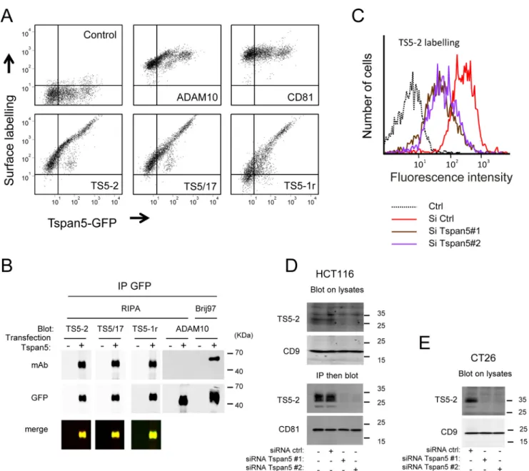
To generate anti-Tspan5 mAbs, we immunized mice twice with U2OS cells stably expressing Tspan5-GFP and twice with a Tspan5-GFP immunoprecipitate. Because the human, mouse, and rat Tspan5 molecules are completely identical, Tspan5 knock-out mice were used. Hybridomas were screened by indi-rect labeling of live U2OS cells stably expressing Tspan5-GFP and flow cytometry analysis. Out of more than 3000 clones tested, we isolated nine hybridomas stably secreting antibodies that stained U2OS-Tspan5 cells proportionally to the level of Tspan5-GFP expressed by the cells. Three examples are shown in Fig. 1A. As a control, the labeling by the CD81 antibody did not change according to the GFP signal, and the labeling by the anti-ADAM10 mAb 11G2 reached a plateau, as described pre-viously (8, 19). The characteristics of these antibodies are shown in Table 1. To validate that these antibodies indeed rec-ognize Tspan5, GFP-Tspan5 was immunoprecipitated from U2OS/Tspan5 cells using GFP trap beads after lysis in RIPA buffer (Fig. 1B). This lysis buffer is known to dissociate from tetraspanins most if not all the associated proteins. Indeed, under this condition, ADAM10 was no longer cipitated with Tspan5, although it is strongly co-immunopre-cipitated after lysis with Brij 97 (Fig. 1B). All antibodies tested recognized by Western blotting the immunoprecipitated Tspan5 GFP (see three examples in Fig. 1B), as shown by the strong signal perfectly overlapping with the signal obtained with an anti-GFP antibody.
The mAb TS5-2 was selected for further studies because in the initial characterization it gave one of the strongest signals in
Western blotting and flow cytometry. To validate that it recog-nized the endogenous Tspan5, we turned to colon cancer HCT116 cells that showed the strongest surface staining among various cell lines tested (data not shown). Silencing Tspan5 in these cells by two previously validated siRNAs (19) reduced by 70 – 80% the staining by TS5-2 in flow cytometry experiments (Fig. 1C). In addition, the mAb TS5-2 recognized by Western blotting endogenous levels of Tspan5 in HCT116 and CT26 cells (a human and a mouse colon cancer cell line, respectively) but not in Tspan5-silenced cells (Fig. 1, D and E). Moreover, this mAb immunoprecipitated a major⬃27–34-kDa band and a fainter⬃22-kDa thin band that were recognized by Western blotting by the TS5-2 mAb (Fig. 1D). None of these bands were visualized after silencing Tspan5 further indicating that they both correspond to Tspan5 (possibly to different conforma-tions or glycosylated forms) and that TS5-2 only recognizes Tspan5 in these cells.
Specificity of Tspan5 antibodies and demonstration that they bind to the LEL
TspanC8 tetraspanins are characterized not only by the pres-ence of 8 cysteines in the larger extracellular domain but also by the presence of specific residues not present in other tetraspa-nins (8). It was therefore important to test whether these anti-Tspan5 antibodies recognized other TspanC8. In a first set of experiments, we tested by Western blotting whether these anti-bodies recognized Tspan14, Tspan15, or Tspan33 in the lysates of U2OS cells stably expressing GFP-tagged versions of Tspan15, Tspan14, or Tspan33 (19), or after GFP immunopre-cipitation. As shown in Fig. 2A and summarized in Table 1, none of the mAbs recognized these three tetraspanins.
Similarly, none of these mAbs recognized Tspan17 or Tspan10 by Western blotting after transfection in HeLa cells (Fig. 2B). However, two antibodies, TS5/17 (Fig. 2) and 20E2 (Table 1), strongly stained Tspan17-transfected cells, either by flow cytometry analysis (data not shown) or immunostaining of saponin-permeabilized U2OS cells grown on coverslips (Fig. 2C and Table 1). As shown previously in HeLa cells, Tspan10 was mainly intracellular after transfection in U2OS cells. None of our antibodies recognized this tetraspanin (data not shown).
We then used Tspan5/Tspan15 chimeric molecules to deter-mine whether our antibodies recognize the small or the large extracellular domain. As summarized in Table 1, and exempli-fied for three of them in Fig 2C, all antibodies recognized, as shown by flow cytometry, a chimera in which the LEL of Tspan15 was replaced with that of Tspan5 (Ts15LEL5) but not the reverse chimera (Ts5LEL15). Thus, all antibodies bind to epitopes present in the LEL of Tspan5.
Absence of major intracellular pool of Tspan5
Using flow cytometry, we have tested the surface expression of Tspan5 in ⬃25 hematopoietic or non-hematopoietic cells lines. In all cell lines tested, the surface expression level of Tspan5 was low as compared with that of ADAM10 or CD81 (data not shown). This, together with the fact that some tetras-panins such as CD63 are mainly intracellular proteins (2), prompted us to compare the expression levels of endogenous Tspan5 at the cell surface and in intracellular compartments.
at INSERM on June 12, 2017
http://www.jbc.org/
For this purpose, the surface pool of Tspan5 was first labeled using a combination of the anti-Tspan5 mAb TS5-2 and an anti-mouse polyclonal antibody coupled to Alexa Fluor 568 (Fig. 3, top, red). In the second step, the cells were incubated, in the presence of saponin to permeabilize them, with a combina-tion of the Tspan5 mAb and an anti-mouse polyclonal antibody coupled to Alexa Fluor 647. In this experiment, the surface pool of Tspan5 was labeled with the two secondary antibodies, whereas the internal pool was labeled only with the Alexa Fluor 647-coupled secondary antibody (Fig. 3, green). As a control for the non-specific intracellular signal, the Tspan5 mAb was replaced by a control IgG2a in the second step (Fig. 3, middle
panels). As shown in Fig. 3, there was a weak intracellular signal with TS5-2 that was higher than that observed with the control IgG2a mAb, indicating the existence of an intracellular pool of Tspan5. This signal is much lower than the intracellular signal observed for CD63 (Fig. 3, right panels). Therefore, Tspan5 lacks a strong intracellular pool. Similar experiments were performed on U2OS cells (data not shown). The higher nonspecific intracellular signal observed with the IgG2a con-trol mAb precluded determining whether there was an intra-cellular pool of Tspan5 in this cell line. We can conclude, however, that this cell line does not have a strong intracellu-lar pool of Tspan5.
Figure 1. Characterization of new anti-Tspan5 mAb. A, flow cytometry analysis of U2OS cells expressing Tspan5 GFP and stained or not with mAbs to
ADAM10, CD81, or three anti-Tspan5 mAbs. B, U2OS cells expressing Tspan5 GFP were lysed in RIPA or Brij 97 lysis buffer as indicated, before immunoprecipitation (IP) of Tspan5 using GFP trap beads. The samples were analyzed by Western blotting using the indicated antibodies. Note that the interaction of Tspan5 with ADAM10 is disrupted in RIPA buffer. C, binding of mAb TS5-2 to HCT116 was analyzed by flow cytometry 3 days after transfection with a control siRNA or two siRNA targeting Tspan5. D, HCT116 cells were lysed 3 days after transfection with a control siRNA or two Tspan5 siRNAs. In the top panels, the cells were lysed directly in Laemmli buffer, before Western blotting using a combination of anti-Tspan5 mAb TS5-2 and a secondary antibody. In the bottom panel, the cells were lysed in Brij 97 buffer, and Tspan5 was immunoprecipitated using TS5-2. The presence of Tspan5 in the immunoprecipitate was determined using a combination of biotin-labeled TS-2 mAb and streptavidin. E, mouse colon CT26 cells were lysed 3 days after transfection with a control siRNA or two to Tspan5 siRNAs. The cells were lysed directly in Laemmli buffer, before Western blotting using a combination of anti-Tspan5 mAb TS5-2 and a secondary antibody.
at INSERM on June 12, 2017
http://www.jbc.org/
Figure 2. Specificity of selected anti-Tspan5 mAbs. A, U2OS cells stably expressing or not GFP-tagged Tspan5, Tspan14, Tspan15, and Tspan33 were lysed
in RIPA buffer before immunoprecipitation (IP) of the transfected protein with GFP trap beads. The recognition of the transfected tetraspanin by the different mAbs was tested by Western blotting using both the cell lysate or the GFP-trap immunoprecipitate (IP). To control for the expression of the different transfected tetraspanins, the membrane was also incubated with an anti-GFP mAb. B, HeLa cells were transiently transfected with Tspan5, Tspan17, Tspan10, or CD9 and lysed 2 days later in Laemmli buffer. To control for the expression of the different transfected tetraspanins, the membrane was also incubated with an anti-GFP mAb. C, U2OS cells were transiently transfected with plasmids coding Tspan17 or Tspan5 and immunolabeled 2 days later with mAb TS5-2 and TS5/17. Note that the mAb TS5/17 also recognizes Tspan17 but TS5-2 does not (bar: 10m). D, U2OS cells were transiently transfected with plasmids coding the GFP-tagged chimeric tetraspanin Ts15LEL5, in which the LEL of Tspan15 was replaced by that of Tspan5 and the reverse chimera Ts5LEL15. The panel shows flow cytometry analysis of the binding of selected anti-Tspan5 mAb to the transfected cells.
Table 1
Characterization of anti-Tspan5 mAbs
The binding of the mAbs to Tspan5, Tspan14, Tspan15, and Tspan33 was analyzed using U2OS cells stably expressing these molecules. The binding to the two chimeric molecules Tspan15LEL5 (in which the LEL of Tspan15 is replaced by that of Tspan5) and the reciprocal construct Tspan5LEL15 was analyzed by flow cytometry after transient transfection. The binding to Tspan10 and Tspan17 was analyzed by indirect immunofluorescence after saponin permeabilization of cells grown on coverslips. The binding to the Tspan5 mutants RDD and NIYF was analyzed by indirect immunofluorescence after Triton X-100 permeabilization of cells grown on coverslips. We did not identify the subclass of mAb 10G5, which is not an IgA, IgM, Ig1, Ig2a, Ig2b, or Ig3. ND means not determined.
Clone Alias Sub class
Antibody binds to:
T5 T10 T14 T15 T17 T33 Ts15 LEL5 Ts5 LEL15 RDD NIYF
16B8 TS5-2 IgG2a Yes No No No No No Yes No Yes No
12E1 TS5-1r IgG1 Yes No No No No No Yes No Yes No
10G11 TS5/17 IgG2b Yes No No No Yes No Yes No Yes Yes
8H12 TS5-3 IgG2a Yes No No No No No Yes No Yes No
8B1 IgG2a Yes No No No No No Yes No Yes No
13G1 IgG2b Yes No No No Weak No Yes No No No
20E2 IgG2b Yes No No No Yes No Yes No Yes Yes
10G5 ND Yes ND No No ND No Yes No ND ND
at INSERM on June 12, 2017
http://www.jbc.org/
Endogenous Tspan5 interacts with ADAM10 and is a component of the tetraspanin web
We then tested whether endogenous Tspan5 interacted with ADAM10, with other tetraspanins, and with selected compo-nents of the tetraspanin web. In a first set of experiments, sev-eral cell lines cells were surface-labeled with biotin before immunoprecipitations. All cells express Tspan5 at the mRNA level, but the most prominent TspanC8 expressed by these cells is Tspan15 in PC3 cells and Tspan14 in U2OS and HCT116 cells (8). As shown in Fig. 4A, the Tspan5 mAb precipitated in all cell types, but at different levels several biotin-labeled pro-teins that co-migrated with propro-teins co-immunoprecipitated with the prototypal tetraspanin CD81, including CD9 and CD81, or1 integrin subunits. Although ectopically expressed Tspan5 is biotin-labeled, we did not detect Tspan5 in the Tspan5 immunoprecipitation after biotin labeling. This may be because the fraction of Tspan5 associated with ADAM10 may not be efficiently biotin-labeled (data not shown). The presence of Tspan5 in Tspan5 and ADAM10 immunoprecipitates was validated by incubating the same membrane with biotin-la-beled TS5-2 mAb, yielding an additional band of 25–30 kDa corresponding to Tspan5.
Similar experiments were performed without biotin labeling, allowing analysis of the composition of the different immuno-precipitates by Western blotting using biotin-labeled antibod-ies (Fig. 4B). Overall, the data show that endogenous Tspan5 co-immunoprecipitates and/or is co-immunoprecipitated with ADAM10, other tetraspanins such as CD9, CD81, and CD151, as well as the 1 integrin subunit or CD9P-1. The apparent molecular weight of Tspan5 is higher in HCT116 cells than in
PC3 and U2OS cells (Fig. 4C). This is likely to be due to different glycosylation patterns, as will be addressed later.
Tspan5 interacts with ADAM10 in mouse organs
Initial attempts to characterize the tissue distribution of Tspan5 by immunohistochemistry in the mouse did not suc-ceed, probably because of the low abundance of Tspan5. As an alternative approach, various tissues were lysed in Brij 97 lysis buffer, and immunoprecipitations were performed with the mAb TS5-2 (Fig. 5). The presence of Tspan5 was determined by Western blotting using a combination of TS5-2 mAb conju-gated to biotin and streptavidin. This approach revealed that the major sites of Tspan5 expression at the protein level were the brain, cerebellum, eye, kidney, lung, uterus, and intes-tine. The high expression of Tspan5 at the protein level in the brain, lung, and kidney is consistent with its expression at the RNA level (21). However, in this previous study Tspan5 RNA was found to be present at the same level in the liver and the heart than in the lung, which contrasts with our data because we found no expression or very little expression of Tspan5 pro-tein in these organs. In the tissues where the expression of Tspan5 is high, including the brain, ADAM10 was co-immuno-precipitated with Tspan5.
An antibody that does not recognize Tspan5 associated with ADAM10 reveals that the majority of Tspan5 is associated with ADAM10 in U2OS and HCT116 cells
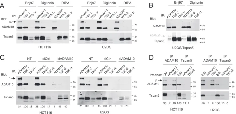
Previous studies have shown that the binding of certain anti-tetraspanin CD151 antibodies is prevented by the interaction with integrins that conceal the epitope, and as a consequence these antibodies only recognize the fraction of CD151 not asso-ciated with integrins (22–26). In our initial screening, we observed that TS5-1r failed to co-immunoprecipitate ADAM10 from U2OS-N1 cells (data not shown), suggesting that it may be such an antibody. As a first step to validate this hypothesis, immunoprecipitations in various detergents were performed using either TS5-1r or TS5-2. After cell lysis with RIPA, a con-dition that disrupts the interaction of ADAM10 with Tspan5, both antibodies immunoprecipitated similar amounts of Tspan5, both in HCT116 cells and U2OS cells (Fig. 6A). In contrast, after lysis with Brij 97 or digitonin, the interaction with ADAM10 was preserved, and TS5-1r precipitated only a small fraction of Tspan5. Notably, the major band co-immuno-precipitated with TS5-1r in HCT116 cells after Brij 97 or digi-tonin lysis had a slightly lower molecular weight than the bulk of Tspan5. Importantly, TS5-1r failed to immunoprecipitate ADAM10 under all conditions tested (except, in some experi-ments, for a slight amount in HCT116 cells after lysis with Brij 97, a detergent that preserves indirect interactions in the tet-raspanin web), confirming our initial observation. The inability of TS5-1r to co-immunoprecipitate ADAM10 is not a conse-quence of the lower immunoprecipitation of Tspan5 after Brij 97 or digitonin lysis (Fig. 6A) because this mAb also failed to co-immunoprecipitate ADAM10 in U2OS cells overexpressing Tspan5, in which it efficiently immunoprecipitated this tetras-panin (Fig. 6B).
The above experiments indicated that the mAb TS5-1r effi-ciently immunoprecipitates Tspan5, but only under conditions
Step 1: Before perm. mAb + GAM 568 merge Tspan5 CD63 IgG2a CD63 Tspan5 Tspan5 Step 2: With perm. mAb + GAM 647
Figure 3. Confocal microscopy analysis of the intracellular pool of Tspan5 and CD63. HCT116 cells were grown for 2 days on coverslips before
incubation with anti-Tspan5 (TS5-2) or CD63 mAb for 1 h at 4 °C. After wash-ing and fixation with paraformaldehyde, the cells were incubated with an anti-mouse polyclonal antibody coupled to Alexa 568 to visualize the surface pool of the tetraspanin (red). In Step 2, the cells were incubated with either the same primary antibody or a control IgG2a mAb in the presence of saponin to permeabilize the cells, and subsequently with an mouse polyclonal anti-body coupled to Alexa 647 (green). In this experiment, the surface pool of Tspan5 or CD63 is labeled with the two secondary antibodies, whereas the internal pool is labeled only with the Alexa 647-coupled secondary antibody (green). This experiment has been performed twice with similar outcomes.
Perm., permeabilization; GAM, goat anti-mouse antibody. Bar, 10m.
at INSERM on June 12, 2017
http://www.jbc.org/
disrupting the interaction with ADAM10. To validate that the low recognition of Tspan5 by TS5-1r after Brij 97 or digitonin lysis is due to the interaction with ADAM10, we next analyzed the effect of ADAM10 silencing on the ability of TS5-1r to immunoprecipitate Tspan5 after Brij 97 lysis (Fig. 6B). Surpris-ingly, silencing ADAM10 in HCT116 and U2OS cells with two different siRNAs (Fig. 6C, 7A) changed the migration profile of Tspan5; the main⬃27–34-kDa diffuse band disappeared, and two bands of slightly higher and lower molecular weight appeared or were reinforced. The lower molecular weight band (⬃27 kDa) co-migrated with the band recognized by TS5-1r in untreated Brij 97 or digitonin-lysed cells. Both bands were
sim-ilarly immunoprecipitated with mAbs TS5-1r and TS5-2 after ADAM10 silencing, showing that the low level of Tspan5 immunoprecipitation by TS5-1r in control cells is a conse-quence of ADAM10 expression and that ADAM10 prevents the binding of this mAb to Tspan5. As a consequence, and consis-tent with the finding that TS5-1r does not co-immunoprecipi-tate ADAM10, the fraction of Tspan5 immunoprecipico-immunoprecipi-tated by TS5-1r under conditions preserving the interaction with ADAM10 (⬃15–20%, excluding the 22-kDa but not the 27-kDa band, Fig. 6C) corresponds to the fraction not associated with ADAM10. To confirm this finding, Tspan5 and ADAM10 were immunoprecipitated after antibody-mediated depletion Blot:Streptavidin Streptavidin +TS5-2 Int. α Int. β1, CD9P-1 ADAM10 EWI-2 CD9 CD81 Tspan5 Tspan5 HCT116 PC3 U2OS 25 Blot: U2OS PC3 (KDa) 130 55 170 70 100 35 45 25 130 55 170 70 100 35 45 25 HCT116
B
A
C
IP: IP: 35 HCT116 55 35 Tspan5 ADAM10 IP: 25 100 130 55 100 35 25 25 U2OS Int. β1 CD9 p. m.Figure 4. Endogenous Tspan5 associates with ADAM10 and other tetraspanins. PC3, U2OS, and HCT116 were lysed in Brij 97, and immunoprecipitations
(IP) were performed as indicated on the top of each lane. A, surface proteins were labeled with biotin before lysis and were visualized using Alexa 680-labeled streptavidin (top). In the 2nd step, the membrane was incubated with biotin-labeled TS5-2 mAb and again with Alexa 680-labeled streptavidin to confirm the presence of Tspan5 in the immunoprecipitates and compare its molecular weight with the other proteins present in the different immunoprecipitations (bottom). B, immunoprecipitations were performed from non-labeled cells. The composition of the immunoprecipitates was analyzed by Western blotting using various biotin-labeled mAbs. The mature (m.) and proform (p.) forms of ADAM10 are indicated by arrows. C, comparison of the migration profile of Tspan5 immunoprecipitated from PC3, U2OS, and HCT116 cells. Int., integrin.
35 25 25 20 35 25 IgG2a Tspan5 IP CD81 Blot CD81 70 55 ADAM10 IP Tspan5 Blot:
Figure 5. Tspan5 associates with ADAM10 in mouse organs. Mouse organs were lysed, and immunoprecipitations (IP) were performed using Tspan5 or
CD81 antibodies. The presence of the target antigen was analyzed by Western blotting using biotin-labeled mAbs. Because Tspan5 co-migrates with a nonspecific band, the membranes were incubated with an irrelevant biotin-labeled mAb of the same subclass (IgG2a) before incubation with the Tspan5 mAb. The presence of ADAM10 in the Tspan5 immunoprecipitates was analyzed using a polyclonal antibody to ADAM10. This experiment has been done twice with similar outcomes.
at INSERM on June 12, 2017
http://www.jbc.org/
of these proteins in U2OS or HCT116 cell lysates. As shown in Fig. 6D, removal of ADAM10 using the mAb 11G2 diminished by 80 – 85% the amount of Tspan5 present in the lysate confirming that the majority of Tspan5 is associated with ADAM10. In contrast, depletion of Tspan5 had no effect or a modest effect on the amount of ADAM10 immunoprecipitated with the mAb 11G2.
ADAM10 regulates Tspan5 exit from the ER and glycosylation
The above data showed that silencing ADAM10 (Fig. 6C) changed the migration profile of Tspan5, with the main band disappearing at the profit of two bands of slightly higher and lower molecular weight (⬃27 and 34 kDa). We then investi-gated whether these new bands corresponded to different gly-coforms of Tspan5 (Fig. 7A). After digestion of Tspan5 immu-noprecipitates with PNGase F, which removes all N-linked glycans, only a band of lower molecular weight was observed in all samples (⬃24 kDa), both in HCT116 cells and U2OS cells. Thus, the change in Tspan5 molecular weight observed upon ADAM10 silencing corresponds to a change in glycosylation. The⬃27-kDa band observed after silencing ADAM10 was sen-sitive to EndoH (Fig. 7A), an enzyme that removes high man-nose structures, which are trimmed in the Golgi, indicating that this band corresponds to a fraction of Tspan5 retained in the ER
in the absence of ADAM10. In contrast, the higher⬃34-kDa band is not sensitive to EndoH and therefore corresponds to a fraction of ADAM10 having egressed from the ER. Because this band is efficiently immunoprecipitated with TS5-1r (Fig. 6C), it corresponds to a fraction of Tspan5 not associated with ADAM10. Thus, the interaction with ADAM10 modifies the glycosylation of Tspan5 in the Golgi.
Retention of Tspan5 in the ER in the absence of ADAM10 should be associated with a reduction of Tspan5 surface expres-sion levels. Indeed, depletion of ADAM10 by RNA interference reduced ADAM10 surface expression levels by⬎70%, as deter-mined by flow cytometry (Fig. 7B), and decreased Tspan5 expression levels (as determined by the labeling with mAb TS5-2) by⬃40%. Of note, in these experiments, the binding of TS5-1r was lower than the binding of TS5-2 in both cell lines, but it increased almost to the level observed for TS5-2 after ADAM10 silencing, further indicating that ADAM10 expres-sion prevents TS5-1r binding to Tspan5 at the cell surface and that Tspan5 is to a large extent associated with ADAM10 at the cell surface.
Previous studies have shown that TspanC8 tetraspanins reg-ulate the exit of ADAM10 from the ER (8, 10, 11). We now demonstrate that reciprocally ADAM10 regulates Tspan5 exit
C
35 25 35 25 55 70 55 70 Tspan5 RIPA Brij97 Digitonin Blot: IP: ADAM10 HCT116A
Brij97 Digitonin RIPAU2OS p. 35 25 35 25 55 70 55 70 HCT116 U2OS Tspan5 siADAM10 NT siCtrl Blot: IP: ADAM10 siADAM10 NT siCtrl p. ADAM10 Tspan5 IP ADAM10 IP Tspan5 Preclear: p. 35 25 55 70 HCT116 35 25 55 70 IP ADAM10 IP Tspan5 U2OS
D
55 70 45 55 Brij97 Digitonin Blot: IP:B
ADAM10 Tspan5 ADAM10 U2OS/Tspan5Figure 6. The majority of Tspan5 molecules associates with ADAM10. A, HCT116 cells were lysed in the presence of Brij 97, digitonin, and RIPA buffer
before immunoprecipitations (IP) with the anti-ADAM10 mAb 11G2 or the anti-Tspan5 mAbs TS5-2 and TS5-1r. The presence of Tspan5 and ADAM10 in the immunoprecipitates was visualized by immunoblotting using biotin-labeled TS5-2 and 11G2 mAbs. Note that TS5-1r does not co-immunoprecipi-tate ADAM10 and that its ability to immunoprecipico-immunoprecipi-tate Tspan5 is poor under lysis conditions preserving the interaction of Tspan5 with ADAM10. The band corresponding to the proform (p.) of ADAM10 is indicated. B, U2OS-N1 cells stably expressing GFP-tagged Tspan5 were lysed in the presence of Brij 97 or digitonin before immunoprecipitations with the anti-ADAM10 mAb 11G2 or the anti-Tspan5 mAbs TS5-2 and TS5-1r. The presence of Tspan5 and ADAM10 in the immunoprecipitates was visualized by immunoblotting using biotin-labeled TS5-2 and 11G2 mAbs. Note that TS5-1r does not co-immunoprecipitate ADAM10. C, HCT116 and U2OS cells were transfected or not with a control siRNA or an siRNA targeting ADAM10 and lysed 3 days later in Brij 97. Immunoprecipitations were performed as indicated at the top of each lane. The presence of Tspan5 and ADAM10 in the immunopre-cipitates was visualized by immunoblotting using biotin-labeled TS5-2 and 11G2 mAbs. Note the change of Tspan5 molecular weight upon ADAM10 silencing and the better immunoprecipitation of Tspan5 by TS5-1r. The band corresponding to the proform (p.) of ADAM10 is indicated. D, HCT116 or U2OS-N1 cells were lysed in digitonin and were subjected to two rounds of immunoprecipitation with a control mAb or the anti-ADAM10 mAb 11G2 or the anti-Tspan5 mAb TS5-2. Each sample was then separated in two for immunoprecipitations with the anti-ADAM10 and the anti-Tspan5 mAbs. The presence of Tspan5 and ADAM10 in the immunoprecipitates was visualized by immunoblotting using biotin-labeled TS5-2 and 11G2 mAbs. Except for
B, the experiments in this figure have been done three times with similar outcomes.
at INSERM on June 12, 2017
http://www.jbc.org/
from the ER, suggesting that the formation of the complex is necessary for the ER exit of both partners. However, the Tspan5 mAbs did not immunoprecipitate the proform of ADAM10, the form of ADAM10 present in this compartment (Figs. 4 and 6). This does not exclude the possibility that they interact in the ER, because once the complex is formed, it may quickly exit the ER, and ADAM10 may be rapidly processed by pro-protein convertases in the Golgi. To determine whether the two pro-teins associate in the ER, immunoprecipitations were per-formed after treatment of cells with brefeldin A, to inhibit the egress of ADAM10 and Tspan5 to the Golgi. As shown in Fig. 7C, the band corresponding to the proform of ADAM10 was reinforced after BFA treatment in the ADAM10 immunopre-cipitate, and was co-immunoprecipitated with Tspan5. Recip-rocally, the ⬃27-kDa band corresponding to the immature form of Tspan5 was strongly reinforced after BFA treatment and was efficiently co-immunoprecipitated with ADAM10. Thus, Tspan5 and ADAM10 interact in the ER.
Analysis of TspanC8-specific motifs in Tspan5 LEL
Because all TspanC8 tetraspanins interact with ADAM10, we hypothesized that the residues conserved in the LEL of all TspanC8 tetraspanins and not in other tetraspanins would mediate the interaction with ADAM10. Sequence analysis uncovered two TspanC8-specific motifs, present in mamma-lian, C. elegans, and D. melanogaster TspanC8 tetraspanins (Fig. 8A) as follows: Y(R/q/y)DDXD(L/qf/)(Q/r/k) between the two first predicted helix of the LEL (YRDDIDLQ in Tspan5, site 1; X corresponds to any residue, and lowercase letters indicate alter-natives found in only one or two TspanC8); NXY(F/h) (NIYF in Tspan5, site 2), immediately before the first of the two TspanC8-specific extra cysteines. Three overlapping mutations were generated for site 1 (RDD3 AAA, DID 3 AAA, and DLQ 3 AAA), whereas the three conserved residues of site 2 were changed to alanines (NIYF3 AIAA).
The ability of GFP-tagged versions of the different mutants to interact with ADAM10 was determined after transfection in
35 25 35 25 EndoH Control PNGase siRNA: IP TS5-2; Blot Tspan5
A
U2OS HCT116 siCtrl siADAM10Fluorescence intensity
ADAM10 HCT116 U2OSNum
ber
of
cells
TS5-2 TS5-1r 35 25 55 70 ADAM10 Tspan5 Ctrl BFA Blot: IP: p.C
B
Figure 7. ADAM10 facilitates Tspan5 exit from the ER. A, HCT116 and U2OS cells were transfected or not with a control siRNA or an siRNA targeting ADAM10
and grown for 3 days before lysis in Brij 97, and immunoprecipitations with mAb TS5-2 were performed. The immunoprecipitated proteins were treated or not with PNGase F or EndoH, as indicated before electrophoresis and immunoblotting using biotin-labeled TS5-2 mAb. Note that the lower molecular weight form of Tspan5 reinforced after ADAM10 silencing is EndoH-sensitive. B, binding of mAb TS5-2 and TS5-1r to HCT116 and U2OS cells, as well as that of the anti-ADAM10 mAb 11G2, was analyzed by flow cytometry 3 days after transfection with a control siRNA or an siRNA targeting ADAM10. Note the decrease of the binding of TS5-2 and the increase of the binding of TS5-1r. The dashed line corresponds to the labeling with only the secondary reagent. C, HCT116 cells treated or not with BFA were lysed using Brij 97 before immunoprecipitations with the anti-ADAM10 mAb 11G2 or the anti-Tspan5 mAb TS5-2 and TS5-1r. The presence of Tspan5 and ADAM10 in the immunoprecipitates was visualized by immunoblotting using biotin-labeled TS5-2 and 11G2 mAbs. Note that TS5-2 co-immunoprecipitates the proform of ADAM10 after BFA treatment and that ADAM10 co-immunoprecipitates the immature form of Tspan5. The experi-ments in this figure have been done twice.
at INSERM on June 12, 2017
http://www.jbc.org/
HeLa cells and immunoprecipitation with GFPtrap beads. As shown in Fig. 8B, the DID and DLQ mutants stimulated an increase in total ADAM10 expression level and co-immuno-precipitated ADAM10, whereas the RDD and NIYF mutants did not. Flow cytometry analysis indicated that the DID and DLQ mutants but not the RDD and NIYF mutants promoted an increase in surface ADAM10 expression level (Fig. 8C and data not shown). However, there was no staining of the NIYF mutant (data not shown), and only little staining of the RDD mutant with mAb TS5-2, despite GFP expression. This indicated that these two mutants do not or minimally reach the plasma mem-brane or alternatively were poorly recognized or not recognized by the mAbs.
This prompted us to analyze the subcellular distribution of these two mutants (Fig. 9, A and B). After transfection in U2OS
or HeLa cells, both mutants showed reticulated and perinuclear labeling typical of an ER labeling. The RDD mutant was recog-nized by all Tspan5 mAbs except one, whereas the NIYF mutant was only recognized by the two Tspan5/17 antibodies (Fig. 9B and Table 1). Confirming the retention in the ER, the signal of the RDD and NIYF mutants strongly overlapped with the signal obtained with an mCherry-tagged sec61 construct (Fig. 9A). Similarly, mutation of YDD (corresponding to RDD in Tspan5) in Tspan15 also yielded ER retention of the mutant (Fig. 9A).
TS5-2 and TS5-3 inhibit Notch signaling
We have previously shown that silencing Tspan5 in U2OS-N1 or PC3 cells reduced ligand-induced Notch signaling (8, 19). To test by an independent approach the role of Tspan5 in the regulation of Notch, we tested whether our Tspan5 mAbs could
Helix A Helix B --- ---CD151 YQ--QLNTELKENLKDTMTKRYHQPGHEAVTSAVDQLQQEFH-CCG--SNNSQDWRDSEWIRSQEAGG---RVVPDSCC Tspan10 WG--PLQDSLEHTLRVAIAH-YQDDPDLRFLL--DQVQLGLR-CCG--AASYQDWQQNLYFNCS--SP-GVQACSLPASCC Tspan15 RN--QTIDFLNDNIRRGIEN-YYDDLDFKNIM--DFVQKKFK-CCG--GEDYRDWSKNQYHDCSAPGP---LACGVPYTCC (Dm)Tsp3A PQ--YMNTFLEKQFTHKIIHSYRDDPDLQNFI--DFAQQEFK-CCGLSNSGYQDWSKNEYFNCS--SP-SVEKCGVPYSCC (Dm)Tsp86D PQ--YMNSFLEYQFTDKIIHSYRDDSDLQNFI--DFAQQEFN-CCGLSNAGYQDWSKNEYFNCS--SP-SVERCGVPYSCC Tspan33 SD--KARGKVSEIINNAIVH-YRDDLDLQNLI--DFGQKKFS-CCG--GISYKDWSQNMYFNCSEDNP-SRERCSVPYSCC (Ce)Tsp12 RD--QLDNYIRNLLNDVVVG-YRDDPDLQLLI--DSMQETWM-CCG--INGADDWDRNTYFSIEAREVASPEAGGVPFSCC (Dm)Tsp26A KDKGWIKDQATEGLKAFIRH-YREDADQQNLI--DWIQEDWLQCCG--IDGPKDWDSNNYFNCSSIAIGSREACGVPFSCC Tspan14 QD--WVRDRFREFFESNIKS-YRDDIDLQNVI--DSLQKANQ-CCG--AYGPEDWDLNVYFNCSGASY-SREKCGVPFSCC Tspan17 KD--WIRDQLNLFINNNVKA-YRDDIDLQNLI--DFAQEYWS-CCG--ARGPNDWNLNIYFNCTDLNP-SRERCGVPFSCC Tspan5 KD--WIKDQLYFFINNNIRA-YRDDIDLQNLI--DFTQEYWQ-CCG--AFGADDWNLNIYFNCTDSNA-SRERCGVPFSCC RDD ---AAA---DID ---AAA---DLQ ---AAA---NIYF ---AIAA---Helix E ---CD151 K---TVVALCGQRDHASN---IYKVEGGCITKLETFIQEHLR Tspan10 IDPREDG-ASVNDQCGFGVLRLD---ADAAQRVVYLEGCGPPLRRWLRANLA Tspan15 IR---NTTEVVNTMCGYKTIDKE---RFSVQDVIYVRGCTNAVIIWFMDNYT Tsp3A INATDISSGLVNIMCGYGVQNAP---VPEATKLIWTSGCIEIVRVWAEHNLY Tsp86D INATDISSGLVNIMCGYGVQVRS---VAAASKRIWTSGCIEIVRVWVERNLY Tspan33 LPTPDQA--VINTMCGQGMQAFD---YLEASKVIYTNGCIDKLVNWIHSNLF Tsp12 I--NSSKLEFKNYFCGHGVRLKPESHMAAHLAAQRVMAHTASIYTEGCLPKLQLWLNNNML Tsp26A R--RRPQEVIKNKQCGYDVRKEG---YGMELSKIIYEKGCVQAGEEWMEHNLI Tspan14 V--PDPAQKVVNTQCGYDVRIQ---LKSKWDESIFTKGCIQALESWLPRNIY Tspan17 V--RDPAEDVLNTQCGYDVRLK---LELEQQGFIHTKGCVGQFEKWLQDNLI Tspan5 T--KDPAEDVINTQCGYDARQK---PEVDQQIVIYTKGCVPQFEKWLQDNLT ADAM10 +TS5-2 GFP S ur fac e l abel lin g ADAM 10 DID 2-5 S T l ort n o C 10 1 10 2 10 3 10 1 10 2 10 3 RDD 10 1 10 2 10 3 10 1 10 2 10 3 10 10 10 1 2 3 10 1 10 2 10 3 Tspan5 IP ADAM10 IP GFP IP TS5-2 Lysate 55 100 55 100 55 100 GFP ADAM10 p. m. Transfection:
C
A
B
present in most tetraspanins >80% conservation in TspanC8 >60% conservation in TspanC8 Conservative substitutions TspanC8-specific cysteines
Figure 8. Mutations of TspanC8-specific motifs in the LEL of Tspan5 abolish the interaction with ADAM10. A, sequence alignment of the LEL of CD151
and of the different Homo sapiens (Tspan), D. melanogaster (Tsp), and C. elegans (Tsp-12) TspanC8 tetraspanins. The residues present in most tetraspanins are in pink. The residues conserved in TspanC8 are also indicated: red,⬎80% conservation; blue, ⬎60% conservation; green, conservative substitutions. The two additional cysteines that are the hallmark of TspanC8 are in yellow. B, HeLa cells were transfected with GFP-tagged Tspan5 or the different mutants as indicated at the top of each lane. Two days later, the cells were lysed in Brij 97, and immunoprecipitations were performed as indicated. The presence of ADAM10, Tspan5, or the mutants in the immunoprecipitates was analyzed by Western blotting. The mature (m.) form and the proform (p.) of ADAM10 are indicated. C, HeLa cells were transfected with plasmids encoding GFP-tagged Tspan5 or the RDD and DID mutants. Two days later, the surface expression of ADAM10 and the binding of the anti-Tspan5 mAb TS5-2 to the cells were analyzed by flow cytometry. The experiments in this figure have been done twice.
at INSERM on June 12, 2017
http://www.jbc.org/
modulate this activity, using an assay in which U2OS-N1 cells are transiently transfected with a luciferase Notch reporter and co-cultured with OP9 cells expressing or not the Notch ligand DLL1. Neither TS5-1r nor TS5/17 significantly inhibited Notch signaling. In contrast, TS5-2 and TS5-3 had a modest effect, reducing Notch signaling by⬃15–20% (Fig. 10A).
Because both Tspan5 and Tspan14, the two major TspanC8 tetraspanins expressed by these cells, positively regulate Notch signaling, the effect of these mAb was also tested on cells depleted of Tspan14. Whereas Tspan14 alone did not signifi-cantly reduce Notch signaling, treatment of Tspan14-silenced cells with TS5-2 or TS5-3 reduced Notch signaling to the level observed after silencing both Tspan5 and Tspan14 (⬃ 50% of the control, Fig. 10, B and C).
Tspan15 and Tspan33 inhibit Notch signaling by competing with endogenous TspanC8 for the interaction with ADAM10
We have previously shown that expression of Tspan15 and Tspan33 in U2OS-N1 cells led to a strong reduction of ligand-induced Notch signaling (19). In the absence of antibodies to the endogenous TspanC8 tetraspanins (mainly Tspan14 and Tspan5), and because expression of Tspan15 and Tspan33 stimulated a strong increase in ADAM10 surface expression levels, it was not possible to determine whether Tspan15 and Tspan33 acted by competing with endogenous Tspan5 and Tspan14 for the associ-ation with ADAM10 or through a different mechanism. The avail-ability of anti-Tspan5 mAbs, and notably of an antibody that rec-ognizes Tspan5 not associated with ADAM10, has opened the way to discrimination between these two hypotheses.
As shown in Fig. 11, the staining of cells transfected with GFP-tagged Tspan14, Tspan15, or Tspan33 with the mAb TS5-1r increased proportionally to the level of GFP. However, the staining of these cells by TS5-1r remains very low as
com-pared with the staining of cells transfected with Tspan5. In con-trast, the labeling by two other Tspan5 mAbs, TS5-2 and TS5-3, slightly diminished as a function of the GFP signal, strongly suggesting a lower surface expression of Tspan5 in these cells. This diminution was more pronounced with the mAb TS5-3, and no diminution of signal was observed for TS5/17. The rea-son why the diminution of the staining by the three mAbs is not of the same magnitude is unknown. This could be due to the fact that some of these mAbs may have slightly different affin-ities for the free and ADAM10-associated forms of Tspan5.
Because TS5-1r only recognizes Tspan5 not associated with ADAM10, the better staining of the cells expressing high levels of GFP-tagged Tspan14, Tspan15, or Tspan33 strongly sug-gests that these transfected tetraspanins compete with endog-enous Tspan5 for the association with ADAM10. However, even if TS5-1r does not recognize these tetraspanins by West-ern blotting, one cannot exclude based solely on these data the possibility that TS5-1r could bind to these tetraspanins with a low affinity. To validate that the higher TS5-1r labeling indeed reveals the presence of a higher free Tspan5 fraction, we immu-noprecipitated ADAM10 and Tspan5, using both TS5-2 and TS5-1r, from digitonin lysates of the different transfectants. As shown in Fig. 11B, TS5-2 immunoprecipitated less ADAM10 from all transfected cells than from parental cells, and recipro-cally ADAM10 immunoprecipitated less Tspan5. This is due at least in part to the diminution of the Tspan5 level. Importantly, this was associated with a higher level of Tspan5 immunopre-cipitation by TS5-1r and a change of the migration profile of Tspan5 that resembles that observed after silencing ADAM10 (Fig. 6C). Of note, not all cells express high levels of the trans-fected tetraspanins (Fig. 11A), which attenuates the changes observed in this experiment.
Figure 9. Two Tspan5 mutants that do not interact with ADAM10 are retained in the endoplasmic reticulum. A, HeLa cells were co-transfected with
plasmids encoding the indicated GFP-tagged Tspan5 or Tspan15 mutants and a plasmid encoding mCherry-tagged Sec61, an ER marker. The images were acquired by confocal microscopy. B, immunofluorescence microscopy analysis of HeLa cells transfected with GFP-tagged Tspan5 mutants RDD and NIYF and stained with mAbs TS5-2 and TS5/17 after Triton X-100 permeabilization. Bar: 10m.
at INSERM on June 12, 2017
http://www.jbc.org/
Discussion
In this paper we describe the generation of the first high quality Tspan5 mAbs and use them to get new insights into the functional implications of the Tspan5-ADAM10 complexes.
A set of anti-Tspan5 mAbs recognizing different epitopes
Because the human, mouse, and rat Tspan5 molecules are identical, we immunized Tspan5 knock-out mice to produce these antibodies. These antibodies were able to stain cells expressing minute amounts of Tspan5-GFP (see Fig. 1A), sug-gesting that they have a strong affinity for Tspan5. Thus the low staining of non-transfected cells actually reflects a low expres-sion level. Analysis of the binding to chimeric Tspan5/Tspan15 chimeras indicated that all mAbs bind to epitopes present in the LEL of Tspan5. To our knowledge, all anti-tetraspanin mAbs able to bind to their target in a native conformation at the cell surface bind to an epitope present in the LEL, and none to epitopes in the small extracellular loop. This is consistent with the model of tetraspanins proposed by Seigneuret (27) in which the small extracellular loop is concealed by the LEL. A crystal structure of the full-length CD81 molecule has recently been published, but the small extracellular domain could not be resolved (28).
The mAbs produced in this study recognize at least three different epitopes. Several of these mAbs, like TS5-2, recognize Tspan5 and no other TspanC8 tetraspanins and are able to co-immunoprecipitate ADAM10. Three mAbs also recognize another TspanC8 tetraspanin, Tspan17, which is consistent with these being the two most closely related TspanC8 tetras-panins, sharing 70% identity at the amino acid level and even 79% in the large extracellular domain. Finally, we also show that one of these mAbs, TS5-1r, recognizes an epitope concealed by the interaction with ADAM10. This is supported by two lines of evidence. 1) TS5-1r never co-immunoprecipitates ADAM10 under conditions preserving this interaction (Brij 97, digito-nin), in contrast to other antibodies, even after overexpression of Tspan5. 2) The immunoprecipitation of Tspan5 by TS5-1r and the binding of this mAb to the cell surface are increased following ADAM10 silencing by RNAi. TS5-1r constitutes the second example of an anti-tetraspanin mAb recognizing the “free” pool of its target tetraspanin. The first example was the CD151 mAb TS151r (followed by several other anti-CD151 mAbs), which was similarly shown to be unable to bind to CD151 in the presence of the integrin␣31 and subsequently to require three residues involved in the interaction with the integrin for binding (22, 23).
The majority of Tspan5 is associated with ADAM10
An antibody that does not recognize the fraction of Tspan5 molecules constitutes a unique tool to evaluate the fraction of free Tspan5, i.e. not directly associated with ADAM10, and to investigate quantitative changes in the level of interaction. In this regard, we found that TS5-1r immunoprecipitated from both HCT116 and U2OS cells ⬃15–20% of the amount of Tspan5 immunoprecipitated by TS5-2, consistent with immu-nodepletion experiments in which removal of ADAM10 from the cell lysates removed⬃85% of Tspan5. Flow cytometry anal-ysis to compare the binding of TS5-2 and TS5-1r indicated that the majority of Tspan5 is associated with ADAM10 at the sur-face of a large variety of cell lines (Fig. 7B and data not shown). In the immunodepletion experiments, Tspan5 depletion had no effect or a modest effect on the amount of ADAM10
immu-C
A
B
0 50 100 150 200 Notch activ ity (% of control) siRNA ctrl : siRNA Tspan5 : siRNA Tspan14 : + -+ -+ -+ + Stimulation: OP9+/-DLL1***
Notch activ ity (% of control) mAb: OP9/DLL1 OP9 0 50 100 150***
**
0 50 100 150 TS5-2 TS5-3 TS5-1rTS5/17 TS81 IgG2a Notch activ ity (% of control) mAb: Stimulation: OP9+/-DLL1 si Tspan14 si Ctrl**
*
*
**
*
Figure 10. Effect of Tspan5 mAbs on ligand-induced Notch signaling. A,
Notch activity in U2OS-N1, measured using a luciferase reporter assay. U2OS-N1 cells treated or not with the indicated mAbs were co-cultured with OP9-DLL1 cells for 20 –24 h to activate Notch signaling or with parental OP9 cells to determine the basal level of luciferase production. The graph shows the mean⫾ S.E. of 3–7 independent experiments in duplicate. The data are expressed as a percentage of the signal observed for control U2OS-N1 cells. B, Notch activity, measured using a luciferase reporter assay, of U2OS-N1 cells treated with the indicated siRNAs. Notch was activated by incubation with OP9-DLL1 cells. The graph shows the mean⫾ S.E. of six independent experi-ments performed in duplicate. C, Notch activity, measured using a luciferase reporter assay, of U2OS-N1 cells treated with a control siRNA or an siRNA to Tspan14, and the indicated mAbs. The graph shows the mean⫾ S.E. of 3–6 independent experiments performed in duplicate. ***, p⬍ 0.001; **, p ⬍ 0.01; *, p⬍ 0.05 as compared with control.
at INSERM on June 12, 2017
http://www.jbc.org/
noprecipitated with the mAb 11G2, indicating that the majority of ADAM10 is not associated with Tspan5. We have previously demonstrated that silencing Tspan14 in HCT116 impaired the surface expression of ADAM10 (8). It is likely that in this cell line ADAM10 is mostly associated with Tspan14.
ADAM10 regulates the exit of Tspan5 from the endoplasmic reticulum and its glycosylation
We previously showed that Tspan5, like other TspanC8 tet-raspanins, controlled the exit of ADAM10 from the ER (8). We now demonstrate that, reciprocally, the exit of Tspan5 from the ER is facilitated by the interaction with ADAM10. Indeed, a large fraction of Tspan5 is retained in the ER after ADAM10 silencing, as shown by acquisition of EndoH sensitivity, yielding a lower level of Tspan5 at the cell surface. Quantification by Western blotting indicated that the total amount of Tspan5 was diminished after ADAM10 silencing. There was no diminution of Tspan5 mRNA as shown by RT-qPCR (data not shown). It is possible that a fraction of ADAM10 retained in the ER has not acquired a conformation recognized by the antibodies or has
been degraded through the ER-associated degradation path-way. Alternatively, the half-life of Tspan5 may be shorter when it is not associated with ADAM10.
The regulation by ADAM10 of Tspan5’s exit from the ER is consistent with the finding that the majority of Tspan5 mole-cules is associated with ADAM10. However, two lines of evi-dence suggest that Tspan5 can exit from the ER without being associated with ADAM10. First, transfection of Tspan5 in U2OS cells yields a large surface pool of Tspan5 that is not associated with ADAM10 as shown by the recognition by TS5-1r (Fig. 1A). Second, a fraction of free Tspan5 is observed at the cell surface after ADAM10 silencing, as shown, again, by the increased binding of TS5-1r (Fig. 7B). This suggests that ADAM10 accelerates the exit of Tspan5 from the ER without being absolutely required. In this regard, ADAM10 is first syn-thesized as a proform, which undergoes a proteolytic cleavage in the Golgi by proprotein convertases (29). Our Tspan5 mAbs do not co-immunoprecipitate the ADAM10 proform, except if the egress to the Golgi is prevented by a treatment with BFA, suggesting that once Tspan5 and ADAM10 have interacted
Surface labelling GFP 101 102 103 104 101 102 103 104 101 102 103 104 101 102 103 104 101 102 103 104 101 102 103 104 101 102 103 104 101 102 103 104 101 102 103 104 101 102 103 104 101 102 103 104 Tspan15 Tspan14 Tspan5 Tspan33 U2OS-N1 TS5-2 TS5/17 TS5-1r TS5-3 ADAM10 CD81
A
35 25 55 100 Tspan5 - Tsp14 Blot: IP: ADAM10 U2OSB
p. Tsp15 Tsp33 Transfection GFP 55 100 U2OS –N1 cells transfected with :Figure 11. Ectopically expressed TspanC8 tetraspanins compete with endogenous Tspan5 for the association with ADAM10. A, flow cytometry analysis
of U2OS cells stably expressing GFP-tagged Tspan5, Tspan14, Tspan15, or Tspan33 and stained or not with mAbs to ADAM10, CD81, or four anti-Tspan5 mAb. Note the increase in TS5-1r binding proportionally to the GFP signal and the slight decrease of the binding of TS5-2 and TS5-3. B, U2OS cells stably expressing or not GFP-tagged Tspan14, Tspan15, or Tspan33 were lysed in digitonin before immunoprecipitations (IP) with the anti-ADAM10 mAb 11G2 or the anti-Tspan5 mAbs TS5-2 and TS5-1r. The presence of Tspan5 and ADAM10 in the immunoprecipitates was visualized by immunoblotting using biotin-labeled TS5-2 and 11G2 mAbs. The band corresponding to the proform (p.) of ADAM10 is indicated. All experiments in this figure have been done three times with similar outcomes.
at INSERM on June 12, 2017
http://www.jbc.org/
in the ER they are quickly transported to the Golgi where ADAM10 is cleaved.
There are now several examples of tetraspanins regulating the exit of their partner proteins from the ER. Besides the exam-ple of ADAM10, the best characterized examexam-ple is that of CD81 regulating the trafficking of CD19 (30). In contrast, the only example of a protein that influences the exit from the ER of its partner tetraspanin is the urothelial plaque component uro-plakin (UP) II, which is essential for the exit of UPIa from the ER after transfection in HEK 293 cells. In the same study, the closely related tetraspanin UPIb did not need the interaction with UPIII to leave the ER (31).
Silencing ADAM10 not only reinforced a Tspan5 band cor-responding to an EndoH-sensitive form, but also a band of higher molecular weight resistant to this glycosidase, and there-fore corresponding to a fraction of Tspan5 having egressed from the ER. PNGase F treatment indicated that this band cor-responds to a different glycoform of Tspan5. Thus the interac-tion with ADAM10 modifies the glycosylainterac-tion of Tspan5, and this constitutes to our knowledge the first example in which a protein regulates the glycosylation of its partner tetraspanin at a post-ER level. Again, the reverse situation has been described. For example, the glycosylation of CD19 and that of the light chain of the␣3 integrin subunit are affected at a post-ER level by their interaction with CD81 and CD151, respectively (30, 32).
Essential role of TspanC8-specific residues in the LEL of TspanC8
The interaction of TspanC8 tetraspanins with ADAM10 has recently been shown to involve the extracellular domain of ADAM10. Consistent with this result, the LEL of Tspan14 was shown to contribute to the interaction with ADAM10 (20). We reasoned that because all TspanC8 tetraspanins interact with ADAM10, the residues responsible for this interaction would be conserved in the LEL of all TspanC8 tetraspanins but not in other tetraspanins. We identified two conserved motifs, located immediately after the first predicted helix of the LEL (RDD) and immediately before the first of the two TspanC8-specific extra-cysteines (NIYF), the mutation of which precludes the interac-tion with ADAM10. However, these two mutants were retained in the ER. This is surprising because previous mutations in the LEL of tetraspanins did not have such an effect. Indeed, it was shown that mutation of cysteines in the LEL of UPIb or in CD151, which strongly affect the conformation of this domain, replacing the LEL of CD151 by a Myc tag, or large deletions in CD81 LEL did not impair trafficking to the plasma membrane (33–35). In addition, mutation in CD81 of the residues (VVD) corresponding to RDD in Tspan5 did not affect its conforma-tion and cell-surface expression (36).
The intracellular retention of the mutants could be due to the loss of interaction with ER proteins that facilitate its egress from the ER or, alternatively, to alterations of their conformation and recognition by chaperones that form a part of the ER quality control system (37). Our mAbs may help to differentiate between these hypotheses. The NIYF mutant was not recog-nized by most Tspan5 mAbs except for the two anti-Tspan5/Tspan17 mAb. This may be due either to the fact that
the NIYF motif is recognized by most antibodies or indicate a major change of its conformation. In contrast, the RDD Tspan5 mutant was recognized by all but one mAb, indicating no major conformation defect. However, by immunofluorescence micro-scopy, the staining by these antibodies was reduced in compar-ison with the staining of WT Tspan5 (i.e. the ratio of Tspan5 antibody labeling/GFP signal was reduced, data not shown), indicating that in the ER only a fraction of Tspan5 RDD is labeled by the mAbs. The recognition of the RDD mutant by TS5-2 was even more impaired by Western blotting, following Brij 97 lysis and GFPtrap immunoprecipitation (Fig. 8B). These data, together with the fact that a minor fraction of RDD reaches the cell surface where it is recognized by TS5-2 (prob-ably efficiently as this surface fraction was not detectable by immunofluorescence microscopy), suggest that the RDD motif is dispensable for the correct folding of Tspan5 but facilitates and stabilizes this folding. This may be a general feature of TspanC8 because the same mutation in Tspan15 similarly causes retention of the protein in the ER.
Our data show that TspanC8-specific LEL motifs contribute to the interaction with ADAM10 and to the exit from the ER, probably by facilitating proper folding. Because previous muta-tions in other tetraspanins did not have such consequences, the folding and ER exit of TspanC8 might be a more complex pro-cess than that of other tetraspanins. The role of these motifs in the folding does not exclude the possibility that they could also directly contribute to the interaction of TspanC8 with ADAM10. Also, it is possible that ADAM10 facilitates the fold-ing of Tspan5 and other TspanC8.
Tspan5, TspanC8, and Notch signaling
We previously reported that Tspan5 and Tspan14 positively regulated Notch signaling in U2OS-N1 cells (8). Surprisingly, contrasting with our previous study, silencing Tspan5 or Tspan14 separately did not significantly reduce Notch signal-ing. The reason for this discrepancy is unknown, and it may be due to a change in culture conditions (i.e. a different batch of FCS). However, silencing Tspan5 and Tspan14 together reduced Notch signaling by⬃50%, confirming our initial con-clusion that both Tspan5 and Tspan14 positively regulate Notch signaling in U2OS-N1 cells and suggesting that Tspan5 and Tspan14 can compensate for each other in this function. The role of Tspan5 in Notch signaling is further confirmed by the finding that the mAbs TS5-2 and TS5-3 inhibit to some extent ligand-induced Notch signaling. The effect is small in untreated cells, but it reached ⬃50% in cells depleted of Tspan14, consistent with the necessity of targeting both tetras-panins for a maximal effect on Notch signaling. This is consis-tent with in vivo data in flies showing that silencing only one of the three TspanC8 tetraspanins produced mild developmental defects, in contrast to the depletion of all three molecules (8). The inhibitory effect of TS5-2 on Notch signaling is not due to a change in ADAM10 expression (data not shown), and it is unlikely to be due to a dissociation of the Tspan5-ADAM10 complex because TS5-2 recognizes Tspan5 associated with ADAM10, in contrast to TS5-1r, which does not inhibit Notch signaling.
at INSERM on June 12, 2017
http://www.jbc.org/
In this study, we provide evidence that ectopically expressed Tspan15 and Tspan33 compete with endogenous Tspan5 for the association with ADAM10. Although we could not test it, it is likely that Tspan15 and Tspan33 also compete with Tspan14. Therefore, the reduced ability of U2OS-N1 cells stably express-ing Tspan15 or Tspan33 to support ligand-induced Notch sig-naling (19) is likely to be due to the replacement of ADAM10-Tspan5 (and ADAM10-Tspan14) complexes functional for Notch signaling by ADAM10-Tspan15 or ADAM10-Tspan33 complexes. Of note, the diminution of Tspan5-ADAM10 com-plexes was observed not only using a biochemical approach but also by flow cytometry, using the mAb TS5-1r that binds only to free Tspan5 molecules and permits the quantification of the free Tspan5 pool in the different transfectants at the single cell level, highlighting the usefulness of this mAb.
Conclusions
In conclusion, this study describes the production of the first mAbs against the evolutionarily conserved Tspan5 tetraspanin, and their use to characterize several aspects of this tetraspanin. These mAbs allow revealing that the inhibition of Notch signal-ing observed after overexpression of Tspan15 or Tspan33 is due to a competition with the endogenous Tspan5 and possibly endogenous Tspan14. They unravel some properties of Tspan5 that are unusual for tetraspanins such as the regulation of the exit from the ER by its partner protein ADAM10, or the key role of two conserved motifs in the LEL in maintaining the interac-tion with ADAM10 and a proper conformainterac-tion, allowing the exit from the ER. Finally, the ability of some of them to inhibit Notch signaling confirms the role of Tspan5 in this process. These mAbs will be useful for further studies addressing the role of Tspan5.
Experimental procedures
Antibodies, plasmids, and mutagenesis
The mAbs directed to mouse CD81 (MT81) and CD9 (4.1F12) as well as human ADAM10 (11G2), CD9 (TS9), CD81 (TS81), CD63 (TS63), CD151 (TS151), CD9P-1 (1F11), and CD55 (12A12) have been previously described (38 – 41). The rabbit polyclonal antibody to the cytoplasmic domain of ADAM10 was provided by P. Saftig (11). The rabbit polyclonal anti-GFP antibody was from Santa Cruz Biotechnology. The plasmid encoding mCherry-Sec61 was a gift from Gia Voeltz (Addgene plasmid catalog no. 49155) (42). The plasmids encoding the various TspanC8 fused to GFP were previously described (8). The chimeric Tspan5/Tspan15 constructs (Ts15LEL5 in which the LEL of Tspan15 is replaced by that of Tspan5 and the reciprocal construct Ts5LEL15) and the vari-ous Tspan5 or Tspan15 mutants used in this study were gener-ated using the overlap extension method in which two overlap-ping PCRs are used as a second PCR template (43). The swaps in the chimeras were made at residues conserved in Tspan5 and Tspan15. The sequences of the swap sites are
Tspan5-LAFV-FRNQT-Tspan15 and Tspan15-MDNYTIVAGI-Tspan5 for
Ts5LEL15 and Tspan15-VALTFKDWI-Tspan5 and Tspan5-QDNLTIMAGI-Tspan15 for Ts15LEL5 (the conserved amino acid where the swap was made is shown in boldface). The siRNA targeting Tspan14 targets the sequence
CUCGCUGU-UGCAGAUAUUU. Its efficiency has been tested on U2OS cells stably expressing Tspan14-GFP (data not shown). The other siRNAs used in this study were previously described (19). They target the following sequences: siTspan5 #1, GACCAGCU-GUAUUUCUUUA; siTspan5 #2, GCUGAUGAUUGGAA-CCUAA; siADAM10 #1, GGAUUAUCUUACAAUGUGG; siADAM10 #2, AGACAUUAUGAAGGAUUAU; and control siRNA, UUUGUAAUCGUCGAUACCC.
Cell culture and generation of cells expressing tetraspanins
OP9 cells expressing or not the human Notch ligand DLL-1 (OP9-DLL-1) (44) were cultured in␣-minimum Eagle’s medium supplemented with 10% FCS and antibiotics. The human osteo-sarcoma cell line U2OS expressing human Notch1 (U2OS-N1) (45) as well as the prostate carcinoma cell line PC3, the colon carcinoma cell line HCT116, the cervical carcinoma cell line HeLa and the murine colon carcinoma cell line CT26 were cul-tured in DMEM supplemented with 10% FCS and antibiotics. These cell lines have been previously described in terms of TspanC8 mRNA expression levels (8, 19). U2OS-N1 cells stably expressing GFP-tagged tetraspanins have also been previously described in the same studies. Transient transfection of cells was performed using FuGENE HD (Promega).
Generation of anti-Tspan5 mAbs
Tspan5 knock-out mice in a C57/Bl6 genetic background were injected intraperitoneally twice with 107U2OS cells stably expressing Tspan5-GFP. For the two subsequent immuniza-tions, U2OS cells expressing Tspan5-GFP were lysed in Brij 97 before immunoprecipitation of Tspan5 using GFP-trap beads (Chromotek). The beads were injected i.p., and 5 days after the last injection, the spleen cells were fused with P3X63AG8 mouse myeloma cells (5⫻ 107cells) according to standard
techniques and distributed into 96-well tissue culture plates. After ⬃10 days, hybridoma culture supernatants were har-vested and tested for the staining of U2OS cells expressing Tspan5-GFP by flow cytometry.
Flow cytometry analysis
Cells were detached with accutase, washed twice in complete DMEM, and incubated for 30 min at 4 °C with 10g/ml pri-mary antibody or hybridoma supernatant. After three wash-ings, the cells were incubated for 30 min at 4 °C with an Alexa 647-conjugated F(ab⬘)2goat anti-mouse antibody The cells
were analyzed using an Accuri C6 flow cytometer (BD Biosciences).
Analysis of Notch activity
This analysis was performed as described previously (45); U2OS-N1 cells were seeded at a concentration of 25,000 cells/ cm2. RNA interference was performed at the time of plating using INTERFERin威 (Polyplus transfection). Cells were trans-fected 24 h later with the CSL reporter and Renilla plasmids using FuGENE HD (Promega). 24 h later, cells were co-cultured with OP9 or OP9-DLL1 at 35,000 cells/cm2. The mAbs were
added to U2OS-N1 cells 4 h before the beginning of the co-cul-ture. The activities of firefly and Renilla luciferases were deter-mined using a dual-luciferase reporter assay (Promega)
at INSERM on June 12, 2017
http://www.jbc.org/