HAL Id: hal-01793124
https://hal.archives-ouvertes.fr/hal-01793124
Submitted on 16 May 2018
HAL is a multi-disciplinary open access
archive for the deposit and dissemination of
sci-entific research documents, whether they are
pub-lished or not. The documents may come from
teaching and research institutions in France or
abroad, or from public or private research centers.
L’archive ouverte pluridisciplinaire HAL, est
destinée au dépôt et à la diffusion de documents
scientifiques de niveau recherche, publiés ou non,
émanant des établissements d’enseignement et de
recherche français ou étrangers, des laboratoires
publics ou privés.
Cadaveric study comparing the biomechanical properties
of grafts used for knee anterolateral ligament
reconstruction
Karine Wytrykowski, Pascal Swider, Nicolas Reina, Jérôme Murgier,
Jean-Michel Laffosse, Philippe Chiron, Étienne Cavaignac
To cite this version:
Karine Wytrykowski, Pascal Swider, Nicolas Reina, Jérôme Murgier, Jean-Michel Laffosse, et al..
Cadaveric study comparing the biomechanical properties of grafts used for knee anterolateral ligament
reconstruction. Arthroscopy: The Journal of Arthroscopy and Related Surgery, WB Saunders, 2016,
vol. 32 (n° 11), pp. 2288-2294. �10.1016/j.arthro.2016.03.004�. �hal-01793124�
O
pen
A
rchive
T
OULOUSE
A
rchive
O
uverte (
OATAO
)
OATAO is an open access repository that collects the work of Toulouse researchers and
makes it freely available over the web where possible.
This is an author-deposited version published in:
http://oatao.univ-toulouse.fr/
Eprints ID: 19949
To link to this article: DOI: 10.1016/j.arthro.2016.03.004
URL:
http://dx.doi.org/10.1016/j.arthro.2016.03.004
To cite this version:
Wytrykowski, Karine and Swider, Pascal and
Reina, Nicolas and Murgier, Jérôme and Laffosse, Jean-Michel and
Chiron, Philippe and Cavaignac, Étienne Cadaveric study
comparing the biomechanical properties of grafts used for knee
anterolateral ligament reconstruction. (2016) Arthroscopy: The
Journal of Arthroscopic & Related Surgery, vol. 32 (n° 11). pp.
2288-2294. ISSN 0749-8063
Any correspondence concerning this service should be sent to the repository
administrator:
staff-oatao@listes-diff.inp-toulouse.fr
Cadaveric Study Comparing the Biomechanical
Properties of Grafts Used for Knee Anterolateral
Ligament Reconstruction
Karine Wytrykowski, M.D., Pascal Swider, Ph.D., Nicolas Reina, M.D.,
Jérôme Murgier, M.D., Jean Michel Laffosse, M.D., Ph.D., Philippe Chiron, M.D., Ph.D., and
Etienne Cavaignac, M.D.
Purpose: To measure the biomechanical properties (maximum load, stiffness, and elongation) of the anterolateral ligament (ALL), gracilis, and iliotibial band (ITB) within the same subject. Methods: Thirteen unpaired knees were used (7 women, 6 men). The donors had a mean age at death of 54 years (range: 37 to 70 years). The mechanical properties of two types of ALL grafts were evaluated: ITB and two-strand gracilis. The mechanical properties of ALL were also measured. Validated methods were used to perform the tensile tests to failure and to record the results. Student’s t-test was used to compare the various samples. Results: The maximum load to failure was 141 N (!40.6) for the ALL, 200.7 N (!48.7) for the gracilis, and 161.1 N (!27.1) for the ITB. Only the gracilis had a significantly higher failure load than ITB and ALL (P ¼ .001 and P ¼ .03). The stiffness was 21 N mm#1(!8.2) for the ALL, 131.7 N mm#1(!43.7) for the gracilis,
and 39.9 N mm#1(!6) for the ITB. The elongation at failure was 6.2 mm (!3.2) for the ALL, 19.9 mm (!6.5) for the
gracilis, and 20.8 mm (!14.7) for the ITB. Conclusions: The gracilis had the highest maximum load to failure. The ITB’s mechanical properties most closely resemble those of the ALL. Clinical Relevance: The biomechanical properties of each potential ALL graft can be factored in when deciding which type of graft to use.
C
ritical analysis of published results after anterior cruciate ligament (ACL) reconstruction reveals that rotational instability persists in a number of cases, even though the measured functional outcomes are good.1Many large population studies with a high level of evidence have found that retear rates range from 1.8% to 14% after isolated ACL reconstruction.1-4 However, these studies used revision rate as the endpoint; this underestimates the actual retear rates5as many patients who retear their ACL do not undergo revision surgery.Although anatomical ACL reconstruction restores the knee’s internal rotational stability and anterior
translation at time zero in a cadaver model,6 several meta-analyses have found that a large percentage of patients have a positive pivot shift postoperatively,7,8 which leads to lower patient satisfaction and greater functional instability.9,10
Over the past 15 years, ACL reconstruction tech-niques have seen many changes that have improved patients’ function and rotational stability.5For example, the double-bundle and anatomical ACL reconstruction methods were developed to better restore the ACL’s footprint, anatomy, and biomechanics. Many publica-tions on these topics exist.11-13 In particular, the resulting kinematics were found to be very similar to those of the intact ACL at time zero.6 However, these methods are also plagued by a significant retear rate.5 As Spencer et al.5explained: “These findings have led researchers to re-examine the peripheral structures of the knee, with the emergence of the anterolateral lig-ament (ALL) as a key structure for further investigation.”
Studies on this topic have mainly focused on the ALL’s function and appearance on imaging; however, there is increasing evidence that the ALL plays an important role in the knee joint.14-16Functionally, the ALL appears to assist the ACL in controlling internal
From the Service de Chirurgie Orthopédique et de Traumatologie, Hôpital P.P.R. (K.W., N.R., J.M., J.M.L., P.C., E.C.); and Laboratoire de Bio-mécanique, Université Paul Sabatier (P.S., J.M.L.), Toulouse, France.
The authors report that they have no conflicts of interest in the authorship and publication of this article.
Received November 24, 2015; accepted March 2, 2016.
Address correspondence to Etienne Cavaignac, M.D., Institut de l’appareil locomoteur, CHU Rangueil, 1, Avenue Jean Poulhès TSA 50032, 31059 Toulouse Cedex 9, France. E-mail:cavaignac.etienne@gmail.com
rotation and anterior translation.17-20 In light of the ALL’s proposed role in rotational stability, several au-thors have proposed performing anatomical recon-struction of this structure.21 These anatomical reconstructions use a two-strand gracilis graft.
The concept of anterolateral capsule reconstruction is not new; several authors have previously proposed performing lateral extra-articular tenodesis (LET) to better control rotational stability.22-25 Hewison et al.26 recently showed through a meta-analysis that the rate of positive pivot shift was significantly reduced after combined ACL reconstruction and LET. The LET pro-cedure is most often performed with iliotibial band (ITB) or gracilis grafts.26
The purpose of this study was to measure the biomechanical properties (maximum load, stiffness, and elongation) of the ALL, gracilis, and ITB within the same subject. We hypothesized that the gracilis and ITB would have a higher maximum load than the ALL.
Methods
Fifteen fresh-frozen cadaver knees (8 women, 7 men) were obtained from the pathology department of the Université Paul Sabatier (Toulouse, France). The donors had a mean age at death of 54 years (range: 37 to 70 years). The cadavers were stored at 4$C. The 15
cadaver knees were evaluated for signs of arthritis and restrictions by the lead author (E.C.). Any knees meeting one of the following exclusion criteria were not used: wounds or macroscopic signs of intra-articular lesions (Outbridge > grade 3, osteophytes in the intercondylar notch); no ACL; signs of cruciate or collateral ligament instability; less than 130$ passive
flexion as measured with a goniometer.
As a consequence, two specimens were excluded because of macroscopic signs of intra-articular lesions and lack of ACL. This resulted in 13 knees (7 women, 6 men) being used in the study. This study was approved by our facility’s institutional review board (No. 01-0121).
Graft Harvesting
All grafts were harvested at our university’s anat-omy laboratory. A midline skin incision was per-formed. The gracilis was identified in the lower part of the incision after opening the sartorius aponeurosis. These tendons were detached from their muscle bodies with an open tendon stripper, and then cut from their tibial attachment at the periosteum. The two-strand gracilis grafts had a mean diameter of 3.2 mm (standard deviation: 0.3, range: 2.6 to 3.8 mm). The knee was then dissected to identify the ALL and harvest the ITB. This was carried out using elements of the protocols described by Claes et al.27 and Cavaignac et al.28 (for the ALL) and Christel and Djian29(for the ITB). Once the ITB was identified,
it was separated from the biceps femoral tendon, and then a 7.5-cm long by 12-mm wide graft was har-vested by detaching it from Gerdy’s tubercle.
A varus load was placed on the knee to help locate the lateral collateral ligament, which was then sepa-rated from the joint capsule and cut mid-substance. In each knee, the ALL was identified as a fibrous struc-ture having a tibial insertion midway between Ger-dy’s tubercle and the fibular head, and a femoral insertion proximal and posterior to the lateral femoral epicondyle; its dissection has been described previ-ously.28Next, the knee was internally rotated to place tension on the ALL; the ligament was separated from the joint capsule and lateral meniscus. Finally, the entire length of the ALL was dissected from its tibial insertion to its femoral insertion. Both menisci were resected.
All of the knee’s muscle and skin tissues were excised, including the patella and extensor mecha-nism, from the distal half of the thigh to the proximal half of the lower leg. Great care was taken to ensure that the previously dissected ALL was not damaged on the lateral side of the knee. At this point, the medial collateral ligament, ACL, and posterior cruci-ate ligament were left intact. This kept the knee ar-ticulated, thereby reducing the risk of damaging the ALL during transport. The femur, tibia, and fibular were cut with a motorized saw through the mid-shaft of each bone.
Graft Preparation
The ITB and ALL bone constructs did not require any special preparation. The gracilis tendon was folded into two and each end was sutured to itself using No. 2 Vicryl (polyglactin 910), to form a two-strand graft.21 Graft Preservation
The prepared grafts were stored at #4$C in a cold
freezing solution containing saline and 10% dimethyl sulfoxide. They were removed from the freezer the evening before testing and kept at room temperature (21$C) for at least 12 hours.
Graft Fixation
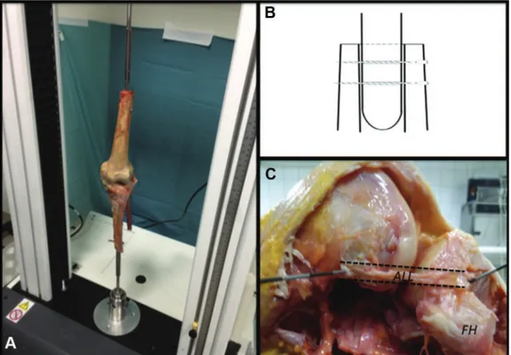
The ITB and gracilis grafts were tested in isolation without any bony attachments. The ends of the ITB and gracilis grafts were placed in two serrated jaws, ac-cording to a previously validated method30(Fig 1). The ALL was tested in situ with its bony attachments. Two 12-mm-diameter rods were press-fit into the distal tibia shaft and then the rods securely attached to the mate-rials testing system using two screws on each side31 (Fig 2). Just before starting the tests, the knee was disarticulated by cutting the medial collateral ligament, medial collateral ligament, and ACL to allow the ALL to be tested in isolation.
Measurements
Each set of grips was attached to a materials testing system (Instrom 3300, Instron, Canton, MA) (Fig 2) to apply tensile loads. Measurements were performed using the system’s software (BlueHill, Instrom SA France, Elancourt, France).
Each graft was preloaded to 10 N, and then cycled 100 times between 50 and 200 N at 0.5 Hz. A tensile test was then performed using a 10 mm min#1 crosshead speed until the graft failed. This sequence is a standard, validated test protocol.30The maximum load at failure (N), elongation at failure (mm), and linear stiffness (N mm#1) were automatically measured by the soft-ware during the failure test (Fig 3).
Graft preparation, preservation, and fixation were performed by K.W., N.R., and E.C. Measurements were performed by K.W., P.S., and J.M.
Statistical Analysis
The statistical analysis was performed with Excel 2011 (Microsoft, Redmond, WA) and XLSTAT 2011 (Addinsoft SARL, Paris, France) software packages. The normal distribution of the measured variables was verified using the Shapiro-Wilk test and the homoge-neity of variances was verified using Fisher’s f-test and Levene’s test to ensure that the conditions had been met for parametric testing. The significance threshold was set at P < .05. The descriptive analysis consisted of mean, median, and standard deviation values. A comparative analysis was performed using the paired Student’s t-test.
Results
The ALL had an average maximum load to failure of 141 N (range: 90 to 210 N). Its stiffness and elongation at failure were 21 N mm#1 (range: 9 to 34 N mm#1)
and 6.2 mm (range: 1.1 to 10.5 mm), respectively. The ALL failed mid-substance in all the specimens.Table 1
summarizes the biomechanical properties of the ALL, gracilis, and ITB.
Maximum Load at Failure
The gracilis had the highest maximum load of the three constructs, with an average of 200.7 N (!48.7)(Table 2). The only statistically significant dif-ferences were between the gracilis and ALL, and the gracilis and ITB (P ¼ .001 and P ¼ .03).
Stiffness
The gracilis was stiffer (131.7 ! 43.7 N mm#1) than the ITB (39.9 ! 6 N mm#1) and the ALL (21 !
8.2 N mm#1)(Table 2). All of these differences were
statistically significant (P ¼ .0001, P ¼ .002, and P ¼ .04, respectively).
Elongation at Failure
The elongation at failure of the ITB (20.8 ! 14.7 mm) and gracilis (19.9 ! 6.5 mm) was significantly higher than that of the ALL (6.2 ! 3.2 mm)(Table 2). There was no significant difference between the ITB and gracilis.
Discussion
Our hypothesis was confirmed: when the gracilis and ITB are prepared in the configuration used for LET, they have a higher maximum tensile load than the ALL. This difference was statistically significant for the gracilis. A two-strand gracilis graft had a significantly higher maximum load at failure and stiffness than the ALL.
Our results are consistent with previously published results. For the ALL, the average maximum load of 141 N (!40 N) and average stiffness of 21 N mm#1 (!8.2) found in our study are substantially the same as found in the only other published study on ALL
Fig 1. Graft fixation method shown on specimen iliotibial band (ITB) 5. The graft was gripped at both ends using two serrated jaw clamps. This test configuration has been previ-ously validated.30
biomechanics. On 15 knees, Kennedy et al.18found an average maximum load of 175 N (!62 N) and stiffness of 20 N mm#1(!7.9). The strength values for the grafts
were consistent with the findings of a published study using the same methodology. In that study, the maximum load at failure of a four-strand gracilis
construct was 416.4 N, which is twice that of the maximum load that a two-strand gracilis construct can withstand.30Sajovic et al.32have shown that doubling a tendon increases its maximum load at failure by two.
Claes et al.33performed a cadaver study to determine the biomechanical role of the ALL. By selectively cut-ting the ALL in knees with either an intact or transected ACL, they were able to show a significant increase in
Fig 2. Method used to test the anterolateral ligament (ALL). The cadaver specimen is attached to the materials testing system (Instron 3300, Instron, Canton, MA) rods and screws. (A) General view, (B) close-up view of the 12-mm intramedullary rods that were press-fit into the bone and securely attached using two screws. (C) Photograph of the ALL after dissection. In each knee, the ALL was identified as a fibrous structure having a tibial insertion midway between Gerdy’s tubercle and fibular head (FH), and a femoral insertion proximal and posterior to the lateral femoral epicondyle.
Fig 3. A sample load-elongation curve, here for specimen anterolateral ligament (ALL) 4. The cyclic loading, elastic deformation, and plastic elongation phases can be made out.
Table 1. Descriptive Statistics for the Biomechanical Properties of the Anterolateral Ligament, Gracilis Graft, and Iliotibial Band Graft Obtained During an Elongation to Failure Test
Minimum Maximum Mean
Standard Deviation ALL Maximum load 90 210 141 40.6 Stiffness 9 34 21 8.2 Elongation 1.1 10.5 6.2 3.2 Gracilis Maximum load 121.8 260.3 200.7 48.7 Stiffness 65 195 131.7 43.7 Elongation 11.6 35 19.9 6.5 ITB Maximum load 110.3 219.4 161.1 27.1 Stiffness 29 48 39.9 6.0 Elongation 2.8 39.2 20.8 14.7 ALL, anterolateral ligament; ITB, iliotibial band.
the tibia’s internal rotation under the femur. They also noted that its contribution to controlling rotation mainly occurred with the knee flexed at 30$to 90$. In
addition, they showed that the ALL must be ruptured for a grade 3 pivot shift to occur in a knee with a damaged ACL.
Parsons et al.19 reported that the ALL is the main restraint for internal rotation of the tibia under the fe-mur starting at 35$ knee flexion. Starting at 35$, the
ACL contributes significantly less to controlling internal rotation. This same study describes the nearly nonex-istent contribution of the ALL to stopping anterior tibial translation; the ACL performs this duty. This finding was consistent for all knee flexion positions. Spencer et al.5 confirmed the ALL’s antirotational function, although they minimized it. They stated that the ALL only stops the tibia’s internal rotation by 2$. However,
this result was obtained during a simulation of the initial portion of the pivot shift test on knees in full extension.
LET is thought to be analogous to the ALL in function, in terms of controlling anterolateral rotational laxity; however, the two differ anatomically.26 Kittl et al.34 measured the length change patterns and isometry in lateral extra-articular reconstructions. The MacIntosh reconstruction method35 appeared to be the most iso-metric. They concluded that “a graft attached proximal to the lateral femoral epicondyle and running deep to the lateral collateral ligament will provide desirable graft behavior, such as it will not suffer excessive tightening or slackening during knee flexion.”34 Kittl et al. showed that the ALL, as described by Claes et al.,27 was not isometric. Spencer et al.5 showed that LET (modified Lemaire technique) provides better sta-bility control, especially in rotation, than anatomical ALL reconstruction. The ALL is not the only structure
that contributes to controlling anterolateral laxity.5The posterior horn of the lateral meniscus and the menis-cocapsular portions of the medial meniscus are also involved in controlling rotational laxity in a knee with an ACL tear.36,37 Similarly, Terry and LaPrade38 showed that the biceps femoral, the ITB, and the anterolateral capsule play a role in anterolateral stabil-ity that is by no means insignificant.
The optimal graft tension and position are also debated.5 This is particularly true when the gracilis is used, because it is six times stiffer than the ALL. Excessive graft tension can place greater pressure on the lateral compartment and limit range of motion, which can lead to premature osteoarthritis and joint stiffness.39-41 Graft fixation in the over-reduced posi-tion (i.e., external rotaposi-tion) seems to overly constrain the knee’s movement, whereas fixation at 70$ flexion and neutral rotation does not.5
Limitations
Our study has certain limitations. The same type of fixation could not be used for all the constructs tested. We chose not to detach the ALL from its bone insertion so as to test it in its entirety. The fixation devices we used require that a certain length of tissue be placed inside the jaws of the clamps. If the bony attachments had been removed from the ALL, the remaining liga-ment tissue would not have been long enough for this testing protocol.
Secondly, like Kennedy et al.,18we believe that “this loading protocol does not reproduce the physiologic orientation of the forces experienced by the ALL and its attachments and therefore cannot be used to make clinical conclusions regarding the physiologic ACL/ALL injury mechanism.” Given the monoaxial tensile load applied to the construct, we did not feel it was necessary
Table 2. Comparison of the Biomechanical Properties of the Anterolateral Ligament, Gracilis, and Iliotibial Band During an Elongation to Failure Test
Paired Differences
P Mean Standard Deviation Std. Error
95% Confidence Interval for the Difference Lower Upper Maximum load ALLdG* #59.7 54.9 15.2 #92.9 #26.5 .001 ALLdITB #20.1 46.4 12.9 #48.1 7.9 .16 GdITB* 39.6 58.1 16.1 4.4 74.7 .03 Stiffness ALLdG* #110.7 41.9 11.6 #136.1 #85.4 .0001 ALLdITB* #18.8 9.8 2.7 #24.8 #12.9 .002 GdITB* 91.9 41.8 11.6 66.6 117.2 .04 Elongation ALLdG* #13.8 6.6 1.8 #17.8 #9.8 .03 ALLdITB* #14.6 13.8 3.9 #22.9 #6.2 .01 GdITB #0.8 17.9 4.9 #11.6 10.1 .9
ALL, anterolateral ligament; G, gracilis; ITB, iliotibial band.
to flex the knee at 30$. Axial traction moves the ALL’s
two-attachment point away from each other until the ligament fails; flexing the knee would not change this condition. Moreover, our results were the same as those reported by another group.18 The fixation method is also another basic consideration, as it can affect the results of tensile test.30The grips used during the testing were validated previously.30 Here also, our values are consistent with those found previously.30 The ITB could not be tested while still attached to Gerdy’s tubercle, because the ITB had to be resected to expose the ALL.18,27
The age of the specimens in this study was clearly higher than the age of patients who typically undergo ACL reconstruction procedures. The effect of age was evaluated on 82 patellar tendons taken from donors between 17 and 54 years of age.42These tendons were tested at strain rates of either 10%/s or 100%/s. The modulus of elasticity was lower only in the older ten-dons tested at 100%/s. The other biomechanical prop-erties were not altered by age.42
Conclusions
The gracilis had the highest maximum load to failure. The ITB’s mechanical properties most closely resemble those of the ALL.
Acknowledgment
The authors wish to thank Joanne Archambault, Ph.D., for the editorial assistance provided during the preparation of this manuscript.
References
1.Chouliaras V, Ristanis S, Moraiti C, Stergiou N, Georgoulis AD. Effectiveness of reconstruction of the anterior cruciate ligament with quadrupled hamstrings and bone-patellar tendon-bone autografts: An in vivo study comparing tibial internal-external rotation. Am J Sports Med 2007;35:189-196.
2.Mariscalco MW, Flanigan DC, Mitchell J, et al. The in-fluence of hamstring autograft size on patient-reported outcomes and risk of revision after anterior cruciate liga-ment reconstruction: A Multicenter Orthopaedic Out-comes Network (MOON) Cohort Study. Arthroscopy 2013;29:1948-1953.
3.van Eck CF, Schkrohowsky JG, Working ZM, Irrgang JJ, Fu FH. Prospective analysis of failure rate and predictors of failure after anatomic anterior cruciate ligament reconstruction with allograft. Am J Sports Med 2012;40: 800-807.
4.Webster KE, Feller JA, Leigh WB, Richmond AK. Younger patients are at increased risk for graft rupture and contralateral injury after anterior cruciate ligament reconstruction. Am J Sports Med 2014;42:641-647. 5.Spencer L, Burkhart TA, Tran MN, et al. Biomechanical
Analysis of simulated clinical testing and reconstruction of
the anterolateral ligament of the knee. Am J Sports Med 2015;43:2189-2197.
6.Kondo E, Merican AM, Yasuda K, Amis AA. Biome-chanical comparison of anatomic double-bundle, anatomic single-bundle, and nonanatomic single-bundle anterior cruciate ligament reconstructions. Am J Sports Med 2011;39:279-288.
7.Mohtadi NG, Chan DS, Dainty KN, Whelan DB. Patellar tendon versus hamstring tendon autograft for anterior cruciate ligament rupture in adults. Cochrane Database Syst Rev 2011:CD005960.
8.Prodromos CC, Joyce BT, Shi K, Keller BL. A meta-analysis of stability after anterior cruciate ligament reconstruction as a function of hamstring versus patellar tendon graft and fixation type. Arthroscopy 2005;21:1202. 9.Ayeni OR, Chahal M, Tran MN, Sprague S. Pivot shift as an outcome measure for ACL reconstruction: A systematic review. Knee Surg Sports Traumatol Arthrosc 2012;20: 767-777.
10.Lee MC, Seong SC, Lee S, et al. Vertical femoral tunnel placement results in rotational knee laxity after anterior cruciate ligament reconstruction. Arthroscopy 2007;23: 771-778.
11.Colombet P, Robinson J, Christel P, et al. Morphology of anterior cruciate ligament attachments for anatomic reconstruction: A cadaveric dissection and radiographic study. Arthroscopy 2006;22:984-992.
12.Siebold R, Ellert T, Metz S, Metz J. Femoral insertions of the anteromedial and posterolateral bundles of the ante-rior cruciate ligament: Morphometry and arthroscopic orientation models for double-bundle bone tunnel placementdA cadaver study. Arthroscopy 2008;24: 585-592.
13.Siebold R, Ellert T, Metz S, Metz J. Tibial insertions of the anteromedial and posterolateral bundles of the anterior cruciate ligament: Morphometry, arthroscopic landmarks, and orientation model for bone tunnel placement. Arthroscopy 2008;24:154-161.
14.Caterine S, Litchfield R, Johnson M, Chronik B, Getgood A. A cadaveric study of the anterolateral liga-ment: Re-introducing the lateral capsular ligament. Knee Surg Sports Traumatol Arthrosc 2015;23:3186-3195. 15.Hughston JC, Andrews JR, Cross MJ, Moschi A.
Classifi-cation of knee ligament instabilities. Part II. The lateral compartment. J Bone Joint Surg Am 1976;58:173-179. 16.Vieira EL, Vieira EA, da Silva RT, Berlfein PA, Abdalla RJ,
Cohen M. An anatomic study of the iliotibial tract. Arthroscopy 2007;23:269-274.
17.Dodds AL, Halewood C, Gupte CM, Williams A, Amis AA. The anterolateral ligament: Anatomy, length changes and association with the Segond fracture. Bone Joint J 2014;96-B:325-331.
18.Kennedy MI, Claes S, Fuso FA, et al. The anterolateral ligament: An anatomic, radiographic, and biomechanical analysis. Am J Sports Med 2015;43:1606-1615.
19.Parsons EM, Gee AO, Spiekerman C, Cavanagh PR. The biomechanical function of the anterolateral ligament of the knee. Am J Sports Med 2015;43:669-674.
20.Vincent JP, Magnussen RA, Gezmez F, et al. The antero-lateral ligament of the human knee: An anatomic and
histologic study. Knee Surg Sports Traumatol Arthrosc 2012;20:147-152.
21. Sonnery-Cottet B, Thaunat M, Freychet B, Pupim BH, Murphy CG, Claes S. Outcome of a combined anterior cruciate ligament and anterolateral ligament reconstruc-tion technique with a minimum 2-year follow-up. Am J Sports Med 2015;43:1598-1605.
22. Ireland J, Trickey EL. Macintosh tenodesis for antero-lateral instability of the knee. J Bone Joint Surg Br 1980;62: 340-345.
23. Lemaire M, Combelles F. [Plastic repair with fascia lata for old tears of the anterior cruciate ligament]. Rev Chir Orthop Reparatrice Appar Mot 1980;66:523-525 [in French]. 24. Losee RE, Johnson TR, Southwick WO. Anterior
sublux-ation of the lateral tibial plateau. A diagnostic test and operative repair. J Bone Joint Surg Am 1978;60:1015-1030. 25. Strickler FP. A satisfactory method of repairing crucial
ligaments. Ann Surg 1937;105:912-916.
26. Hewison CE, Tran MN, Kaniki N, Remtulla A, Bryant D, Getgood AM. Lateral extra-articular tenodesis reduces rotational laxity when combined with anterior cruciate ligament reconstruction: A systematic review of the literature. Arthroscopy 2015;31:2022-2034.
27. Claes S, Vereecke E, Maes M, Victor J, Verdonk P, Bellemans J. Anatomy of the anterolateral ligament of the knee. J Anat 2013;223:321-328.
28. Cavaignac E, Wytrykowski K, Reina N, et al. Ultrasono-graphic identification of the anterolateral ligament of the knee. Arthroscopy 2016;32:120-126.
29. Christel P, Djian P. [Anterio-lateral extra-articular tenodesis of the knee using a short strip of fascia lata]. Rev Chir Orthop Reparatrice Appar Mot 2002;88:508-513 [in French].
30. Pailhe R, Cavaignac E, Murgier J, Laffosse JM, Swider P. Biomechanical study of ACL reconstruction grafts. J Orthop Res 2015;33:1188-1196.
31. Cavaignac E, Carpentier K, Pailhe R, Luyckx T, Bellemans J. The role of the deep medial collateral liga-ment in controlling rotational stability of the knee. Knee Surg Sports Traumatol Arthrosc 2015;23:3101-3107. 32. Sajovic M, Vengust V, Komadina R, Tavcar R, Skaza K.
A prospective, randomized comparison of semitendinosus
and gracilis tendon versus patellar tendon autografts for anterior cruciate ligament reconstruction: Five-year follow-up. Am J Sports Med 2006;34:1933-1940.
33. Claes S. The pivot shift unraveled: Why we disagree with Dr. Fu. http://www.vumedi.com/video/the-pivot-shift-unraveled-why-we-disagree-with-dr-fu/. Accessed Febu-rary 1, 2016.
34.Kittl C, Halewood C, Stephen JM, et al. Length change patterns in the lateral extra-articular structures of the knee and related reconstructions. Am J Sports Med 2015;43:354-362.
35.Amirault JD, Cameron JC, MacIntosh DL, Marks P. Chronic anterior cruciate ligament deficiency. Long-term results of MacIntosh’s lateral substitution reconstruction. J Bone Joint Surg Br 1988;70:622-624.
36.Bhatia S, LaPrade CM, Ellman MB, LaPrade RF. Meniscal root tears: Significance, diagnosis, and treatment. Am J Sports Med 2014;42:3016-3030.
37.Tanaka M, Vyas D, Moloney G, Bedi A, Pearle AD, Musahl V. What does it take to have a high-grade pivot shift? Knee Surg Sports Traumatol Arthrosc 2012;20: 737-742.
38.Terry GC, LaPrade RF. The biceps femoris muscle complex at the knee. Its anatomy and injury patterns associated with acute anterolateral-anteromedial rotatory instability. Am J Sports Med 1996;24:2-8.
39.Guenther D, Rahnemai-Azar AA, Fu FH, Debski RE. The biomechanical function of the anterolateral ligament of the knee: Letter to the editor. Am J Sports Med 2015;43: NP21-22.
40.Anderson AF, Snyder RB, Lipscomb AB Jr. Anterior cru-ciate ligament reconstruction. A prospective randomized study of three surgical methods. Am J Sports Med 2001;29: 272-279.
41.Noyes FR, Barber SD. The effect of an extra-articular procedure on allograft reconstructions for chronic rup-tures of the anterior cruciate ligament. J Bone Joint Surg Am 1991;73:882-892.
42.Blevins FT, Hecker AT, Bigler GT, Boland AL, Hayes WC. The effects of donor age and strain rate on the biome-chanical properties of bone-patellar tendon-bone allo-grafts. Am J Sports Med 1994;22:328-333.