HAL Id: ensl-00000006
https://hal-ens-lyon.archives-ouvertes.fr/ensl-00000006
Submitted on 7 Apr 2006
HAL is a multi-disciplinary open access
archive for the deposit and dissemination of
sci-entific research documents, whether they are
pub-lished or not. The documents may come from
teaching and research institutions in France or
abroad, or from public or private research centers.
L’archive ouverte pluridisciplinaire HAL, est
destinée au dépôt et à la diffusion de documents
scientifiques de niveau recherche, publiés ou non,
émanant des établissements d’enseignement et de
recherche français ou étrangers, des laboratoires
publics ou privés.
Products of the Thyroid Hormone Receptor a Locus
Jacques Samarut, Karine Gauthier, Michelina Plateroti, Clare B. Harvey,
Graham R. Williams, Roy E. Weiss, Samuel Refetoff, Ames F. Willott,
Victoria Sundin, Jean-Paul Roux, et al.
To cite this version:
Jacques Samarut, Karine Gauthier, Michelina Plateroti, Clare B. Harvey, Graham R. Williams, et
al.. Genetic Analysis Reveals Different Functions for the Products of the Thyroid Hormone Receptor
a Locus. Molecular and Cellular Biology, American Society for Microbiology, 2001, 21 (14),
pp.4748-4760. �ensl-00000006�
Copyright © 2001, American Society for Microbiology. All Rights Reserved.
Genetic Analysis Reveals Different Functions for the Products
of the Thyroid Hormone Receptor
␣ Locus
KARINE GAUTHIER,1MICHELINA PLATEROTI,1CLARE B. HARVEY,2GRAHAM R. WILLIAMS,2
ROY E. WEISS,3SAMUEL REFETOFF,3,4JAMES F. WILLOTT,5VICTORIA SUNDIN,6
JEAN-PAUL ROUX,7LUC MALAVAL,7MASAHIRO HARA,1
JACQUES SAMARUT,1*ANDOLIVIER CHASSANDE1
Laboratoire de Biologie Mole´culaire et Cellulaire de l’Ecole Normale Supe´rieure, UMR 5665 CNRS, LA 913 INRA, 69364 Lyon cedex 07,1and INSERM U369, Faculte´ de me´decine, RTH Laennec, Lyon,7France; ICSM
Molecular Endocrinology Group, Division of Medicine and MRC Clinical Sciences Centre, Imperial College School of Medicine, Hammersmith Hospital, London, United Kingdom2; Departments of
Medicine3and Pediatrics,4University of Chicago, Chicago, Illinois 60637; Department of
Psychology, University of South Florida, Tampa, Florida 336205; and Department
of Psychology, Northern Illinois University, Dekalb, Illinois 601156
Received 27 November 2000/Returned for modification 13 February 2001/Accepted 15 April 2001
Thyroid hormone receptors are encoded by the TR␣ (NR1A1) and TR (NR1A2) loci. These genes are
transcribed into multiple variants whose functions are unclear. Analysis by gene inactivation in mice has provided new insights into the functional complexity of these products. Different strategies designed to modify the TR␣ locus have led to strikingly different phenotypes. In order to analyze the molecular basis for these
alterations, we generated mice devoid of all known isoforms produced from the TR␣ locus (TR␣0/0). These mice
are viable and exhibit reduced linear growth, bone maturation delay, moderate hypothermia, and reduced thickness of the intestinal mucosa. Compounding TR␣0and TRⴚmutations produces viable TR␣0/0ⴚ/ⴚ
mice, which display a more severe linear growth reduction and a more profound hypothermia as well as impaired hearing. A striking phenotypic difference is observed between TR␣0/0and the previously described
TR␣ⴚ/ⴚmice, which retain truncated TR⌬␣ isoforms arising from a newly described promoter in intron 7. The
lethality and severe impairment of the intestinal maturation in TR␣ⴚ/ⴚmice are rescued in TR␣0/0animals.
We demonstrate that the TR⌬␣ protein isoforms, which are natural products of the TR␣ locus, are the key
determinants of these phenotypical differences. These data reveal the functional importance of the non-T3-binding variants encoded by the TR␣ locus in vertebrate postnatal development and homeostasis.
Thyroid hormone receptors (TRs), encoded by the TR␣ and
TR loci, mediate the action of triiodothyronine (T3) in all vertebrates. These nuclear proteins activate or repress the transcription of target genes by recruiting several protein com-plexes which modify the structure of chromatin (15). In mam-mals, T3 exerts homeostatic and developmental functions. Thyroid hormone (TH) deprivation leads to developmental (growth retardation, impaired neurogenesis) (8, 23, 36) or met-abolic (reduced oxygen consumption, bradycardia, weakness, fatigue) disorders (13). Point mutations in the TR gene result in syndromes known as resistance to thyroid hormone (30). Recently, the introduction of mutations in the genes encoding these receptors in mice has allowed further understanding of the mechanisms mediating the physiological actions of T3 (17, 19). The TR locus encodes the TR1 and TR2 receptors. A new receptor, TR3, and its non-DNA-binding TR⌬3 variant have recently been described. The latter isoforms are proposed to arise from a novel promoter located upstream of the exons encoding the DNA binding domain (41). Genetic invalidation of the TR2 gene leads to pituitary resistance to TH with elevated thyrotropin (TSH) and thyroxine (T4) levels in serum
(1). Knocking out all TR1, TR2, and TR3/⌬3 genes re-produces this phenotype (10, 14) but also induces the loss of auditory function, due to delayed expression of a potassium channel in the cochlea (11), and results in impaired transcrip-tional response to T3 in liver (38). The TR␣ locus encodes the ubiquitous TR␣1 receptor and a splice variant, TR␣2, which cannot bind T3 and behaves as a weak inhibitor of TRs in transfection assays (21, 22). We have previously shown that, in a limited number of tissues, the TR␣ locus also encodes two transcripts, previously characterized by Northern blotting, RNase protection, and reverse transcription (RT)-PCR anal-ysis in embryonic stem (ES) cells, driven by a promoter located in intron 7, which generate TR⌬␣1 and TR⌬␣2 proteins. These proteins do not bind DNA or T3; hence, they are inhib-itors of the activities of TRs and retinoic acid receptors in transfection assays (6). In vivo genetic modifications of the
TR␣ locus result in different phenotypes, according to the precise location of the mutation. In TR␣⫺/⫺mice, the
expres-sion of TR␣1 and TR␣2 transcripts is abolished, but the TR⌬␣ transcripts are still expressed (12). These mice suffer from hypothyroidism and display several developmental defects, in-cluding growth retardation, altered intestinal development (28), impaired T- and B-lymphocyte maturation (3), expansion of cartilage regions in long bones, and finally growth arrest and death shortly after weaning (12). Mice which lack the TR␣1 and TR⌬␣1 products (TR␣1⫺/⫺) have been reported to exhibit
* Corresponding author. Mailing address: Laboratoire de Biologie Mole´culaire et Cellulaire, CNRS UMR 5665 ENS LA 913 INRA, Ecole Normale Supe´rieure, 46 alle´e d’Italie, 69364 Lyon Cedex 07, France. Phone: 33 4 72 72 81 71. Fax: 33 4 72 72 85 36. E-mail: Jacques .Samarut@ens lyon.fr.
mild hypothyroxinemia, bradycardia, and lower body temper-ature, but they display normal growth and life span (39). The lethal phenotype of TR␣⫺/⫺mice compared to the viability of
TR␣1⫺/⫺mice might be attributed to the deeper
hypothyrox-inemia of the former. The involvement of TH as the cause of this phenotypic discrepancy can be ruled out. Indeed, although TR␣⫺/⫺⫺/⫺ and TR␣1⫺/⫺⫺/⫺ mice both lack the known
TRs and have very high serum levels of TH, TR␣⫺/⫺⫺/⫺mice
die upon weaning (14), whereas TR␣1⫺/⫺⫺/⫺mice are viable
(16). Hence, the phenotypic differences must be ascribed to structural differences between the TR␣ mutant alleles. They could be attributed either to the TR␣2 isoform, which is ex-pressed in TR␣1⫺/⫺but not in TR␣⫺/⫺mice, or to the balance
between the TR␣ and TR⌬␣ isoforms. In an attempt to deter-mine the role of the non-T3-binding products, we have gener-ated a new mouse strain harboring a targeted deletion de-signed to prevent the expression of all transcripts from the TR␣ locus. In this paper we describe the phenotype of these TR␣0/0
mutant mice and point out similarities and striking differences with previously described TR␣1⫺/⫺ and TR␣⫺/⫺ mice. This
comparative analysis brings a new piece in the genetic analysis of the complex TR␣ locus and sheds new light on the respective functions of the TR␣ products in body growth, bone matura-tion, thermogenesis, audimatura-tion, and intestinal maturation.
MATERIALS AND METHODS
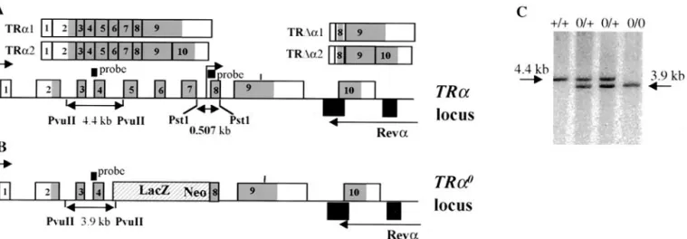
Targeted disruption of TR␣ gene. The TR␣0-targeting vector was constructed
using pGNA as starting plasmid (gift from P. Brulet, Institut Pasteur, Paris, France). The 3⬘ arm homologous to TR␣ was amplified by PCR from ENS ES cell DNA (14), using the Expand High Fi system (Roche). The 5⬘ arm was cloned from aEMBL4 library (gift from J. P Magaud). The thymidine kinase gene was inserted downstream of the 3⬘ arm.
ENS ES cells were electroporated with 40g of linearized targeting vector and then selected with 250g of G418 (Gibco-BRL) per ml and 0.2 M ganciclovir. Targeted clones were confirmed by Southern blot analysis using PvuII enzyme and a 900-bp PCR fragment extending from the 3⬘ end of exon 3 to the 5⬘ end of exon 4 of TR␣ as a probe. Two positive clones were injected into blastocysts of C57BL/6 recipient mice. TR␣0/⫹animals were derived in an inbred 129sv
background. TR␣0/0mice were obtained by intercrossing heterozygous animals.
Mouse breeding and handling were carried out in a certified animal facility, according to procedures approved by the local animal care and use committee. Purification of RNA, Northern blotting RNase protection assay, and RT-PCR analysis.Total RNA from distal small intestine or pituitary was isolated by the improved acid-guanidine-phenol-chloroform method. For Northern blot analy-sis, single-stranded DNA probes were generated by a 30-cycle reaction using Taq DNA polymerase from Eurobio using 10 ng of TR␣ (exon 8), growth hormone (GH), or hypoxanthine phosphoribosyltransferase (HPRT) PCR fragments as a template, and 20 ng of a specific antisense primer: oligonucleotides␣15A (CA GCCTGCAGCAGAGCCACTTCCG), GHa (GTCAAACTTGTCATAGGTT TG), and HPRTa (CACAGGACTAGAACACCTGC) were used as primers to generate the TR␣, GH, and HPRT probes, respectively.
RNase protection was performed using reagents from Ambion (Austin, Tex.) according to the manufacturer’s instructions. HPRT and TR␣ cDNA fragments were generated by RT-PCR and inserted into a pGEMt easy vector (Promega). Oligonucleotides HPRTs (GCTGGTGAAAAGGACCTCT) and HPRTa (see above) were used to generate the HPRT fragment, and TR␣Cs (CTCTGTGA TCCTGCTGTTCCACAG) and TR␣1a (CGACTTTCATGTGGAGGAAG) were used to generate the TR␣ cDNA.
RT was performed as described in reference 12. cDNA (0.05g) was used for each PCR with Eurobio Taq. To allow the detection of TR␣ gene products (TR␣1, TR␣2, TR⌬␣1, and TR⌬␣2) by RT-PCR, we used a sense primer within exon 8 (␣2⬘, CTGCCTTGCGAAGACCAGATC) and an antisense primer within exon 9 (␣3, GCGGTGGTTGACGTAGTGCTC). All the oligonucleo-tides were purchased from MWG (Ebersberg, Germany).
Morphological analysis of long bones and growth plate cartilage.Skin and muscle were dissected from forelimbs and hindlimbs of 3-week-old wild-type and TR␣0/0null mice. For anatomical analysis of long bones, one upper and one
lower limb from each animal were fixed in 80% ethanol for 1 to 4 days and transferred to 95% ethanol for a further 1 to 8 days, followed by acetone for 1 to 2 days at 4°C. Fixed limbs were stained with alizarin red and alcian blue 8GX (0.3% alcian blue in 70% ethanol [1 ml]–0.1% alizarin red in 95% ethanol [1 ml]–glacial acetic acid [1 ml]–70% ethanol [17 ml]) for 2 days at 37°C and transferred sequentially for 2- to 4-day periods through solutions of 1% KOH, 1% KOH–20% glycerol, 1% KOH–40% glycerol, 1% KOH–60% glycerol, and 1% KOH–80% glycerol at 4°C until, finally, destained limbs were stored in 100% glycerol at 4°C prior to analysis and photography under a dissecting microscope. In these preparations, mineralized bone is stained by alizarin red and cartilage is stained by alcian blue 8GX. For mineralization analysis, tibias were dissected out, fixed in 70% alcohol, embedded in paraffin, and cut (7m), and undecalcified sections were stained according to the standard protocol (26). For histological analyses, the paired limbs were fixed in 10% neutral buffered formalin for 4 to 6 days and subsequently decalcified for 2 days in 10% formic acid–10% neutral buffered formalin at 20°C. Limbs were embedded in paraffin and 3-m sections were cut, deparaffinized in xylene, and rehydrated. Sections were stained with hematoxylin and eosin or alcian blue 8GX and van Gieson to visualize cartilage matrix mucopolysaccharides in blue and collagen-containing bone osteoid in red, according to standard histological protocols.
Body temperature.The system we used, ELAMS (Electronic Laboratory An-imal Monitoring System by BioMedic Data Systems, Inc., Seaford, Del.), con-sisted of implantable programmable temperature transponders (IPTT-100) and a portable data scanner (DAS-5007). The transponders were implanted subcu-taneously in the backs of three 8- to 12-week-old males from each genotype. Animals were maintained under anesthesia with midazolam. All animals were kept in a climate-controlled room with 12-h alternating light and dark cycles. Temperature measurements were initiated 3 days after the implantation and were recorded in duplicate at 10 a.m., while the animals were moving freely in their cage with the lid removed, and the scanner was placed within 5 cm of the transponder. Body temperature monitoring continued for up to 7 days.
Temperature was measured rectally at the same time in TR␣0/0⫺/⫺by means
of a digital recording with a thermistor system. Values were not significantly different from those recorded by the transponder (data not shown).
Growth measurements. Male mice anesthetized with methoxyflurane were weighed and measured (nose to tail base) weekly from 2 to 9 weeks of age. Mice were housed five per cage, and food and water were provided ad libitum. The numbers of mice used from each genotype were as follows: wild type, 19 to 29; TR⫺/⫺, 6 to 17; and TR␣0/0, 10 to 22.
Hormone measurements.Serum T4 and TSH were measured by radioimmu-noassays as previously described (29).
Hearing tests.The auditory brainstem response (ABR) was used to assess the auditory sensitivity by a procedure that has been described in detail elsewhere (35). Tucker-Davis Technologies (Gainesville, Fla.) hardware and software (AeP and Siggen packages) were employed. Briefly, mice were anesthetized with so-dium pentobarbital (Nembutal, 1.2l per g of body weight intraperitoneally [i.p.]) and chlorprothixene (Taractan, 0.5l/g i.p.) and kept warm with a heating pad. The average response (waveform) per 100 presentations of tone bursts (1 ms rise and fall; 3 ms duration; rate, 21 Hz; frequencies of 4, 8, 12, 16, and 24 kHz) was recorded using a subdermal needle electrode placed in the scalp. Each tone frequency was presented in descending intensity steps beginning at 80-dB sound pressure level and ending when a visually discernible ABR waveform could no longer be detected. Thresholds were then determined to within 5 dB for each frequency (35). The numbers of mice (60 to 90 days old) tested from each genotype were as follows: wild type, 7; TR⫺/⫺, 9; TR␣0/0, 7; and combined
TR⫺/⫺TR␣0/0, 4.
Morphological staining and immunohistochemistry. Three-week-old mice were killed by cervical dislocation and the small intestines were immediately removed. Proximal jejunums and distal ilea were collected separately and fixed in Carson’s solution overnight at 4°C. They were then embedded in paraffin and 5-m sections were applied on polylysine-coated slides. For morphological ob-servations, after dewaxing and rehydration, the slides were stained with hema-toxylin and eosin.
Immunochemistry experiments were performed with a monoclonal antibody (Novocastra Laboratories) for Ki-67 detection. Polyclonal antibody PAI-211 (Affinity Bioreagents, Inc.) for TR␣1 and TR⌬␣1 protein localization and an-other recognizing the N-terminal part of both TR␣1 and TR␣2 proteins (gen-erous gift of D. Baas [4]) were used in combination with secondary biotinylated antibody and streptavidin-peroxidase detection system (Histomouse; Zymed). The tissue was counterstained lightly with hematoxylin. Before incubation with the primary antibody, the slides were immersed in 0.01 M citrate buffer, pH 6, and microwaved for 15 min.
Cells, plasmids, transfections, and Western blotting.HeLa cells were main-tained in Dulbecco modified Eagle medium supplemented with 10% fetal calf serum. The plasmids containing the cDNAs of human TR␣1 (pSG5hTR␣1), rat TR1 (pSG5rTR1), mouse TR⌬␣1 (pSG5mTR⌬␣1), FlagTR⌬␣1 (CMV-FmTR⌬␣1), and mouse TR⌬␣2 (pSG5mTR⌬␣2) have been described previ-ously (6). HeLa cells were transfected using a calcium phosphate procedure as previously described (7) and harvested after 48 h. Lactacystin was a gift from Satoshi Omura (Tokyo, Japan). Western blotting was performed as previously described (6). We used the no. 21 polyclonal antibody (gift of J. Ghysdael) as described in reference 5 to detect TR␣1 and TR⌬␣1 proteins and the M2 anti-Flag antibody (IBI) to detect the Flag-TR⌬␣1 protein.
RESULTS
The TR␣0/0mutation abolishes the production of all
prod-ucts from the TR␣ locus. To generate mice deprived of any
TR␣ gene product (TR␣1 and TR␣2 and their truncated coun-terparts TR⌬␣1 and TR⌬␣2), a lacZ neor cassette (12) was
introduced by homologous recombination between exon 4 and exon 8 of the TR␣ gene (Fig. 1A) to generate the TR␣0allele
(Fig. 1B). This removed the DNA binding region of TR␣1 and TR␣2, preventing the production of a chimeric protein that could act in a dominant-negative manner upon TR. It also removed the internal promoter in intron 7 which has been shown to drive the transcription of TR⌬␣1 and TR⌬␣2 (6). The TR␣0mutated allele was introduced into ES cells by
ho-mologous recombination. The structure of the modified allele was checked by Southern blotting (Fig. 1C). Two independent positive clones were injected into host blastocysts. Heterozy-gous mice were derived from an inbred 129Sv genetic back-ground. They were then intercrossed to obtain TR␣0/0mice.
We checked for the expression of TR␣ transcripts in wild-type, TR␣0/0, and previously described TR␣⫺/⫺mice (12). This
analysis was performed on intestine RNA where the four TR␣ isoforms have been shown to be expressed. Northern blot anal-ysis (Fig. 2A) showed that all four transcripts could be identi-fied in the distal ileum of wild-type mice. In TR␣⫺/⫺ mice,
TR␣1 and TR␣2 RNAs were undetectable, whereas the TR⌬␣1 and TR⌬␣2 transcripts were still detected. In TR␣0/0
mice, neither TR␣ nor TR⌬␣ transcripts were detected. In agreement with these data, RT-PCR did not allow the detec-tion of transcripts containing exons 8 and 9 in the intestines of TR␣0/0 mice (Fig. 2B). RNase protection analysis (Fig. 2C)
revealed that the TR␣1 and TR␣2 isoforms, which are highly expressed in wild-type samples, are almost undetectable in TR␣⫺/⫺and TR␣0/0samples. Occasionally, the 448- and
386-nucleotide fragments were detected in the mutant mice, cor-responding to transcription reading through the stop sites within the lacZ neor cassette. It is noteworthy that RNase
protection showed that wild-type ileum contained similar amounts of TR␣1 and TR␣2 transcripts. Underrepresentation of the large TR␣1 transcript in Northern blot experiments may have been caused by poor transfer onto nylon membranes. Two protected fragments, whose sizes correspond to the TR⌬␣1 and TR⌬␣2 isoforms, were detected in wild-type and TR␣⫺/⫺but not in TR␣0/0samples. It is thus concluded that in
the distal ileum of wild-type mice, four TR␣ isoforms are expressed, including TR⌬␣1 and TR⌬␣2, which are expressed at much lower levels than TR␣1 and TR␣2. The TR␣0
muta-tion completely abolishes the transcripmuta-tion from the TR␣ locus, in contrast with the TR␣ mutation which still allows the pro-duction of TR⌬␣ transcripts, as previously described (Fig. 2D).
TR␣0/0 and TR␣0/0ⴚ/ⴚ mice display normal embryonic
development and are viable.It has previously been shown that TR␣⫺/⫺and TR␣⫺/⫺⫺/⫺mice were born in Mendelian
pro-portions but died after weaning (12, 14). Lethality was found in 129SV, BALB/c, and C57BL/6 genetic backgrounds after eight backcrosses. When TR␣0/⫹mice were intercrossed, among 346
born pups, 98 (28.3%), 170 (49%), and 78 (22.5%) were, re-spectively, wild-type, heterozygous, and homozygous mutant progenies. Unlike the TR␣⫺/⫺mice, all TR␣0/0mice survived
over the weaning period and the mortality in the adult popu-lation was not different from that observed in the wild type. Male and female TR␣0/0mice were fertile. Viability and
fer-tility were observed in C57BL/6 and 129SV backgrounds. TR␣0/0mice were crossed with TR⫺/⫺mice to obtain TR␣0/⫹
FIG. 1. Targeted disruption of the TR␣ gene by homologous recombination. (A) Structure of the TR␣ gene and of the various isoforms encoded by this gene. The two transcription start sites are indicated by upper arrows. The differential splice site in exon 9 is indicated by a vertical bar. Exons are numbered starting downstream to the distal promoter. Grey, coding regions. The structure of the transcripts is shown. (B) The targeted TR␣0
allele contains a deletion extending from the middle part of intron 4 to the beginning of exon 8. The probe used for Southern blot analysis and the size of the fragment detected after digestion with PvuII are indicated. (C) Detection of wild-type (⫹/⫹), heterozygous (0/⫹), and homozygous (0/0) littermates by Southern blot analysis.
⫺/⫺progenies. These mice were then intercrossed and their
progenies contained TR␣0/0⫺/⫹ and TR␣0/⫹⫺/⫺ animals
that were all viable. Because of the low fertility displayed by TR␣0/⫹⫺/⫺animals, only TR␣0/0⫺/⫹mice were used to
fur-ther generate double-homozygous TR mutants (TR␣0/0⫺/⫺).
The proportion of TR␣0/0⫺/⫺ progenies was 19.5%. This
slight deviation from Mendelian ratios may reflect marginal embryonic lethality or occasional improper maternal care for TR␣0/0⫺/⫺newborn mice. However, this result indicates that
TRs are not absolutely required for embryonic viability. TR␣0/0⫺/⫺mice survived beyond weaning time and were
vi-able for at least six months. Only occasionally were progenies obtained from TR␣0/0⫺/⫺intercrossings or from TR␣0/0⫺/⫺
males or females mated with wild-type partners, suggesting that these animals have a very low fertility.
Deregulation of the pituitary-thyroid axis in TR␣0/0ⴚ/ⴚ
mice.The serum levels of T3 and T4 are strictly controlled by
the direct hormonal feedback on TSH-releasing hormone (TRH) and TSH secretion, in the hypothalamus and the pitu-itary, respectively (20, 24, 29). We and others have previously demonstrated that TR plays a key role in this feedback loop since TR⫺/⫺mice display high T3, T4, and TSH levels (11, 14,
1). The role of TR␣ in this regulation has not been clearly established. TR␣1⫺/⫺ mice demonstrated only a slight
de-crease of T4 and TSH concentrations in males (39), whereas TR␣⫺/⫺mice presented a progressive but severe reduction of
circulating T4 and T3 between the third and the fourth week after birth (12). In TR␣0/0adult males, basal concentration of
T4, but not T3 or TSH, is slightly but significantly reduced compared to wild-type mice. Furthermore, TSH and T4 sup-pression by increasing doses of T3 is more profound in TR␣0/0
than in wild-type mice (25). In adult TR␣0/0⫺/⫺ mice, the
concentrations of serum T4, T3, and TSH were increased 14-fold, 13-14-fold, and more than 200-fold respectively, compared to
FIG. 2. Expression of the different transcripts encoded by the TR␣ locus in the distal ilea of the different mutants. (A) A Northern blot assay was performed using 20g of total RNA isolated from the distal ilea of animals carrying the indicated genotypes. The radiolabeled probe hybridized to both TR␣ and TR⌬␣ transcripts. A glyceraldehyde-3-phosphate dehydrogenase or a hypoxanthine phosphorybosyl transferase probe was used as an internal control. (B) Representative semiquantitative RT-PCR analysis of mRNAs encoded by the TR␣ locus in wild-type (WT), TR␣⫺/⫺, and TR␣0/0mice. The primers used allowed the amplification of both TR␣ and TR⌬␣ transcripts. HPRT was used as an internal control.
(C) RNase protection assay. Twenty micrograms of total distal ileum RNA from either wild-type, TR␣0/0, or TR␣⫺/⫺mice was mixed with 75 fmol
of HPRT and 75 fmol of TR␣ RNA probes. The sizes of the protected TR␣ fragments and the positions of the TR␣ transcript isoforms are indicated. (D) Summary of the TR␣ and TR⌬␣ mRNA expression in the distal ileum of each genotype.
those of wild-type mice. These levels are comparable with those previously reported for TR␣1⫺/⫺⫺/⫺animals (11-, 30-,
and 60- to 160-fold, respectively) (16) or 3-week-old TR␣⫺/⫺
⫺/⫺mice (7-, 20-, and more than 100-fold, respectively) (14).
Reduced growth rate and delayed bone maturation in TR␣0/0and TR␣0/0ⴚ/ⴚmice.The growth of TR␣0/0males and
their wild-type littermates was monitored over a two-month period. TR␣0/0mice showed a reduced increase of body weight
compared to wild-type animals, at all ages analyzed, in both 129Sv (not shown) and C57BL/6 genetic backgrounds (Fig. 3A). The time course of linear growth displayed similar differ-ences (Fig. 3B), showing that alteration of body growth fully accounts for the weight differences and that the TR␣0mutation
does not result in an abnormal weight-to-length ratio. TR⫺/⫺
mice displayed no difference in weight or length growth com-pared to that of the wild type (Fig. 3A and B)
The growth of TR␣0/0⫺/⫺mice was further reduced
com-pared to the TR␣0/0 mutants, though no difference could be
observed at birth. Weight reductions of 50 and 30% were observed in mice at weaning time and in adult mice, respec-tively, compared to wild-type mice (Fig. 3C).
The synthesis of GH has been shown to be regulated by TH and to be decreased in TR␣1⫺/⫺⫺/⫺mice (16), which display
marked growth retardation. We compared the expressions of the GH mRNA in the pituitary glands of wild-type, TR␣0/0,
and TR␣0/0⫺/⫺mice. As shown in Fig. 3D, the amount of GH
RNA was not altered in TR␣0/0 mice but was markedly
re-duced in double-knockout animals. These data suggest that the reduced growth observed in TR␣0/0mice may not be due to a
reduced production of GH.
Since linear growth was affected in TR␣0/0mice, we studied
endochondral ossification in their long bones. For this purpose, anatomical and histological analyses were performed. In 3-week-old TR␣0/0animals at weaning, the ends of metatarsals
and phalanges of the foot, the condyles and growth plate of the femur, and the growth plates of the tibia and fibula stained positively for cartilage with alcian blue (Fig. 4A). In wild-type mice, alcian blue staining was markedly diminished and re-placed by alizarin red staining in these areas, indicating that bone formation and mineralization were more advanced than in TR␣0/0animals. Furthermore, ossification of the patella was
delayed in TR␣0/0mice relative to that in wild-type mice (Fig.
4A). Similar evidence of delayed bone formation and miner-alization was seen in the upper limbs of 3-week-old TR␣0/0
mice (data not shown). Goldner staining of undecalcified lon-gitudinal sections of tibias revealed that the ossification of the epiphyses was delayed in TR␣0/0 mice, and the density of
trabecular bone in the metaphyses was markedly lower than in control mice (Fig. 4B). Histological analyses demonstrated that tibial growth plates from TR␣0/0mice were grossly
disor-ganized compared to those of wild-type animals (Fig. 4B, C, and D), and similar findings were observed in other growth plates throughout the upper and lower limbs of TR␣0/0mice
(data not shown). In particular, proliferating growth plate
FIG. 3. Comparison of growth in wild-type and TR␣- and TR-deficient mice. Weight (A) and length (B) were measured weekly from 2 to 9 weeks and plotted as means⫾ standard deviations. The numbers of animals are given in Materials and Methods. ⴱ, P ⬍ 0.01 for wild-type and TR␣0/0mice. (C) Body weights of wild-type, TR␣0/0, and TR␣0/0⫺/⫺mice (in grams). (D) Northern blot analysis of pituitary GH mRNA from
wild-type, TR␣0/0, and TR␣0/0⫺/⫺mice. One to two micrograms of total RNA from individual pituitaries was loaded. Single-stranded DNA probes
chondrocytes failed to form discrete columns and the hyper-trophic zone was markedly diminished and morphologically indistinct in TR␣0/0mice compared to wild-type mice,
suggest-ing that hypertrophic chondrocyte differentiation failed to progress normally in TR␣0/0mice. Furthermore, the degree of
alcian blue staining of the growth plate cartilage matrix in TR␣0/0mice was diminished and patchy compared to that in
wild-type animals, supporting the view that epiphyseal growth plate architecture is disorganized and endochondral ossifica-tion is disrupted in TR␣0/0 mice (Fig. 4C and D). In TR␣0/0
⫺/⫺mice, a similar phenotype was observed (data not shown).
These alterations were similar to those described previously for TR␣⫺/⫺and TR␣⫺/⫺⫺/⫺mice (12, 14) and are reminiscent of
abnormalities in epiphyseal growth plates of hypothyroid rats (34).
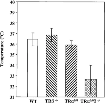
TR␣0/0ⴚ/ⴚ mice display severe hypothermia. TH plays a
crucial role in the control of thermogenesis, and hypothyroid-ism results in decreased oxygen consumption and heat produc-tion (13). We examined the temperatures of adult wild-type and mutant mice, using both a rectal probe and a temperature-sensitive probe implanted under the skin. The two techniques provided similar data. The mean body temperature of TR⫺/⫺
mice was similar to that of wild-type mice, but it was 0.4°C lower in TR␣0/0 mice (Fig. 5). Interestingly, the mean body
temperature of TR␣0/0⫺/⫺ animals was dramatically
de-creased (⫺4°C) relative to that of the wild type (Fig. 5). These data suggest functional redundancy between the TR and the TR␣ loci in the control of basal body temperature.
Decrease of auditory sensitivity in TR␣0/0mice and deafness
in TR␣0/0ⴚ/ⴚmice.Several authors have described deafness
FIG. 4. Morphological appearance of lower limbs and tibial growth plates in 3-week-old wild-type (wt) and TR␣ gene knockout (␣0/0) animals.
(A) Lower-limb skeletal mounts stained with alizarin red and alcian blue 8GX showing feet (left) and knee joint including lower femur and upper tibia with fibula (right); magnification,⫻8. Red staining shows mineralized bone and blue shows cartilage growth plates; the patella is shown to the right of each knee joint and is stained red in the wt, indicating that mineralization is complete, but blue in TR␣0/0, demonstrating the persistence
of cartilage. (B) Longitudinal medial bone sections stained with modified Goldner of TR␣⫹/⫹(wt) and TR␣0/0mice. Mineralized matrix is stained
in green. E, epiphysis; D, diaphysis; horizontal arrows, growth plate. (C and D) alcian blue 8GX and van Gieson staining of proximal tibial growth plates from wt and TR␣0/0mice; a section through the whole growth plate (magnification,⫻200) is shown in panel C and a section containing the
distal half of the growth plate (magnification,⫻400) is shown in panel D. Black arrows, resorption front which separates the growth plate from underlying primary spongiosum; e, epiphysis; ps, primary spongiosum; rz, reserve zone chondrocytes; pz, proliferative zone; hz, hypertrophic zone.
in mice lacking both TR1 and TR2, but not in mice lacking TR2 only or TR␣1 (1, 11), suggesting that TR1 is the only mediator of this T3-dependent function. However, the role of TR␣2 and the effect of the double knockout on the integrity of hearing have not been documented. We therefore measured the ABR threshold in TR␣0/0and TR␣0/0⫺/⫺mice. Whereas
at low or middle frequencies TR␣0/0mice had a normal
hear-ing threshold, they exhibited a marked loss of sensitivity at high tone frequencies (Fig. 6). In TR␣0/0⫺/⫺ mice, the loss of
hearing threshold was significantly more severe than that ob-served in TR⫺/⫺animals. These data suggest that, although
the TR1 receptor is the major mediator of T3 action, in addition the integrity of the TR␣ gene is required to achieve full function of the auditory apparatus.
The intestinal phenotype is mildly altered in TR␣0/0 mice
compared to that of TR␣ⴚ/ⴚ mice. In mammals, the small
intestine is lined by a continuously renewable population of epithelial cells. The pluripotent crypt stem cells give rise to immature cells that migrate and differentiate in the villus com-partment and die after exfoliation at the villus tip. It was previously demonstrated that TR␣⫺/⫺mice displayed a strong
impairment of postnatal intestinal development (14, 28). It is noteworthy that the intestine is one of the few organs where the TR⌬␣ and the TR␣ isoforms are coexpressed. In order to differentiate functions of the TR␣1/␣2 and TR⌬␣1/⌬␣2 isoforms in the intestine epithelium, we carried out a compar-ative investigation of morphofunctional parameters in TR␣0/0,
TR␣⫺/⫺, and wild-type mice.
The size of the small intestine mucosa was reduced in TR␣0/0 mutants compared to that of wild-type animals. This
was mainly due to the reduced lengths of the villi, resulting from a decreased number of epithelial cells per crypt-villus axis (Fig. 7A and B). These features were more strongly affected in the TR␣⫺/⫺ mice (Fig. 7C). The proliferation and
differenti-ation parameters of the epithelial cells also indicated a stron-ger impairment in TR␣⫺/⫺ than in TR␣0/0 mice (27a). In
contrast to the TR␣⫺/⫺mutant animals that displayed a
thin-ning of the smooth muscular layers (12, 28), TR␣0/0mutants
appeared normal in this respect. Taken together, these data strongly suggest that the severity of the intestinal phenotype correlates mostly with the persistence of the TR⌬␣ transcripts and not with the absence of TR␣1 and TR␣2 isoforms.
The intestinal phenotype is more severe in TR␣ⴚ/ⴚⴚ/ⴚ
than in TR␣0/0ⴚ/ⴚmice. It was previously shown that
com-bining the TR␣⫺and TR⫺mutations in TR␣⫺/⫺⫺/⫺mice
results in a more dramatic alteration of the structure of the
FIG. 5. Body temperature was measured at 10 a.m. with implant-able transponders for 7 days in at least 3 male mice aged 8 to 12 weeks for each of the wild-type (WT) TR⫺/⫺, TR␣0/0, and TR␣0/0⫺/⫺
genotypes. Each bar represents a mean value (⫾ standard deviation). Body temperatures of TR␣0/0and TR␣0/0⫺/⫺animals were
statisti-cally different from those of wild-type control animals (P⬍ 0.001 and
P⬍ 0.001, respectively, by the Student t test).
FIG. 6. ABR threshold in wild-type and TR gene knockout mice. The average response per 100 presentations of tone bursts (1 ms rise and fall; 3 ms duration; rate, 21 Hz; frequencies of 4, 8, 12, 16, and 24 kHz) were recorded. The numbers of animals are given in Materials and Methods. At all frequencies the difference between wild-type and TR⫺/⫺and TR␣0/0⫺/⫺mice was P⬍ 0.001. Solid lines, P values for differences between
distal ileum than that observed in TR␣⫺/⫺mice (14, 28). When
we analyzed the distal ilea of TR␣0/0⫺/⫺ versus those of
TR␣0/0 mice, no obvious morphological difference was
ob-served (Fig. 7B and D) and no statistically significant difference was found in the number of epithelial cells per crypt-villus axis. In contrast, in the ilea of TR␣⫺/⫺⫺/⫺ mice, the number of
epithelial cells was further decreased compared with that of TR␣⫺/⫺ mice (Fig. 7C and E) and the morphology was
dra-matically altered (Fig. 7E), in agreement with previous obser-vations. The number of proliferating cells in the crypts of TR␣0/0⫺/⫺mice (8⫾ 1) was decreased to the same extent as
in TR␣0/0animals (7⫾ 1). In contrast, this number was more
reduced in TR␣⫺/⫺⫺/⫺(2⫾ 1) than in TR␣⫺/⫺animals (5⫾
1). The analysis of epithelial cell differentiation markers also demonstrated similar features in TR␣0/0and TR␣0/0⫺/⫺
an-imals but showed a stronger impairment in TR␣⫺/⫺⫺/⫺than
in TR␣⫺/⫺mice. In summary, the knockout of the TR gene
does not worsen the phenotype observed in TR␣0/0mice, but
clearly enhances the alterations observed in TR␣⫺/⫺ mice.
These data rule out functional redundancy between TRs in the control of postnatal development of the intestine. They suggest instead that the expression of the TR⌬␣ isoforms, which is
deleterious in the absence of TR␣1 and TR␣2, has even more toxic effects in the absence of all TRs.
Identification of the TR⌬␣ proteins in the distal ileum by
immunohistochemistry.The intestinal epithelium is composed of proliferating and undifferentiated cells which form the crypt compartment and of mature cells located in the villi. In order to analyze the presence of the TR⌬␣ proteins in the intestinal mucosa and their pattern of expression along the crypt-villus axis, we performed immunocytochemical analysis. Since TR⌬␣ proteins do not contain any peptide sequence which would differentiate them from their complete TR␣ counterpart, we had to compare the patterns in distal ilea from wild-type, TR␣⫺/⫺, and TR␣0/0mice, using the following antisera:
anti-C␣1 raised against the common C-terminal part of TR␣1 and TR⌬␣1, anti-C␣2 raised against the common C-terminal part of TR␣2 and TR⌬␣2 but different from TR␣1, and anti-N␣ raised against the N-terminal part common to TR␣1 and TR␣2. No staining was obtained with the anti-C␣2 antibody in any genotypes (data not shown), whereas the expression of TR␣2 and TR⌬␣2 mRNAs has been demonstrated by semi-quantitative RT-PCR (28), Northern blotting, and RNase pro-tection (this study) in wild-type mice. As this antiserum was
FIG. 7. Morphological appearance of distal small intestine in 3-week-old animals. Sections (5m) from wild-type (A), TR␣0/0(B), TR␣⫺/⫺(C),
TR␣0/0⫺/⫺(D), and TR␣⫺/⫺⫺/⫺(E) mice were stained with hematoxylin and eosin. The numbers at the right of each picture indicate the number
of epithelial cells per crypto-villus axis. Statistical analysis indicated that the number of the epithelial cells in TR␣⫺/⫺, TR␣0/0⫺/⫺, and
TR␣⫺/⫺⫺/⫺animals was significatively decreased compared to that in wild-type mice (P⬍ 0.001, P ⬍ 0.05, and P ⬍ 0.0001, respectively, by the
Student t test; n⫽ 5). In addition, the number of epithelial cells was also significantly decreased in TR␣⫺/⫺⫺/⫺compared to TR␣⫺/⫺mice (P⬍
used successfully in immunocytochemical detection of TR⌬␣2 and TR␣2 proteins in transfected cells (data not shown), the failure to detect these proteins in the small intestine probably results from their very low concentration in this tissue.
Using the anti-N␣ antiserum, we observed a nuclear labeling in muscle layers of wild-type mice (Fig. 8A to D), but no staining was observed in the epithelium or mesenchyme (Fig. 8B and C). No positive cells were detected in TR␣0/0 or
TR␣⫺/⫺mice (Fig. 8E to H and I to L). By semiquantitative
RT-PCR, it was previously shown that in smooth muscle layers only the TR␣1 RNA was detected (28). Consequently, the signal detected with the anti-N␣ antibody in this tissue can be attributed to TR␣1. In the same experiment, the TR␣1 mRNA was found in the epithelium and connective tissues, whereas the TR␣2 mRNA was found only in the epithelium (28); there-fore, we assume that the corresponding proteins are expressed in these tissue regions but at levels undetectable by this tech-nique.
Using the anti-C␣1 antibody, we detected a strong nuclear signal in the differentiated epithelial cells of the villi in wild-type mice (Fig. 9A and B). Only a faint signal could be de-tected in the undifferentiated epithelial cells of crypts, in the lamina propria, and in smooth muscle cells (Fig. 9C and D). However, unlike the signal detected with the anti-N␣ reagent, this staining persisted in TR␣⫺/⫺ mice (Fig. 9I to L), which
retain the expression of TR⌬␣ transcripts, and disappeared in TR␣0/0mice (Fig. 9E to H). These observations demonstrate
that the protein labeled by the anti-C␣1 reagent is TR⌬␣1. They also suggest that, unlike the N␣ antibody, the anti-C␣1 antibody is unable to recognize the TR␣1 protein, pre-sumably due to the small amount of this protein in the distal ileum, and to the reduced accessibility of the target epitope in the tight conformation of the ligand binding domain.
Alto-gether, these data indicate that in wild-type mice, the TR␣1 protein is mainly located in the nuclei of smooth muscle cells, whereas the TR⌬␣1 protein is mainly present in the nuclei of differentiated epithelial cells. It is worth noting that in TR␣⫺/⫺
undifferentiated and differentiated epithelial cells have similar nuclear labeling.
The activity of TR⌬␣ proteins is down-regulated by TR␣1
via the proteasome pathway.Our data show that the deleteri-ous activity of the TR⌬␣ isoforms is triggered by the inactiva-tion of the TR␣1 and TR␣2 genes. To clarify the molecular mechanisms which could account for this gene inactivation-dependent activity, we tested the possibility that TR␣1 and TR␣2 could interfere with the synthesis or degradation of the TR⌬␣ isoforms, thereby modulating their activity. For this purpose, we measured the expression of the TR⌬␣1 protein in HeLa cells after cotransfection of a TR⌬␣1 expression vector together with either an empty vector or a vector expressing either TR␣1 or TR␣2. Figure 10 shows that, while the amount of TR⌬␣1 transcripts was unaffected by the coexpression of the TR␣1 protein (Fig. 10C), the amount of TR⌬␣1 protein was reduced by increasing amounts of TR␣1 (Fig. 10A). This effect was independent of the presence of T3 (data not shown). In contrast to these data, the expression of small amounts of TR␣2 did not produce any decrease in the amount of TR⌬␣1 protein (Fig. 10B). It is noteworthy that expression of TR1 did not change the amount of TR⌬␣1 (data not shown). In order to show that degradation, and not the rate of protein synthesis, accounted for the reduction in the amount of the TR⌬␣1 protein, we tested the abilities of different permeant protease inhibitors to prevent this effect. When cotransfected cells were treated with lactacystin, a potent inhibitor of the proteasome pathway (9), the amount of TR␣1 protein was not changed, but the amount of TR⌬␣1 protein was considerably
FIG. 8. Immunolocalization of TR␣1/TR␣2 proteins. The pattern of expression of TR␣ proteins of distal ileum was analyzed in paraffin-embedded sections (5m) in wild-type (A to D), TR␣0/0(E to H), and TR␣⫺/⫺(I to L) 3-week-old mice. The antibody used recognizes the
N-terminal part common to TR␣1 and TR␣2 (anti-N␣) on wild-type sections (B to D), TR␣0/0(F to H), and TR␣⫺/⫺(J to L). Technical controls
included the use of phosphate-buffered saline or nonimmune serum instead of the primary antibody. (A, E, and I) Phase contrast low magnifi-cation; bar, 30m; (B to D, F to H, and J to L) bright field high magnification; bar, 70 m. ce, crypt epithelium; ve, villus epithelium; ct, connective tissue; cml, circular muscle layer; lml, longitudinal muscle layer.
increased, reaching levels higher than those observed in the absence of TR␣1 (Fig. 10D). These data suggest that TR␣1 triggers the degradation of the TR⌬␣1 protein through a pro-teasome-dependent pathway.
DISCUSSION
The TR␣ locus encodes the TR␣1 receptor, whose targeted inactivation results in bradycardia and a slight decrease in body temperature (39). This locus also generates the TR␣2 variant, which cannot bind T3, poorly heterodimerizes with RXRs, and does not contain any transactivation domain. Transfection studies have shown that the TR␣2 protein can inhibit the transcriptional activity of TRs. The TR␣2 transcript is present in all mammalian tissues and is generally more abundant than the TR␣1 transcript, at least at the level of whole organ anal-ysis (22). Despite these many data, the in vivo function of the TR␣2 protein remains unknown. In addition to TR␣1 and TR␣2, we have identified the TR⌬␣1 and TR⌬␣2 transcripts, which are expressed in murine embryonic stem cells and in intestine, lung, and brain tissues of adult mice. The proteins encoded by these transcripts do not bind T3 or DNA but inhibit the transcriptional activity of several nuclear receptors when they are associated with RXR partners (6). Their in vivo function is unknown. Several genetic modifications of the TR␣ locus which have been generated in a mouse now provide tools with which to obtain insights into the functions of these differ-ent products. The goal of this discussion is to draw some conclusions from the comparison of the phenotypes observed in the different TR␣ gene knockout mice. For the sake of clarity, we have indicated in Fig. 2C the TR␣ isoforms which are expressed or abolished in the different knockout strains.
TR␣1 and TRs cooperate to regulate TSH production. Abolishing a TR1, TR2, TR3 and TR⌬3, or TR2 only has been shown to result in altered TH-dependent feedback regulation of TSH production by T3 (1, 37). In contrast, abol-ishing the TR␣1/⌬␣1 isoforms in TR␣1⫺/⫺mice (R. E. Weiss,
O. Chassande, P. E. Macchia, K. Cua, J. Samarut, and S. E. Refetoff, submitted for publication), TR␣1/␣2 in TR␣⫺/⫺mice
(12), or TR␣1/␣2/⌬␣1/⌬␣2 in TR␣0/0mice (25) results in mild
alterations in the serum concentrations of TH and TSH, indi-cating that the TR␣ isoforms may play minor roles in this feedback control. However, TR␣1⫺/⫺mice display decreased
T4 and TSH levels, suggestive of secondary hypothyroidism, while TR␣0/0mice have decreased T4 but normal TSH levels,
indicating increased sensitivity of the pituitary to TH. More-over, careful examination of central responses to varying doses of T3 in TR␣0/0mice reveals increased sensitivity to TH (25).
Altogether, these data suggest that the TR␣2 isoform weakly antagonizes the activity of TRs in thyrotroph cells. In the absence of any true TRs, in TR␣1⫺/⫺⫺/⫺(16), TR␣⫺/⫺⫺/⫺
(14), and TR␣0/0⫺/⫺mice, TSH reaches very high levels
de-spite large amounts of TH, independently of the presence of non-T3-binding TR isoforms. This shows that in thyrotroph cells, TR␣2 could modulate TSH production only in the pres-ence of a TR-mediated T3 response.
TR␣1, TR␣2, and TRs cooperate to maintain basal body
temperature.Mice lacking the TR␣1/⌬␣1 isoforms (TR␣1⫺/⫺)
have a body temperature 0.5°C lower than that of wild-type mice (18). We show here that TR␣0/0mice display a similar
deficit, suggesting that abolishing TR␣2 does not alter ther-mogenesis, at least under basal conditions. TR⫺/⫺mice have
normal body temperatures suggesting that the contribution of the TR isoforms in the T3-dependent basal thermogenesis is
FIG. 9. Immunolocalization of TR⌬␣1 protein. Its pattern of expression in distal ileum was analyzed in paraffin-embedded sections (5 m) in wild-type (WT) (A to D), TR␣0/0(E to H), and TR␣⫺/⫺(I to L) 3-week-old mice. The antibody recognized the C-terminal sequence common to
TR␣1 and TR⌬␣1 (anti-C␣I) on WT (B to D), TR␣0/0(F to H) and TR␣⫺/⫺(J to L). Technical controls included the use of phosphate-buffered
saline or the antibody preincubated with an excess of the recognized peptide. (A, E, and I) Phase contrast low magnification; bar, 30m; (B to D, F to H, and J to L) bright field high magnification; bar, 70m. ce, crypt epithelium; ve, villus epithelium; ct, connective tissue; cml, circular muscle layer; lml, longitudinal muscle layer. The open arrow in C indicates the stronger nuclear staining of cells outside the crypts.
minor or compensated for by TR␣1. Surprisingly, the compar-ison between body temperature deficits observed in TR␣1⫺/⫺
⫺/⫺(⫺ 0.5°C) and TR␣0/0⫺/⫺ (⫺4°C) mice reveals
poten-tially important thermogenic functions for the TR␣2 isoform. Since no difference in body temperature is observed between TR␣1⫺/⫺and TR␣0/0mice, this function is unraveled only in
the absence of functional TR. The above data lead to the conclusion that the effects of TH on basal body temperature are mediated by the TR␣ and TR nuclear receptors, that TR␣1, TRs, and TR␣2 isoforms exercise redundant functions in the control of thermogenesis, and that the persistence of only one of these isoforms is sufficient to maintain body tem-perature within a physiologic range.
TR␣2 is essential for normal body growth and bone
matu-ration.Previous publications and our data have shown that in TR␣⫺/⫺⫺/⫺, TR␣1⫺/⫺⫺/⫺, and TR␣0/0⫺/⫺mice, the total
absence of functional TH receptors results in a reduced growth rate and delayed bone maturation, independently of the pres-ence of non-TH-binding isoforms from the TR␣ locus. This consensus is in agreement with the bone maturation delay
observed in hypothyroid rats (34). This simple observation contrasts with the complexity resulting from the analysis of the functions of individual TR␣ and TR isoforms. Previous re-ports indicate that TR⫺/⫺ and TR␣1⫺/⫺mice have normal
growth rates and bone maturation (10, 14, 16, 39). In contrast, combining some of the TR mutations results in a profound disruption of these processes, revealing functional redundan-cies between TR isoforms. One redundancy clearly occurs with TR␣1 and TR, which are both expressed in bone (2, 5, 31, 34) and which in combined but not individual knockouts result in bone maturation delay. The selective abolishing of TR␣1 in TR␣1⫺/⫺mice does not result in obvious impairment of linear
growth or bone maturation. However, in TR␣⫺/⫺ (12) and
TR␣0/0 (this paper) mice, devoid of both TR␣1 and TR␣2
isoforms, growth retardation and delayed skeletal develop-ment are observed. The skeletal phenotype of TR␣0/0 mice
includes retarded ossification, failure of progression of hyper-trophic chondrocyte differentiation, and disorganization of epiphyseal growth plate architecture, all features similar to those seen in hypothyroidism (34). Importantly, circulating
FIG. 10. Regulation of the stability of TR⌬␣1 protein in HeLa cells. (A) Effects of increasing amounts of the expression vector pSG5hTR␣1 (0, 1.5, 5, and 15g) on the TR⌬␣1 protein (plasmid CMV-FmTR⌬␣1, 0.3 g) as analyzed by Western blotting. HeLa cells were lysed after 48 h of transient transfection. The no. 21 antibody diluted 1:1,000 revealed both TR␣1 and FTR⌬␣1 proteins as indicated by the arrows. (B) Effects of increasing amounts of the expression vector pSG5hTR␣2 (0, 1.5, 5, and 15 g) on the TR⌬␣1 protein (plasmid CMV-FmTR⌬␣1, 0.3 g) as analyzed by Western blotting. (C) Effect of cotransfection in HeLa cells of 15g of pSG5hTR␣1 and 0.3 g of CMV-FmTR⌬␣1 on FTR⌬␣1 mRNA expression analyzed by RT-PCR. RNA was prepared from HeLa cells after 48 h of transient transfection. HPRT was used as internal control. (D) Effect of lactacystin addition (10⫺5M) in culture medium of HeLa cells transiently transfected with 0.3g of CMV-FmTR⌬␣1 and/or
15g of pSG5hTR␣1 expression vectors. The crude protein lysate was analyzed by Western blotting and the FTR⌬␣1-specific band was revealed with an anti-Flag antibody diluted 1:1,000. The bands in the middle and upper parts of the picture represent nonspecific staining. In transient transfection experiments a total of 15g of DNA/dish has been used; when necessary the pSG5 empty vector has been added.
basal TH concentrations in TR␣0/0mice were only mildly
im-paired and GH concentrations were unchanged compared to those of wild-type mice, supporting the notion that the deletion of the TR␣ gene fully accounts for this phenotype. On consid-eration of the TR isoforms that are expressed in the different knockout strains (Fig. 2C), the skeletal phenotypes described can be proposed to result primarily from the abolishing of TR␣2, or the concomitant abolishing of both TR␣1 and TR␣2. Only when data concerning the TR␣2-specific mutation are available will it be possible to test this hypothesis further. Nevertheless, our data reveal the important role of TR␣2 in body growth and maturation of long bones. An interesting observation is that in bone, abolishing TR␣2, an isoform that is known to weakly inhibit T3 responses in vitro, paradoxically results in a phenotype which is similar to the skeletal conse-quences of profound hypothyroidism. This situation contrasts with the increased T3 response observed in the pituitary of TR␣0/0mice (25). The alteration of growth and bone
matura-tion observed in TR␣1⫺/⫺⫺/⫺and TR␣0/0⫺/⫺mice could be
attributed to impaired GH synthesis since these animals exhibit decreased amounts of GH transcripts in pituitary (reference 16 and the present study). In TR␣0/0and TR␣⫺/⫺mice, however,
the GH transcript is expressed at a normal level; therefore, the bone phenotype may be the direct consequence of the TR␣ mutation within bone cells.
Deafness caused by loss of the TR gene is not rescued by
deletion of the TR␣ gene. In addition to confirming the impor-tance of TR in achieving normal hearing function, the present studies demonstrate that TR␣ may also play a role in the development of normal hearing. In fact, a reduced hearing ability was observed in TR␣0/0mice compared to that of
wild-type mice at higher frequencies. Although some elevation of ABR thresholds at 24 kHz is typical of C57BL/6J mice older than 2 to 3 months, the observed hearing loss in the TR␣0/0
mice is highly significant. Moreover, the TR␣0/0⫺/⫺ mouse
was more severely deaf than the TR⫺/⫺mouse. While these
observations confirm the important role of TR for the devel-opment of auditory function (12), they have also identified a contribution of TR␣ to the full integrity of hearing.
Unbalanced expression of TR⌬␣ isoforms results in
im-proper intestinal development and precocious lethality. We have previously described the lethal phenotype of TR␣⫺/⫺
mice. The TR␣⫺mutation abolishes the TR␣1 and TR␣2
tran-scripts but retains TR⌬␣ trantran-scripts. We show in this study that TR⌬␣ transcripts are expressed in the small intestines of wild-type and TR␣⫺/⫺but not of TR␣0/0mice. Accordingly, using
anti-␣1 C-terminal antibody, we detect the TR⌬␣ isoforms in the intestinal epithelial cells of wild-type and TR␣⫺/⫺but not
TR␣0/0 mice. This is the first time that the specificity of the
staining observed in an immunocytochemical analysis of TRs is assessed by genetic arguments. Therefore, analysis of the prod-ucts of the TR␣ locus reveals that the only molecular difference between the TR␣⫺/⫺and TR␣0/0mutations is the expression
of the TR⌬␣ isoforms. Immunocytochemical analysis further shows that the expression of the TR⌬␣1 protein is expanded in the TR␣⫺/⫺mice, extending all along the crypt and the villus.
In vivo observations show that abolishing TR␣1 and TR␣2 in TR␣⫺/⫺ mice confers deleterious effects on the TR⌬␣
iso-forms, suggesting that, under normal conditions (wild-type mice), the TR␣ products down-modulate the activity of the
TR⌬␣ isoforms. Transfection experiments suggest that one mechanism by which TRs could control the activity of TR⌬␣ proteins is by triggering their rapid removal from the cells through the proteasome-mediated degradation. This mecha-nism would explain the increased intensity and extended ex-pression domain of the TR⌬␣1 protein in the absence of TR␣1 in the intestines of TR␣⫺/⫺ mice. The mechanism by which
TR⌬␣ proteins exert their toxic effects in the developing in-testine is unclear. The persistence of a severe intestinal phe-notype upon inactivation of the TR gene in TR␣⫺/⫺⫺/⫺
mice implies that the molecular targets of the TR⌬␣ proteins are not only the TRs. We have previously suggested that other nuclear receptors, such as retinoic acid receptors, could be antagonized by TR⌬␣ products.
At the level of the whole organism, the lethal phenotype observed in TR␣⫺/⫺and TR␣⫺/⫺⫺/⫺mice is indeed due to
the persistence of TR⌬␣ isoforms since it can be rescued by their abolition in TR␣0/0 and TR␣0/0⫺/⫺ mice. Thus, the
severe intestinal abnormalities and the lethality observed in TR␣⫺/⫺and TR␣⫺/⫺⫺/⫺mice are consequences of the
per-sistence of TR⌬␣ isoforms, which are no longer under the control of the TR␣1 protein. These genetic and molecular analyses suggest that the TR⌬␣ isoforms have powerful, dose-dependent toxic effects and that their level of expression must be tightly controlled. Altogether, these genetic analyses show that the non-T3-binding isoforms encoded from the TR␣ locus have important developmental and homeostatic functions.
In addition, our present and previously published data allow the prediction that a potential homozygous mutation in the
TR␣ locus in humans, which would abolish all TR␣ isoforms, would be expected to result in a mild phenotype mainly char-acterized by (i) increased sensitivity to TH in the thyrotroph and liver and (ii) reduced sensitivity to TH in heart and bone, manifested as bradycardia, growth retardation, and bone mat-uration delay. Mutations restricted to the N-terminal moiety of the gene, sparing the TR⌬␣ isoforms, are expected to have severe pathologic consequences.
ACKNOWLEDGMENTS
We thank Christelle Morin, Nadine Aguilera, and Djamel Belgarbi for animal handling, J. P. Magaud, J. Ghysdael, P. Brulet, and S. Omura for kindly providing genomic libraries, antibodies, plasmids, and lactacystin, respectively. We thank F. Flamant and J. M. Vanacker for helpful advice.
This work was supported in part by CNRS and grants from Fonda-tion pour la Recherche Me´dicale and Human Frontier Scientific Pro-gram, and by grants from NIH (DK 15070 [S.R.], AG07554 [J.F.W.], MH61090 [J.F.W.]) and the Seymour J. Abrams Thyroid Research Center. G.R.W. was supported by an MRC Career Establishment grant (G9803002) and a Wellcome Trust Project grant (050570). M. Hara was supported by an FRM fellowship.
REFERENCES
1. Abel, E. D., M.-E. Boers, C. Pazos-Moura, E. Moura, H. Kaulbach, M. Zakaria, B. Lowell, S. Radovick, M. C. Liberman, and F. Wondisford.1999. Divergent roles for thyroid hormone receptor isoforms in the endocrine axis and auditory system. J. Clin. Investig. 104:291–300.
2. Abu, E. O., S. Bord, A. Horner, V. K. K. Chatterjee, and J. E. Compston. 1997. The expression of thyroid hormone receptors in human bone. Bone 21:137–142.
3. Arpin, C., M. Pihlgren, A. Fraichard, D. Aubert, J. Samarut, O. Chassande, and J. Marvel.2000. Effects of T3R␣1 and T3R␣2 gene deletion on T and B lymphocyte development. J. Immunol. 164:152–160.
4. Baas, D., C. Fressinaud, M. E. Ittel, A. Reeber, D. Dalenc¸on, J. Puymirat, and I. L. Sarlie`vre.1994. Expression of thyroid hormone receptor isoforms
in rat oligodendrocyte cultures. Effect of 3,5,3⬘-triiodo-L-thyronine.
Neuro-sci. Lett. 176:47–51.
5. Ballock, R. T., B. C. Mita, X. Zhou, D. H.-C. Chen, and L. M. Mink. 1999. Expression of thyroid hormone receptors isoforms in rat growth plate car-tilage in vivo. J. Bone Miner. Res. 14:1550–1556.
6. Chassande, O., A. Fraichard, K. Gauthier, F. Flamand, C. Legrand, P. Savatier, V. Laudet, and J. Samarut.1997. Identification of transcripts ini-tiated from an internal promoter in the c-erbA␣ locus that encode inhibitors of retinoic acid receptor-␣ and triiodothyronine receptor activities. Mol. Endocrinol. 11:1278–1290.
7. Desbois, C., D. Aubert, C. Legrand, B. Pain, and J. Samarut. 1991. A novel mechanism of action for v-erbA: abrogation of the inactivation of transcrip-tion factor AP-1 by retinoic acid and thyroid hormone receptors. Cell 67: 731–740.
8. Dussault, J. H., and J. Ruel. 1987. Thyroid hormones and brain develop-ment. Annu. Rev. Physiol. 49:321–334.
9. Fenteany, G., R. F. Standaert, W. S. Lane, S. Choi, E. J. Corey, and S. L. Schreiber.1995. Inhibition of proteasome activities and subunit-specific ami-no-terminal threonine modification by lactacystin. Science 268:726–731. 10. Forrest, D., E. Hanebuth, R. J. Smeyne, N. Everds, C. L. Stewart, J. M.
Wehner, and T. Curran.1996. Recessive resistance to thyroid hormone in mice lacking thyroid hormone receptor beta: evidence for tissue-specific modulation of receptor function. EMBO J. 15:3006–3015.
11. Forrest, D., L. C. Erway, L. Ng, R. Altschuler, and T. Curran. 1996. Thyroid hormone receptor beta is essential for development of auditory function. Nat. Genet. 13:354–357.
12. Fraichard, A., O. Chassande, M. Plateroti, J. P. Roux, J. Trouillas, C. Dehay, C. Legrand, K. Gauthier, M. Kedinger, L. Malaval, B. Rousset, and J. Samarut.1997. The TR␣ gene encoding a thyroid hormone receptor is essential for post-natal development and thyroid hormone production. EMBO J. 16:4412–4420.
13. Franklyn, J. A. 2000. Metabolic changes in hypothyroidism, p. 833–836. In L. E. Braverman and R. D. Utiger, (ed.), The thyroid. Lippincott, Williams and Wilkins, Philadelphia, Pa.
14. Gauthier, K., O. Chassande, M. Plateroti, J. P. Roux, C. Legrand, B. Pain, B. Rousset, R. Weiss, J. Trouillas, and J. Samarut.1999. Different functions for the thyroid hormone receptors TR␣ and TR in the control of thyroid hormone production and post-natal development. EMBO J. 18:623–631. 15. Glass, C. K., and M. G. Rosenfeld. 2000. The coregulator exchange in
transcriptional functions of nuclear receptors. Genes Dev. 14:121–141. 16. Go¨the, S., Z. Wang, L. Ng, J. M. Kindblom, A. Campos Barros, C. Ohlsson,
B. Vennstro¨m, and D. Forrest.1999. Mice devoid of all known thyroid hormone receptors are viable but exhibit disorders of pituitary-thyroid axis, growth, and bone maturation. Genes Dev. 13:1329–1341.
17. Hashimoto, K., F. H. Curty, P. P. Borges, C. E. Lee, E. D. Abel, J. K. Elmquist, R. N. Cohen, and F. E. Wondisford.2001. An unliganded thyroid hormone receptor causes severe neurological dysfunction. Proc. Natl. Acad. Sci. USA 98:3998–4003.
18. Johansson, C., S. Gothe, D. Forrest, B. Vennstrom, and P. Thoren. 1999. Cardiovascular phenotype and temperature control in mice lacking thyroid hormone receptor-beta or both alpha1 and beta. Am. J. Physiol. 276:H2006– H2012.
19. Kaneshige, M., K. Kaneshige, X.-G. Zhu, A. Dace, L. Garrett, T. A. Carter, R. Kazlauskaite, D. G. Pankratz, A. Wynshaw-Boris, S. Refetoff, B. Wein-traub, M. C. Willinham, C. Barlow, and S.-Y. Cheny.2000. Mice with a targeted mutation in the thyroid hormone receptor gene exhibit impaired growth and resistance to thyroid hormone. Proc. Natl. Acad. Sci. USA 97:13209–13214.
20. Langlois, M.-F., K. Zanger, T. Monden, J. D. Safer, A. N. Hollenberg, and F. E. Wondisford.1997. A unique role of the b2 thyroid hormone receptor isoform in negative regulation by thyroid hormone. J. Biol. Chem. 272: 24927–24933.
21. Lazar, M. A., R. A. Hodin, and W. W. Chin. 1989. Human carboxyl-terminal variant of␣-type c-erbA inhibit trans-activation by thyroid hormone recep-tors without binding thyroid hormone. Proc. Natl. Acad. Sci. USA 86:7771– 7774.
22. Lazar, M. A. 1993. Thyroid hormone receptors: multiple forms, multiple possibilities. Endocr. Rev. 14:184–193.
23. Legrand, J. 1986. Thyroid hormone effects on growth and development, p.
503–534. In G. Hennemann (ed.), Thyroid hormone metabolism. Dekker, Rotterdam, The Netherlands.
24. Lezoualc’h, F., A. H. Hassan, P. Giraud, J. P. Loeffler, S. L. Lee, and B. Demeneix.1992. Assignment of the-thyroid hormone receptor to 3,5,3⬘-triiodothyronine-dependent inhibition of transcription from the thyrotropin-releasing hormone promoter in chick hypothalamic neurons. Mol. Endocri-nol. 6:1797–1804.
25. Macchia, P. E., Y. Takeuchi, T. Kawai, K. Cua, K. Gauthier, O. Chassande, H. Seo, Y. Hayashi, J. Samarut, Y. Murata, R. E. Weiss, and S. Refetoff. 2001. Increased sensitivity to thyroid hormone in mice with complete defi-ciency of thyroid hormone receptor␣. Proc. Natl. Acad. Sci. USA 98:349– 354.
26. Meunier, P. J. 1983. A yearly survey of developments in the field of bone and mineral metabolism. W. A. Peck, Amsterdam, The Netherlands. 27. Nuclear Receptors Committee. 1999. A unified nomenclature system for the
nuclear receptor superfamily. Cell 97:161–163.
27a.Plateroti, M., K. Gauthier, C. Domon-Dell, J.-N. Freund, J. Samarut, and O. Chassande.2001. Functional interference between thyroid hormone recep-tor␣ (TR␣) and natural truncated TR⌬␣ isoforms in the control of intestine development. Mol. Cell. Biol. 21:4761–4772.
28. Plateroti, M., O. Chassande, A. Fraichard, K. Gauthier, J.-N. Freund, J. Samarut, and M. Kedinger.1999. Involvement of T3R␣- and -receptor subtypes in mediation of T3 functions during post-natal murine intestinal development. Gastroenterology 116:1367–1378.
29. Pohlenz, J., A. Maqueem, K. Cua, R. E. Weiss, J. Van Sande, and S. Refetoff. 1999 Improved radioimmunoassay for measurement of mouse TSH in se-rum: strain differences in TSH concentration and thyrotroph sensitivity to thyroid hormone. Thyroid 9:1265–1271.
30. Refetoff, S. 2000. Resistance to thyroid hormone, p. 1028–1043. In L. E. Braverman and R. D. Utiger (ed.), The thyroid. Lippincott, Williams and Wilkins, Philadelphia, Pa.
31. Robson, H., T. Siebler, D. A. Stevens, S. M. Shalet, and G. R. Williams. 2000. Thyroid hormone acts directly on growth plate chondrocytes to promote hypertrophic differentiation and inhibit clonal expansion and cell prolifera-tion. Endocrinology 141:3887–3897.
32. Savatier, P., H. Lapillonne, L. van Grunsven, B. B. Rudkin, and J. Samarut. 1996. Withdrawal of differentiation of inhibitory activity/leukemia inhibitory factor up-regulates D-type cyclins and cyclin-dependent kinase inhibitors in mouse embryonic stem cells. Oncogene 12:309–322.
33. Scanlon, M. F., and A. D. Toft. 1996. Regulation of thyrotropin secretion, p. 220–240. In L. E. Braverman and R. D. Utiger (ed.), Werner and Ingbar’s the thyroid: a fundamental and clinical text, 7th ed. Lippincott-Raven, New York, N.Y.
34. Stevens, D. A., R. P. Hasserjian, H. Robson, T. Siebler, S. M. Shalet, and G. R. Williams.2000. Thyroid hormones regulate hypertrophic chondrocyte differentiation and expression of parathyroid hormone-related protein and its receptor during endochondral bone formation. J. Bone Miner. Res. 15: 2431–2442.
35. Turner, J. G., and J. F. Willott. 1998 Exposure to an augmented acoustic environment alters progressive hearing loss in DBA/2J mice. Hear. Res. 118:101–113.
36. Weiss, R. E., and S. Refetoff. 1996 Effect of thyroid hormone on growth: lessons from the syndrome of resistance of thyroid hormone. Endocrinol. Metab. Clin. N. Am. 25:719–730.
37. Weiss, R. E., D. Forrest, J. Pohlenz, K. Cua, T. Curran, and S. Refetoff. 1997. Thyrotropin regulation by thyroid hormone in thyroid hormone receptor beta-deficient mice. Endocrinology 138:3624–3629.
38. Weiss, R. E., Y. Murata, K. Cua, Y. Hayashi, H. Seo, and S. Refetoff. 1998. Thyroid hormone action on liver, heart and energy expenditure in thyroid hormone receptor deficient mice. Endocrinology 139:4945–4952. 39. Wikstro¨m, L., C. Johansson, C. Salto, C. Barlow, A. Campos Barros, F.
Baas, D. Forrest, P. Thoren, and B. Vennstro¨m.1998. Abnormal heart rate and body temperature in mice lacking thyroid hormone receptor␣1. EMBO J. 17:455–461.
40. Williams, G. R., H. Robson, and S. M. Shalet. 1998. Thyroid hormone actions on cartilage and bone: interactions with other hormones at the epiphyseal plate and effects on linear growth. J. Endocrinol. 157:391–403. 41. Williams, G. R. 2000. Cloning and characterization of two novel thyroid