Development and Application of Tools for Glycan Characterization
by
Nia Beckley
B.S., Chemical Biological Engineering (2007)
Massachusetts Institute of Technology
Submitted to the Department of Biological Engineering
in Partial Fulfillment of the Requirements for the Degree of Masters of Engineering on
Biological Engineeringat the
Massachusetts Institute of Technology
June 2009
© 2009 Massachusetts Institute of Technology
All rights reserved
MASSACHUSETTISIENIIY OF TECHNOLOGY'
AUG
I
LIBRARF
ARCHIVES
Signature of Authe
Department of Biological Engineering
May 15, 2009
Certified By...
Ram Sasisekharan
Thesis Advisor
Accepted By...
LIV
v V...Darrell Irvine
/-,BE
M.Eng Program Co-Director
Accepted By...
Jay Han
BE M.Eng Program Co-Director
Development and Application of Tools for Glycan Characterization
By
Nia Beckley
Submitted to the Department of Biological Engineering
On May 15, 2009, in partial fulfillment of the requirements for the degree of Masters of
Engineering in Biological Engineering
Abstract
Glycans are essential components of all living things because they function as key elements of
cellular membranes and extracellular spaces by mediating cell-cell communication, transduction
pathways, and cellular development, function, and survival. Because glycans are secondary gene
products that depend on the availability of sub-cellular enzymes for synthesis, research on their
structure, synthesis, and biological significance has lagged behind that of DNA and proteins due
to both a lack of appreciation of their importance and the slow pace at which tools are being
developed to study them. In this thesis, three projects focus on the development, application, and
exploration of tools for glycan characterization. The first project resulted in the successful
optimization of an analytical method to isolate and characterize O-linked glycans, on which
relatively few research projects focus because of the limited availability of tools to isolate them
and the need for specific analytical equipment to properly characterize them. Using this
optimized method, the O-linked glycans of bovine mucin and fetuin were successfully profiled,
and the analysis of the former provided the motivation for a second project focused on the
significance of goblet cells and mucins in influenza infection. This project explored the potential
benefits of a glycoprotein direct binding assay as a way to obtain quantitative information about
lectin and influenza hemagglutinin specificities. Using mucins adsorbed to a polystyrene plate, it
was possible to obtain quantitative binding constants for two commonly used lectins. The last
project focused on the isolation and characterization of the cell surface N-linked glycans from
chicken erythrocytes, turkey erythrocytes, and human tracheal epithelial (HTE) cells. Analysis of
these cell types is warranted due to their importance as model systems to study influenza
infection. The results of this project provide a context for future questions about the relevance of
the erythrocyte model system for studying influenza binding specificities. All of these projects
reiterate the importance of the study of glycobiology by showing how both the development and
application of tools to study glycans can provide in new and interesting information about
pathological processes related to human health and disease.
Thesis Supervisor: Ram Sasisekharan
Title: Underwood-Prescott Professor of Biological Engineering and Health Sciences and
Technology
Acknowledgments
I feel privileged to have worked under Professor Ram Sasisekharan for four summers and
nine academic terms. I began in his laboratory as an undergraduate research assistant who was
unsure of what research meant and what career paths were available. Because my experience in
the lab, I have discovered my passion for research in the field of health-related science, and I
would like to thank Professor Sasisekharan for providing me with this opportunity to grow as a
scientist.
Much of my training was possible because of the support and mentorship provided by
Aravind Srinivasan, a past post-doc in the Sasisekharan Laboratory. He has guided me since my
undergraduate years and has provided me with invaluable support and training. I owe much of
my skills and success in glycobiology related research to him, and I am sure that the impact that
he has had on my professional life will be felt for decades to come. I would also like to thank
Karthik Viswanathan, Zachary Shriver, Rahul Raman, Carlos Bosques, Akila Jayaraman,
Ganpan Gao, Pete Wishnok, and Li Li from the Department of Chemistry Instrumentation
Facility for taking the time to patiently answer questions about experimental methods and
research principles. They have all served as excellent mentors, and I have grown tremendously as
a result of their influence. The rest of the Sasisekharan Laboratory was also instrumental to my
development and growth, and I will always cherish their support. I would also like to thank the
National Institutes of Health for their financial support during my graduate years, without which,
none of my successes would have been possible.
I would especially like to thank my parents, whose constant encouragement, support, and
love have always inspired me to do my best and face challenges with energy and determination.
Even from 3,000 miles away, my mother still took interest in my research and my development
as a graduate student by sharing common experiences, dispensing invaluable advice, and
encouraging me to reach for the stars. My father's encouragement and words of affirmation, as
well as the articles he sends to me about anything related to biological research, have kept me
excited about my field, and determined to succeed. Without both of their influences, I would
have never been able to reach this point in my graduate career. My brother's youth and energy
has also kept me optimistic and balanced, which can be crucial at a place like MIT. Lastly, I
would like to thank my boyfriend of 6 years, Michael, for his constant support and undying love,
both of which have positively impacted me in every genre of my life.
Table of Contents
1
INTRODUCTION...131.1 BACKGROUND...13
1.1.1 N-glycan Structure and Synthesis ... 16
1.1.2 0-glycan Structure and Synthesis ... 18
1.1.3 The Role of Glycans in Influenza Infection ... 24
1.1.3 .1 Influ en za A ... 24
1.1.3.2 Hemagglutinin and Host Cell Surface N-Glycans... 27
1.1.4 Tools to study HA-Glycan Interactions... 28
1.1.4.1 HA Agglutination and Inhibition Assays ... 28
1.1.4.2 HA-Glycan Tissue Binding Studies ... 29
1.1.4.3 HA-Glycan Array Studies ... 31
1.1.5 HA and O-linked Glycans ... 33
1.2 THESIS W ORK ... 36
1.3 REFERENCES...39
2 O-GLYCAN ANALYTICAL METHOD DEVELOPMENT ... 42
2.1 0-GLYCAN M S ANALYSIS... 43
2 .1.1 B ackgrou nd ... 4 3 2.1.1.1 M ALDI-TOF M S ... 43
2.1.1.2 Important M ALDI-TOF M S Parameters... 44
2.1.1.3 M ALDI-TOF M S M atrices ... 47
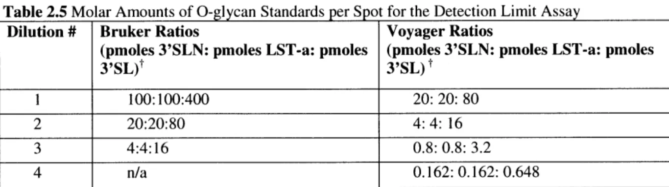
2.1.3 Results and C onclusions... 49
2.1.3.1 Bruker O ptim ization... 49
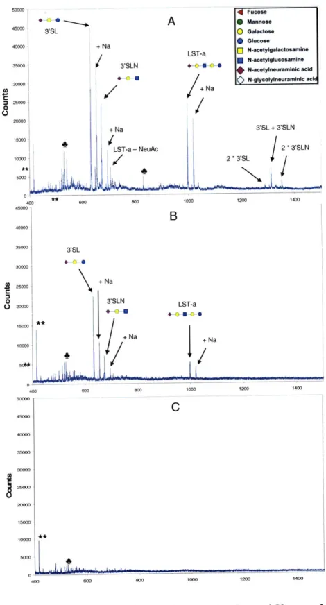
2.1.3.2 V oyager O ptim ization ... 54
2.1.3.3 Detection Limits for Voyager and Bruker Machines ... 58
2.1.3.4 Conclusion...61
2.2 0 -G LY CA N PU RIFICA TIO N ... 62
2.2.1 Background ... 62
2.2.2 M aterials and M ethods ... 64
2.2.3 Results and Conclusions... 65
2.3 O -G LY CA N RELEA SE ... 76
2.3.1 B ackground ... 76
2.3.2 M aterials and M ethods ... 78
2.3.2.1 O ptim ization of Reagent Concentrations ... 78
2.3.2.2 Length of Incubation Studies ... 79
2.3.2.3 Sialic A cid Linkage A nalysis ... 79
2.3.3 Results and C onclusions... 80
2.3.3.1 Reagent Choice & C oncentration Results ... 80
2.3.3.2 Length of Incubation Study Results ... 82
2.3.3.3 Sialic A cid Linkage A nalysis Results ... 90
2.3.3.4 C onclusion...94
2.4 D ISC U SSIO N ... 95
2.5 REFEREN CES...97
3
MUCIN DIRECT BINDING ASSAY EXPLORATION...99
3.1 PRO JECT M O TIV A TIO N ... 99 6
3.2 M ATERIALS AND M ETHODS ... 100
3.2.1 Optim ization of M ucin Binding Conditions...100
3.2.2 Optim ization of M ucin Concentration for Lectin A ssays ... 102
3.2.3 Lectin Dose Response ... 102
3.2.4 Lectin Binding Specificity A ssays ... 102
3.3 RESULTS...103
3.3.1 Optim ization of M ucin Concentration for Lectin A ssays ... 103
3.3.2 Lectin Dose Response A ssay Results...104
3.3.3 Sialic Acid Specificity A ssay Results ... 111
3.4 DISCUSSION ... 114
3.5 REFERENCES...117
4 CELL SURFACE GLYCAN CHARACTERIZATION ... 118
4.1 PROJECT M OTIVATION ... 118
4.2 M ATERIALS AN D M ETHODS ... 119
4.2.1 Red Blood Cell Surface G lycan Extraction...119
4.2.2 Epithelial Cell Surface G lycan Extraction ... 120
4.2.3 Glycan Purification ... 121
4.2.4 Glycan Sialic Acid Linkage Analysis ... 122
4.2.5 Glycan M S Analysis...122
4.3 RESULTS...123
4.3.1 RBC G lycan Analysis ... 123
4.3.2 HTE Glycan Analysis...138
4.4 D ISCUSSION ... 145
4.5 REFERENCES...147 7
List of Figures
FIGURE 1.1 COMMON GLYCOCONJUGATES ON THE CELL SURFACE...15
FIGURE 1.2 PRECURSOR MOLECULES AND DOL-P GLYCOSYLTRANSFERASES INVOLVED IN THE BIOSYNTHESIS OF THE N-GLYCAN PENTASACCHARIDE CORE ... 16
FIGURE 1.3 THE EIGHT CORE STRUCTURES OF O-LINKED GLYCANS... 18
FIGURE 1.4. TYPES OF EXTENSIONS FOR O-LINKED GLYCAN ARMS ... 20
FIGURE 1.5. STRUCTURE OF A TYPICAL SECRETED MUCIN ... 21
FIGURE 1.6. CO-STAINING OF TRACHEAL TISSUE SECTIONS WITH CON A/JACALIN AND SNA-I /JA C A L IN ... 3 0 FIGURE 1.7. CONCEPTUAL DIAGRAM OF THE BIOTINYLATED GLYCAN ARRAY... 32
FIGURE 1.8. HEMATOXYLIN AND EOSIN STAIN OF A HUMAN PSEUDOSTRATIFIED COLUMNAR EPITHELIAL LINING OF THE TRACHEA ... 34
FIGURE 1.9. COSTAINING OF TRACHEAL AND ALVEOLAR TISSUE SECTIONS WITH HINI A/SOUTH CAROLINE/I/18 HA AND H3N2 A/MOSCOW/10/99 HA... 35
FIGURE 2.1. MALDI-TOF MASS SPECTRA FOR RESOLVED AND UNRESOLVED PROTEIN SPECIES...45
FIGURE 2.2. O-GLYCAN STANDARD MS METHOD OPTIMIZATION ON THE BRUKER MALDI-TOF ... 51
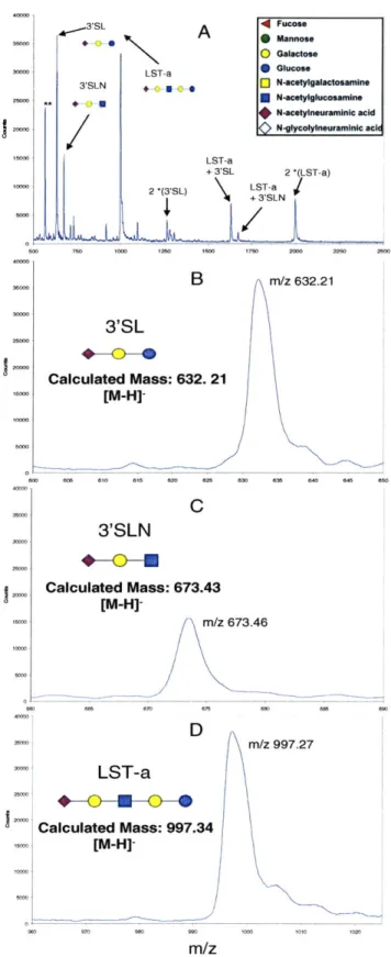
FIGURE 2.3 O-GLYCAN STANDARD MS SPECTRA ON THE VOYAGER MALDI-TOF ... 57
FIGURE 2.4. O-GLYCAN STANDARD MOLAR DETECTION LIMIT ON THE BRUKER AND VOYAGER M A L D I-T O F M A C H IN E S ... 59
FIGURE 2.5. SUPELCO PURIFICATION OF ACIDIC O-GLYCAN STANDARDS... 67
FIGURE 2.6. GELOADER PURIFICATION OF O-GLYCAN STANDARDS... 70
FIGURE 2.7. THE PURIFICATION LIMIT OF GELOADER MICRO-PURIFICATION COLUMNS ... 73
FIGURE 2.8. MALDI MS SPECTRA FOR FETUIN 0-GLYCANS TREATED WITH DIFFERENT B-ELIMINATION REAGENT COMBINATIONS...81 FIGURE 2.9. THE EFFECT OF P-ELIMINATION INCUBATION LENGTH ON FETUIN 0-GLYCAN
S P E C T R U M ... 8 3 FIGURE 2.10. THE EFFECT OF $-ELIMINATION INCUBATION LENGTH ON THE BOVINE MUCIN
0-G L Y C A N SPE C T R U M ... 86
FIGURE 2.11. SIALIC ACID LINKAGE ANALYSIS OF BOVINE FETUIN ... 92
FIGURE 2.12. SIALIC ACID LINKAGE ANALYSIS OF BOVINE MUCIN ... 93
FIGURE 3.1. OPTIMIZATION OF MUCIN CONCENTRATION FOR LECTIN ASSAYS...104
FIGURE 3.2. SNA AND JACALIN DOSE RESPONSE BINDING ASSAY ON BOVINE FETUIN ... 106
FIGURE 3.3. LINEARIZED HILL EQUATIONS USING FRACTIONAL SATURATION VALUES FOR SNA AND JACALIN ON BOVINE FETUIN ... 107
FIGURE 3.4. SNA AND JACALIN DOSE RESPONSE BINDING ASSAY ON BOVINE MUCIN ... 109
FIGURE 3.5. LINEARIZED HILL EQUATIONS FOR FRACTIONAL SATURATION VALUES OF SNA AND JACALIN BOUND TO BOVINE M UCIN...110
FIGURE 3.6. EFFECT OF 37C INCUBATION ON LECTIN BINDING TO MUCIN...112
FIGURE 3.7. SIALIC ACID BINDING SPECIFICITY FOR SNA BOUND TO FETUIN AND MUCIN...114
FIGURE 4.1 .VOYAGER MALDI-TOF MS SPECTRA OF CRBC AND TRBC NEUTRAL CELL SURFACE G L Y C A N S ... 124
FIGURE 4.2. VOYAGER MALDI-TOF MS SPECTRA OF ACIDIC CRBC AND TRBC CELL SURFACE G L Y C A N S ... 12 5 FIGURE 4.3. SIALIC ACID LINKAGE ANALYSIS OF CRBC ACIDIC GLYCANS ... 133
FIGURE 4.4. SIALIC ACID LINKAGE ANALYSIS OF TRBC ACIDIC GLYCANS...136
FIGURE 4.5. VOYAGER MALDI-TOF MS SPECTRA OF HTE CELL SURFACE GLYCANS...139
List of Tables
TABLE 1.1 COMMON ANIMAL MONOSACCHARIDE NAMES, ABBREVIATIONS, FORMULAS, AND
R E SID U E M A SSE S ... 14 TABLE 2.1 VOYAGER MALDI-TOF MS PARAMETERS USED IN THE ANALYSIS OF ACIDIC GLYCANS
... 4 9
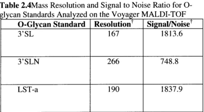
TABLE 2.2 BRUKER MALDI-TOF MS PARAMETERS USED IN THE ANALYSIS OF ACIDIC GLYCANS.. 49 TABLE 2.3. MASS RESOLUTION AND SIGNAL TO NOISE RATIO FOR O-GLYCAN STANDARDS
ANALYZED ON THE BRUKER MALDI-TOF... ... 53
TABLE 2.4 MASS RESOLUTION AND SIGNAL TO NOISE RATIO FOR O-GLYCAN STANDARDS
ANALYZED ON THE VOYAGER MALDI-TOF ... ... 55
TABLE 2.5 MOLAR AMOUNTS OF O-GLYCAN STANDARDS PER SPOT FOR THE DETECTION LIMIT
A S S A Y ... 5 8
TABLE 2.6 PROFILED O-GLYCANS OF BOVINE SUBMAXILLARY MUCIN ... 85
TABLE 2.7 REPORTED GLYCAN COMPOSITIONS FOR BOVINE MUCIN FROM FIVE SEPARATE
S T U D IE S ... 8 9
TABLE 3.1. DISSOCIATION CONSTANT CALCULATED FOR SNA AND JACALIN ON BOTH FETUIN
AND SYNTHETIC GLYCAN ARRAYS. ... 107
TABLE 3.2. DISSOCIATION CONSTANT CALCULATED FOR SNA AND JACALIN ON BOVINE MUCIN
AND SYNTHETIC GLYCAN ARRAYS. ... 110
TABLE 4.1. MOST PROMINENT NEUTRAL GLYCAN PEAKS OF TURKEY AND CHICKEN RED BLOOD
C E L L S ... 1 2 6
TABLE 4.2. TEN MOST PROMINENT ACIDIC GLYCAN PEAKS OF TURKEY AND CHICKEN RED B L O O D C E L L S ... 127
TABLE 4.3. EXPERIMENTAL MALDI-MS OUTCOMES USED TO CHARACTERIZE THE SIALIC ACID
LINKAGES WITHIN A GLYCAN POPULATION AFTER SIALIDASE S TREATMENT...131
TABLE 4.4. COMPARISON OF PROMINENT GLYCAN SPECIES PRESENT IN SIALIDASE A AND S
TREA TED C R B C SA M PLES. ... 134 TABLE 4.5. MOST PROBABLE GLYCAN COMPOSITIONS OF THE TEN MOST PROMINENT ACIDIC
C R B C SU RFA C E G LY CA N S...135 TABLE 4.6. COMPARISON OF PROMINENT GLYCAN SPECIES PRESENT IN SIALIDASE A AND S
TREA TED TR BC SA M PLES. ... 137 TABLE 4.7. MOST PROBABLE GLYCAN COMPOSITIONS OF THE TEN MOST PROMINENT ACIDIC
TRBC SURFACE GLYCANS BASED ON SIALIC ACID LINKAGE ANALYSIS ... 138 TABLE 4.8. MOST PROBABLE GLYCAN COMPOSITIONS OF THE NEUTRAL HTE SURFACE GLYCANS
... 1 4 0
TABLE 4.9. MOST PROBABLE GLYCAN COMPOSITIONS OF THE TEN MOST PROMINENT ACIDIC HTE
SU R FA C E G L Y C A N S ... 140
TABLE 4.10. COMPARISON OF PROMINENT GLYCAN SPECIES PRESENT IN SIALIDASE A AND S TREA TED H TE SA M PLES...144 TABLE 4.11. MOST PROBABLE GLYCAN COMPOSITIONS OF THE TEN MOST PROMINENT ACIDIC
Chapter 1
1 Introduction
1.1
BACKGROUND
The majority of membrane-bound and cellular species are glycosylated, making glycosylation one of the most common types of post-translational modification. Glycoconjugates, or biological species having covalently linked carbohydrates called glycans, function as important components of cellular membranes and extracellular spaces by mediating cell-cell communication, cell transduction pathways, and cellular development, function, and survival [1]. Their non-carbohydrate portions (aglycones) are often transcribed under the genetic control of the cell, while their carbohydrate moieties are enzymatically created and later added via the concerted action of a series of glycosidases and glycosyltransferases.
Because their carbohydrate moieties are added via non-template synthesis, glycoconjugates have many different carbohydrate compositions and conformations, and thus have diverse molecular properties and functions. For example, the influenza virus recognizes and interacts with specific glycan residues on glycoproteins in the bronchial epithelium in order to infect these cells [1, 3, 4]. Variations in protein glycoforms can be strong indicators of a change in the virulence of a disease because cell surface binding can be increased or decreased. Thus, the isolation and characterization of glycoconjugates and their glycan moieties in their normal and altered forms could provide important information about the molecular mechanisms of disease pathogenesis.
Since the function of a glycoconjugate can sometimes be greatly attributed to its glycan moieties, many studies have focused on the characterization of glycan structures. Glycans are technically defined as linear or branched carbohydrates composed of monosaccharides linked by glycosidic bonds via a
hydroxyl group. The most common monosaccharides found in animals are shown in Table 1.1 with their abbreviations, residue formulas and masses. These monosaccharide units can be neutral or negatively charged (acidic).
Table 1.1 Common Animal Monosaccharide Names, Abbreviations, Formulas, and
Residue Masses
Monosaccharide Examples Residue Residue Masst
Formula
Deoxyhexose Fucose (Fuc) C6HI004 146.078
Hexose Glucose (Glu), C6H2005 162.053
Mannose, (Man),
Galactose (Gal)
HexNac GalNAc, GlcNAc C8H13NO5 203.079
N-acetyl neuraminic acid NeuAc C1 1H17NO8 291.095
N-glycoyl neuraminic acid NeuGc C1 1H17NO9 307.090
Residue masses are .monoisotopic mass calculated based on C= 12.000000amu, H= 1.007825amu, N = 14.003074amu, O= 15.994915amu ). These residue masses do not include water, which is typically lost in the condensation reaction involving two monosaccharides. Since the terminal monosaccharide is only involved in one condensation reaction, the mass of one water molecule must be added to this residue mass.
Glycans composed of at least 2 or more monosaccharide units are called oligosaccharides, while polysaccharides tend to be composed of many repeating oligosaccharide units. The 6 major classes of glycoconjugates include proteoglycans, glycolipids, lipopolysaccharides, peptidoglycans, and
glycoproteins. Shown in Figure 1.1 are some of the major glycoconjugates, all of which play important roles in the functionality of a cell's surface and extracellular environments.
Glycoprotein
N-linked
Glycan 0-linked 03 Glycophospholipid
a3 a6 glycan 13 4 0-linked -- Legend glycan 4a Glycosphingolipid
0
0 GicNAc
0~a
4
N
$r/hr'
er.-
$4
N H2
[0 GaINAc
OUTSIDE
P
*
HexA
VXyl
@GICA
Gal
INSIDE
J3
Glycoprotein 'Ser/Thr"Figure 1.1 Common Glycoconjugates on the Cell Surface. Figure adapted from Varki et al 1999 [1].
This thesis focuses on secreted and membrane-bound glycoproteins, or proteins carrying one or more nitrogen-linked (N-linked via Asparagine) or oxygen-linked (0-linked via Serine or Threonine) glycan. Secreted glycoproteins possess a variety of important biological functions which are often mediated by their glycan moieties. They can serve as structural molecules (collagen), lubricants (mucins), transport molecules (ceruloplasmin and tranferrin), hormones (thyroid stimulating hormone), immunologic molecules, and enzymes (5). Cell membrane associated glycoproteins, on the other hand, are involved in cell-cell, virus-cell, bacterium-cell, and hormone-cell interactions (5). For both classes of molecules, much is still unknown with respect to the structures and functions of their glycan moieties.
Although they appear to differ only by their linkage to the protein backbone, N- and 0-linked glycans differ tremendously in structure, synthesis, and biological significance. Because of their size and
susceptibility to enzymatic removal, the majority of studies on glycans have focused on N-glycans and
their biological significance. However, there has been an increased interest in O-linked glycans because of the potential roles they play in several biological processes.
1.1.1 N-glycan Structure and Synthesis
The formation of a nitrogen-linked glycan (N-glycan) starts with the synthesis of an oligosaccharide precursor linked to a lipid Dol-P (Figure 1.2). This dolichol lipid structure, which resides in cellular compartments other than the endoplasmic reticulum (ER), is linked via a pyrophosphate linkage to an oligosaccharide containing 14 monosaccharides [I]. This procedure first involves the transfer of two GlcNAc residues to the Dol-P precursor, followed by the addition of five mannose residues, all of which takes place in the cytosol. From here the structure is flipped such that the oligosaccharide faces the lumen of the ER and is further modified with 4 mannose and 3 glucose residues [1]. This structure is then transferred to an asparagine residue on a nascently translated protein in a high energy reaction carried out
by the oligosaccharyltransferase (OST) complex [1]. Typically, the oligosaccharide precursor is
transferred to an asparagine residue that is a part of the consensus sequence Asn-X-Thr/Ser (where X is any amino acid except for proline) [1].
Mannose
GIcNAc
1 c IV ' V Extensive Processing
SFlip
ccurs in the cytosol
|Occursnthe
Lumen of the ER andGolgiFigure 1.2 Precursor Molecules and Dol-P Glycosyltransferases involved in
the Biosynthesis of the N-glycan Pentasaccharide Core. Figure adapted from
After being linked to an asparagine residue, the oligosaccharide precursor, which is high-mannose type because of the prevalence of unsubstituted terminal mannose residues, is then trimmed by a-mannosidases in the ER and Golgi until it is converted into the pentasaccharide core (consisting of two GlcNac and 3 Man residues) traditionally associated with N-glycans (Figure 1.2) [1]. This
pentasaccharide core is the subject to diversification by way of exposure to several different
glycosyltransferases which can result in the elongation, branching, or termination of the N-glycan chains.
Whether secreted or membrane-bound, N-glycosylated proteins have a diverse array of functions, many of which are dependent on either the local or systemic presence of N glycans in specific peptide regions [6]. According to the SWISS-PROT database, it is estimated that 90% of all of the reported glycoproteins, which make up half of the total number of reported eukaryotic proteins, are N-glycosylated
[6]. Although the majority of these proteins have not been studied in the context of their glycosylation, it
is thought that N-glycans play crucial roles in their protein signaling processes, stabilization, immune responses, orientation, rigidity, and protein turnover, since they are typically exposed on the surface and have flexible or elongated arms that can extend 3nm or further into the solvent [6].
The different functionalities of N-glycans can be broadly classified into three categories. The first category deals with the role of N-glycans in the protein folding and quality control processes that occur in the ER. These specific roles became evident after an N-glycosylation inhibitor called tunicamycin was applied to cell culture. The resultant studies showed that many peptides were retained in the ER due to improper folding, and were eventually targeted for degradation [7, 8]. Another correlation between N-glycosylation and proper protein folding was drawn with studies on the Aichi strain influenza virus hemagglutinin, which contains only 6 N-glycosylation sites. Without only one of the six glycans (N81),
HA cannot form any disulfide bonds because of a destabilizing effect that prevents disulfide bond
oxidation, and thus fails ER protein folding quality controls [9]. The second category deals with the role of N-glycans in the intracellular transport and targeting processes that occur in the ER, Golgi, and trans-Golgi compartments. The importance of this function is evident in the trafficking of acid hydrolase
precursors, which are N-glycosidically linked to mannose-6-phosphate and ultimately targeted to the trans-Golgi and lysosomal compartments [6,10]. Lastly, N-glycans play important roles in the function and stability of mature proteins that have been extensively processed in the Golgi. By far, this category has the largest number of specific N-glycan functions since every mature protein can have a different function. Regardless, studies have shown that glycosylated proteins are typically more stable, soluble, and resistant to proteases when compared to their non-glycosylated counterparts. It is hypothesized that N-glycans confer these properties to the stabilized folded protein by reducing the degrees of (mobile) freedom in the unfolded protein [6].
1.1.2 0-glycan Structure and Synthesis
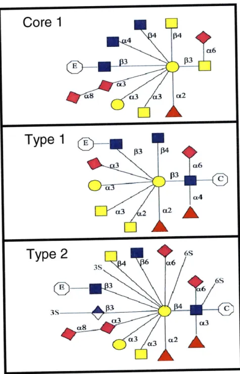
Oxygen-linked glycans (0-glycans) are formed through the addition of a monosaccharide residue, usually a GalNac, to either a serine or threonine residue via an a-O-glycosidic linkage [1]. Unlike N-glycan synthesis, O-N-glycan synthesis does not require a lipid-linked oligosaccharide precursor in the synthesis initiation event. Instead, a GalNac-transferase initiates synthesis in the Golgi Body by adding a GalNAc residue to a threonine/serine residue [I].
.3il o-Ser/Thr ;A3o-SerThr
p13 p3 Core 1 Core 2 p6 o-SerThr qo-Ser/Thr @3 @r3 Core 3 Core 4 0S o-er/Thr Core 5 Core 6 o-Ser/Thr
o-ser/Thr
Core 7 Core 8Key: MGlcNAc COGaINAc *Gal
Figure 1.3 The Eight Core Structures of O-linked Glycans. Figure from the Consortium for Functional Glycomics [11].
From this point, the attachment of an additional GalNAc, GlcNAc, or Gal residue determines what is termed as the "core structure" of the O-glycan (Figure 1.3) [1]. In total, there are currently 8 core
structures, 4 of which are largely prevalent (Cores 1-4). The existence of many core structures as well as the difference in amino acid linkage distinguish N- and 0- glycans, since N-glycans are always linked to an asparagine residue and always contain a common pentasaccharide core region. Furthermore, unlike N-glycans, whose asparagine residue is always a part of the peptide consensus sequence Asn-X-Ser/Thr (where X is any amino acid except proline), 0-glycans have no consensus sequence, even though proline residues seem to be abundant in the -I and +3 positions in heavily O-glycosylated proteins [1].
After the formation of the O-glycan core structure, additional monosaccharides (GaINAc, GlcNAc, Gal, Fuc, or NeuAc) can be added by specific glycosyltransferase enzymes to elongate the glycan in a linear or branched fashion, although the latter is much less common [1]. The number of possible glycan structures, however, is limited by the rules of the O-glycan biosynthetic pathways (i.e. the selectivity of the glycosyltransferases); in other words, certain transferases will only elongate a glycan chain according to the presence of specific terminal monosaccharides linked in specific ways (Figure 1.4). On the whole, 0-glycans mainly exist as short linear chains with sialic acid or fucose residues in the terminal position. However, longer O-glycans that contain repeating Gal-GlcNAc-Gal (polylactosamine) units do exist. For example, linear polylactosamines can be subjected to further modification in humans to form the ABO Blood Group Antigens, which essentially describe 3 different types of terminal oligosaccharide groups present on the glycoproteins on the surface of red blood cells [1, 8].
Figure 1.4. Types of Extensions for O-linked Glycan Arms. Core 1 extensions are
unique only of Core 1 structures. They may be elongated in a Type 1 or 2 fashion only after the addition of the appropriate Core 1 extension. Type 1 and 2 extensions may be applied to any Core structure, and can include both monosaccharide and
adduct (sulphate/phosphate) additions.
=Extension,
=Core,
= NeuAc,
=
GlcNAc,n
= GaINAc,0
= Glucose,A
= Fucose,=
GlcA, S= Sulphate (S04). Figures adapted from the Consortium of Functional Glycomics [11].Although less understood than N-glycosylation, 0-glycosylation has become equally important to our understanding of tissue-specific regulation and disease progression. A number of studies have helped to better elucidate potential roles of O-glycans, mostly in the context of mucins, a family of large and heavily 0-glycosylated secreted and cell surface proteins primarily implicated in the maintenance of mucous membranes (Figure 1.5). Since these membranes help to regulate epithelial absorption and secretion, it is thought that mucins, which are usually larger than 200kDa and rich in serine/threonine repeats, regulate these processes through their dense water-retaining O-glycan coatings and form a viscous gelatinous material via mucin disulfide bond cross-linkages and other non-covalent interactions (1,12). These O-glycan coatings tend to look different in various tissues in the body, and since
glycosylation is not template driven, the same peptide backbone can have drastically different functions in different tissues.
S S S S
S S s s
H2N" COOH
Figure 1.5. Structure of a Typical Secreted Mucin. The apomucin peptide
backbone is rich in serine/threonine repeats which give rise to dense regions of 0-glycans. Mucins form their thick gelatinous structure through non-covalent interactions as well as disulfide bonds. Note that a small number of N-linked glycans (red) can also be present on the apomucin backbone Figure adapted from Toribara et al 2001[13].
Of the two classes of mucins, secreted and membrane bound, the former seems to be implicated
mostly in the protective function of viscous mucus gel, while the latter does not form oligomeric
complexes and is implicated in a variety of functions ranging from cellular adhesion to scaffold formation [14]. The broadest definition of a mucin is a protein comprised of at least 50% O-glycans by mass, which leads to the inclusion of rarely studied non-epithelial mucins, which might serve as ligands for selectins and facilitate lymphocyte trafficking [15].
Even though the bulk of mucin structure is dominated by O-glycans, the protein backbone
(apomucin), which is encoded by at least 19 human genes (MUCl-20), has gained just as much attention because it is variably expressed in different epithelial tissues throughout the body. All mucins have dense O-glycan coatings, but their protein backbones differ in the number of tandem repeats of a defined number of nucleotides [14], which allows for a large variability in mucin size. Overall, it is thought that the size and length of the mucins are not as important as the generic protein scaffold that they provide for oligosaccharide attachment, which can dramatically improve substrate binding affinities due to the
multivalent nature of their dense glycan coating [14].
Interestingly, diseased tissues are noted for changes in both the level of mucin expression and the structural characteristics of the attached glycans. These mucins may express O-acetylated, sulphated, truncated, elongated or more sialylated glycan structures in addition to being over- or under- expressed
themselves. Studies of mucins from diseased tissues have offered much insight on their pathological and physiological roles. For example, MUC1, a membrane-associated phospho-mucin that protects cells by binding to pathogens, is aberrantly expressed in many carcinomas and metastatic lesions [1]. It is thought to play a role in tumor progression and metastasis by destabilizing cell-cell and cell-matrix interactions, thus enabling cancerous cells to escape from the epithelial layer and into surrounding tissues [1]. Furthermore, tumor cells that overexpress MUC 1 are more likely to be resistant to natural killer and cytotoxic T cell mediated death [1]. On the other hand, mucins from airways affected by bronchitis, asthma, or cystic fibrosis are constitutively expressed, and are produced in excess due to goblet cell
hyperplasia and submucosal gland hypertrophy. Even though it is clear that these mucins express glycans that are short, branched, and sulfated, we do not completely understand if these structures contribute to the pathology of the disease [1].
Still, the study of mucins, glycans, and their biological roles face many challenges. Firstly, it is important to understand that the analysis of the structural and functional role of mucins and their glycans in diseased tissues is hindered by a lack of analogous mucin from normal tissues. Therefore, proof of speculations about the unique structures of glycans and their specific role in disease pathophysiology has been difficult to obtain. Studies aimed at identifying and characterizing both sets of samples will
ultimately provide more insight into the molecular basis for disease pathogenesis. Secondly, because glycans make up the majority of the structure of a mucin and critically determine its function, it is necessary to be able to characterize these structures in a way that links form to function. This means that studies of phenomena like cell binding, mucin aggregation, and lymphocyte activation will be best supported with supplemental structural analysis of the glycans of the species being analyzed. Currently, most of these studies are done separately, such that a full understanding of mucin form and function is not achieved. Lastly, unlike most glycosylated proteins, the study of mucins from diseased tissues is limited to a few organs. For example, currently the membrane bound MUCI has only been characterized and described in lung, breast, and renal cancer [16]. The study of other tissues, diseased or normal, will greatly expand our understanding of how the same mucin functions in different environments. Furthermore, these studies could identify new potential therapeutic targets for many diseased tissues, since it is suspected that most tissues express at least one type of mucin [16].
Even though definitive comparisons of oligosaccharide structures from normal and diseased tissues are not currently possible, some studies do shed light on the physiological roles of specific mucins in various tissues. Salivary mucins, for example, both adhere to and protect tooth enamel demineralization caused by organic acids, as well as defend against oral microbes in the local environment of epithelial cells [17]. Other studies have suggested that mucins on the surface of leukocytes facilitate cell adhesion
events [4]. More specifically, the overexpression of core 2 branched 0-glycans correlated to a reduction of interactions between leukocytes and antigen-presenting cells [7], which are key to the proper function of the immune system. Conversely, the elimination of a particular mucin gene leukosialin from the mouse genetic code resulted in hyperimmune responses, since the proteins and their 0-glycan coatings are absent
[6]. Pulmonary mucins, on the other hand, line the region spanning from the nasal passages to the
respiratory bronchioles, and are thought to encapsulate particulate matter and pathogens in order to protect the lower airways and alveoli from obstruction [18].
1.1.3 The Role of Glycans in Influenza Infection
1.1.3.1 Influenza A
The potential pandemic threat of the Influenza A virus, which is a major cause of the yearly flu epidemic, has instigated the investment of billions of dollars in research surrounding its pathogenesis [1]. This virus, which is categorized based on its surface antigens hemagglutinin (HA) and neuraminidase
(NA), affects approximately 15% of the U.S. population each year, and results in about 36,000 deaths and
more than 200,000 hospitalizations [19]. Even though Influenza A subtypes mainly infect and transmit between wild aquatic birds, two human adapted strains of the virus named HINI and H3N2 are the primary subtypes circulating in the human population, and are thus responsible for the yearly influenza outbreaks and epidemics. The real threat from this virus comes from its potential to mutate and become
highly pathogenic and virulent, such that its spread would cause serious global health concerns. These
pathogenic Influenza A viruses, which either acquire mutations in key viral proteins or obtain new gene segments through reassortment, efficiently transmit from human-to-human, and cause a high mortality rate. In the last century, three major pandemics were caused by influenza viruses: the 'Spanish flu' of 1918 caused
by the HINI subtype, the 'Asian flu' of 1957 caused by the H2N2 subtype, and the 'Hong Kong flu' of 1968
caused by the H3N2 subtype [20-25]. Based on the estimated mortality rate of the 1918 Spanish flu pandemic, epidemiological studies have projected that a pandemic in the present time would affect roughly 75 million people worldwide [19]. Furthermore, the H5NI strain of Influenza A, otherwise known as bird flu, is known to have the
highest mortality rate, with greater than 60% of infected individuals succumbing to death [20-25]. However, other Influenza A subtypes, such as H2N2, H5N2, H7N7, and H9N2, have all adapted to infect humans and also present the realistic threat of a new flu pandemic equal in impact, or even greater than, that of the
H5NI Spanish influenza. Fortunately, many of these virus subtypes have not acquired the ability to
efficiently transmit between humans; as a result, influenza outbreaks have been relatively localized around the world [25,26]. However, because a serious potential pandemic threat still exists, studies on influenza infectivity and pathogenesis represent the first steps in global preparedness to an outbreak of virulent subtypes.
On a molecular level, Influenza A is a virus of the Orthomyxoviridae family of viruses and contains 8 single-stranded negative-sense RNA strands that encode for 11 viral proteins [27]. Three of these proteins, hemagglutinin (HA), neuraminidase (NA), and matrix protein (M1), are present on the surface of influenza viruses. The other encoded proteins include RNA polymerase subunits (PB 1, PB2, PA), nucleoprotein (NP), nonstructural proteins (NS I and NS2), a proton-selective ion channel protein (M2) and a pro-apoptotic PB I -F2 protein. However, only three of the RNA strands contain the genetic information encoding the three proteins that are key to the first step in the viral infection (HA, NA, and PB I polymerase). HA is mediator of the first step of viral infection and is responsible for binding to terminal sialylated glycans projected from human respiratory epithelia. HA also contributes to the intracellular release of virion particles in the host cell by way of fusion of the virus with the host cell membrane [28]. Inside the cell, host proteases cleave the viral HA precursor into two subunits linked by a disulfide bond) at a specific amino acid residue (which go on to play a critical role in infection [28]. Neuraminidase, on the other hand, is responsible for cleaving viral cell surface glycan sialic acids, and also has a role in viral entry and the release of mature virions [28]. Each Influenza subtype is characterized by the type of HA and NA present on the viral coating. For Influenza A in particular, 16 types of HA and 9 types of NA have been identified.
Antigenic shift and drift in Influenza viruses are responsible for the yearly appearance of new and more resistant strains of the virus. Antigenic shift occurs when two or more different strains of virus
infect the same cell and are susceptible to genomic segmentation. Since different strains may contain drastically different genetic information, antigenic shift can result in major genetic changes. Antigenic
drift, on the other hand, is caused by the error prone virus-encoded RNA-dependent RNA polymerases, which frequently make minor genetic errors in the form of amino acid substitutions (1/104 bases per replication cycle). Overall, while antigenic drift is a continuous process and leads to gradual changes in surface antigens, antigenic shift is an occasional occurrence leading to significant changes in antigen presentation on the virus surface. Since HA and NA are crucial to the virus' ability to initially infect the host cell, genetic changes in these proteins have a large impact on the efficiency and malevolence of the virus. In particular, changes in the HA and NA subtype due to antigenic shift and drift may result in the increased ability of the virus to escape vaccination or neutralizing antibody produced by the host immune system [28]. Some studies have also suggested that mutations in these proteins can lead to human adaptation, since the HA-host cell interactions are purely governed by the properties of the protein-glycan binding sites [3]. More
specifically, the crossover from birds to humans is believed to be associated specifically with the ability of HA to switch its binding preference from u2-3 to o2-6 sialylated glycan receptors [29-36].
Even though our understanding of the virus' pathogenecity has improved, we have yet to fully grasp how the virus could acquire the ability to effectively transmit between humans. Understanding this process is key to our ability to develop improved vaccine strategies that rely less on a specific antigen, which often changes rapidly due to antigenic drift, and capitalizes on the host's acquired immune
responses, which can most effectively remove infected cells after the initial assault [28]. Furthermore, we cannot begin to address the issue of viral transmission efficiency if we do not have a firm grasp on the molecular basis of the initial virus-host cell binding events prior to infection.
Thus far, we understand that there are three stages in the influenza infection cycle. The first step is the attachment of HA to x2-3 sialylated and ax2-6 sialylated glycan receptors of the host cells for birds and humans, respectively [37-39]. This initial binding event is followed by the endocytosis of the virus and the structural modification of HA, which aids in the fusion of the viral membrane with the endosomal membrane. From here, the
ion channel activity of Ml is activated, and viral RNA is transported into the host nucleus, where it is replicated and transcribed. After glycosidic modification in the Golgi Bodi, the newly synthesized HA and NA proteins are free associate with newly synthesized viral RNA transcripts to form the virion. These virions then present themselves on the outer surface of the cell, and are released from the cell surface via the action of NA. At this point, these progeny virions can infect other host cells and repeat the entire process.
1.1.3.2 Hemagglutinin and Host Cell Surface N-Glycans
Since the hemagglutinin protein is essential to the initial viral infection events, many researchers have focused on this protein as a means of both understanding the molecular mechanism of viral infection and identifying novel influenza vaccine targets. More specifically, with respect to Influenza A, researchers have focused on the genetic changes in HA that would enable H5NI, which normally infects birds, to infect and transmit efficiently between humans. Genetic changes in the hemagglutinin binding site in particular can lead to a change in the sialic acid binding specificity, and ultimately improve the efficiency of virus infection and transmission, since this binding event appears to be the key determinant of whether or not a particular virus can efficiently infect humans [3,28]. For example, avian influenza viruses of the
H5NI subtype only bind to 2-3 linked sialic acids bound to a penultimate galactose residue (on N-glycans), while their human counterparts bind to 2-6 linked sialic acids linked to a penultimate galactose residue [28]. This difference explains the differences in viral infectivity of birds and humans, since human and bird tracheal epithelial tissues mainly express glycans that have terminal a2-6 and a2-3 linked sialic acids, respectively [28]. Through H5NI HA binding assays on glycan with different sialic acid linkage types and lengths, research has shown that a2-6 linked sialic acids preceded by repeating lactosamine units (Gal-GlcNAc) provide a larger 3-D space over which the glycan can move after it is bound to the
viral HA [3]. Analyses such as these will become important in accurately describing the binding specificities of different HA subtypes, and ultimately identifying therapeutic or vaccine targets that will actually confer protection against specific virus subtypes.
Besides understanding HA's binding specificity, researchers have also attempted to characterize the glycans on the relevant host cell surfaces so that the binding of a particular HA to a particular host cell type can be predicted [3]. This piece of the puzzle may prove to be the most important, since the HA binding pocket of an influenza virus that effectively transmits between humans will have had to mutate to specifically fit the glycan topologies present on the human respiratory tract cell surfaces. Previous
research has shown that the cell surface N-glycans of human bronchial epithelial cells (HBE) are mostly composed of N-linked glycans which contain long repeating lactosamine units capped with a a2-6 linked sialic acid [3]. Therefore, it is postulated that the binding pocket of avian H5N1 strains must mutate to be able to bind glycans of this topology in order to infect human bronchial epithelial cells.
1.1.4 Tools to study HA-Glycan Interactions
1.1.4.1 HA Agglutination and Inhibition Assays
Avian influenza viruses are able to agglutinate erythrocytes (red blood cells, RBCs) through the binding of HA to receptors, and therefore provide an experimental system with which researchers have classified wild-type and mutant influenza A viruses, screened potential HA inhibitors, and identified new
vaccine targets [3]. In this agglutination assay, if viral particles are present at a high enough concentration, a diffuse lattice of linked erythrocytes will form, preventing the erythrocytes from precipitating as a small pellet at the bottom of a well in a microtiter plate. RBCs from species such as chicken, turkey, equine, and guinea pigs are typically used in this assay, and because these erythrocytes have a mixture of ou2-6 and o2-3 linked sialic acids, their respective cell surface glycans are de-sialylated using sialidase enzymes, and then re-sialylated using specific o2-6sialyl-tranferases [40].
The agglutination assay is the primary tool used to classify HA subtypes by sialic acid specificities, screen potential HA inhibitors, and identify potential vaccine candidates because of the ease with which it can be performed and the low cost of acquiring all of the necessary materials [40]. However, some researchers have postulated that these assays may not be the best way to study HA-glycan interactions because they might not describe the HA-glycan interactions that are relevant in the influenza infection process [3]. It is possible that the glycans modified by simple de-sialylation and re-sialylation of
erythrocyte cell surface glycans do not represent the natural glycan ligands present on the cell surfaces of human tracheal and bronchial epithelial cells. Ultimately, the success of this tool depends on the overlap between erythrocyte and human upper airway cell surface glycans, both of which are still relatively unexplored. It will therefore be necessary to compare and contrast the glycan topologies present on both the surfaces of commonly used erythrocytes and human tracheal and bronchial epithelial cells in order to begin to draw conclusions about HA binding specificities in humans.
1.1.4.2 HA-Glycan Tissue Binding Studies
HA-glycan tissue binding studies have helped to reveal some information about the glycans present on the surface of the human tracheal and bronchial epithelia. Typically, de-paraffinized and re-hydrated human upper airway tissue sections are incubated with labeled lectins, each of which bind to specific terminal glycan residues and can ultimately be detected by fluorescence microscopy after the addition of the appropriate reagent, such as Alexa fluor 546 streptavidin [3] (Figure 1.6). These studies have revealed that human tracheal and bronchial epithelia predominantly express x2-6 sialylated glycans; these glycans are omnipresent in the nasal mucosa, on epithelial cells of paranasal sinuses and the pharynx, and in the trachea and bronchi [43]. Knowledge of the glycans present on these surfaces is specifically important in the context of influenza infections because they are the first to be exposed to the free virion. On the other hand, glycans in the human deep lung alveolar cells, as well as those present at the infection site in birds, are mainly a2-3 sialylated [43]. Knowledge of the sialic acid residues present on these surfaces is beneficial to our understanding of how non-adapted avian influenza subtypes infect humans; for
individuals who breathe in large amounts of virion will be susceptible to virion aggregation, virion descent into the lower airways, and infection of these surfaces.
Figure 1.6. Co-staining of Tracheal Tissue Sections with Con A (red)/Jacalin (green)
[top two panels] and SNA-I (red)/Jacalin (green) [bottom two panels]. For both the top
and bottom panels, the figures on the left are at 25x magnification, while the figures on the right are shown a 63x magnification. Con A binds branched a -mannosidic structures, which includes high-mannose type, hybrid type and biantennary complex type N-linked glycans. SNA binds a2-6 linked Neu5Ac residues that are linked to a penultimate Gal residue. Jacalin binds to a number of residues containing GalNAc, with the strongest binding occurring to the motif (Sialic Acid) Galpl-3GalNAcal-Serine/Threonine. In the top Figure, Jacalin binding is distinct and localized to the goblet cells expressing O-linked glycans, while the regions of Con A binding correspond to ciliated cells expressing N-linked glycans (white arrow). SNA, however, extensively binds to both the goblet cells (co-stain with Jacalin in yellow) and the ciliated cells expressing N-glycans (red) [3].
While these studies have helped to reveal the global distribution of the terminal sialic acid residues present on the surface of human respiratory tract tissues, they still do not help to elucidate the types of glycan compositions present on these surfaces. Ultimately, in order to fully and specifically describe HA-glycan interactions, it is necessary to analyze the structural compositions of the HA-glycans present on these cells so that our understanding of HA-glycan specificities can be improved.
1.1.4.3 HA-Glycan Array Studies
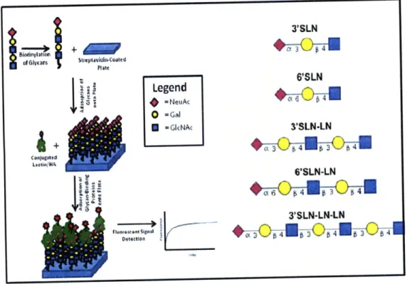
While the tissue binding studies can provide useful information about the terminal glycan moieties present on cell surfaces, other more specific arrays have helped to further describe HA-glycan
specificities. An ELISA-like assay using biotinylated a2-3 and a2-6 sialylated glycans bound to a streptavidin coated plate have been used to determine virus receptor specificity (Figure 1.7). These assays are especially relevant to the study of HA binding to cell surface N-glycans because they theoretically present glycan moieties in a manner that is similar to the way in which cell surface glycoproteins present these same moieties. In these assays, recombinantly expressed HA viruses in their natural and mutant forms from H1, H3, and H5 subtypes have already been classified according to their binding preferences to x2-3 and a2-6 sialylated glycans of different lengths [3]. These studies have specifically shown that HIN1, for examples, binds strongly to the 6'SLN-LN glycan, but not to
3'SLN, 6'SLN, 3'SLN-LN, or 3'SLN-LN-LN [3]. This means that this HA subtype specifically requires long a2-6
sialylated glycan arms for binding, and that the glycan topology or composition, not just the sialic acid linkage type, is important in the study of HA-glycan interactions. Furthermore, the effects of sulfation and fucosylation on HA binding have also been determined using these assays. This assay has also been used to quantify the
multivalent HA-glycan interactions, and explain how subtle mutations in HA amino acid sequences led to quantitative differences in glycan binding. The results of studies such as these ultimately show how mutations in HA proteins could lead to human adaptation of the virus by changing HA-glycan binding specificities.
3'SLN d~lymSIteptavidin-.Coated Plait 6'SLN Legend =NeuAt 0 -cGal -Gl<NAc 3'SLN-LN 6'SLN-LN 3 SLN-LN-LN
Figure 1.7. Conceptual Diagram of the Biotinylated Glycan Array. The five types of
synthetic biotinylated glycans (left) were absorbed into separate wells on a streptavidin coated plate, and exposed to a glycan-binding protein labeled with a tag that would allow for
its detection by fluorescent signal (either directly or indirectly) (right).
The direct binding assay can provide important quantitative binding affinities, such as the dissociation constant, Kd. There are several ways to conduct the assay by using different reporter proteins and labeled lectins, but for the work performed in this thesis, a FITC-conjugated protein of interest (lectin or hemagglutinin) is added to the array at various concentrations in a dose-dependent manner. The wells are then incubated with HRP-conjugated anti-FITC antibody, and then exposed to hydrogen peroxide (H202)
and Amplex reagent so that a brightly fluorescent product is created [41]. The experimental rationale behind the calculation of these binding constants is that the fluorescent signal for each well is a direct readout for the number of lectins bound to the glycans. Since both the fluorescent signal and the rate of HRP reaction decrease as the substrate is used up, the most accurate indication of the number of lectins in each well is the initial slope of the fluorescent curve (in units of fluorescence per time). For each lectin concentration, a unique initial slope can be obtained, and the fractional saturation of lectins on glycans
(the concentration-specific initial slope divided by the maximum initial slope) can be calculated. From here, the Hill equation, which describes multivalent kinetic interactions, can be used to calculate the apparent dissociation constant, Kd.
[HlA]"
[AJ" + Kj P]
In this equation, y is the fractional saturation, and n is the cooperativity factor. The linear form of this equation,
log( - = * log[M I A]) - logKi), [21
can be used to calculate Kd by plotting the log of the lectin or HA concentration against the log(y/1-y).
The negative of the y-intercept is the log (Kd), while the slope is the cooperativity factor. To obtain accurate quantitative numbers, it is essential that enough data points be collected so that the trendline can be realistically determined (R2> .90). While the cooperativity factor is important in understanding the
strength of polyvalent interactions, an analysis of the cooperativity factors of different lectins is beyond the scope of this project because other factors complicate this analysis (glycan spacing, lectin
conformation, etc).
Overall, these studies have helped to identify specific types of glycan moieties present on N-glycan arms that are important in HA binding. The combination of the glycan array and tissue binding studies
therefore provide a strong experimental system for exploring HA-glycan interactions, and would be even better complimented with additional analytical studies on the glycan structures themselves.
1.1.5 HA and 0-linked Glycans
Although less understood than N-glycosylation, 0-glycosylation has become equally important to our understanding of tissue-specific regulation and disease progression. Of recent interest is the
pathophysiological role of mucins, if any, in influenza infection. Mucins play a critical role in the integrity of the human tracheal and bronchial epithelia because the viscous gel layer they create protects against shear stress and chemical damage, and traps particulate matter and microorganisms that are eventually removed by the "sweeping" abilities of the ciliated epithelium [14-18]. In the human upper respiratory tract, mucins are secreted from goblet cells, which are cylindrical "goblet"-shaped cells found scattered in between ciliated epithelial cells (Figure 1.8). Their nuclei and cellular organelles all sit on the basal side of the epithelia, while their apical side extends into the lumen and releases membrane-bound
secretory granules filled with mucus. The secretion of mucins occurs via two pathways, constitutive and stimulated secretion, the latter of which is typically enhanced by irritating stimuli like dust, smoke, or microorganisms [14-18]. Goblet cells also have an abundance of mucins on the surface of their membranes that are mediators of membrane binding events (adhesion/cell-cell communication). In addition to their muco-protective and signal transducing functions, these cells also have the ability to differentiate into other cell types, such as ciliated epithelial cells, giving them a "stem cell-like" quality [14-18].
Trachea H&E
goblet cells cilia
b 0ent merane cala
Figure 1.8. Hematoxylin and Eosin Stain of a Human Pseudostratified Columnar
Epithelial Lining of the Trachea. Goblet cells filled with secretory granules are interspersed between ciliated epithelia cells, which sit above a basement membrane nestled by basal cells [42].
Since we still don't fully understand the events that take place or know the cell participants in the influenza infection process, the study of goblet cells and their mucins could shed light on these events, as well as help researchers elucidate some pathophysiological roles of mucins. Firstly, previous tissue binding experiments have shown that some humanized HA subtypes preferentially bind goblet cells over ciliated epithelial cells, suggesting that goblet cells may play a stronger role in viral infectivity than previously thought (Figure 1.9) [3]. These results have led to the hypothesis that goblet cell mucins are key mediators of viral binding, since they represent a biological "template" onto which many "ligands" (glycans) can be attached. This "template" ultimately allows for strong multivalent interactions, which can enhance viral infectivity.
Trachea Alveolus
Trachea Alveolus
Figure 1.9. Costaining of Tracheal and Alveolar Tissue Sections with HINI A/South Caroline/1/18 HA (top) and H3N2 A/Moscow/10/99 HA (bottom). Both HAs stain green against the propidium iodide staining in red. The HINI ha seems to only stain goblet cells in the tracheal tissue, while the H3N2 HA seems to stain the entire apical side of the membrane. Neither HA heavily stains the alveolar tissues.
Lastly, very little is known about the protective functions of mucins in the context of influenza infection. It is possible that like many other diseased tissues, influenza-infected tissues overexpress specific kinds of mucin, either on the cell surface or in secretory granules, so that influenza binding to the host cell surface is impeded. Conversely, just like cancer cells, infected host cells might also aberrantly express certain mucins in order to impede protective immune responses. Confirmation of all of these potential roles of mucins requires an understanding of both the mucin glycan structures and the
mucin-HA binding interactions. These two components allow researchers to relate structure to function, and
could potentially highlight novel vaccine or therapeutic targets.
1.2 THESIS WORK
The study of the role of N- and 0-linked glycans in influenza infection and pathogenesis is still young and presents exciting new areas of experimental exploration. This thesis describes design, optimization
and application work that aims to further elucidate the roles of these glycans in a manner than acknowledges the significance of their structures or specific compositions. The following specific objectives were met:
1. The development, optimization, and application of a method for releasing, purifying, and
analyzing 0-linked glycan structures on glycoproteins. The structural information obtained from the above analysis will serve as the foundation on which conclusions can be made about the role of glycans in disease pathogenesis. More specifically, the knowledge of the structural motifs of specific mucins provided the motivation for the exploration of a direct binding assay that utilizes these mucins to obtain quantitative information on the 0-glycan binding specificities of
2. The isolation and characterization of chicken erythrocyte, turkey erythrocyte, and human tracheal cell surface N-glycans as a means for scrutinizing the ability of the RBC agglutination assay to capture the HA-glycan interactions that are key determinants of virus adaptation.
The first objective was broken two major projects; one project focused on the design and optimization of an O-glycan analytical method, while the other focused on the use the information obtained in the former to design a direct binding assay that yields quantitative information on lectin or HA O-glycan binding specificities. The O-glycan analytical method consisted of three mini-projects (0-glycan MS
analysis optimization, purification optimization, and release optimization). This subdivision was
necessary in order to effectively explore all potential options for each process, and evaluate the processes that were effective in attaining the desired goal of maximal O-glycan detection. The completed analytical method was then utilized to study the types of terminal and core sialic acid linkages present on mucin 0-linked glycans.
The direct binding assay exploration, on the other hand, sought to corroborate the information obtained from synthetic glycan arrays and tissue binding assays. Previous research has suggested that certain HA isotypes preferentially bind to the mucin-rich goblet cells in the trachea and bronchia instead
of the N-glycan rich ciliated epithelial cells. The potential significance of this preference is completely unknown, but it is hypothesized that this binding preference is due to a high affinity or multivalent
interaction between the O-glycan mucin coatings on the goblet cells and the HA isotype. In the context of the study of the interactions between O-linked glycans and HA, this assay would be more specific than the tissue binding assay because of the presence of characterized glycans, and also more relevant than the glycan array, since O-glycans are rarely presented in a biological setting as single isolated species. Therefore, the use of intact mucins with characterized glycan structures would enable researchers to capture the natural spacing of 0-linked glycans and its potential effect on multivalent binding.
![Figure 1.1 Common Glycoconjugates on the Cell Surface. Figure adapted from Varki et al 1999 [1].](https://thumb-eu.123doks.com/thumbv2/123doknet/14731789.573122/15.918.142.787.112.554/figure-common-glycoconjugates-cell-surface-figure-adapted-varki.webp)