TECHNICAL NOTE - SPINE
Biomechanical comparison of sagittal-parallel versus non-parallel
pedicle screw placement
Mazda Farshad&Nadja A. Farshad-Amacker&
Elias Bachmann&Jess G. Snedeker&Samuel L. Schmid
Received: 29 August 2014 / Accepted: 15 September 2014 / Published online: 27 September 2014 # Springer-Verlag Wien 2014
Abstract
Background While convergent placement of pedicle screws in the axial plane is known to be more advantageous biome-chanically, surgeons intuitively aim toward a parallel place-ment of screws in the sagittal plane. It is however not clear whether parallel placement of screws in the sagittal plane is biomechanically superior to a non-parallel construct. The hypothesis of this study is that sagittal non-parallel pedicle screws do not have an inferior initial pull-out strength com-pared to parallel placed screws.
Methods The established lumbar calf spine model was used for determination of pull-out strength in parallel and non-parallel intersegmental pedicle screw constructs. Each of six lumbar calf spines (L1-L6) was divided into three levels: L1/ L2, L3/L4 and L5/L6. Each segment was randomly instru-mented with pedicle screws (6/45 mm) with either the stan-dard technique of sagittal parallel or non-parallel screw place-ment, respectively, under fluoroscopic control. CT was used to verify the intrapedicular positioning of all screws. The maxi-mum pull-out forces and type of failure were registered and compared between the groups.
Results The pull-out forces were 5,394 N (range 4,221 N to 8,342 N) for the sagittal non-parallel screws and 5,263 N
(range 3,589 N to 7,554 N) for the sagittal-parallel screws (p=0.838). Interlevel comparisons also showed no statistically significant differences between the groups with no relevant difference in failure mode.
Conclusion Non-parallel pedicle screws in the sagittal plane have at least equal initial fixation strength compared to parallel pedicle screws in the setting of the here performed cadaveric calf spine experiments.
Keywords Biomechanical comparison . Sagittal non-parallel . Pedicle screw . Pull-out force
Introduction
Posterior pedicle screw instrumentation has become the most frequently used surgical technique in the treatment of spinal disorders. However, pull-out of the screws can be a concern and is a major contributor to instrumentation failure in osteo-porotic spines [5,7,14]. Technical options to minimize po-tential screw fixation failure by loosening and pull-out are: use of pedicle screws with an outer cylindrical and inner conical configuration with a V-shaped thread [10], cannulated pedicle screws with polymethylmethacrylate augmentation [2, 14] and expansive pedicle screws [10,11]. Intraoperatively, not only can the choice of the implant affect the mechanical stability of the pedicle screw-rod construct, but also the place-ment and alignplace-ment of the screws themselves. “Hubbing” should be avoided during insertion [12]. Cortical violation decreases pull-out forces, particularly if the lateral cortex of the pedicle is affected [4]. If the screws are redirected after breach of the lateral wall, their maximal insertion torque, seating torque, screw loosening force and axial pull-out strength are largely decreased [15]. Redirection should be avoided if possible, but is often performed if the surgeon is not satisfied with the parallelism of the screws in the lateral
M. Farshad (*)
:
S. L. SchmidOrthopaedic Surgery, Balgrist University Hospital, University of Zürich, Forchstrasse 340, 8008 Zürich, Switzerland
e-mail: [email protected] N. A. Farshad-Amacker
Institute of Diagnostic and Interventional Radiology, University Hospital of Zürich, University of Zürich, Rämistrasse 100, 8091 Zürich, Switzerland
E. Bachmann
:
J. G. SnedekerLaboratory for Orthopaedic Biomechanics, Balgrist University Hospital, University of Zürich, Forchstrasse 340, 8008 Zürich, Zürich, Switzerland
fluoroscopic image. Although it is known that convergence of the screws in the axial plane increases the biomechanical stability [1,6], the optimal placement of the screws in the sagittal plane is not clear. However, the surgeon intui-tively tends to aim for radiographic parallelism of the screws in the sagittal plane. The hypothesis of this study is that sagittal non-parallel pedicle screws do not have inferior initial pull-out strength compared to parallel placed screws. If so, this would add to the reasoning for not redirecting sagittal non-parallel screws.
Materials and methods
The established calf spine model for screw pull-out testing [3,
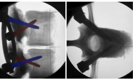
9] was selected to answer the research question asked here. Six lumbar spines (L1-L6) from calves (age, 4–5 months; mean weight, 117±12 kg) were divided into three levels, L1/L2, L3/L4 and L5/L6, so that 18 segments were available for testing. Randomly, nine samples were selected for sagittal-parallel screw placement (“P”) and nine for sagittal non-parallel (= sagittal open and closed angle) (“N”) screw place-ment (Figs. 1, 2 and 3) so that for each lumbar level, six segments where available in the P and six in the N group.
The holes were drilled with a 3-mm drill under fluoroscop-ic imaging aiming at intersegmental parallelism in the sagittal plane for the P group. For the N group, the aim was to drill the holes in 30° convergence on one side (sagittal closed angle) and in 30° divergence (sagittal open angle) on the contralateral side (Figs.2and3and Table1). Convergence of at least 60° was desired in the axial plain (Table1) in both the N and P groups. Attention was paid not to violate the pedicle during drilling. Subsequently, 6/45-mm pedicle screws were inserted and connected with a 6-mm rod. Computer tomography was used to exclude intraosseous pathologies such as hematomas and to assure intrapedicular placement of all screws before biomechanical testing (Fig.4).
Pull-out testing was performed with a universal testing machine (Zwick-Roell, Zwick GmbH, Ulm, Germany). The segments were fixed with a steel rod (16 mm) connected to the testing machine and inserted through the spinal canal (Fig.5) to avoid the influence of polymer resin fixation on pull-out strength [13]. Each end of the two rods was connected to a high-tension steel cable to allow both sides to be independent of each other (Fig. 5). Pull-out testing was initiated with a preload of 100 N and a constant dis-placement rate of 1 mm/s. The maximum pull-out force was quantified, and the type of failure was documented by two observers. Two types of failure were distin-guished: screw pull-out versus vertebral/pedicle fracture, respectively.
Statistical analyses
Descriptive statistics were used to report means, standard deviations and ranges of data, where adequate. Intergroup comparison of insertion angles and pull-out forces was done using two-tailed Student’s t-test assuming parametric data, with a p-value of <0.05 defined as statistically significant.
Results
The maximal pull-out force was similar with 5,394 N (range 4,221 N to 8,342 N) in the N group compared to 5,263 N (range 3,589 N to 7,554 N) in the P group (p=0.838), with no relevant differences if individual segments were compared (Table2).
The mean maximum pull-out forces per calf differed from 4,108 N to 7,377 N with each lumbar calf spine, providing three segments for testing. Each lumbar calf spine with its three segments was randomized to receive either a sagittal-parallel construct for L1/2, then a sagittal non-parallel construct for L3/ 4 and finally again a sagittal-parallel construct for L5/6 (P-N-P),
Fig. 1 Fluoroscopic lateral and axial images of a vertebral segment, instrumented with the standard sagittal parallel screw technique
or first a sagittal non-parallel construct for L1/2 followed by a sagittal-parallel construct for L2/3 and sagittal non-parallel con-struct for L5/6 (N-P-N). There was no difference within the lumbar calf spines with a P-N-P sequence (5,321±1,790 N) compared to an N-P-N sequence (5,335±708 N).
The reason for failure of fixation was fracture of the pedicle or the vertebra in five (56 %) cases with a sagittal-parallel screw construct with a pull-out force of 5,526±1,443 N. In the other four (44 %) cases with a sagittal-parallel screw con-struct, the pedicle screws pulled out without an osseous frac-ture with a pull-out force of 4,935 N±871 N. With sagittal non-parallel screws, a fracture of the pedicle or the vertebra occurred in six (67 %) cases (mean of maximum pull-out force 5,911 N±1,553 N), and pull-out of pedicle screws occurred in three (33 %) cases (mean of maximum pull-out force 4,360± 76 N N). In the sagittal non-parallel screw constructs, the sagittal closed angle side failed four times (6,221±1,733 N), and the sagittal open-angle side failed five times with a non-significantly lower force of 4,732±858 N (p=0.189).
Discussion
While it is well known that convergence of the pedicle screws in the axial plane adds to the biomechanical stability of the
instrumentation construct, most surgeons aim at radiographic parallelism in the sagittal plane. Often, the screws are redirected if not parallel enough in the sagittal plane for the sake of radiological aesthetics. The aim of this in vitro biome-chanical study was to investigate whether the pull-out strength of sagittal non-parallel screws is inferior, equal or superior to sagittal parallel screws. We were able to document the non-inferiority of the pull-out strength of sagittal non-parallel screws compared to parallel screws in a cadaveric calf lumbar spine model. This observation has an important intraoperative impact as it counteracts the surgeon’s intuition to place the screws absolutely parallel in the sagittal plane. Some surgeons even redirect screws to achieve radiographic parallelism in the sagittal plane, even though it is well known that redirecting screws decreases the biomechanical stability [15]. One would expect that sagittal intersegmental divergence (sagittal open-angle positioning) of the screws would add to the biomechanical stability because of better bone qual-ity near the endplates, but it was reported previously that pedicle screws placed straight forward have similar pull-out failure characteristics compared to screws placed in an upward trajectory [8]. This is in concor-dance with our observation of the non-inferiority of sagittal intersegmental closed angle screws achieving at
Fig. 2 Fluoroscopic lateral and axial images of a vertebral segment, instrumented with the alternative sagittal non-parallel screw technique
Fig. 3 Posterior view of instrumented vertebral segments with the standard sagittal parallel screw and the alternative sagittal non-parallel screw technique
least the same pull-out strength as sagittal open-angle screws.
There are several aspects to consider before directly trans-lating the results of this study to daily surgery. First, in the human spine, anatomical landmarks often limit the free choice of sagittal angulation of the screws. While intersegmental convergence (closed angle) of the screws is limited by the mechanical conflict of the head of the screw to the adjacent facet joint, intersegmental divergence (open angle) is limited by avoiding perforation of the endplates with the tips of the screws. Second, we used a calf spine model. While established in numerous previous studies on the pull-out strength of pedicle screws, the model has important limitations. The interspecies anatomy of the vertebrae is different, and the pedicles of the calf are significantly larger than human verte-bral pedicles. However, the bone quality of different calf spines is more equal than those of cadaveric human spines of different ages. In this study, computer tomography was
used to rule out intraosseous pathologies such as hematomas that could influence the fixation strength of the screw and introduce bias. Further, the spinal segments were randomly assigned to the different groups in order to eliminate selection bias. Third, we assessed only primary stability by applying axial pull-out forces and cannot report on behavior of the different constructs with cyclic loading or angulated forces as this was not the research question of this study and is the subject of further research. Specifically, it was not the aim to
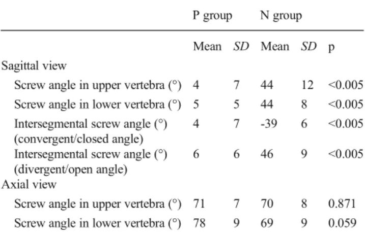
Table 1 Screw angles in the sagittal and axial plane in the sagittal-parallel (P) group and the sagittal non-sagittal-parallel screw (N) group
P group N group Mean SD Mean SD p Sagittal view
Screw angle in upper vertebra (°) 4 7 44 12 <0.005 Screw angle in lower vertebra (°) 5 5 44 8 <0.005 Intersegmental screw angle (°)
(convergent/closed angle)
4 7 -39 6 <0.005 Intersegmental screw angle (°)
(divergent/open angle)
6 6 46 9 <0.005 Axial view
Screw angle in upper vertebra (°) 71 7 70 8 0.871 Screw angle in lower vertebra (°) 78 9 69 9 0.059
Fig. 4 Three-dimensional reconstruction of computer tomography im-ages of a vertebral segment, instrumented with sagittal non-parallel screws
Fig. 5 Setup for biomechanical testing of pull-out strengths with a universal testing machine (Zwick-Roell, Zwick GmbH, Ulm, Germany). The segments are fixed with a steel rod (16 mm) connected to the testing machine and inserted through the spinal canal. Each end of the two rods is connected to a high-tension steel cable
Table 2 Maximal pull-out forces of the sagittal-parallel (P group) and sagittal non-parallel screws (N group) in Newton
P group N group
Mean SD Mean SD p Overall pull-out force 5,263 1,193 5,394 1,453 0.838 Level 1/2 5,559 1,728 5,376 922
Level 3/4 5,495 484 5,670 2,317 Level 5/6 4,735 1,357 5,135 1,419
show the superiority but the non-inferiority of sagittally not perfectly parallel screws to counteract the surgeon’s intuition of desiring radiographic parallelism in the sagittal plane.
Conclusion
Non-parallel pedicle screws in the sagittal plane have at least equal initial fixation strength compared to parallel pedicle screws in the setting of the here performed cadaveric calf spine experiments.
Presentation at conferences None. Conflicts of interests None.
Source of funding Internal institutional resources.
References
1. Barber JW, Boden SD, Ganey T, Hutton WC (1998) Biomechanical study of lumbar pedicle screws: does convergence affect axial pullout strength? J Spinal Disord 11:215–220
2 . C h a n g M - C , K a o H - C , Yi n g S - H , L i u C - L ( 2 0 1 3 ) Polymethylmethacrylate augmentation of cannulated pedicle screws for fixation in osteoporotic spines and comparison of its clinical results and biomechanical characteristics with the needle injection method. J Spinal Disord Tech 26:305–315
3. Chaudhari R, Zheng X, Wu C, Mehbod AA, Transfeldt EE, Winter RB (2011) Effect of number of fusion levels on S1 screws in long fusion construct in a calf spine model. Spine 36:624–629
4. Costa F, Villa T, Anasetti F, Tomei M, Ortolina A, Cardia A, La Barbera L, Fornari M, Galbusera F (2013) Primary stability of pedicle screws depends on the screw positioning and alignment. Spine J 13: 1934–1939
5. DeWald CJ, Stanley T (2006) Instrumentation-related complications of multilevel fusions for adult spinal deformity patients over age 65: surgical considerations and treatment options in patients with poor bone quality. Spine 31:S144–151
6. Hadjipavlou AG, Nicodemus CL, Al-Hamdan FA, Simmons JW, Pope MH (1997) Correlation of bone equivalent mineral density to pull-out resistance of triangulated pedicle screw construct. J Spinal Disord 10:12–19
7. Halvorson TL, Kelley LA, Thomas KA, Whitecloud TS, Cook SD (1994) Effects of bone mineral density on pedicle screw fixation. Spine 19:2415–2420
8. Higashino K, Kim J-H, Horton WC, Hutton WC (2012) A biome-chanical study of two different pedicle screw methods for fixation in osteoporotic and nonosteoporotic vertebrae. J Surg Orthop Adv 21: 198–203
9. Kang DG, Lehman RA, Bevevino AJ, Gaume RE, Purcell RL, Dmitriev AE, Lenke LG (2014) Pedicle screw "hubbing" in the immature thoracic spine: a biomechanical and micro-computed to-mography evaluation. J Pediatr Orthop
10. Kim Y-Y, Choi W-S, Rhyu K-W (2012) Assessment of ped-icle screw pullout strength based on various screw designs and bone densities—an ex vivo biomechanical study. Spine J 12:164–168
11. Liu D, Shi L, Lei W, Wei M-Q, Qu B, Deng S-L, Pan X-M (2013) Biomechanical comparison of expansive pedicle screw and polymethylmethacrylate-augmented pedicle screw in osteoporotic synthetic bone in primary implantation: an Experimental study. J Spinal Disord Tech
12. Paik H, Dmitriev AE, Lehman RA, Gaume RE, Ambati DV, Kang DG, Lenke LG (2012) The biomechanical effect of pedicle screw hubbing on pullout resistance in the thoracic spine. Spine J 12:417– 424
13. Pfeiffer M, Gilbertson LG, Goel VK, Griss P, Keller JC, Ryken TC, Hoffman HE (1996) Effect of specimen fixation method on pullout tests of pedicle screws. Spine 21:1037–1044
14. Ponnusamy KE, Iyer S, Gupta G, Khanna AJ (2011) Instrumentation of the osteoporotic spine: biomechanical and clinical considerations. Spine J 11:54–63
15. Stauff MP, Freedman BA, Kim J-H, Hamasaki T, Yoon ST, Hutton WC (2014) The effect of pedicle screw redirection after lateral wall breach—a biomechanical study using human lumbar vertebrae. Spine J 14:98–103