HAL Id: tel-02984773
https://tel.archives-ouvertes.fr/tel-02984773
Submitted on 1 Nov 2020HAL is a multi-disciplinary open access archive for the deposit and dissemination of sci-entific research documents, whether they are pub-lished or not. The documents may come from teaching and research institutions in France or abroad, or from public or private research centers.
L’archive ouverte pluridisciplinaire HAL, est destinée au dépôt et à la diffusion de documents scientifiques de niveau recherche, publiés ou non, émanant des établissements d’enseignement et de recherche français ou étrangers, des laboratoires publics ou privés.
The chemokine CCL5, a key regulator of
neuroinflammation and type 2 diabetes associated with
diet-induced obesity
Katharina Stobbe
To cite this version:
Katharina Stobbe. The chemokine CCL5, a key regulator of neuroinflammation and type 2 diabetes associated with dietinduced obesity. Molecular biology. COMUE Université Côte d’Azur (2015 -2019), 2019. English. �NNT : 2019AZUR6010�. �tel-02984773�
La chimiokine CCL5, un régulateur clé
de la neuroinflammation et du diabète
de type 2 associé à l’obésité
nutritionnelle
Katharina STOBBE
Institut de Pharmacologie Moléculaire et Cellulaire
Présentée en vue de l’obtentiondu grade de docteur en Sciences de la Vie et de la Santé
d’Université Côte d’Azur
Dirigée par : Dr Carole Rovère
Soutenue le : 31 Octobre 2019
Devant le jury, composé de :
Pr Jacques NOEL, Président
IPMC-Université Côte d’Azur, Valbonne, France
Dr Nicolas CHARTREL, Rapporteur
DC2N-Université de Rouen, France
Pr Denis RICHARD, Rapporteur
IUCPQ-Université Laval, Québec, Canada
Dr Annabelle REAUX-LE GOAZIGO, Examinatrice
Institut de la Vision-Sorbonne Université, Paris, France
Dr Ariane SHARIF, Examinatrice
JPArc-Université de Lille, France
Dr Carole ROVERE, Directrice de thèse
IPMC-Université Côte d’Azur, Valbonne, France
Dr Jean-Louis NAHON, Co-directeur de thèse
IPMC-Université Côte d’Azur, Valbonne, France
La chimiokine CCL5, un régulateur clé de la
neuroinflammation et du diabète de type 2
associé à l’obésité nutritionnelle
Jury :
Président du Jury
Jacques Noël, Professeur, IPMC-Université Côte d’Azur, Valbonne, France
Directeurs de Thése
Carole Rovère, Docteur, IPMC-Université Côte d’Azur, Valbonne, France Jean-Louis Nahon, Docteur, IPMC-Université Côte d’Azur, Valbonne, France
Rapporteurs
Nicolas Chartrel, Docteur, DC2N-Université de Rouen, France Denis Richard, Professeur, IUCPQ-Université Laval, Québec, Canada
Examinatrices
Annabelle Réaux-Le Goazigo, Docteur, Institut de la Vision-Sorbonne Université, Paris, France
Résumé
Le mode de vie occidental favorise le développement de l’obésité et du diabète de type 2. Le lien entre l'obésité et le diabète de type 2 est bien établi au niveau épidémiologique. Toutefois, ce lien reste encore mal défini au niveau étiologique. Les marqueurs du diabète de type 2, tels que l’hyperglycémie et la résistance à l'insuline et à la leptine, peuvent résulter d'un état pro-inflammatoire chronique du tissu adipeux, associé à une sécrétion de cytokines (IL-1β, IL-6, TNF-a) et chimiokines (RANTES/CCL5, MCP-1/CCL2) diabétogènes. Ces médiateurs pro-inflammatoires sécrétés seraient les promoteurs de l’inflammation systémique et de la neuroinflammation dans l’hypothalamus, région importante du cerveau, qui contient des réseaux neuronaux impliqués dans le contrôle du métabolisme énergétique et du comportement alimentaire.
Nous nous sommes intéressés à l’impact de régimes riches en lipides sur le développement de l'obésité nutritionnelle et les paramètres métaboliques associés, d’autre part sur la réponse inflammatoire dans l'hypothalamus et le tissu adipeux. Nous nous sommes focalisés sur le rôle de la chimiokine CCL5 et de son récepteur CCR5. Notre hypothèse est que la chimiokine CCL5 régule l’activité des neurones de l’hypothalamus impliqués dans le contrôle de l’homéostasie énergétique et de l’homéostasie glucidique. Nous proposons d’étudier le rôle de la chimiokine CCL5 dans le contrôle de la balance énergétique, l’obésité, le diabète de type 2 et la neuropathie périphérique diabétique, une des complications diabétiques les plus fréquentes, affectant jusqu'à la moitié des patients atteints de diabète de type 2.
Dans ma thèse, nous avons testé les effets à long terme (16 semaines) du régime hyperlipidique apportant 40% de lipides, comparé au régime standard (contenant 5% de lipides), sur le développement de l'obésité chez les souris invalidées pour le gène de CCL5 (souris CCL5-/-) ou le gène de son récepteur CCR5 (CCR5-/-) en
comparaison aux souris sauvages (contrôles). Dans un premier temps, nous avons confirmé la présence de CCL5 et de son récepteur CCR5 dans l'hypothalamus par hybridation in situ (technologie RNAScope®) chez les souris contrôles. Nous avons alors montré que les souris CCL5-/- et CCR5-/- semblent plus résistantes à l’obésité et
avoir une homéostasie du glucose moins perturbée que les souris sauvages. Pour évaluer l'implication du CCL5 dans la douleur neuropathique associée au diabète, la sensibilité à la douleur thermique des souris CCL5-/- et CCR5-/- a été mesurée chez
chaque groupe de souris. Les souris CCL5-/- sous régime hyperlipidique semblent
moins sensibles à la douleur thermique que les souris contrôles. De plus, les souris CCL5-/- semblent présenter une expression génique des neuropeptides
hypothalamiques modifiée par rapport aux souris contrôles.
Nos résultats suggèrent d’une part que l'absence de CCL5 et de son récepteur CCR5 protège contre le développement de l'obésité et le diabète de type 2, d’autre part que l'absence du CCL5 abolit la sensibilité accrue à la douleur thermique observée sous régime hyperlipidique. Ainsi, la cascade de signalisation CCL5/CCR5 pourrait représenter une nouvelle cible thérapeutique pour lutter contre l’obésité.
Mots clés : Obésité, Comportement alimentaire, Homéostasie glucidique, Régimes hyperlipidiques, Inflammation, Cytokine, Chimiokine, Signalisation de CCL5, Hypothalamus, Neuropeptides.
Abstract
Obesity is defined by the excessive accumulation of body fat and accompanied by chronic low-grade inflammation of peripheral metabolic tissues, especially of adipose tissue. Adipocytes secrete inflammatory mediators such as cytokines and chemokines, which can act at the cerebral level and modulate neuronal activity. The hypothalamus is an important region of the brain, which contains neural networks involved in the control of energy metabolism and feeding behavior. Emerging evidence indicates that inflammation occurs also at the level of the hypothalamus. Our recent results showed that the chemokines can be involved in the deregulation of energy homeostasis.
CCL5 is a chemoattractant cytokine well known for its role in cerebral and peripheral inflammation. Together with one of its cognate receptors, CCR5, it also contributes to neural function and diseases such as obesity, type 2 diabetes and neuropathic pain. We were interested in the inflammatory response of the hypothalamus and different adipose tissues to high-fat diet and its role in the development of diet-induced obesity. In particular, we are focusing on the role of the previously identified chemokine CCL5 and its receptor CCR5, in the central inflammation associated with the deregulation of energy metabolism and the pathogenesis of obesity.
In this study, we tested the long-term effects of an obesogenic high-fat or standard diet on the development of obesity in adult CCL5-/-, CCR5-/- and wild-type mice. After
16 weeks of feeding, animals were sacrificed and peripheral and cerebral tissues collected. Metabolic parameters, locomotor activity, expression levels of pro-inflammatory mediators and peptides involved in feeding behavior were measured. We discovered that both CCL5-/- and CCR5-/- mice seem to be protected from
weight gain and the associated impairment of glucose metabolism compared to WT mice. To evaluate the implication of CCL5 in neuropathic pain associated with diabetes, thermal pain sensitivity of CCL5-/- and CCR5-/- mice was measured in both
conditions. Remarkably, in high fat diet condition, CCL5-/- mice displayed higher
tolerance to heat pain compared to control mice.
Furthermore, CCL5-/- mice show a different expression pattern of inflammatory
markers and hypothalamic neuropeptides compared to control mice. In addition, we used RNA in situ hybridization (RNAScope® Technology) to verify the cellular localization of CCL5 and its receptor CCR5 in the hypothalamus of wild-type mice. Our results indicate that the absence of CCL5 and its receptor CCR5 protects against the development of obesity and type 2 diabetes and the absence of CCL5 abolishes the increased thermal pain sensitivity observed under high fat diet challenge. Thus, the CCL5 signaling cascade could represent a new putative target for the development of therapeutic strategies.
Keywords: Obesity, Feeding behavior, Glucose homeostasis, High fat diet, Inflammation, Cytokine, Chemokine, CCL5 signaling, Hypothalamus, Neuropeptides.
University Côte d’Azur – UFR Sciences
Ecole Doctorale de Sciences de la Vie et de la Santé
THESIS
Submitted for the degree of
Doctor of Philosophy in Sciences
by the University of Côte d’Azur
Specialty: Cellular and Molecular Interactions
Presented by
Katharina Stobbe
THE CHEMOKINE CCL5, A KEY
REGULATOR OF NEUROINFLAMMATION
AND TYPE 2 DIABETES ASSOCIATED WITH
DIET-INDUCED OBESITY
Thesis directed by Dr Carole Rovere
Defended on the 31 October 2019
Before the Jury: Pr Jacques NOEL
Dr Nicolas CHARTREL Pr Denis RICHARD
Dr Annabelle REAUX-LE GOAZIGO Dr Ariane SHARIF Dr Carole ROVERE Dr Jean-Louis NAHON Président Rapporteur Rapporteur Examinatrice Examinatrice Directrice de thèse Co-directeur de thèse
Acknowledgments
I would like to kindly thank the jury members, Professor Jaques Noël for accepting to be the president of my committee, as well as the reporters, Dr. Nicolas Chartrel and Professor Denis Richard, for taking the time to evaluate my manuscript. Furthermore, I would like to kindly thank Dr. Annabelle Reaux-Le Goazigo and Dr. Ariane Sharif who agreed to be my examiners, and evaluate my work. I would also like to thank warmly my co-director and team leader Dr. Jean Lois Nahon to have welcomed me to be part of his team and gave me access to the laboratory and research facilities.
I would like to thank LABEX SIGNALIFE and the University of Côte d’Azur for their finanancial support to undertake this project.
I would like to express my sincere gratitude to Dr. Carole Rovere, my thesis director, who has accepted to be my thesis supervisor along this journey and warmly and kindly welcomed me to be part of this team. Thank you for all the opportunities that you have given me, from being part of this exciting project, to the many national and international conferences that I have had the opportunity to participate and learn from as well as all the great memories that I have made. Thank you equally for all the support and patience, especially in the last and most stressful part of my PhD, as well as for all the help with French administration that I had trouble adjusting to. I am also grateful for the possibility you gave me to learn and improve my French within these years and who made me discover and raise my mood with many French delights.
Thank you equally to the other members of this team to François and Nadege, for their support and help throughout these years, to have been there with an open ear for my questions. Thank you Nadege, also for not letting me forget my german language.
In this sense, I would like also to thank all the BTS students Myriam, Melanie and Léa, whom I had the pleasure to meet and with whom I gained some first experiences in tutoring. Thank you also for all the help with my experience during your stay.
A special gratitude goes out to Celine, who as my colleague and friend taught me many things, not limited to scientific experiments. I would like to thank you for always being there for me, supporting me and believing in me and cheering me up in difficult moments.
To Clara, thank you for your help with some last experiments but also with your informatics skills that helped me a lot with formatting this manuscript. I am
sorry I did not have more time to get to know you better. I wish you all the best for your PhD.
A special mention to Nicolas Blondeau for the stimulating discussions, his help with the project and his humor.
My special thanks are extended to the staff of the animal facility and the imaging platform for their help and support throughout these years.
I would also like to thank all the other people in the IPMC, who I had the pleasure to meet, talk and share moments of laughter with and for making my years of PhD pleasant everyday.
To Ahmed, for your help with experiments when I needed a hand, for your encouragement and the stimulating discussions.
To Wejdane, Alex, Ligia and Paula who either from the beginning or only recently have shared my journey as fellow PhD students, for sharing special moments in the institute during lunch or coffe breaks, or outside the institute and for sharing the stress of writing and the last few months of PhD with me and always being there with an open ear and patience.
A special gratitude goes equally out to Maria, whom I have had the pleasure to get to know through the SIGNALIFE program. Thank you for becoming my friend and being there for me during the amazing and the less amazing moments and for listening to me rambling and your encouraging words and chocolate wine.
I am also thankful for many of the amazing SIGNALIFE people, who have made this journey special and worthwile.
With a special mention to my dear friends, Doreen, Theodora, and Neil for always being there for me and believing in me. Thank you for your friendship and your support despite the distance and your positivity and kind words of encouragement.
Last but not the least, I would like to thank my family, in particular my parents, without whom all this would not be possible.
List of Publications
Original articles
2019 Stobbe K., Cansell C., Negm A., Le Thuc O., Sanchez C., Colson C., Pisani D., Gautier N., Rékima S., Nédélec E., Devaux N., Brau F., Bénani A., Blondeau N., Amri Z., Noël J., Nahon J.L., Rovère C. Role Of CCL5 Signaling In The Establishment Of Diet-Induced Obesity And Type 2. In preparation.
2019 Negm A., Stobbe K., Deval E., Linguiglia E., Rovère C., Noël J. Lipid-rich diet consumption induce thermal pain through the activation of ASIC3 channels. In preparation.
2019 Cansell C.,Stobbe K., Le Thuc O., Mosser CA., Ben-Fradj S., Leredde J., Lebeaupin C.,Debayle D., Fleuriot L., Brau F., Devaux N., Benani A., Audinat E., Blondeau N., Nahon JL., Rovère C. Post-prandial hypothalamic inflammation displays an exacerbated response to a single high-fat meal and involves both GFAP-positive and microglial cells. Submitted.
2016 Le Thuc O., Cansell C., Bourourou M., Denis R.G.P., Stobbe K., Devaux N., Guyon A., Cazareth J., Heurteaux C., Rostène W., Luquet S., Blondeau N., Nahon JL., Rovère C. Central CCL2 signaling onto MCH neurons mediates metabolic and behavioral adaptation to inflammation. EMBO Rep. Oct 12. pii: e201541499. IF 2015/16: 7,739
Reviews
2019 Le Thuc O., Stobbe K., Rovère C. Inflammation hypothalamique et deregulation de la balance énergétique : rôle des chimiokines. La lettre des Neurosciences 56, 20-23.
2017 Le Thuc O., Stobbe K., Cansell C., Nahon JL., Blondeau N., Rovère C. Hypothalamic Inflammation and Energy Balance Disruptions: Spotlight on Chemokines. Front. Endocrinol. doi.org/10.3389/fendo.2017.00197. IF 2017: 3,675
Participation in National and
International Conferences
Oral communications
July 2019 Oral communication at the 7th International Mediterranean
Neuroscience Conference in Marrakech, Morocco.
May 2019 Oral communication at the international colloquium “NeuroFrance 2019” in Marseille, France.
September 2018 Oral communication and prize for best oral communication at the 2nd thematic seminar of the french Society of
Experimental Neuroendocrinology (SNE), Paris, France. May 2018 Oral communication and prize for best oral communication
at the JEDN 2018, Nice, France.
September 2017 Oral communication at the 42nd conference of the SNE, Dijon, France.
Poster presentations
July 2018 Poster presentation at the 9th International Congress of Neuroendocrinology (ICN), Toronto, Canada.
Invited chair of symposium "The Gut-Brain Axis and Metabolism" at the 9th ICN, Toronto, Canada.
Member of the young ambassador's committee for the organization of the 9th ICN.
May 2017 Poster presentation at the Keystone Symposium Z5 “Neuronal Control of Appetite, Metabolism and Weight”, Copenhagen, Denmark.
November 2016 Poster presentation and best poster prize at the LABEX SIGNALIFE Student Conference/Retreat, Nice, France. October 2016 Poster presentation and best poster prize at the 41st
Page | 1
Table of Contents
TABLE OF CONTENTS ... 1 ABBREVIATIONS ... 4 LIST OF FIGURES ... 7 LIST OF TABLES ... 9 INTRODUCTION ... 111. HYPOTHALAMUS AND THE REGULATION OF ENERGY BALANCE ... 11
1.1. THE HYPOTHALAMUS ... 14 1.1.1. The Arcuate Nucleus ... 15 1.1.2. The Ventromedial Hypothalamic Nucleus ... 22 1.1.3. The Paraventricular Nucleus ... 23 1.1.4. The Lateral Hypothalamus ... 24 1.1.5. The Dorsomedial Nucleus ... 25 1.1.6. The Caudal Brainstem ... 26
1.2. BRAIN REGULATION OF ENERGY EXPENDITURE ... 27
2. OBESITY ... 28
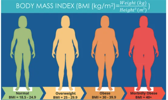
2.1. DEFINITION, FACTS AND GENERALITIES ... 28
2.2. GENETIC CAUSES OF OBESITY ... 30
2.3. OBESITY AND INFLAMMATION ... 33 2.3.1. Inflammation ... 34 2.3.2. Peripheral Inflammation in Obesity ... 35 2.3.3. Central Inflammation in Obesity ... 43 2.3.4. Hypothalamic Gliosis ... 50 3. GLUCOSE HOMEOSTASIS ... 51
3.1. CENTRAL REGULATION OF GLUCOSE HOMEOSTASIS ... 52
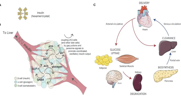
3.2. PANCREATIC REGULATION OF GLUCOSE HOMEOSTASIS ... 53
3.2.1. Glucagon ... 56 3.2.2. Somatostatin ... 57 3.2.3. PP Peptide ... 57 3.2.4. Insulin ... 58 3.2.4.1. Insulin Receptor Expression ... 58 3.2.4.2. Peripheral Insulin Action ... 59 3.2.4.3. Central Insulin Action ... 63 3.2.5. Gut Hormones: Incretins ... 65 3.3. DIABETES MELLITUS (DM) ... 66 3.3.1. Type 1 Diabetes Mellitus (T1DM) ... 69 3.3.2. Type 2 Diabetes Mellitus (T2DM) ... 73 3.3.2.1. Different Stages of T2DM ... 74 3.3.2.2. Pathophysiology ... 76 4. NEUROPATHIC PAIN ... 83 4.1. DIABETIC NEUROPATHY (DN) ... 86
Page | 2
4.2. PATHOPHYSIOLOGICAL MECHANISMS ... 88
5. CHEMOKINES ... 91
5.1. STRUCTURE ... 92
5.2. CHEMOKINE RECEPTORS ... 94
5.3. LIGAND‐RECEPTOR INTERACTION AND SIGNALING ... 95
5.4. FUNCTION ... 95
5.1. CHEMOKINES FUNCTION IN THE CNS ... 98
5.2. ROLE OF CHEMOKINES IN OBESITY AND COMORBIDITIES ... 100
5.2.1. CCL5/RANTES ... 101 5.2.1.1. Expression ... 103 5.2.1.2. Receptor Interaction and Synergistic Effects With Other Chemokines ... 104 5.2.2. Receptors of CCL5 ... 107 5.2.2.1. C‐C Motif Chemokine Receptor 1 (CCR1) ... 107 5.2.2.2. C‐C Motif Chemokine Receptor 3 (CCR3) ... 107 5.2.2.3. G Protein‐coupled Receptor 75 (GPR75) ... 108 5.2.2.4. Duffy Antigen Receptor For Chemokines (DARC) ... 109 5.2.2.5. Chemokine‐binding Protein 2 (CCBP2) ... 110 5.2.2.6. C‐C Motif Chemokine Receptor 5 (CCR5) ... 111 5.2.2.6.1. Signaling ... 113 5.2.2.6.2. Regulation ... 114 5.2.3. Role of CCL5/CCR5 in Peripheral Inflammation in Obesity ... 115 5.2.4. Role of CCL5/CCR5 Signaling in Insulin Signaling and Diabetes ... 123 5.2.5. Role of CCL5/CCR5 Signaling in Neuropathic Pain ... 128 5.2.6. Role of CCL5 Signaling in NASH/NAFLSD ... 129 5.2.7. Role of CCL5/CCR5 Signaling in HIV ... 130 OBJECTIVES ... 133 EXPERIMENTAL STRATEGY ... 135
MATERIALS AND METHODS ... 139
1. ANIMAL PROCEDURES ... 139
2. BLOOD AND TISSUE COLLECTION ... 139
3. DIETS ... 140
4. GLUCOSE TOLERANCE TEST (GTT)/INSULIN TOLERANCE TEST (ITT) ... 141
5. MICROCOMPUTED TOMOGRAPHY ANALYSIS ... 142
5.1. ADIPOSE TISSUE HISTOLOGY ... 142
5.2. PLASMA TRIGLYCERIDE (TG) AND GLYCEROL CONTENTS ... 142
6. RNA ISOLATION AND QUANTITATIVE QPCR ... 143
7. CYTOKINES, CHEMOKINES AND HORMONES QUANTIFICATION ... 145
8. IMMUNOHISTOCHEMICAL ANALYSIS ... 145
9. SINGLE‐MOLECULE RNA IN SITU HYBRIDIZATION ... 146
10. STEREOTAXIC CANNULA GUIDE PLACEMENT ... 146
10.1. COMPOSITION OF SOLUTIONS FOR ICV INFUSION ... 147
10.2. CHRONIC ICV INFUSIONS ... 148
11. HARGREAVES’ TEST OF THERMAL PAIN SENSITIVITY ... 150
Page | 3
RESULTS ... 151
1. THE EFFECTS OF LIPID NATURE ON THE DEVELOPMENT OF OBESITY ... 151
1.1. THE EFFECT OF LIPID NATURE ON THE DEVELOPMENT OF OBESITY AND FOOD INTAKE ... 151
1.2. THE EFFECTS OF LIPID NATURE ON HYPOTHALAMIC NEUROPEPTIDE EXPRESSION ... 154
1.3. THE EFFECTS OF LIPID NATURE ON GLUCOSE HOMEOSTASIS ... 156
1.4. THE EFFECT OF LIPID NATURE ON HFD‐INDUCED CENTRAL INFLAMMATION ... 157
1.5. THE EFFECTS OF LIPID NATURE ON THERMAL PAIN SENSITIVITY ... 159
2. ROLE OF CHEMOKINE CCL5 IN THE DEVELOPMENT OF OBESITY ... 160
2.1. CHEMOKINE CCL5 DEFICIENCY REDUCES HFD‐INDUCED BW GAIN AND FOOD INTAKE. ... 160
2.2. CHEMOKINE CCL5 HAS A NEUROMODULATORY EFFECT ON HYPOTHALAMIC NEURONS ... 163
2.3. CHEMOKINE CCL5 DEFICIENCY IMPROVES GLUCOSE METABOLISM IN OBESE MICE ... 166
2.4. THE EFFECT OF CCL5 ON CENTRAL INFLAMMATION AND HYPOTHALAMIC GLIOSIS ... 170
2.5. THE EFFECT OF CCL5 ON PERIPHERAL AND SYSTEMIC INFLAMMATION ... 174
2.6. CCL5 DEFICIENCY PARTLY PROTECTS FROM HFD‐INDUCED INCREASE IN NEUROPATHIC PAIN SENSITIVITY ... 178
2.7. HYPOTHALAMIC EXPRESSION OF CCL5 AND CCR5 ... 184
3. ROLE OF CHEMOKINE RECEPTOR CCR5 IN THE DEVELOPMENT OF OBESITY ... 187
3.1. CHEMOKINE RECEPTOR CCR5 DEFICIENCY PARTLY PROTECTS AGAINST HFD‐INDUCED DEVELOPMENT OF OBESITY. ... 187
3.2. CHEMOKINE RECEPTOR CCR5 HAS A NEUROMODULATORY EFFECT ON HYPOTHALAMIC NEURONS ... 189
3.3. CCR5 AFFECTS GLUCOSE METABOLISM IN OBESE MICE ... 192
3.4. THE EFFECT OF CCR5 ON CENTRAL INFLAMMATION AND HYPOTHALAMIC GLIOSIS ... 196
3.5. THE EFFECT OF CCR5 DEFICIENCY ON SYSTEMIC INFLAMMATION ... 199
3.6. THE EFFECT OF CCR5 DEFICIENCY ON DIABETES‐ASSOCIATED NEUROPATHIC PAIN ... 201
3.7. THE EFFECT OF CCL5 AND CCR5 ON SUBCUTANEOUS AT MORPHOLOGY ... 202
3.8. THE EFFECT OF CHRONIC ICV INFUSIONS OF DIFFERENT CONCENTRATIONS OF CCL5 ... 204
DISCUSSION ... 210
1. THE EFFECT OF LIPID NATURE ON DIET‐INDUCED OBESITY ... 210
2. THE ROLE OF CHEMOKINE CCL5 IN THE DEVELOPMENT OF DIET‐INDUCED OBESITY ... 212
3. CCL5 AND CCR5 MRNA EXPRESSION IN SUBPOPULATION OF CELLS IN THE NUCLEI OF THE HYPOTHALAMUS .. 214
4. THE EFFECT OF CCL5/CCR5 SIGNALING ON HFD‐INDUCED BODY WEIGHT GAIN AND FEEDING BEHAVIOUR ... 217
5. HYPOTHALAMIC NEUROMODULATION BY CCL5/CCR5 SIGNALING ... 219
6. THE EFFECT OF CCL5/CCR5 SIGNALING ON GLUCOSE HOMEOSTASIS ... 223
7. THE ROLE OF CCL5/CCR5 SIGNALING IN HFD‐INDUCED CHANGES IN ADIPOSE TISSUE ... 235
8. THE ROLE OF CCL5/CCR5 SIGNALING IN HFD‐INDUCED INFLAMMATION ... 236
9. THE ROLE OF CCL5/CCR5 SIGNALING IN THERMAL PAIN SENSITIVITY ... 243
CONCLUSION & PERSPECTIVES ... 249
REFERENCES ... 252
Page | 4
Abbreviations
2-DG 2-deoxy-D-glucose 3V third ventricle 5-HT serotonin or, 5-hydroxytryptamine 7TM seven transmembrane domain ACTH adrenocorticotropic hormoneACKR1 “atypical” chemokine receptor 1
AD Alzheimer’s disease AGE advanced glycation end
products
AgRP agouti-related peptide AMPK 5' adenosine
monophosphate-activated protein kinase
pAMPK phosphorylated AMPK ANS autonomic nervous system ARC arcuate nucleus
ASICs acid-sensing ion channels
AT adipose tissue
ATP adenosine triphosphate BAT brown adipose tissue BBB blood-brain-barrier
BNDF brain-derived neurotrophic factor
BMI body mass index
BW body weight
CART cocaine- and
amphetamine-regulated transcript CCBP-2 chemokine-binding protein 2 CCK cholecystonkinin cCKRs “conventional” chemokine receptors CCL5 CC-chemokine ligand 5; RANTES, “Regulated upon activation, normal T-cell expressed and secreted” CGI-58 comparative gene
identification-58 protein CGRP calcitonin gene-related
peptide
CNS central nervous system
CRH corticotrophin-releasing hormone
CTLA-4 cytotoxic T lymphocyte-associated antigen-4 gene DAG diacylglycerol
DARC “duffy antigen receptor for chemokines” also known under the acronym “atypical chemokine receptor 1” (ACKR1) DCs dendritic cells
DHA docosahexaenoic acid DIO diet-induced obesity DM diabetes mellitus DMN dorsomedial nucleus DN diabetic neuropathy DNL de novo lipogenesis DPN length-dependent diabetic polyneuropathy/distal symmetric polyneuropathy/diabetic polyneuropathy DPP4 dipeptidyl peptidase 4 DRG dorsal root ganglia
EAE experimental autoimmune encephalomyelitis
EE energy expenditure
eNOS endothelial nitric oxide synthase
ER endoplasmic reticulum ERa estrogen receptor alpha FFAs free fatty acids
FAs fatty acids
FOXO1 factor Forkhead box O1 GABA γ-aminobutyric acid GAD6 glutamate decarboxylase GAG glycosaminoglycans GDM gestational diabetes
mellitus
GFAP glial fibrillary acidic protein
GHSR growth hormone
secretagogue receptor GIP gastric inhibitory
polypeptide
GIT gastrointestinal tract GLP-1 glucagon-like peptide 1 GLUT glucose transporter
Page | 5 GS glycogen synthase GPCR G protein-coupled receptor GPR103 G protein-coupled receptor 103 GPR75 G protein-coupled receptor 75 GRK G protein-coupled receptor kinase
GTT glucose tolerance test iGTT intraperitoneal GTT oGTT oral GTT
HbA1c increased glycated hemoglobin A1c HFD high fat diet
HPA hypothalamic-pituitary-adrenal axis HPT hypothalamic-pituitary-thyroid axis IA-2 insulinoma-associated protein 2
IASP International Association for the Study of Pain
Iba1 ionized calcium binding adaptor molecule 1 ICV Intracerebroventricular IDF International Diabetes
Federation
IFG impaired fasting glucose IFN interferons
IFN-γ interferon γ
IGT impaired glucose tolerance IGF insulin-like growth factor IKKβ inhibitor of κB kinase β
IL interleukin IL-1β interleukin-1β IL-6 interleukin-6 IL-8 interleukin-8 IL-10 interleukin-10 IP intraperitoneal IR Insulin receptor
IRS insulin receptor substrate IRTK insulin receptor tyrosine
kinase
ITT insulin tolerance test JAK janus kinase
JNK c-Jun N-terminal kinase KATP ATP-dependent potassium
channels KO knockout LDL low-density lipoproteins LH lateral hypothalamus LPS lipopolysaccharide β-LPH β-lipotropin
LOX1 Lectin-like oxidized LDL receptor 1
MAPK mitogen-activated protein kinase MBH mediobasal hypothalamus MCH melanin-concentrating hormone MCR melanocortin receptor MC3R melanocortin receptor 3 MC4R melanocortin receptor 4 MCHR1 melanin-concentrating hormone receptor 1 ME median eminence MHC major histocompatibility complex
MRI magnetic resonance
imaging
MS multiple sclerosis
a-MSH a-melanocyte-stimulating hormone
mTOR mammalian target of rapamycin
NASH non-alcoholic liver steatosis NAFLD non-alcoholic fatty liver
disease
NF-κB nuclear factor kappa-light-chain-enhancer of
activated B cells NK natural killer cells NKT natural killer T cells
NO nitric oxide
NOD non-obese diabetic (mouse model)
NPY neuropeptide Y
NTS nucleus tractus solitaries; nucleus of the solitary tract ob/ob leptin-deficient mice PAI-1 plasminogen activator 1 PBN parabrachial nucleus PC pro-hormone convertases PI3K phosphatidylinositol 3-kinase PKC protein kinase C PLC phospholipase C PMN polymorphonuclear neutrophils
PNS peripheral nervous system PNL partial sciatic nerve ligation
Page | 6 POMC proopiomelanocortin
PP pancreatic polypeptide PPARγ peroxisome
proliferator-activated receptor γ PUFA polyunsaturated fatty acid PVN paraventricular nucleus PYY peptide YY
RANTES Regulated upon Activation Normal T cells Expressed and Secreted
RAGE receptor for advanced glycation end products ROS reactive oxygen species SAA serum amyloid A
SD standard diet
SDF-1 stromal cell-derived factor 1
SFAs saturated fatty acids SF-1 steroidogenic factor-1 SNS sympathetic nervous
system
SOCS3 suppressor of cytokine signaling-3
SREBP1 sterol regulatory element-binding protein
STATs signal transducer and activator of transcription STAT3 signal transducer and
activator of transcription-3
T1DM type 1 diabetes mellitus T2DM type 2 diabetes mellitus
TG triglycerides
TGF-a tumor growth factor-a TGF-β tumor growth factor β TLR4 toll-like receptor 4 TRH thyrotropin-releasing
hormone
TRPA1 transient receptor potential cation channel subfamily A member 1
TRPM8 transient receptor potential cation channel subfamily M member 8
TRPV1 transient receptor potential cation channel subfamily V member 1
UCP-1 uncoupling protein-1 UPR unfolded protein response
VMH ventromedial
hypothalamus
VTA ventral tegmental area WAT white adipose tissue
WHO World Health Organization
WT wild-type
ZDF Zucker diabetic fatty rat ZNT8 zinc transporter 8
Page | 7
List of Figures
FIGURE 1:ENERGY BALANCE REGULATION BY THE CNS. ... 13
FIGURE 2:HYPOTHALAMIC CONTROL OF FOOD INTAKE BEHAVIOR. ... 16
FIGURE 3:BODY MASS INDEX. ... 29
FIGURE 4:HFD-INDUCED ALTERATIONS IN WAT ASSOCIATED WITH OBESITY AND INSULIN RESISTANCE. . 37
FIGURE 5:INFLAMMATORY EFFECTS OF HFD ON DIFFERENT TISSUES IN DIO. ... 42
FIGURE 6:GLUCOSE METABOLISM:GLUCOGENOLYSIS AND GLUCONEOGENESIS. ... 52
FIGURE 7:ANATOMICAL LOCATION OF THE HUMAN PANCREAS. ... 55
FIGURE 8:INSULIN ACTION IN THE WHOLE BODY. ... 61
FIGURE 9:INSULIN ACTION IN PERIPHERAL METABOLIC ORGANS. ... 62
FIGURE 10:INSULIN AND LEPTIN SIGNALING PATHWAY IN THE BRAIN. ... 64
FIGURE 11 :DIAGNOSTIC CRITERIA OF DM. ... 68
FIGURE 12:DIFFERENT STAGES OF T1DM. ... 70
FIGURE 13:PATHOGENESIS OF T1DM. ... 72
FIGURE 14:DIFFERENT STAGES OF T2DM DEVELOPMENT. ... 75
FIGURE 15:MECHANISM BEHIND INSULIN RESISTANCE IN T2DM. ... 76
FIGURE 16:MECHANISM OF PERIPHERAL INSULIN RESISTANCE IN METABOLIC ORGANS. ... 79
FIGURE 17 :HYPERGLYCEMIA-INDUCED GLUCOTOXICITY PATHWAYS. ... 82
FIGURE 18:DIFFERENT TYPES AND PATTERNS OF DN. ... 87
FIGURE 19:MECHANISM BEHIND THE PATHOPHYSIOLOGY OF DN. ... 90
FIGURE 20:CHEMOKINE STRUCTURE AND CHARACTERISTIC FEATURES EXEMPLIFIED BY MONOMERIC HUMAN CXCL12. ... 93
FIGURE 21 :THE SIGNALING CASCADE OF CCL5 COUPLING TO GPR75. ... 109
FIGURE 22 :STRUCTURE OF CCR57TM CHEMOKINE RECEPTOR. ... 112
FIGURE 23:SIGNAL TRANSDUCTION PATHWAYS OF CCR5. ... 114
FIGURE 24:SCHEMATIC REPRESENTATION OF OUR EXPERIMENTAL APPROACH FOR THE FIRST EXPERIMENT. ... 136
FIGURE 25:SCHEMATIC REPRESENTATION OF THE EXPERIMENTAL APPROACH EMPLOYED IN THIS STUDY. ... 137
FIGURE 26:SCHEMATIC REPRESENTATION OF UNILATERAL CANNULA PLACEMENT IN THE LATERAL VENTRICLE. ... 148
FIGURE 27:PROTOCOL OF CHRONIC INJECTIONS. ... 149
FIGURE 28:THE EFFECTS OF LIPID NATURE ON BODY WEIGHT GAIN AND LEPTINEMIA. ... 153
FIGURE 29:THE EFFECTS OF LIPID NATURE ON HYPOTHALAMIC NEUROPEPTIDE EXPRESSION. ... 155
FIGURE 30:THE EFFECTS OF LIPID NATURE ON GLUCOSE HOMEOSTASIS. ... 157
FIGURE 31:THE EFFECT OF LIPID NATURE AND COMPOSITION ON HYPOTHALAMIC INFLAMMATION AND GLIOSIS. ... 158
FIGURE 32:THE EFFECT OF LIPID NATURE AND COMPOSITION ON THERMAL PAIN SENSITIVITY AT 12W OF DIET. ... 159
FIGURE 33:EFFECT OF CCL5 DEFICIENCY ON DIO DEVELOPMENT. ... 162
FIGURE 34:EFFECT OF CCL5 DEFICIENCY ON HYPOTHALAMIC NEUROPEPTIDE EXPRESSION COMPARED BETWEEN DIETS. ... 164
FIGURE 35:EFFECT OF CCL5 DEFICIENCY ON HYPOTHALAMIC NEUROPEPTIDE EXPRESSION COMPARED BETWEEN GENOTYPES. ... 165
Page | 8
FIGURE 36:GLUCOSE HOMEOSTASIS PERTURBATIONS IN WT AND CCL5-/- MICE AFTER 8 AND 16 WEEKS
OF SD OR HFD. ... 167
FIGURE 37:EFFECT OF CCL5 DEFICIENCY ON HYPOTHALAMIC INSULIN SIGNALING. ... 169
FIGURE 38:EFFECT OF CCL5 DEFICIENCY ON HYPOTHALAMIC NEUROINFLAMMATION COMPARED BETWEEN DIETS. ... 171
FIGURE 39:EFFECT OF CCL5 DEFICIENCY ON HYPOTHALAMIC NEUROINFLAMMATION COMPARED BETWEEN GENOTYPES. ... 172
FIGURE 40:EFFECT OF CCL5 DEFICIENCY ON DIO ASSOCIATED HYPOTHALAMIC GLIOSIS. ... 173
FIGURE 41:EFFECT OF CCL5 DEFICIENCY ON PERIPHERAL INFLAMMATION COMPARED BETWEEN DIETS. ... 175
FIGURE 42:EFFECT OF CCL5 DEFICIENCY ON PERIPHERAL INFLAMMATION COMPARED BETWEEN GENOTYPES. ... 176
FIGURE 43:EFFECT OF CCL5 DEFICIENCY ON SYSTEMIC PERIPHERAL INFLAMMATION. ... 177
FIGURE 44:EFFECT OF CCL5 DEFICIENCY ON THERMAL PAIN SENSITIVITY IN DIO MICE. ... 178
FIGURE 45:EFFECT OF CCL5 DEFICIENCY ON INFLAMMATORY MARKER EXPRESSION IN DRG COMPARED BETWEEN DIETS. ... 180
FIGURE 46:EFFECT OF CCL5 DEFICIENCY ON INFLAMMATORY MARKER EXPRESSION IN DRG COMPARED BETWEEN GENOTYPES. ... 181
FIGURE 47:EFFECT OF CCL5 DEFICIENCY ON INFLAMMATORY MARKER EXPRESSION IN SPINAL CORDS COMPARED BETWEEN DIETS. ... 182
FIGURE 48:INFLAMMATORY MARKER EXPRESSION IN SPINAL CORDS OF DIOWT AND CCL5-/- MICE COMPARED BETWEEN GENOTYPES. ... 183
FIGURE 49:HYPOTHALAMIC EXPRESSION OF CCL5 AND RECEPTOR CCR5 IN SD-FED WT MICE. ... 185
FIGURE 50:HYPOTHALAMIC EXPRESSION OF CCL5 AND RECEPTOR CCR5 IN SD VS HFD-FED WT MICE. ... 186
FIGURE 51:EFFECT OF CCR5 DEFICIENCY ON DIO DEVELOPMENT. ... 188
FIGURE 52:CCR5 DEFICIENCY HAS A NEUROMODULATORY EFFECT ON HYPOTHALAMIC NEURONS. ... 190
FIGURE 53:CCR5 DEFICIENCY HAS A NEUROMODULATORY EFFECT ON HYPOTHALAMIC NEURONS. ... 191
FIGURE 54:GLUCOSE HOMEOSTASIS PERTURBATIONS IN WT AND CCR5-/- MICE AFTER 8 AND 16 WEEKS OF SD OR HFD. ... 193
FIGURE 55:EFFECT OF CCR5 DEFICIENCY ON HYPOTHALAMIC INSULIN SIGNALING. ... 195
FIGURE 56:EFFECT OF CCR5 DEFICIENCY ON HYPOTHALAMIC NEUROINFLAMMATION COMPARED BETWEEN DIETS. ... 197
FIGURE 57:EFFECT OF CCR5 DEFICIENCY ON HYPOTHALAMIC NEUROINFLAMMATION COMPARED BETWEEN GENOTYPES. ... 198
FIGURE 58:EFFECT OF CCR5 DEFICIENCY ON DIO ASSOCIATED HYPOTHALAMIC GLIOSIS. ... 199
FIGURE 59: EFFECT OF CCR5 DEFICIENCY ON SYSTEMIC PERIPHERAL INFLAMMATION. ... 200
FIGURE 60:EFFECT OF CCR5 DEFICIENCY ON THERMAL PAIN SENSITIVITY IN DIO MICE. ... 201
FIGURE 61:EFFECT OF CCL5 AND CCR5 DEFICIENCY ON AT MORPHOLOGY AND LIPOLYSIS. ... 203
FIGURE 62:EFFECT OF ICV ADMINISTRATION OF DIFFERENT CONCENTRATIONS OF CCL5 ON CHANGES IN BW. ... 206
FIGURE 63:EFFECT OF ICVCCL5 AND ANTAGONIST METCCL5 ADMINISTRATION ON CHANGES IN BW, FOOD INTAKE AND THERMAL PAIN SENSITIVITY. ... 207
FIGURE 64:EFFECT OF ICVCCL5 AND ANTAGONIST ADMINISTRATION ON GLUCOSE HOMEOSTASIS. 209 FIGURE 65:PROPOSED MECHANISM OF ACTION OF CCL5 IN THE REGULATION OF ENERGY BALANCE IN THE HYPOTHALAMUS. ... 251
Page | 9
List of Tables
TABLE 1:COMPOSITION OF DIETS USED FOR THE FIRST PART OF THIS STUDY. ... 135
TABLE 2:COMPOSITION OF THE DIETS USED IN THE SECOND PART OF THE EXPERIMENT. ... 141
Page | 11
Introduction
1. Hypothalamus and the Regulation of Energy Balance
Energy, in the form of adenosine triphosphate (ATP), is the life force for every existing organism, whether they are unicellular or multicellular organisms. Different from autotrophs, humans and animals are heterotrophs that require external organic sources for energy production which they acquire through nutrition. Nutrition is mainly based on four macromolecules that can be divided into proteins, lipids, carbohydrates and nucleic acids and constitute sources of energy for cellular processes. While it seems that the decision to eat, what, when and how much to eat is made deliberately by humans, it is yet a highly complex and regulated process with the aim to maintain energy balance, and controlled by different areas of the brain, most notably and famous of which is the hypothalamus (Morton, Meek and Schwartz, 2014). The brain is the center of regulation of many processes including energy balance. The brain maintains energy balance by adjusting energy intake to match energy output, through regulating food intake and energy expenditure (EE). EE can be subdivided into basal metabolic rate, thermogenesis and locomotor activity. To be able to do this complex task, it is dependent on both peripheral and central signals that keeps it informed about the nutritional or energy state of the body, previous experiences, taste, emotions associated with food and other factors that are encoded as memories (Fig. 1). The brain is constantly monitoring the energetic state and receives various information via different sensors, integrates them, compares it to the target set point and with previous experiences and elicits a response that results in adaptive behavior with the aim to keep the body in homeostasis (Fig. 1). In this sense, by adjusting feeding behavior and EE, the brain can maintain the body weight (BW) of an organism within a set range. Because of the dependence on external sources of energy, it is not surprising that the energy balance regulatory system consists of redundant pathways that assures
Page | 12 energy intake without fail (Schwartz et al., 2000; Zheng and Berthoud, 2008; Yuan, Xiong and Guan, 2013; Morton, Meek and Schwartz, 2014). The brain receives information about nutrient availability and stored energy in form of humoral, mechanical stimuli and neuronal signals from innervations of different organs via the enteric and autonomous nervous system (Fig. 1). Ghrelin, a gut hormone, is the only known orexigenic or “hunger” signal that is secreted by the empty stomach to stimulate feeding in the central nervous system (CNS) (Fig. 1). It is secreted often in anticipation of food, with the highest increase in plasma levels detectable right before meals and decreases when nutrition reaches the duodenum (Zheng and Berthoud, 2008).
In contrast, leptin is a satiety-inducing hormone or one among many adipokines released by white adipose tissue (WAT) in proportion to the state of adiposity, and thus informs the brain about the amount of long-term energy stores secured in the body (Fig. 1). However, although its serum level is dependent on fat mass, plasma leptin levels can vary during fasting and feeding. While fasting transiently but rapidly suppresses leptin, feeding rapidly increases leptin levels (Zheng and Berthoud, 2008). Leptin binds to leptin receptor that is expressed on neurons in the hypothalamus, brain endothelial cells, astrocytes and tanycytes. Leptin exerts its anorexigenic effect by activating anorexigenic-, and inhibiting orexigenic neurons in the hypothalamus to regulate energy balance as well as systemic lipid and glucose metabolism (Freire-Regatillo et al., 2017).
Another anorexigenic hormone that induces satiety and a decrease in food intake is insulin, a hormone secreted by the endocrine pancreas in response to increases in blood glucose levels. Together with the incretins that are secreted by enteroendocrine cells and induce the secretion of insulin by the pancreas in proportion to incoming glucose, it maintains stable glucose levels and leads to glucose uptake by different metabolic tissues. In the brain, insulin binds to insulin receptor (IR) on neurons, astrocytes and endothelial cells to
Page | 13 induce different effects including the increase in EE, a decrease in food intake and energy storage as well as the regulation of systemic glucose levels (Freire-Regatillo et al., 2017).
Figure 1: Energy balance regulation by the CNS. (Adapted from (Morton, Meek and Schwartz, 2014)).
Furthermore, the stomach can transmit signals about incoming fats and proteins via the release of cholecystokinin (CCK) and peptide YY (PYY). In addition to that, it informs the brain about incoming food via receptors stimulated by mechanical distention of the stomach and transmitting the signal via vagal afferents. Glucose sensing is mediated by taste receptors in the mouth, incretins and 5-hydroxytryptamine (5-HT or serotonin) released by enteroendocrine cells in the intestines. While some of these signals act on vagal sensory neurons that innervate the portal vein and liver, that can sense the incretin glucagon-like peptide-1 (GLP-1) and circulating glucose levels, to
Page | 14 transmit information to the brain, others can act directly in the brain (Zheng and Berthoud, 2008). Such sensors and receptors for nutrition-related molecules can also be found in several brain areas. The brain is protected by a blood-brain barrier (BBB) that limits the access to the brain. However, specific transport systems are in place to enable the passage of nutrients and important metabolic signals (Zheng and Berthoud, 2008). In addition to that, the brain contains a few areas, the so-called circumventricular organs, that are characterized by increased permeability and fenestrated capillaries, and include the median eminence (ME) in the hypothalamus and the area postrema in the medulla oblongata (Ganong, 2000). Feeding is regulated by three interacting brain systems in the brain, two of which are responsible for sensing and integrating physiological signals and circulating humoral signals from the periphery (hypothalamus), and neuronal signals from nerves innervating the gut (brainstem) and thereby control satiety, while the third neuronal circuit, which receives inputs from the other two systems, controls motivation for food foraging and feeding (Myers and Olson, 2014).
1.1. The Hypothalamus
The hypothalamus is one of the most important regions of the brain that participates in the control of energy metabolism and feeding regulation by controlling adaptive behaviours like food intake or suppression and EE as well as adipose tissue (AT) function, in response to circulating nutritional and hormonal cues from the periphery (Leibowitz and Wortley, 2004; Berthoud and Morrison, 2008).
Being composed of highly heterogenic groups of neurons and glial cells such as tanycytes in the ME, astrocytes, microglia cells and some macrophages, it constitutes a highly interactive and complex neural-glia network (Leibowitz and Wortley, 2004; Morton, Meek and Schwartz, 2014; Freire-Regatillo et al., 2017). With its placement around the 3V, it is well positioned for its task in regulating energy balance as it is in contact with the cerebrospinal fluid of the 3V and fenestrated microvessels in the ME where specialized neurons and
Page | 15 glia cells can sense different nutrients, hormones and other important factors from the peripheral circulation and respond rapidly to metabolic changes and nutrient flux (Murphy and Bloom, 2006; Morton, Meek and Schwartz, 2014). In addition to these humoral factors it receives also neuronal input from other brain regions and via the vagal afferents that inform the brain about the nutritional state of the body, which is processed by the brai, which then elicits an adaptive response as behavioral, autonomic and endocrine output through changes in neuronal modulation (Berthoud and Morrison, 2008).
The hypothalamus consists, beside a circumventricular organ at the base of the 3V termed the ME, of several nuclei, including the arcuate nucleus (ARC), ventromedial hypothalamic nucleus (VMH), paraventricular nucleus (PVN), the dorsomedial nucleus (DMN) and the lateral hypothalamus (LH) (Fig. 2) (Berthoud and Morrison, 2008; Roh, Song and Kim, 2016).
1.1.1. The Arcuate Nucleus
The ARC is one of the nuclei in the ventral part of the mediobasal hypothalamus (MBH) and lies adjacent to each side of the 3V. It contains mainly two populations of neurons, which co-express either the orexigenic neuropeptides agouti-related peptide (AgRP) and neuropeptide Y (NPY), or the anorexigenic neuropeptides pro-opiomelanocortin (POMC) and cocaine- and amphetamine-regulated transcript (CART) (Fig. 2).
These two neural populations express leptin and IRs and thus, can receive peripheral signals like leptin, insulin and ghrelin as well as nutrients and transmit the information to second order neurons in the PVN and LH to regulate food intake and EE. They also send projections to other nulcei of the hypothalamus like the VMH and the perifornical area. AgRP/NPY neurons secrete both γ-aminobutyric acid (GABA) as well as the orexigenic neuroendocrine peptides AgRP and NPY to stimulate food intake and reduce EE (Schwartz et al., 2000).
Page | 16 Figure 2: Hypothalamic control of food intake behavior.
(Adapted from (Le Thuc et al., 2017)).
In contrast to that, POMC neurons produce and secrete the neuroendocrine peptide called a-melanocyte-stimulating hormone (a-MSH), that is derived from the peptide POMC, and acts on second order neurons via melanocortin receptors (MCRs), to reduce food intake and induce EE (Morton, Meek and Schwartz, 2014). Interestingly, both AgRP and a-MSH are competing for the same receptor, called melanocortin 4 receptor (MC4R), yet induce opposing effects by binding to the receptor on second order neurons (Spiegelman and Flier, 2001). Furthermore, ARC neurons have direct and rapid access to blood-borne and peripheral signals, as it is a vascularized area with fenestrated vessels forming the BBB, allowing the passage of hormones and other proteins or nutrients such as free fatty acids (FFA) (Tran et al., 2016). It has been shown that acute injection of palmitate-rich milk fat and 4 weeks of HFD leads to raised and accumulated FFAs and palmitate in the former case in the hypothalamus (Valdearcos et al., 2014; Tran et al., 2016).
Page | 17 NPY and AgRP, peptides co-expressed in neurons of the ARC, are both very similar in their functions and effects, even though they bind to different receptors. NPY exerts its function by binding to different receptor subtypes, namely Y1R, Y5R, and potentially also Y2R, while AgRP belongs to the central melanocortin system and is an antagonist for the MC3R and MC4R receptors (Leibowitz and Wortley, 2004).
These neurons are highly responsive to states of energy deficiencies or increased metabolic demands, such as food deprivation, increased exercise, cold, and pregnancy. Thus during these states, the expression and synthesis of both peptides is increased and mediate the stimulation of food intake to meet the energy demands of the body (Hahn et al., 1998; Chen et al., 1999; Mizuno and Mobbs, 1999). Food deprivation or negative energy balance is mediated by the stomach-derived hormone ghrelin, which can bind the growth hormone secretagogue receptor (GHSR) on NPY/AgRP neurons to increase the expression of both peptides, leading to increased food intake. Experiments using either central or peripheral ghrelin administration in rodents found that it stimulates food intake and c-fos expression (marker for neuronal acitivty) in neurons of the PVN and leads to increased weight gain and fat mass through reduction in lipid utilization (Leibowitz and Wortley, 2004).
Furthermore, NPY and AgRP are also stimulated by the adrenal steroid called corticosterone, which exerts its effect through glucocorticoid type II receptors to stimulate the expression and production of both peptides and ultimately leading to an increase in food intake (Hisano et al., 1988; Stanley et al., 1989; Akabayashi et al., 1994; Tempel and Leibowitz, 1994; Arvaniti, Huang and Richard, 2001; Savontaus, Conwell and Wardlaw, 2002; Makimura et al., 2003). Interestingly, this steroid is implicated in circadian rhythm and thus rises at the onset of the nocturnal feeding period in rodents, but also during energy deficiency, stimulating NPY and AgRP expression (Akabayashi et al., 1994; Tempel and Leibowitz, 1994; Xin-Yun et al., 2002; Leibowitz and Wortley, 2004).
Page | 18 NPY and AgRP are stimulated during states of negative energy balance, when ghrelin and corticosterone are increased and insulin and leptin, hormones correlated with adiposity and energy abundance, are low. In contrast, states of positive energy balance are characterized by low ghrelin and high levels of leptin and insulin. These hormones, in contrast to ghrelin and corticosterone, suppress the expression of NPY and AgRP peptides and inhibit food intake. The effect of these hormones was demonstrated by central injections of leptin and insulin, which decreased NPY and AgRP expression and synthesis, and resulted in a reduction of not only food intake but also meal size and weight gain, while at the same time increasing EE and sympathetic nervous system (SNS) activity (Leibowitz and Wortley, 2004). In accordance with this, the ablation of insulin and leptin in animals renders them hyperphagic and obese. Moreover, mice with genetic forms of obesity like ob/ob mice that lack leptin have strongly activated NPY/AgRP neurons as well as hyperphagia (Schwartz et al., 1996). It thus seems like
NPY/AgRP neurons are inhibited by leptin and insulin through binding to their respective receptors, which are expressed by those neurons.
Another interesting property of NPY/AgRP neurons was revealed in a series of experiments, which showed that NPY and AgRP respond to dietary nutrients, in particular to glucose levels. For example, both NPY and AgRP were decreased upon glucose injection, but increased when glucose levels returned to baseline. However, these peptides do not change to Intralipid-induced increases in circulating lipids. Similarly, pharmacological inhibiting glucose utilization with 2-deoxy-D-glucose (2-DG) increased the expression of both peptides. Strikingly, in rats with access to a low-energy diet high in carbohydrates, NPY and AgRP expression was consistently higher than when they were given a diet high in fat (Leibowitz and Wortley, 2004). Consistent with this, studies performing central injections of NPY or AgRP report increases in levels of corticosterone, food intake, a preferred intake of carbohydrates to fats, reduced EE and thermogenesis, increased lipogenesis and hence elevated body fat accumulation. Interestingly, the administration of both
Page | 19 peptides stimulates food intake, the effect of AgRP seems to last longer than NPY (Leibowitz and Wortley, 2004).
NPY/AgRP neurons that also express GABA can also interact directly with POMC neurons through local axon collaterals and inhibit their neuronal activity (Berthoud and Morrison, 2008).
Studies have identified NPY and AgRP as primary inducers of food intake, because central injection of either of those peptides led to significant increases in food intake, while acute ablation of these peptides in adult rodents induced significant weight loss and hyperphagia. Interestingly, constitutive genetic deletion of either peptide has only a limited effect on weight regulation or leptin sensitivity, most likely due to compensation for their loss by other neuronal networks or pathways during early development (Gropp et al., 2005; Luquet et al., 2005; Berthoud and Morrison, 2008).
It seems that both peptides are redundant in that they act on parallel neuronal circuits inducing the same effect on feeding behavior in particular inducing feeding in situations of food or energy scarcity (Leibowitz and Wortley, 2004).
CART and POMC belong to the peptide family known as the melanocortins that mediate the anorexigenic neuronal response to decrease food intake and increase EE. The expression of these neuropeptides increases in response to rising adiposity signals stimulating these neurons (Singla, 2010).
POMC/CART neurons that are situated in the ARC represent likewise first-order neurons, which respond to blood-borne metabolic signals. There is a debate however, whether POMC/CART neurons are directly stimulated by metabolic signals or indirectly regulated through NPY/AgRP neurons via their inhibitory axonal collaterals (Berthoud and Morrison, 2008).
It seems that POMC/CART neurons express, similar to their neighboring neurons, leptin and IRs and thus can be directly regulated through these metabolic signals. Studies have shown that central leptin administration
Page | 20 stimulates POMC expression and c-fos expression, while POMC expression is markedly reduced in mice with leptin-deficient (ob/ob) mice that develop obesity and hyperphagia, but become normal upon administration of leptin (Leibowitz and Wortley, 2004). Interestingly, while leptin has a stimulatory effect on POMC, insulin, serotonin and ghrelin lead to inhibition. Studies suggest that this might be due to increased inhibition by GABA-expressing axonal collaterals from NPY/AgRP neurons (Leibowitz and Wortley, 2004). Different from NPY/AgRP, POMC does not seem to be affected by dietary intake or components. CART, however, which is co-localized with POMC neurons increases with HFD feeding (Leibowitz and Wortley, 2004).
In addition to that, they appear to be regulated by 5-HT through their 5HT-2C receptor to mediate their anorexigenic effect (Heisler et al., 2002). Similar to NPY/AgRP neurons, POMC/CART neurons target second-order neurons in different hypothalamic nuclei, such as the PVN, LH and VMH as well as autonomic preganglionic neurons in the brain stem and spinal cord (Roh, Song and Kim, 2016).
Apart from the ARC, POMC is expressed in many tissues such as the nucleus tractus solitarius (NTS) in the CNS but also outside the brain such as in the pituitary, the adrenal gland and the intestine among others. It is a polypeptide that upon cleavage by pro-hormone convertases (PC) gives rise to several different peptides of the melanocortin family, defined by which type of PC the tissue produces (Harno et al., 2018). In the pituitary, the expression of PC1/3 but not PC2 results in the sequential cleavage of POMC to yield the peptides adrenocorticotropic hormone (ACTH) and β-lipotropin (β-LPH). In the hypothalamus however, PC1/3 and PC2 are expressed, that further process the resulting ACTH and β-LPH into a-, β- and γ-MSH (melanocyte-stimulating hormone) and β- endorphins.
a-MSH induces a reduction in food intake and increase in EE through binding to melanocortin receptors 3 and 4 (MC3R & MC4R) on neurons in the PVN
Page | 21 and LH of the hypothalamus and the NTS in the brainstem (Coll et al., 2004; Harno et al., 2018).
The importance of MC4R in the regulation of feeding behavior and BW was shown in mice harboring a deletion in this gene because they develop obesity due to hyperphagia and reduced EE (Roh, Song and Kim, 2016). Similarly, mutations in this gene lead to severe obesity with early-onset in humans and account for about 6% of obesity cases (Tao, 2005). Furthermore, mice with deletions in the gene coding for POMC, develop similar to MC4R or leptin receptor deletion, hyperphagia and obesity, while central leptin injection has a reduced effect, when the melanocortin signaling is blocked in neurons (Morton et al., 2006).
Interestingly, POMC neurons mediate also the stimulating effect of leptin on locomotor activity and can thereby modulate EE.
CART is one of the anorexigenic neuropeptides abundantly expressed in the ARC and thus an important regulator of food intake and BW. In addition to that, CART is expressed also in the PVN, DMN, LH of the hypothalamus, but also many different neural circuits outside the hypothalamus as well as the adrenal and pituitary glands (Leibowitz and Wortley, 2004; Lau and Herzog, 2014). Although no receptor has been identified that binds CART, it has been implicated in many different functions apart from energy balance such as reward and addiction, the stress response as well as psychostimulant and neuroendocrine functions (Lau and Herzog, 2014).
The expression of CART in hypothalamic neurons has been found to be regulated by several hormones like leptin, insulin and also by glucocorticoids and food deprivation (Leibowitz and Wortley, 2004).
While leptin administration, hyperleptinemia and chronic exposure to cold stimulate the expression of CART, leptin deficiency such as in ob/ob mice, reduces it. Similarly, CART expression falls in the ARC in states of food deprivation, which is also associated with decreases in serum leptin concentrations. It was suggested that CART might mediate SNS activation by
Page | 22 leptin (Leibowitz and Wortley, 2004). Interestingly, injecting one of the peptides encoded by the gene for CART into the PVN or ARC stimulated the expression of uncoupling protein-1 (UCP-1) revealing its implication in the regulation of EE by modulating thermogenesis. Furthermore, CART has been shown the be part of the regulation of the HPA axis in experiments where application of hypothalamic explants induced the production and secretion of corticotrophin-releasing and thyrotropin-releasing hormone (CRH and TRH)(Lau and Herzog, 2014). Intracerebroventricular (ICV) injection of CART revealed some contradicting results. While some studies reveal decreases in food intake and weight loss as well as inhibition of gastric acid secretion, gastric emptying and increased circulating FFAs after ICV injections, others injecting directly into the ARC report increases in food intake. Inhibiting central CART via antibodies in turn augments nocturnal food intake (Leibowitz and Wortley, 2004).
In a few interesting studies, it was demonstrated that CART expression is sensitive to nutritional lipids. Studies have shown not only that CART expression is increased in the ARC and PVN in HFD-fed and obese rats, compared to low-diet-fed controls, but also increased CART expression in Intralipid administered animals, which raises blood triglycerides (TG) without changing leptin. Furthermore, HFD-feeding in CART-deficient mice leads to hyperphagia, increased weight gain, as well as fat mass, while SD-fed mice exhibit a normal BW. The increase in CART after HFD might be a compensatory mechanisms by the body to sense and react to high energy and dissipate excess energy by increasing EE (Leibowitz and Wortley, 2004).
1.1.2. The Ventromedial Hypothalamic Nucleus
The VMH of the hypothalamus that is situated just above the ARC is known as the satiety center of the hypothalamus because studies stimulating the VMH showed a decrease in food intake, while bilateral lesions in the VMH results in obesity and hyperphagia but also hyperglycemia (Shimizu et al., 1987; Singla, 2010). It reciprocally connects with the ARC and sends further projections to
Page | 23 other areas such as the DMN, LH and areas of the brain stem (Roh, Song and Kim, 2016). Similar to neurons of the ARC, subsets of cells in the VMH also express receptors for leptin and insulin and thus are implicated not only in energy metabolism regulation but also in glucose homeostasis (Fei et al., 1997; Gonzàlez, Reimann and Burdakov, 2009). The VMH contains distinct populations of neurons, that can express apart from leptin receptor and IR also brain-derived neurotrophic factor (BNDF), estrogen receptor alpha (ERa), and steroidogenic factor-1 (SF-1) (Fei et al., 1997; B. Xu et al., 2003; Gonzàlez, Reimann and Burdakov, 2009; Choi et al., 2013). Leptin administration in the VMH was shown to stimulate glucose uptake in peripheral tissues like skeletal muscle, heart and brown adipose tissue (BAT) via the SNS (Haque et al., 1999; Minokoshi, Haque and Shimazu, 1999; Toda et al., 2009). Interestingly, glucose-sensing neurons in the VMH are activated by both low and high glucose and impairing IR signaling in the VMH in fasting or hypoglycemic state stimulates glucagon secretion (Paranjape et al., 2010, 2011). Moreover, studies have shown that the VMH regulates BW and energy homeostasis via controlling the autonomic nervous system (ANS) (Choi et al., 2013).
1.1.3. The Paraventricular Nucleus
The PVN is another nucleus of the dorsorostral part of the hypothalamus situated beside the 3V and contains many different neurons that are implicated in neuroendocrine, autonomic and behavioral functions (Richard and Timofeeva, 2009). It receives many neuronal projections such as from the brainstem, limbic system and the ARC and integrates visceral information about nutrition, emotional and reward-related signals as well as energy balance related signals to exert adaptive responses by sending projections towards different brain areas. For example, It harbors MC4R-expressing secondary neurons that receive terminals from both NPY/AgRP and POMC/CART neurons from the ARC and thus can mediate effects on appetite and thermogenesis (Richard and Timofeeva, 2009; Roh, Song and Kim, 2016). As such it mediates the net catabolic effect of these neurons. However, it can also control fatty acid (FA) oxidation and lipolysis in
Page | 24 peripheral metabolic tissues via sympathetic efferents (Roh, Song and Kim, 2016).
Apart from that, it contains other neurons of the parvocellular neurosecretory system, such as CRH, and TRH as well as the magnocellular neurosecretory system including vasopressin and oxytocin expressing neurons among others. It thus can also mediate different effects via the hypothalamic-pituitary-adrenal (HPA) and the hypothalamic-pituitary-thyroid axis (HPT) axis and is involved in regulating blood pressure, water balance and lactation (Berthoud and Morrison, 2008; Richard and Timofeeva, 2009).
The PVN receives many inputs from various areas including the ARC and has many downstream connections, for example it can regulate neuroendocrine functions via the hypothalamic pituitary axis or control the ANS (Berthoud and Morrison, 2008). Its role in energy balance has been shown in animals harboring lesions in the PVN or mutations in the sim1 gene that is a transcription factor, important for the correct development of the PVN leading to obesity and hyperphagia.
1.1.4. The Lateral Hypothalamus
Different from the VMH, the LH is known as the hunger center of the brain, because stimulation of the LH mediates feeding, while lesions leading to ablation of neurons within the LH have the opposite effect (hypophagia and weight loss) (Singla, 2010; Roh, Song and Kim, 2016).
The second-order neurons of the LH, which receive direct inputs from ARC neurons, express the neuropeptides orexin/hypocretin (ORX), melanin-concentrating hormone (MCH) (Figure 2) and 26RFa among others (Chartrel
et al., 2003, 2016; Leibowitz and Wortley, 2004; Berthoud and Morrison, 2008).
Interestingly, 26RFa has been identified as a ligand for the orphan G protein coupled receptor (GPCR) GPR103 and its expression was found in neurons of the ARC, LH and VMH. Furthermore, this neuropeptide has been shown to act as an incretin but also as an important regulator of food intake and energy balance in the hypothalamus (Prévost et al., 2015; Chartrel et al., 2016).
Page | 25 Additionally, neurons of the LH receive input from areas other than the ARC, namely areas transmitting sensory information like the insular and olfactory cortex as well as areas that integrate reward, motivation but also learning and memory (orbitofrontal cortex, nucleus accumbens, amygdala and ventral tegmental area). Furthermore, LH neurons are target for ascending neurons from brain stem areas such as the NTS, parabrachial nucleus (PBN) and locus coeruleus, which transmit vagal and visceral sensory information from the periphery (Berthoud and Morrison, 2008).
The information that LH neurons receive from the various areas mentioned above is processed, integrated and further transmitted to different areas throughout the brain from the cortex to the spinal cord affecting many different neural networks to elicit the appropriate action (Berthoud and Morrison, 2008).
Orexin is important in the regulation of glucose sensing and sleep-wake cycles and mutations of its receptor can lead to narcolepsy (Roh, Song and Kim, 2016).
Pharmacological and genetic studies have implicated MCH in development of obesity and the regulation of feeding behavior and identified it as a potent orexigenic peptide, mainly expressed in neurons of the posterior LH and zona incerta and projecting to different areas in the brain and hence controlling many different processes (Leibowitz and Wortley, 2004). It can bind the seven transmembrane (7TM) receptor melanin-concentrating hormone receptor 1 (MCHR1), which is widely expressed not only expressed in the ARC and VMH of the hypothalamus but also abundantly expressed in areas outside the hypothalamus that are related to olfactory and reward-related functions (Leibowitz and Wortley, 2004).
1.1.5. The Dorsomedial Nucleus
The DMN is interconnected with the ARC as it receives high amounts of terminals of NPY and a-MSH containing neurons and participates in the regulation of food intake as lesions in the DMN lead to hyperphagia and