HAL Id: tel-01139717
https://tel.archives-ouvertes.fr/tel-01139717v2
Submitted on 14 Apr 2015HAL is a multi-disciplinary open access
archive for the deposit and dissemination of sci-entific research documents, whether they are pub-lished or not. The documents may come from teaching and research institutions in France or abroad, or from public or private research centers.
L’archive ouverte pluridisciplinaire HAL, est destinée au dépôt et à la diffusion de documents scientifiques de niveau recherche, publiés ou non, émanant des établissements d’enseignement et de recherche français ou étrangers, des laboratoires publics ou privés.
Morgan Chabanon
To cite this version:
Morgan Chabanon. Multiscale study of a perfusion bioreactor for bone tissue engineering. Biomechan-ics [physBiomechan-ics.med-ph]. Ecole Centrale Paris, 2015. English. �NNT : 2015ECAP0003�. �tel-01139717v2�
ÉCOLE CENTRALE DES ARTS
ET MANUFACTURES
«ÉCOLE CENTRALE PARIS»
THÈSE
présentée par
Morgan CHABANON
pour l’obtention du
GRADE DE DOCTEUR
Spécialité : Bioingénierie, Biophysique
Laboratoires d’accueil : MSSMat (UMR CNRS 8579), EM2C (UPR CNRS 288)
Multiscale Study of a Perfusion Bioreactor
for Bone Tissue Engineering
soutenue le : 12 janvier 2015 devant un jury composé de :
Mme. Cécile LEGALLAIS (DR) UMR CNRS 7338, BMBI Rapporteure Mr. Michel QUINTARD (DR) UMR CNRS 5502, IMFT Rapporteur Mr. Bruno LE PIOUFLE (Prof. ENS Cachan) UMR CNRS 8029, SATIE Examinateur Mr. Didier LASSEUX (CR, HDR) UMR CNRS 5295, I2M Examinateur Mr. Benoît GOYEAU (Prof. ECP) UPR CNRS 288, EM2C Directeur de Thèse Mr. Bertrand DAVID (CR) UMR CNRS 8579, MSSMat Co-encadrant
R
EMERCIEMENTS
Je souhaite remercier particulièrement Benoît Goyeau pour ses conseils, son encadrement et son soutien. J’ai eu énormément plaisir à débattre et échanger aux sujets de prise de moyenne, de dispersion, d’interfaces fluide/poreux, d’Harley-Davidson, de Gong, de politique et bien d’autres.
Je remercie Bertrand David, grâce à qui ce sujet de thèse a vu le jour, et qui m’a fait confiance pour le mener à bien (et parfois à mal).
Mes remerciements s’adressent également à Hachmi Ben Dia, directeur du MSSMat, et Olivier Gicquel, directeur du EM2C, dont les soutiens ont permis l’aboutissement de cette thèse dans de bonnes conditions.
Je remercie Michel Quintard et Cécile Legallais d’avoir accepté d’être les rapporteurs de cette thèse, ainsi que Bruno Le Pioufle et Didier Lasseux qui ont bien voulu endosser les rôles d’examinateurs.
J’adresse mes chaleureux remerciements à Bruno Le Pioufle, Olivier Français et tous les membres du SATIE-BIOMIS, qui m’ont si souvent accueilli au sein de leur équipe, toujours dans la bonne humeur.
Je souhaite remercier également Eric Perrin pour son support et son humour, ainsi que Hervé Duval pour ses conseils et son soutien. Muchas gracias à Francesco Valdes-Parada, qui a affronté tremblements de terre et chaleurs torrides pour m’aider à finaliser quelques calculs de permeabilité en milieu fibreux. Merci aussi à Benjamin dont la participation à l’exploitation des données expérimentales a été précieuse.
Je tiens à exprimer ma profonde gratitude à tous les membres du MSSMat, qui ont participé de près ou de loin au bon déroulement de ma thèse. Merci donc à Natalie, Nadège, Nicolas, Thomas, Sokona, Gilbert, Farida, Francis... Et je ne voudrais pas oublier les filles de l’ED, Catherine et Emmanuelle, qui m’ont sponsorisé en stylos pointeurs et mug.
Et bien sûr, un grand merci à mes collègues thésards, postdoc ou au autres, avec qui l’amitié s’est développée en delà des murs du laboratoire, Ahlem (par qui je dois commencer sous peines de représailles), Diana, Samuel, Julien, Mathieu, Sofi (Wang), Silvana, Fernando, Gilles, Aurélie, Wenlong ...
Enfin, je remercie mes amis et ma famille qui m’ont supporté aussi bien pendant les hauts que pendant les bas de cette passionnante aventure.
A
BSTRACT
T
ISSUE ENGINEERING represents a promising approach for the production of bonesub-stitutes. The use of perfusion bioreactors for the culture of bone-forming cells on a three-dimensional porous scaffold material, resolves mass transport limitations and provides physical stimuli, increasing the overall proliferation and differentiation of cells. Despite the recent and important development of bioreactors for tissue engineering, the underlying mech-anisms leading to the production of bone substitutes remain poorly understood.
The aim of this thesis is to gain insight on the influence of transport phenomena, on cell and tissue growth within a perfusion bioreactor. To this purpose, a combined modeling and experimental approach is followed.
To start with, a rigorous theoretical framework is developed in order to study the trans-port properties of the bioreactor. Given the hierarchical nature of the system, the multiscale aspect of the problem must be taken into account. Based on the volume averaging theory with closure, mass and momentum transport processes are upscaled from the extracellular matrix scale, to the bioreactor scale. The effective properties of the encountered structures are evaluated, and the influence of the interscale dependencies are emphasized. The resulting macroscopic model includes non-conventional terms, which contributions are evaluated in the case of the bioreactor culture conditions.
Then, cell proliferation and tissue growth are studied both, from an experimental and mod-eling point of view. First, fibroblast cells are cultured on glass beads in a bioreactor, perfused with culture medium at 10mL/min, for up to three weeks. A protocol combining histolog-ical techniques and image analysis allows the quantification of cell and tissue growth as a function of space and time. Second, a theoretical tissue production kinetic is introduced in the multiscale transport model previously developed. Finally, the resolution at the bioreac-tor scale allows to discuss the theoretical and experimental results in regard to the transport phenomena taking place in the perfusion bioreactor.
Key words : Tissue engineering, Perfusion bioreactor, Transports in porous media, Volume averaging, Cell and tissue growth
R
ÉSUMÉ
L
’INGÉNIERIE TISSULAIRE représente une solution prometteuse pour la production desubstituts osseux. L’utilisation de bioréacteurs à perfusion pour cultiver des cellules ostéo-compétentes sur des matrices poreuses, permet de résoudre les limitations dues au transfert de masse, et d’apporter des stimuli physiques améliorant la prolifération et la dif-férenciation cellulaire. Malgré les récents et importants développements des bioréacteurs en ingénierie tissulaire, les mécanismes menant à la production de substituts osseux en bioréac-teurs restent mal compris.
Le but de cette thèse est d’améliorer la compréhension de l’influence des phénomènes de transport, sur la croissance cellulaire et tissulaire dans un bioréacteur à perfusion. Dans cet objectif, une approche combinant modélisation et expérimentation est proposée.
Dans un premier temps, un cadre théorique rigoureux est développé afin d’étudier les pro-priétés de transport du bioréacteur. Etant donné la nature hiérarchique du système, l’aspect multi-échelle du problème doit être pris en compte. En se basant sur la méthode de prise de moyenne volumique avec fermeture, les processus de transport d’espèces et de quantité de mouvement sont homogénéisés à partir de l’échelle de la matrice extracellulaire, jusqu’à l’échelle du bioréacteur. Les propriétés effectives des différentes structures rencontrées sont évaluées, et l’influence des dépendances inter-échelles sont mises en valeur. Le modèle macroscopique obtenu inclut des termes non-conventionnels, dont les contributions sont éval-uées pour les conditions de fonctionnement du bioréacteur.
Dans un second temps, la prolifération cellulaire et la production de tissu sont étudiées d’un point de vue expérimental et théorique. Premièrement, des cellules de type fibroblaste, sont cultivées jusqu’à trois semaines sur des billes de verre, dans un bioréacteur perfusé à 10mL/min. Un protocole combinant des techniques d’histologie et d’analyse d’image, permet de quantifier la croissance de cellules et de tissu en fonction du temps et de l’espace. Deux-ièmement, une cinétique de production de tissu est introduite dans le modèle de transport multiéchelle développé plus tôt. Finalement, la résolution à l’échelle du bioréacteur permet de discuter les résultats expérimentaux et théoriques au regard des phénomènes de transport ayant lieu dans le bioréacteur à perfusion.
Mots clés : Ingénierie tissulaire, Bioréacteur à Perfusion, Transport en milieux poreux, Prise de moyenne volumique, Croissance cellulaire et tissulaire
C
ONTENTS
Introduction 1
I T
HEP
ERFUSIONB
IOREACTOR:
FROMT
ISSUEE
NGINEERINGTO
M
ULTISCALEM
ODELS5
1 Bone Tissue Engineering 7
1.1 Introduction to bone biology . . . 7
1.2 Bone tissue repair . . . 10
1.2.1 Natural healing and remodeling . . . 10
1.2.2 Current clinical treatments . . . 11
1.3 Tissue engineering . . . 12
1.3.1 Scaffold design . . . 13
1.3.2 Cell choice . . . 15
1.3.3 In vitroculture conditions . . . 16
2 Bioreactors for Bone Tissue Engineering 19 2.1 Bioreactors using hydrodynamic stimuli . . . 20
2.1.1 Spinner flask bioreactors . . . 21
2.1.2 Rotating wall bioreactors . . . 22
2.1.3 Perfusion bioreactors . . . 23
2.2 Bioreactors based on other principles . . . 24
2.3 The double porosity bioreactor [David et al., 2011] . . . 26
2.4 Requirements for a translational bioreactor . . . 29
3 Modeling Perfusion Bioreactors 33 3.1 Modeling transport phenomena . . . 33
3.1.1 Modeling momentum transport . . . 34
3.1.2 Modeling mass transport . . . 39
3.2 Modeling cell proliferation . . . 40
3.2.1 Individual based models . . . 41
3.2.2 Continuum models . . . 43
II M
ODELINGM
OMENTUM ANDM
ASST
RANSPORT IN AP
ER-FUSION
B
IOREACTOR51
5 Introduction on the Modeling Approach 53
6 The Extracellular Matrix Scale 57
6.1 Derivation of the ECM transport properties . . . 59
6.2 Computation of the ECM transport properties . . . 62
6.2.1 ECM permeability . . . 62
6.2.2 ECM diffusion-dispersion tensor . . . 66
7 The Cellular Scale 69 7.1 Local description of the cellular scale . . . 69
7.2 Upscaling momentum transport to the tissue scale . . . 73
7.2.1 Theoretical development . . . 74
7.2.2 Computation of the tissue effective permeability . . . 77
7.3 Upscaling mass transport to the tissue scale . . . 80
7.3.1 Theoretical development . . . 80
7.3.2 Computation of the tissue diffusion-dispersion tensor . . . 87
8 The Tissue Scale 91 8.1 Local description of the tissue scale . . . 92
8.2 Upscaling momentum transport to the bioreactor scale . . . 95
8.2.1 Theoretical development . . . 95
8.2.2 Computation of the effective parameters . . . 103
8.3 Upscaling mass transport to the bioreactor scale . . . 110
8.3.1 Theoretical development . . . 111
8.3.2 Computation of the effective parameters . . . 120
III C
ELLG
ROWTH IN AP
ERFUSIONB
IOREACTOR: R
ELATINGE
XPERIMENTAL ANDM
ULTISCALEM
ODELS123
9 Experimental Model of the Perfusion Bioreactor 125 9.1 Presentation of the experimental model . . . 1259.2 Material and methods . . . 128
9.2.1 Cell culture in the bioreactor . . . 128
9.2.2 Histological protocol . . . 130
9.2.3 Image analysis . . . 131
9.3 Observations of cell proliferation . . . 134
9.3.1 Results . . . 134
9.3.2 Discussion . . . 136
10 Macroscopic Model of the Perfusion Bioreactor 141 10.1 Tissue growth model . . . 141
Contents
10.2.1 Tissue effective parameters . . . 144
10.2.2 Bioreactor effective parameters . . . 145
10.3 Resolution at the bioreactor scale . . . 146
General Conclusions 149 Nomenclature 154
IV A
PPENDICES155
A Volume averaging from level IV to III 157 A.1 Averaging momentum transport in the α-phase . . . 157A.2 Averaging mass transport in the α-phase . . . 159
A.3 Closure . . . 161
B Dispersion in bi-disperse hierarchical porous media 163 B.1 Upscaling analysis . . . 164
B.1.1 Mesoscopic Model . . . 164
B.1.2 Macroscopic model . . . 170
B.2 Conclusion . . . 178
B.3 Appendix . . . 179
B.3.1 Derivation of the macroscopic solute transport equation . . . 179
B.3.2 Deviation problem . . . 180 C One equation model of momentum transport in the bioreactor 183
I
NTRODUCTION
L
ARGE BONE DEFECTS can result from high energy traumatic events, ablation due to apathology (for instance bone tumor or infection), or non-healing fractures. In these cases, the natural regeneration ability of bone is insufficient, and grafts are required to fill the defects. Presently, more than 2 million bone grafting procedures are conducted annually in the United States, representing an estimated market of $2.5 billion [McCoy & O’Brien, 2010]. The current "gold standard" surgical procedure is autologous graft, where a volume of bone is harvested from a healthy spot of the patient (typically the iliac crest in the hip), to be implanted in the defect. Although healing rates with this procedure have been reported as high as 60-100%, several drawbacks remain [Calori et al., 2011]. Indeed, the harvesting procedure is a heavy surgical intervention implying risks of complications and additional cost. Moreover the harvested volume is limited and may be inadequate for large defects or multiple sites reconstruction. Other acellular treatments such as allografts or filling material, present lower success rate and heterogeneous bone formation. This motivates the research of alternative bone substitutes and treatments for large bone defects.
Tissue engineering aims to answer this need, by providing an interdisciplinary framework to produce tissue and organs in vitro. In one of its primary approach, bone tissue engineering combines three-dimensional porous materials with osteocompetent cell culture, in order to produce bone substitutes. Whilst this methodology has shown very promising results [Petite et al., 2000], very few clinical studies have been carried [Quarto et al.,2001]. This is mainly due to long and expensive culture periods as well as low reproducibility and heterogeneous substitute production. These limitations may be addressed by the use of more automated culture devices called bioreactors.
Bioreactors for bone tissue engineering are designed to improve mass transport within the forming bone substitute, and provide biophysical stimuli to enhance cell proliferation, differ-entiation and extracellular matrix deposition. Despite an extensive literature on bioreactors for bone tissue engineering, the translation of this technology to clinical applications still faces important challenges. Most importantly, great difficulties remain in increasing the quantities of produced bone substitutes to clinically relevant volumes. This can be partly explained by the empiric development of bioreactors, which has led to gaps in the understanding of tissue growth within these environment. Limitations to the design of relevant bioreactor for clinical applications cannot be addressed without a deeper understanding of the biological, biochem-ical and biophysbiochem-ical phenomena taking place in these systems.
Modeling approaches have been shown to be promising tools to represent and predict mechanisms that are difficult or impossible to observe experimentally. Theoretical and numer-ical studies related to bioreactors, have mainly focused on the development of mass transport, hydrodynamics, or cell growth models at a specific scale. Yet, in order to propose a relevant model for the prediction of substitute production, it appears necessary to take into account the coupling of the different physics, as well as the multiscale aspect of the problem. Addition-ally, in the interest of facilitating the use of such model in bioengineering applications, strong interactions between the modeling and experimental approaches should be maintained.
Objectives of the thesis
The aim of the project, is to gain insight in the driving mechanisms leading to cell prolifer-ation and tissue production within a perfusion bioreactor. It has been experimentally observed that mass transport, as well as flow induced mechanical stimuli, have a critical importance in the development of in vitro bone substitutes. Yet the relative contribution of these phenomena on tissue growth remains to be assessed. To this purpose, the following objectives have been identified
• Establish a theoretical framework taking into account relevant biophysical parameters at different scales in order to model mass and momentum transport phenomena within a perfusion bioreactor.
• Investigate the effective transport properties for a wide range of culture conditions. • Develop an experimental methodology able to quantify the time and space evolution of
biological tissues within a porous medium.
• Propose a model capable of predicting bone substitute production in a perfusion biore-actor. This necessitates to identify and take into account the relevant phenomena influ-encing tissue growth.
• Approach experimental and theoretical representations, in order to improve the under-standing of the coupling between transport processes and tissue formation within the bioreactor.
In an attempt to answer this challenges, the following approach is proposed. Given the hi-erarchical nature of biological systems, and the dependency of cell proliferation on its culture environment, a bioreactor experimental model is set up in order to isolate the main features of the original process. Next, a multiscale theoretical model for mass and momentum transport, is developed by multiple upscalings. This allows the computation of the effective transport properties of the bioreactor at different scales. Based on a rigorous theoretical framework, a cell/tissue growth model is then proposed, which parameters are shown to rely on experimen-tally relevant quantities at the cellular scale. Finally, the coupled resolution of this models at the macroscopic scale, allows the comparison with experimental results of cell growth in the bioreactor.
The present manuscript is organized as follows. A literature review on bioreactors for bone tissue engineering is presented inPart I. This covers the fundamentals of tissue engineering for bone substitutes, as well as the working principles of the most popular bioreactor designs.
Contents
After discussing the remaining challenges to translate bioreactor technologies to clinical ap-plications, the main modeling approaches related to bioreactors are reviewed. Finally, it is shown that a multiscale framework combining transport processes and tissue growth is miss-ing. InPart II, the derivation of such a model for mass and momentum transport is presented. Four scales are described (i) the bioreactor scale, (ii) the tissue scale, (iii) the cellular scale and (iv) the interstitial scale. Three successive upscaling steps are operated from scale (iv) to (i) using the volume averaging method. The closure problems at the interstitial and cel-lular scales are solved for the determination of the associated effective transport properties (permeability, diffusion/dispersion coefficient). The interscale interactions on these effective properties are highlighted. Part IIIis dedicated to the bioreactor scale (i). First an experimen-tal study of the bioreactor is presented, in order to quantify cell and tissue production kinetic in space and time. Then the bioreactor scale transport properties are evaluated at scale (i), and a cell/tissue growth model is derived based on the theory developed in the second part. Finally a macroscopic resolution of the transport process, coupled with tissue growth is pro-posed. This work is concluded by a discussion on the comparison between the experimental and modeling results.
P
ART
I
T
HE
P
ERFUSION
B
IOREACTOR
:
FROM
T
ISSUE
C
HAPTER
1
B
ONE
T
ISSUE
E
NGINEERING
A
S DEFINED by Langer & Vacanti [1993], the discipline of "tissue engineering, appliesthe principles of biology and engineering to the development of functional substitutes
for damaged tissue". More precisely, this field aims to repair, replace or regenerate specific
tissues or organs through the implementation of physical, chemical and biological sciences into materials, devices, systems and clinical strategies. In one of its fundamental approaches, tissue engineering associates cells with a porous scaffold which serves as the structure for three-dimensional tissue development, and which degrades or is resorbed at a defined rate. The cell-scaffold construct is cultured in vitro, in controlled conditions in order to support the nutrition of cells, and possibly provide stimuli (e.g. biochemical, biophysical) to direct and/or enhance cellular activity (e.g. proliferation, differentiation, production of biomolecules and extracellular matrix).
The development of bioreactors for tissue engineering has been motivated by the need to simplify, control and optimize culture conditions of the bioengineered constructs. The use and design principles of bioreactors for bone tissue engineering will be discussed inChap. 2, but first it may be useful to recall the basic concepts of bone biology and tissue engineering.
1.1
Introduction to bone biology
Bones form a complex structure which serves as the mechanical support for the body, protection for vital organs, and attachment sites for the tendons and muscles. In addition to their mechanical role, bones are the place of production of a variety of indispensable cells for the organism. Bone marrow is housed in the head of long bones and in flat bones, and is responsible for the production of red blood cells (hematopoiesis) and lymphocytes. Stem cells are found in bone marrow, mainly in the form of mesenchymal stem cells (MSC) or bone marrow stromal cells (BMSC), and hematopoietic stem cells (HSC). Additionally, bone is the mineral reservoir of the organism and is implied in the regulation of the blood calcium level [Kneser et al.,2006].
Figure 1.1 – Illustration of cortical and trabelcular bone structure1
exist : the cortical bone (also named compact bone), and the trabecular bone (also called cancellous or spongy bone). Although they differ greatly in their microstructure, functions and location, both are made of the same basic mineral (hydroxyapatite crystals) and organic (type I collagen, glycosaminoglycans, osteocalcin, osteonectin, bone sialoprotein) materials [Buckwalter et al.,1995a].
Cortical bone is the strongest part of the bone, having a porosity of less than 0.1 it supports most of the stress of the skeleton. It is located in the diaphysis (central part) of long bones and on the exterior part of short and flat bones. Its characteristic structure is the Harvesian system which is composed of concentric lamellae constituting osteons of around 200µm diameter (Fig. 1.1). The canals present at the center of the osteons are about 40µm diameter and allow blood vessels, nerves and lymphatic fluid to circulate within cortical bone. Lacunae are cavities which host osteocyte cells between the lamellae, and which are linked through a network of canals of 0.2µm diameter called canaliculi. The oriented structure of osteons makes cortical bone an highly anisotropic material [Cowin,2001]. In contrast trabecular bone is isotropic, highly porous (up to 0.95), and has a "rod and plate" characteristic structure. The volume of the cavities houses the main part of the body’s bone marrow where most of the bone metabolic functions happen. Trabecular bone is found at the epiphysis (extremity parts) of long bone and in the core of short bones.
1.1. Introduction to bone biology
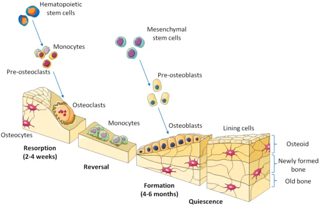
Figure 1.2 – Illustration of bone remodeling sequence supported by cellular activity1
Bone is a dynamic structure constantly developing, regenerating and remodeling. These processes are complexly regulated by a multitude of different growth and transcription fac-tors which coordinate the interactions of cells and matrix in response to external or internal stimuli. Several types of cells can be found in bone tissue, but its functional integrity is ensured by mainly three cell types which play a well defined role in the remodeling pro-cess [Buckwalter et al., 1995b]. Osteoblast cells are responsible for bone matrix deposition, they produce alkaline phosphatase (ALP) which plays a role in bone mineralization. Osteo-clast cells are in charge of bone resorption by producing H+ ions and tartrate-resistant acid
phosphatase (TRAP) which degrade hydroxilapatite and organic matrix respectively. Finaly, osteocytes are mechanosensible cells that regulate the remodeling process by interacting with osteoblasts and osteoclasts via extracellular signaling. Osteocalsts are mature osteoblasts that are embedded within the bone matrix in cavities called lacuanae. They communicate with each other thanks to gap junctions, made possible by a network of long cytoplasmic exten-sions going through the canaliculi. They also participate, although much less than osteoblasts, in the production of bone extracellular matrix.
This three cell types are very functional cells but have a relatively limited proliferative ability. In the scope of cell based therapies for bone reconstruction it may be useful to ex-plore their origins, differentiation and maturation. Stem cells are responsible for the renewal of all tissue in the body during repair and remodeling. Biochemical and biophysical signals
1Illustration built from the free PowerPoint image bank Servier Medical Arthttp://smart.servier.
control their activation, proliferation, migration, differentiation and survival. Stem cells give rise to progenitor cells through asymmetric division, in which a mother stem cell divides into one daughter stem cell and one daughter progenitor cell [Muschler et al., 2004]. That way the overall population of stem cell is conserved while progenitor cells are destined to pro-gressively differentiate into more functional cells. Mesenchymal stem cells (MSCs) have the ability to differentiate into divers connective tissues cells such as osteoblasts (bone), chondro-cytes (cartilage), adipochondro-cytes (adipose tissue), myochondro-cytes (muscles) and even neurons. In the case of bone lineage, they first differentiate into pre-osteoblasts, which have good proliferative capabilities, before to progress into osteoblasts. Osteoblasts are responsible for the deposition of collagen extracellular matrix (ECM) during bone formation phase (Fig. 1.2). After about 40 days, osteoblasts have three possible fates : they may differentiate into ostocytes if they are trapped in the forming bone, they may mature in lining cells on the surface of mature bone, or they may die by apoptosis. Finally, osteocytes and lining cells may survive for more than 20 years in the cortical bone until a new remodeling cycle is initiated in the region they reside [Buckwalter et al., 1995a;Muschler et al., 2004]. Osteoclasts does not originate from MSC but from hematopoietic stem cells (HSC). This later cells are the origin of all blood cells and are located in the bone marrow. They indirectly differentiate into monocytes which, when in presence with osteoblasts and MSCs, can self-fusion to produce osteoclasts [Buckwalter et al.,1995a].
1.2
Bone tissue repair
When damaged, bone has a unique ability to heal and remodel without leaving any scar. The healing of a fracture is a natural physiological process that results in bone union. However 5 to 10% of fractures face difficulties to restore, resulting in delayed union or even non-union [Calori et al., 2011]. In these cases, clinical treatments may be needed in order to restore physiological functionality.
1.2.1
Natural healing and remodeling
The first reaction to bone fracture is an inflamatory response due to damaged blood ves-sels, leading to the formation of an hematoma. Many signaling molecules released during inflammation (e.g. FGFs, BMPs, PDGF) are also involved in new bone formation [Frohlich et al., 2008]. In the three first days MSCs are recruited and start proliferating and differen-tiating into pre-osteoblasts and osteoblasts. During the first week, surrounding soft tissues stabilize the fracture by formation of a callus, which will be transformed in cartilaginous tis-sue during chondrogenesis (Fig. 1.3). Additionally angiogenesis is initiated to provide blood supply in the damaged zone. After the second week cell proliferation decreases, but intense osteoblastic activity continues. The callus is mineralized then resorbed by chondroclast cells while osteoblasts form new woven bone, a disorganized bone characteristic of initial bone formation. Finally the mechanical continuity is restored by the remodeling process in which
1.2. Bone tissue repair
Figure 1.3 – Illustration of stages in bone healing after a fracture
osteocytes, ostoclasts and osteoblasts replace woven bone by lamellar bone [Dimitriou et al.,
2005].
1.2.2
Current clinical treatments
In general, the excellent healing ability of bone, combined with external restoration of alignment and stable fixation, suffice for the reconstruction of most fracture [Frohlich et al.,
2008]. However in certain clinical situations such as high energy traumatic events, large re-sections following a pathology (e.g. tumor, infection), or complex non-union defects, natural bone repair may be too slow or inadequate. For instance, in the case of long bones, critical defects are considered for a length of bone loss of 3cm in the forearm, 5cm in the femur and tibia, and 6cm in the humerus [Calori et al., 2011]. In these challenging orthopedic cases, some form of grafting is required.
The functions of a bone graft are to fill the space of the defect, provide support, and en-hance biological repair. The biological properties of a bone graft are often discussed in terms of osteoinductivity (ability to promote bone formation), osteoconductivity (ability to promote vascularization and cell colonization within the graft), and osetogeniticity (ability to host bone-forming cells within the bone graft) [Giannoudis et al.,2005].
Nowadays, the best graft material is autologous bone graft. The technique consists in harvesting trabecular bone from a healthy site of the patient (usually the iliac crest in the pelvic girdle), and implant it directly in the bone defect. This procedure is widely applied, and the reported successful healing rate is 60 to 100% [Calori et al.,2011]. Autologous bone graft is safe, cheap, available to every surgeon, and present the three desirable properties of a graft material : osteoinductivity , osteoconductivity and osteogeniticity. Nevertheless autologous grafting has significant disadvantages. First the harvesting operation is often associated with complications such as bleeding, hematoma, infections and prolonged pain. Second, the donor site morbidity increases with the amount of harvested bone, and in the case of large or multiple reconstructions, the volume of bone material available is insufficient [Kneser et al., 2006].
Finally, the cost of the additional intervention may be equivalent to the cost of commercially available bone substitutes [Calori et al.,2011].
Allogenic (from a human donor) and xenogenic (from an animal donor) bone grafts are the main alternatives to autologous graft. They present the advantage of avoiding the harvest-ing of bone on the patient and all its possible complications, risks and costs. These grafts have ideal osteoconductivity and biomechanical properties, and are relatively easy to obtain through tissue banks [Kneser et al., 2006]. To avoid immune response, freezing and irra-diation processes are applied to eliminate the cellular phase of the graft. Even though the osteoconductivity is conserved, the process dramatically weaken the osteoinductive capabili-ties of the graft. Moreover risks of viral and bacterial transmission remain [Giannoudis et al.,
2005]. Allografts and xenografts are donor and process dependent, yielding to variable clin-ical results. The lack of osteogenesis delays osteointegration and vascularization, which in large grafts may lead to remaining non-vital regions [Frohlich et al.,2008].
Several other types of material are used for the reconstruction of bone defects. The main material families are biomaterial (e.g. demineralized bone matrix, collagen), ceramic (e.g. calcium phosphate and apatite based cements), bioactive glasse (e.g. silicon-based cements), and non-biologic (e.g. polymers, metals) substrates [Giannoudis et al., 2005; Calori et al.,
2011]. Nevertheless, most of these substitutes are only osteoconductive, there mechanical support can be weak, and present mixed clinical results [Giannoudis et al., 2005]. While the osteoconductivity of a biomaterial is directed by its composition, surface physicochemical properties and internal structure, osteoinductivity can be provided with the aid of osteoin-ducitve substances (e.g. transforming growth factor (TGF-β), insuline-like growth factor (IGF I and II), platelet derived growth factor (PDGF), fibroblast growth factor (FGF), vari-ous bone morphogenic proteins (BMPs)) [Janicki & Schmidmaier,2011]. The use of growth factors in combination with the above filling materials has been tested clinically, but still few studies have yet been reported, and the applications protocols are not yet standardized [Kneser et al.,2006].
Further osteogenicity can be improved with the introduction of osteocompetent cells within the filling material. This is the aim of bone tissue engineering, and the specificities of this field are discussed in the next section.
1.3
Tissue engineering
Tissue engineering is an interdisciplinary field which aims to repair damaged tissues or to restore a defect by combining in vitro biomaterials, cells and/or signaling molecules. If cells are used, expansion and tissue specific cell differentiation may request an in vitro culture period ranging from a couple of days to several months. The obtained constructs must be de-veloped in parallel with well elaborated surgical concepts in order to lead to successful in vivo applications [Kneser et al.,2006]. In this section, the main components for the development of bone substitutes, in the frame of tissue engineering, are reviewed.
1.3. Tissue engineering
1.3.1
Scaffold design
Three-dimensional porous scaffolds play an important role in tissue engineering strategies. During in vitro culture, it serves as a support for cells attachment, migration, proliferation and differentiation and may be a provider of osteoinductive substances. Upon transplantation it is the vehicle for delivery of cells to the defect site. The porous structure lets void for tissue ingrowth and vascularization while maintaining mechanical integrity [Muschler et al.,
2004]. The scaffold must be made from biocompatible material, and should biodegrade and bioresorb at a controlled rate, following new tissue formation. The development of scaffolds for tissue engineering is an intense and prolific research field, and several possibilities are already available for clinical use. In this section, the variety of existing scaffolds is discussed along the following main characteristics : the bulk material composition, the geometrical architecture, the mechanical properties, the surface chemistry and the in vivo degradation properties.
The bulk materials used in bone tissue engineering are inherited from orthopedic clinical practices. It includes biological-derived material (e.g. processed allograft, coral), biological polymers (e.g. collagen, chitin), ceramics or mineral-based material (e.g. tricalcium phos-phate (TCP), hydroxyapatite (HA), calcium phosphos-phate), metals (e.g. titanium, tantalum) and synthetic biodegradable polymers (e.g. polyglycolide (PGA), polylactides (PLLA, PDLA), polycaprolactone (PCL)) [Hutmacher, 2000; Muschler et al., 2004]. Composite scaffolds made of two or more of the above cited material are also in use.
The three-dimensional porous architecture of the scaffold is critical since it influences me-chanical properties, cellular fate, nutrient supply, vascularization and tissue ingrowth. The pore size is usually in the range of 100 - 500µm. Even though the ideal size has been es-timated around 350µm [Murphy et al., 2010], it is believed that larger pore sizes support deeper cell proliferation and tissue penetration [Muschler et al., 2004]. Concerning the pore structure, the geometry can be dictated by the fabrication process (for instance in the case of biological based scaffold), or arbitrary chosen when the synthesis method allows it (Fig. 1.4). Most of these scaffolds are homogeneous porous media with isotropic transport properties. Considering the recent advances in fabrication techniques (e.g. 3D-printing, stereolithogra-phy), it may be possible to produce controlled scaffolds with hierarchical microstructures and oriented channels to guide the patterns of cell migration, fluid flow and diffusion through the construct [Hutmacher, 2000; Khademhosseini et al., 2006; Sprio et al., 2011]. Finally the nanostructural architecture of the scaffold should be considered since the substrate rigidity and rugosity is known to influence cell adhesion, differentiation and migration [Engler et al.,
2006;Yoon et al.,2012].
The required mechanical properties of the scaffold depends on the graft site physiolog-ical load and bone property. For instance, defects on load-bearing long bones require the restoration of high mechanical stability, while the mechanical loads involved in craniofacial reconstructions does not necessitate such mechanical performances [Kneser et al.,2006]. On the other hand, the use of rigid non-degradable material such as metals, protects adjacent tis-sue from mechanical loads, resulting in a change of the stress environment that may lead to a loss of local tissue mechanical properties. Cortical bone has a Young’s modulus of 15-20GPa
Figure 1.4 – Scanning electron microscope (SEM) images of various scaffold structures used in bone tissue engineering. (A) Partial deproteinized bone [Wu et al.,2010]. (B) Natural coral (porites) [David et al.,2014]. (C) Nonwoven PLLA fiber mesh [VanGordon
et al.,2010]. (D) Polymer fiber mesh (PCL) built by fused deposition modeling (FDM) [Hutmacher,2000]. (E) Hydroxyapatite scaffold obtained by foaming method [Sprio et al.,
1.3. Tissue engineering
and a compressive strength of 100-200MPa, while trabecular bone has a Young’s modulus of 0.1-2GPa and a compressive strength of 2-20MPa [Cowin, 2001]. A summary of scaffolds mechanical properties of different materials and architectures can be found in [Bose et al.,
2012]. The scaffold should present similar mechanical properties to the local bone, therefor ceramic based scaffolds are appropriate to replace cortical bone, while polymers may be better suited for trabecular bone. Another key factor related to mechanical properties of the scaffold is the decrease in mechanical support during in vivo resorption. In order to avoid mechanical failure of the implant (fracture), the degradation rate of the scaffold material must match the regeneration rate of the the replacing bone [Hutmacher,2000]. Finally, the mechanical stimuli playing an important role in the differentiation of bone marrow stem cells, the scaffold should transmit an appropriate stress environment within the graft site [Pioletti,2013].
The surface chemistry of the scaffold directs its interactions with cells. Surface properties are influenced by the bulk material, but are mainly dependent on the adsorbed proteins and lipids. These biomolecules that come from coating solutions, culture medium, biological fluids and/or cell metabolism, cover the surface of the scaffold and are the primary mediator of cellular response to the material [Muschler et al.,2004]. This can be put to profit to direct cell attachment, survival, proliferation and differentiation by precoating the scaffold with bioactive proteins such as fibronectin or various growth factors [Bose et al.,2012].
Finally, the degradation properties of the scaffold must be considered with care. First the resorption rate should be control to match tissue formation and mechanical load transfer from the biodegrading scaffold to the tissue. Typical degradation times range from 3 to 12 month depending on the application [Bose et al.,2012]. Second, degradable materials release degradation products within the implantation site. This products, when released too quickly, may influence local pH and and reach toxic concentrations, leading to inflammatory reactions. The control of the pH environment during scaffold resorption can be addressed by the use of composite material. Indeed, synthetic polymer materials (PGA, PDLA, PLLA) release acidic by-products, while ceramic materials (TCP, HA) produce basic resorption products. Therefore a adequate combination of the two types of material could avoid the production of unfavorable environment for the cells [Hutmacher,2000].
1.3.2
Cell choice
Tissue engineering strategies combine three-dimensional scaffolds with cells, in order to build a functional tissue and/or to recruit and attract other cell types toward the construct. The ideal cells should be easily isolated and expended, have stable phenotype of interest and show long term safety.
In the aim of producing bone substitutes, the cell type must present an osteogenic pheno-type, which include mesenchymal stem cells (MSCs) (Fig. 1.5), bone marrow stromal cells (BMSCs), periosteal cells and osteoblasts [Kneser et al.,2006]. MSCs are particularly promis-ing since they have the ability to differentiate into osteoprogenitors and mature osteoblasts, they are easily isolated, expanded, and show a stable phenotype up to 50 population doubling [Frohlich et al.,2008]. Several autologous sources of MSCs are known (e.g. cartilage, fat), but
bone marrow aspirates provide the higher density of stem cells, are relatively safe, and easy to proceed [Muschler et al., 2004; Colnot, 2011]. Nevertheless the number of cells and the quality of the aspirate are patient dependent, with an observed decrease in MSC concentration with age, motivating in vitro cellular expansion before seeding or transplantation.
Figure 1.5 – Mesenchymal stem cells stained with fluorescent dyes2. Nucleus are in blue,
microtubules in green, and actin filaments in red.
During early stages of scaffold and device testing, the use of human or animal immortal-ized cell lines are a reasonable choice (e.g. murine osteoblasts MC3T3-E1 [Cartmell et al.,
2003], murine mesenchymal cells C3H10T1/2 [David et al., 2011], human osteoblast-like cells MG63 [Olivier et al.,2007]). These are populations of cells issued from a multicellular organism, which have lost their natural senescence due to mutation or human intervention. Therefore they can be grown in vitro for prolonged periods. From a practical research point of view, they present the advantages of being easily available and easy to maintain in any basic cell culture facilities. However, as soon as the proof of concept has been established for a given process, more clinical oriented cells must be selected.
1.3.3
In vitro
culture conditions
Once the scaffold and the cell type are selected, the appropriate culture conditions have to be maintain to optimize cell proliferation, differentiation into the appropriate lineage, migra-tion and matrix deposimigra-tion.
The importance of the seeding method must not be underestimated if an homogeneous initial distribution of cells is intended (Fig. 1.6). When the scaffold is introduced in cell suspension and let to rest, sedimentation process drives the cells to the surface. This means that regions with higher ratio of suspension volume to scaffold surface will have a high cell density once attached on the wall. This occurs particularly for heterogeneous porous scaffolds
2Left : https://globalmedicaldiscovery.com/wp-content/uploads/2014/04/
Human-mesenchymal-stem-cell.jpg Right : http://news.softpedia.com/news/ New-Technique-for-Tracking-Stem-Cells-Developed-405076.shtml
1.3. Tissue engineering
(including scaffolds made of multiple porous granules), and on the edge of the scaffolds, close to the bulk cell suspension. It results in an initial heterogenous distribution of the cells within the scaffold, with more cells on the surrounding of the scaffold than in its center. To overcome this issue, multistep seeding [Grayson et al., 2008] or dynamic seeding methods [Vunjak-Novakovic et al.,1998] have been proposed.
Figure 1.6 – Fluorescent microscope pictures of 20µm sections of coral scaffolds seeded with Hoechst stained human MSC (adapted from [Mygind et al.,2007])
Once the cells seeded and attached to the scaffold, the construct is usually maintained
in vitro in a bath of culture medium within an incubator. It has been shown that the first
days/weeks after seeding, cellular adherence, growth and differentiation is dominated by sur-face phenomena due to cell/material interactions [Kommareddy et al.,2010]. Then in a second time, cells produce extracellular matrix (ECM) which allows them to grow three-dimensional. From that point, the pore geometry of the scaffold becomes important since it is shared be-tween forming tissue and culture medium.
When the culture medium is at rest, the culture is said to be in static conditions, as opposed to dynamic culture conditions. The role of culture medium is to provide the necessary nutrient and oxygen levels to support cellular activity. It can be supplemented with osteoinductive substances to promote cell differentiation, division and matrix deposition. In static culture conditions, diffusion is the only transport process, and the cell consumption may induce large concentration gradients leading to regions with nutrient and/or oxygen depletion. This mass transport limitation occurs especially at the core of large constructs, and at high cell density where there is competition for resources [Malda et al., 2004]. Cells in these regions may enter in a quiescent state or in necrosis due to lack of nutrients or hypoxic (lack of oxygen) conditions. Nevertheless, it has been suggested that MSCs can survive for long periods (up to 12 days) in sever hypoxia as long as glucose is available [Deschepper et al., 2011], and that hypoxic conditions may be a stimulus for cell differentiation or migration toward a more favorable environment [Muschler et al.,2004].
Mass transport limitations together with the need to produce more easily larger and cheaper bone substitutes have motivated the development of bioreactors for bone tissue engineering. The next chapter is dedicated to this subject, with a focus on bioreactors using hydrodynamic environments.
C
HAPTER
2
B
IOREACTORS FOR
B
ONE
T
ISSUE
E
NGINEERING
A
S DISCUSSED in the previous chapter, tissue engineering sets the ground principles ofcellular based therapy for the reconstruction of tissues and organs. Nevertheless most of this processes are not yet ready to be used in practical applications because of insuffi-cient substitute quality and reproducibility, high production time and cost, and difficulties to implement in clinical applications [Salter et al., 2012]. By adopting an approach based on process engineering, bioreactors systems are a promising tool to address this issues. In the present chapter, the different principles of bioreactors for bone tissue engineering are exam-ined, and the prerequisites for translation to clinical applications are reviewed. Finally the double porosity bioreactor developed in [David et al.,2011] is detailed.
Bioreactors are generally used to facilitate, monitor and control biological, biochemical and biophysical processes. The parameters influencing cell growth within a bioreactor include temperature, pH, concentration in nutrient, growth factors and oxygen, and biophysical stim-uli. These systems are usually constituted of biologically inert and non-corrosive material to avoid cytotoxic responses. Furthermore the components must be able to support sterilization techniques, and the bioreactor should be convenient to be manipulated in aseptic conditions.
Bioreactor systems must efficiently provide nutrients, oxygen and biophysical stimuli in order to optimize cell proliferation, differentiation and matrix deposition. Possibilities to en-hance mass transport are culture medium flow, integration of membranes or hollow fibers within the scaffold, or pre-vascularization. In the case of bone forming cells, the relevant bio-physical stimuli are mechanical solicitations (hydrodynamic shear stress, mechanical strain) and electromagnetic fields. Each design of bioreactor for bone substitute presented bellow applies one or more of this solutions to produce bone substitutes.
2.1
Bioreactors using hydrodynamic stimuli
The principle behind the application of hydrodynamic culture conditions is to make use of culture medium flow to address mass transport limitations and to apply mechanical stimuli to the cells.
In vitroconstructs are usually not vascularised, and in that case, culture medium is the only
source of oxygen and nutrients. Thus diffusion from the exterior of the construct to its core is the main process responsible for species supply of the inner cells. Simple calculations can show that cells at more than 500-1000 µm in the depth of the construct can be very quickly in hypoxic conditions (lack of oxygen) [Muschler et al.,2004], which could lead to necrosis at the center of the construct. Moreover static culture conditions lead to inhomogeneous con-centrations fields due to local consumption of nutrients in zones of higher cell density. These limitations can be partially solved by the use of hydrodynamic conditions where convective effects enhance mass transport around and within the construct.
In addition to improve mass transport, hydrodynamic culture conditions are a means to stimulate mechanically the cells. Particularly, bone cells have been shown to be more sen-sitive to mechanical strain than other cell types [Meyer et al., 1999]. In everyday life, in
vivomechanical stress stimulates osteocyte and osteoblast cells present in bones, and is thus
a key factor in the control of bone remodeling [Cowin, 2007]. Similarly, in a hydrodynamic bioreactor, fluid flow induced mechanical stimuli influence cell differentiation and prolifer-ation [Glowacki et al., 1998; Bancroft et al., 2002; Cartmell et al., 2003; Raimondi et al.,
2006; Grayson et al., 2010; McCoy & O’Brien, 2010]. In bone tissue engineering studies, the mechanical parameter of interest is the fluid flow induced shear stress [Hung et al.,1995;
Goldstein et al.,2001;Sikavitsas et al.,2003;Raimondi et al.,2006;Leclerc et al.,2006; Ped-ersen et al.,2007, 2010;Jungreuthmayer et al., 2009a;Park et al.,2010;McCoy & O’Brien,
2010;Grayson et al., 2011]. To be more precise, the main known effects of fluid flow influ-encing cell fate, are the deformation of cytoskeleton [Horikawa et al., 2000; McGarry et al.,
2004;Kwon & Jacobs,2007], the activation of stretch-sensitive ion channels in the membrane [Jacobs et al., 2010], and the stimulation of primary cilia [Hoey et al., 2012;Delaine-Smith et al.,2014]. These solicitations induce complex chains of biophysical and biochemical pro-cesses, called mechanotransduction, that lead to cell response [Ingber, 2006]. Since all of these solicitations are considered to be the effect of shear stress, which has the advantage to be easily computed and quantified, the fluid flow mechanical stimuli are assumed to be limited to this stress.
Parameters influencing spacial distribution and intensity of local shear stress within a three-dimensional scaffold are, the bioreactor and scaffold geometries (which include the scaffold porosity, isotropy, pore size and interconnectivity) as well as the fluid flow rate and physical properties. The distribution of the shear stress on the scaffold walls are extremely hard to determine experimentally, however different fluid mechanics models have been successfully developed to address this question (seeSec. 3.1). Concerning the intensity of the shear stress, the values found in the literature vary greatly from a study to another, depending on the cell line, the support material or whether the cells are grown in two or three dimensions
2.1. Bioreactors using hydrodynamic stimuli
[Jungreuthmayer et al.,2009b;McCoy & O’Brien,2010]. For three-dimensional cell cultures, it is observed that a first range of shear stress values yields to an enhanced cell proliferation. Then for increasing values of shear stress, cell division slows down while differentiation and production of extracellular matrix are improved. Finally for very high values, shear stress is damageable for the cell survival and attachment, and fluid flow may wash them off the scaffold [Cartmell et al.,2003;Mygind et al.,2007].
Several bioreactor designs make use of hydrodynamic culture environments to improve cell proliferation, differenciation and matrix deposition [Rauh et al., 2011]. The types of system reviewed below are the spinner flask bioreactor [Vunjak-Novakovic et al.,1998], the rotating wall bioreactor [Freed & Vunjak-Novakovic, 1995], and the perfusion bioreactor [Mueller et al.,1999].
2.1.1
Spinner flask bioreactors
The simplest and cheapest way to provide hydrodynamic stimuli to a biohybrid, is to sus-pend the seeded scaffold in a flask filled with culture medium, and to agitate the liquid using a magnetic bar. The bioreactors based on that principle are called spinner flask bioreactors (Fig. 2.1). The system is placed in an incubator which maintains adequate temperature, hu-midity and CO2 content. Apertures with filters are present at the top of the flask to allow gas
exchange between the bioreactor and the incubator without risks of contamination. The level of applied shear stress is dependent of the stirring speed, which has typical values of 30 to 50rpm for flasks of 120mL [Rauh et al.,2011].
Figure 2.1 – The spinner flask bioreactor. Left : Schematic representation [Rauh et al.,
2011]. Right : Wheaton Celstirrspinner flask
Cultures in spinner flask bioreactors lead to very good results in term of cell proliferation and production of osteogenic markers, compared to static conditions [Sikavitsas et al.,2002;
Mygind et al.,2007]. Using coral scaffold of mean pore size of 200µm and 500µm,Mygind et al.[2007] obtain a better proliferation, differenciation and homogeneity of human MSC in
a spinner flask bioreactor than in static culture. Moreover the 200µm pore constructs showed better osteogenic differentiation than the 500µm pore constructs, while this later had a higher number of cell indicating a better proliferation. This results suggest that lower shear stress (in larger pore size) enhance cell division, while higher shear stress (in smaller pores) stimulates cell differentiation.
Despite its inexpensiveness and good results, the spinner flask bioreactors present several disadvantages. The most important drawback is the complex hydrodynamics in the flask making difficult the precise evaluation of applied levels of shear stress. Moreover the velocity field is inhomogeneous within the bioreactor, leading to stimuli dependent on the location of the construct, and bad repeatability between experiments [Goldstein et al.,2001].
2.1.2
Rotating wall bioreactors
Rotating wall bioreactors have been initially design by the National Aeronautics and Space Administration (NASA) to study cellular growth in micro-gravity environment. It is com-posed of a horizontal cylindrical vessel, put in rotation along its axis my an external motor. The closed bioreactor contains culture medium and constructs in suspension. Rotation of the vessel induces laminar flow by inertial effects (Fig. 2.2). Although mass transport limitations are solved by the culture medium flow, it seems that the level of shear stress remains very low, probably because the constructs follow the movement of the fluid. The results of cell culture in rotating wall bioreactors in the frame of bone tissue engineering are controversial. While some authors find improved cell proliferation and osteogenic markers productions [Botchwey et al., 2001; Song et al., 2008], other observe a decrease in cell survival compared to static conditions [Goldstein et al.,2001;Sikavitsas et al.,2002].
Figure 2.2 – The rotating wall bioreactor. Left : Schematic representation [Rauh et al.,
2011]. Right : One of the first rotating wall bioreactor prototype developed by the NASA1 The divergence in the results obtained with rotating wall bioreactors can partly be ex-plained by the weaknesses of the system. To start with, the material density of the scaffold
1http://science.nasa.gov/science-news/science-at-nasa/1999/msad05oct99_
2.1. Bioreactors using hydrodynamic stimuli
and its size play an important role in the way the construct will be moved by the fluid. If the density of the scaffold is close to the density of the culture medium, the construct will stay in suspension, limiting collisions with the walls or other constructs. But if the density is differ-ent from the culture medium, the construct may have turbuldiffer-ent movemdiffer-ents within the vessel, increasing risks of damages by collisions. Some modifications to the bioreactor have been proposed to address this issue, by fixing the construct to the walls of the vessel [Song et al.,
2008]. This has the advantage of keeping the biohybrid in the peripheric zone of the vessel, where the main hydrodynamic effects take place. Yet, rotating wall bioreactors have limited volume of production, and the heterogeneous levels of shear stress applied to the biohybrides remain a clear restriction to its use in laboratory.
2.1.3
Perfusion bioreactors
The limitations of the previously presented bioreactors motivated the development of per-fusion bioreactors [Yeatts & Fisher, 2011]. This design takes advantage of laminar flow to enhance mass transport and distribute shear stress in the whole construct. The perfusion sys-tems are usually constituted of a chamber in which one or more seeded scaffolds are placed, and through which the culture medium is perfused with the help of pump and tubing sys-tems. Two main categories of perfusion bioreactors are met, direct perfusion systems force the culture medium through the construct, while indirect perfusion systems allow the culture medium to flow around the construct (Fig. 2.3).
In indirect perfusion bioreactors, the culture medium can follow the path of less resistance around the construct, inducing less flow in the core than on the surrounding of the scaffold [Janssen et al.,2006;David et al.,2011]. Shear stress is thus mainly located at the periphery of the biohybrid, and the benefits of the perfusion on mass transport may be limited at the center. This may result in preferential cell proliferation at the surrounding of the scaffold, and hypoxic and starving conditions at the core, leading to necrosis. In contrast, direct perfusion bioreactors enhance mass transport and shear stress distribution within the construct [Bancroft et al.,2002;Cartmell et al.,2003;Grayson et al.,2008,2011]. Nevertheless it necessitates the use of sufficiently permeable scaffolds in order to limit too high local stresses which could harm the cells. Moreover at long culture times, cell proliferation decrease the porosity of the construct, intensifying the level of shear stress which could lead to a maximal state of tissue growth, or even detachment of cell aggregates.
The design of perfusion bioreactors varies widely from a team to another. The culture medium can either flow in a closed loop or go from a reservoir to a waste flask. Perfusion modes can be oscillatory, pulsed or continuous [Jaasma & O’Brien, 2008;Wu et al., 2010]. Additional equipments can be adapted to mechanically stimulate the construct [El Haj et al.,
1990], monitor the mineralization, or measure the concentration of species of interest (oxygen, osteogenic markers...) [Janssen et al., 2006]. Typical continuous flow rates range from 1 to 600mL/h, and the volume of the produced constructs are usually of few milliliters. Yet the transfer to clinic applications requires volumes up to 300mL of bone substitutes.
Figure 2.3 – The perfusion bioreactor. Left : Schematic representation of (A) indirect perfusion, (B) direct perfusion [Rauh et al.,2011]. Right : Photography of a perfusion
bioreactor prototype used in our team.
et al.,2007], but this often leads to higher heterogeneities in cell proliferation. An alternative strategy is to multiply the number of millimeter size scaffolds in the chamber [Janssen et al.,
2006;David et al.,2011]. The resulting construct shows a very good cellular proliferation and differentiation, and the scaffolds are covered with interconnected and dense layer of extracel-lular matrix. However the core of the individual scaffolds still present a lack of proliferative cells, probably due to mass transport limitations. Although, these studies succeed in increas-ing the volume of the final construct of a factor 10, another factor 10 is needed in order to face all clinical needs.
2.2
Bioreactors based on other principles
Bioreactors using hydrodynamic stimuli are the most widely used systems in bone tissue engineering, mainly because it deals with mechanical stimuli as well as mass transport limi-tations. However other types of designs based on different physical and biological principles have been proposed.
Several other possibilities than hydrodynamic culture exist to provide mechanical stim-uli. An intuitive and more biomimetic approach is to strain the scaffold so that the stress is transmitted to the attached cells. Machines originally fabricated for material testing have been adapted to apply compression [El Haj et al.,1990;Wartella & Wayne,2009;Baas et al.,
2.2. Bioreactors based on other principles
of ostegenic markers and matrix deposition compared to unloaded samples. Alternatively, cell stretching systems have been developed to study the effect of tensile strain on bone cells. Cells are embedded in a collagen matrix, which is fixed on a silicon dish. This silicon support is cyclically stretched uniaxially or biaxially using piloted motors [Neidlinger-Wilke et al.,
1994;Ignatius et al.,2005]. Stretching stimulus seems to mainly increase bone cell prolifer-ation. Interestingly cells where found to be oriented along the axis of the applied mechanical stress. Nevertheless, severe drawbacks exist for these direct mechanical stress systems. In-deed, any force producing mechanism that invades the bioreactor (piston, silicon sheet...) are a possible cause of infection. Additionally the scaffold material must transmit the force to the cells, and this implies that they must be strong enough in compression or bending systems, and soft enough in silicon stretching systems. This constraints may lead to incompatibilities with the prerequisites for biodegradation times of the scaffolds [El Haj & Cartmell, 2010]. Finally mass transport limitations are present in this types of bioreactors, and they may have to be coupled with perfusion systems [Bölgen et al.,2008].
Electromagnetic fields have been used in clinical studies to treat various bone patholo-gies [Aaron et al., 2006]. The underlying idea is that in vivo mechanical deformations in bones cause piezoelectricity, generating electric potentials. Vibration and movement of hu-man muscles induce mechanical strains and currents of specific frequencies to which bone cells are sensitive. Bioreactor systems based on this principle use magnetic coils to gener-ate pulsed electromagnetic fields of controlled intensity to stimulgener-ate growing constructs [ Bo-damyali et al., 1998]. The results show enhanced osteogenesis [Schwartz et al., 2008; Sun et al., 2010] and in certain cases better proliferation [Tsai et al., 2007] compared to control samples. Although electromagnetic fields systems are non-invasive, which is an advantage to reduce contamination risks, the high cost of the equipment is a clear limitation.
Instead of trying to reproduce in vitro the ideal biochemical and biophysical conditions of bone growth, in vivo bioreactors tend to make use of a host organism to supply the adequate nutrients, growth factor, and physiological environment to the construct. This method consists of implanting a cell-loaded scaffold material within a living organism, in order to initiate vascularization and the creation of more mature bone structures. Different animal models have been developed [Petite et al., 2000;Stevens et al., 2005;Holt et al., 2005] resulting in vascularized neo-bone formation. A recent exploratory study has been carried on a man as an
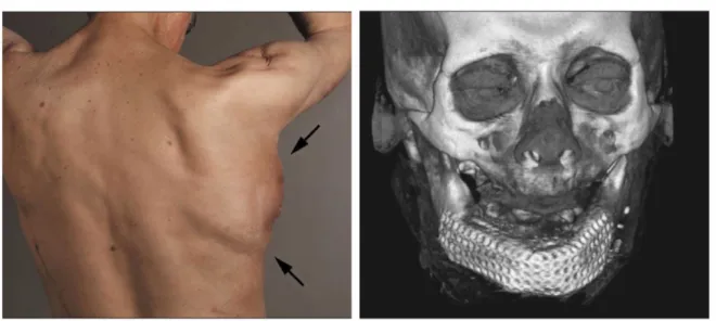
in vivo bioreactor to replace a large part of his mandible [Warnke et al., 2004]. A titanium
cage filled with bone mineral scaffold, autologous bone marrow and growth factor (BMP-7), has been implanted in one of the patient’s dorsal muscle (right latissimus dorsi muscle). After 7 weeks the construct was transplanted to repair the mandibular defect (Fig. 2.4). The formation of new bone leaded in an improvement of the patient quality of life [Warnke et al.,
2006], until his death from heart failure 15 month after the mandible replacement. Although the application of in vivo bioreactor concept has shown success in some individual cases, they currently imply very heavy, risky and expansive surgical operations.
Figure 2.4 – Left : Dorsal view of mandibular replacement 3 weeks after implantation. Arrows point to the implantation site. Right : 3D computed tomography scan after transplantation of the in vivo cultured mandibular substitute. Figures adapted from [Warnke
et al.,2004]
2.3
The double porosity bioreactor [David et al., 2011]
In the frame of the present thesis, the double porosity bioreactor developed byDavid et al.
[2011] is studied. We seek to analyze the effects of culture medium perfusion on mass trans-port as well as on cell proliferation and tissue development. In this section the bioreactor’s principal characteristics and results are presented.
The primary goal of the development of the double porosity bioreactor is to produce clin-ically relevant volumes of bone substitute in a limited time. This necessitates to enhance the proliferation and differentiation of osteo-competent cells within a bioresorbable three-dimensional scaffold. The design is partly inspired from fluidized bed reactors [Legallais et al., 2000; David et al., 2004] to tackle mass transport limitations and to provide optimal mechanical stimuli to the growing construct.
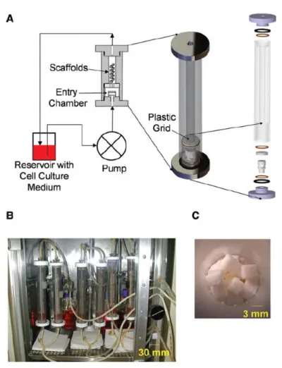
The bioreactor system is composed of a vertical cylindrical tube (inside diameter 3.3cm, height 21cm) containing the construct, and connected in a closed loop to a culture medium flask. Perfusion is ensured by a peristaltic pump and silicon tubing (Fig. 2.5). Except for the pump, the whole system is placed in an incubator and maintained at 37◦C during culture. The
silicon tubing are permeable to gas exchange between the culture medium and the incubator environment, providing oxygen and CO2 during the whole duration of the experiment.
Cul-ture medium flow rate has been tested for values ranging between 1 to 100mL/min, and best results in terms of proliferation has been found for 10mL/min. The flask of culture medium is changed every three days in aseptic conditions.
The scaffold is constituted of a stack of cube shaped decellularized coral (approximately 140 microporous granules of 3×3×3mm3). This material has been widely used as bone
sub-2.3. The double porosity bioreactor [David et al., 2011]
Figure 2.5 – Experimental set-up of the double perfusion bioreactor. (A) Schema of the set-up. (B) Several bioreactors perfused in parallel inside an incubator. (C) A stack of coral
scaffold cubes
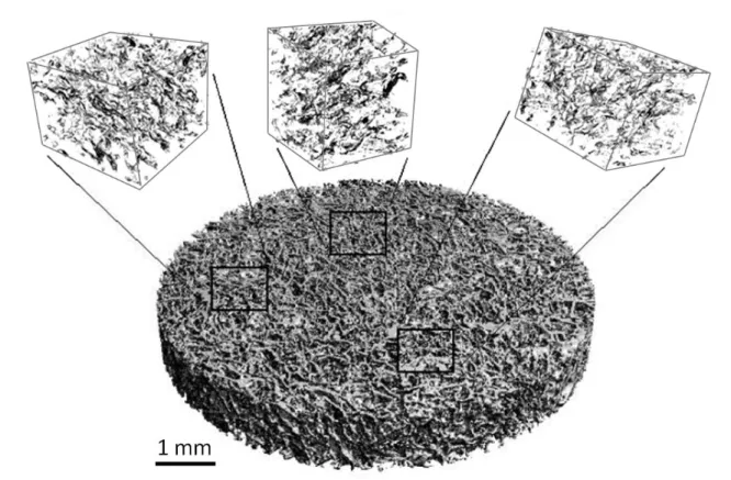
stitute for the past 10 years in orthopedic and maxilofacial surgery. It has the advantage of being biocompatible, osteoconductor, and bioresorbable. Two types of corals with different geometrical properties have been used : Porites which has a porosity of roughly 50% and mean pore size of approximately 80µm, and Acropora which presents a porosity of 12% for a mean pore size of 500µm. The scaffold cubes are staked randomly in the bioreactor, allowing the culture medium to flow in the space between the cubes, and assuring a relative isotropy. The double porosity composed of the inner coral porosity, and the staking of the cubes, gives its name to the bioreactor.
In the original article, the cell line used for the experiments are pluripotent stem cells derived from mouse embryos (C3H10T1/2) and transfected with green fluorescent protein (GFP). Coral cubes are precoated with culture medium before to be seeded with 106cell/mL
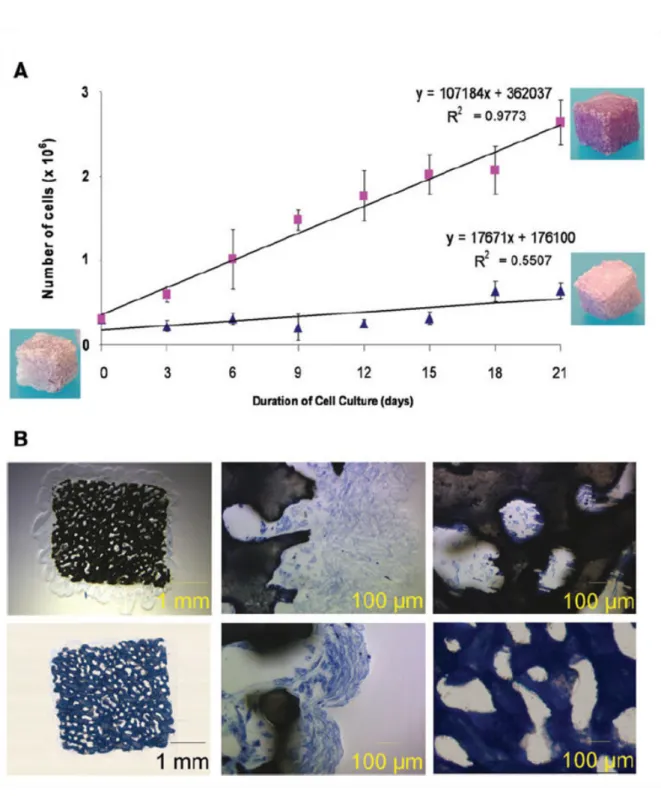
cell suspension. Cells are kept at rest 24 hours in an incubator to allow proper attachment, and are then transfered in the bioreactor. At a flow rate of 10ml/min, the wall shear stress in the scaffold is roughly estimated between 0.01 and 10mPa. Cell number per cube is evaluated after 3, 6, 9, 12, 15, 18 and 21 days of culture by a destructive method. Basically each
con-Figure 2.6 – Effect of culture medium fluid flow on cell proliferation. (A) Cell proliferation under static conditions (blue triangles), and under 10mL/min perfusion (pink squares). (B)
Histological slices (Stevenel blue) of biohybrid cultured under 10mL/min perfusion for 3 weeks (top line) and in static conditions (lower line). Middle pictures show the periphery of

![Figure 1.6 – Fluorescent microscope pictures of 20µm sections of coral scaffolds seeded with Hoechst stained human MSC (adapted from [Mygind et al., 2007])](https://thumb-eu.123doks.com/thumbv2/123doknet/14643405.735602/30.892.204.745.270.445/figure-fluorescent-microscope-pictures-sections-scaffolds-hoechst-stained.webp)
![Figure 2.1 – The spinner flask bioreactor. Left : Schematic representation [Rauh et al., 2011]](https://thumb-eu.123doks.com/thumbv2/123doknet/14643405.735602/34.892.143.808.666.958/figure-spinner-flask-bioreactor-left-schematic-representation-rauh.webp)
![Figure 2.3 – The perfusion bioreactor. Left : Schematic representation of (A) indirect perfusion, (B) direct perfusion [Rauh et al., 2011]](https://thumb-eu.123doks.com/thumbv2/123doknet/14643405.735602/37.892.118.720.138.519/figure-perfusion-bioreactor-schematic-representation-indirect-perfusion-perfusion.webp)
![Figure 3.1 – Regular scaffold used in [Boschetti et al., 2006]. A : SEM image of a PDLLA scaffold made by particulate leaching](https://thumb-eu.123doks.com/thumbv2/123doknet/14643405.735602/49.892.88.753.140.378/figure-regular-scaffold-boschetti-pdlla-scaffold-particulate-leaching.webp)