HAL Id: hal-01248034
https://hal.archives-ouvertes.fr/hal-01248034
Submitted on 23 Dec 2015
HAL is a multi-disciplinary open access archive for the deposit and dissemination of sci-entific research documents, whether they are pub-lished or not. The documents may come from teaching and research institutions in France or abroad, or from public or private research centers.
L’archive ouverte pluridisciplinaire HAL, est destinée au dépôt et à la diffusion de documents scientifiques de niveau recherche, publiés ou non, émanant des établissements d’enseignement et de recherche français ou étrangers, des laboratoires publics ou privés.
CLEANING LIVING HARD-SHELLED
FORAMINIFERA
L Rossignol, C Dupuy, Py Pascal, P Debenay
To cite this version:
L Rossignol, C Dupuy, Py Pascal, P Debenay. HYDROBIA ULVAE: A DEPOSIT-FEEDER FOR CLEANING LIVING HARD-SHELLED FORAMINIFERA. Journal of Foraminiferal Research, Cush-man Foundation for Foraminiferal Research, 2007, �10.2113/gsjfr.37.1.8�. �hal-01248034�
1
HYDROBIA ULVAE: A DEPOSIT-FEEDER FOR CLEANING
2
LIVING HARD-SHELLED FORAMINIFERA
3
4
5
L. ROSSIGNOL1, C. DUPUY1, PY. PASCAL1 AND J. –P. DEBENAY2 6
7
1 CRELA, UMR 6217, Université de La Rochelle, Pôle Sciences, AV. Michel Crépeau, 17042 8
La Rochelle Cedex, France
9 10
2 Département de Géologie, Université d’Angers, UPRES EA 2644, 2 Bd Lavoisier, 49045 11
Angers Cedex, France
12 13 14
Keywords: mudflat, Hydrobia ulvae, hard-shelled foraminifera, extraction
15 16 17 18 19 20 21 22 23 contact: [email protected] 24 25 26
1 2 3 4 5 6 7 8 9 ABSTRACT 10
This study proposes a new method for fast and inexpensive extraction of a large number of
11
living foraminifera for laboratory cultures. The method is a significant improvement over
12
current extraction methods, which are highly time-consuming. Several treatments were
13
designed to test the method. Sediment bearing foraminifera from Brouage Mudflat (Atlantic
14
coast of France) was washed through a 50-m sieve and distributed in glass Petri dishes with
15
20, 40 and 80 specimens of Hydrobia ulvae, a common gastropod from European intertidal
16
mudflats. As a control experiment, one dish was treated similarly but maintained without
17
Hydrobia. After two days, most of the sediment in the Hydrobia treatments was compacted
18
into small cylindrical gastropod feces and the tests of living benthic foraminifera (Ammonia
19
tepida and Haynesina germanica) were clean and easily visible. Additional experiments
20
showed that the foraminifera were not ingested by Hydrobia ulvae, and could be picked
21
quickly and easily.
22 23 24 25 26 27 28 29 30 31 32
1 2 3 4 5 INTRODUCTION 6
Laboratory studies using living foraminifera for biological and ecological investigation
7
have been used for more than a half-century and provide important, complementary data to
8
field-base studies (e.g., Myers, 1935; Le Calvez, 1938; Jepps, 1942; Arnold, 1954). They have
9
been increasingly used for ecological and environmental studies (e.g., Bradshaw, 1961;
10
Bender and Hemleben, 1988; Bijma and others, 1990; Stouff and others, 1999; Khare and
11
Nigam, 2000; Heinz and others, 2002). These studies require separation of live individuals
12
from the sediment without harming them and efficient techniques to differentiate live and
13
dead individuals. Vital staining, such as the use of fluorescent probes, associated with direct
14
observation of cytoplasm and reticulopodial network allow distinguishing live individuals
15
from dead ones (Murray and Bowser, 2000, Bernhard, 2000). However, before using these
16
methods, it is first necessary to isolate individuals from the sediment. In samples containing a
17
high proportion of mud, the tests are hard to discern, even more so because they are often
18
hidden in small-particle agglutinated cysts, making observation quite difficult. The sediment
19
must be sieved to concentrate the foraminifera before observation (e.g., Bowser and others,
20
1992; Linke and others, 1995; Moodley and others, 2000), but even after sieving, the tests are
21
still scattered among the coarser sediment particles and incased in their cysts. The
22
foraminifera may be separated from the sediment by using their negative geotaxis, which
23
makes them crawl up the walls of their dishes or microscope slides put in the dishes (Arnold,
24
1974; Anderson and others, 1991; Bernhard, 2000). However, not all foraminiferal species
25
exhibit such behavior (Bernhard, 2000), and infaunal species must be cleaned and picked out
26
from the sediment with a brush or pipette. This is highly time consuming and may harm
27
living individuals (Anderson and others, 1991; Carey, 1993).
This study proposes a new harmless biological technique for concentrating living
hard-1
shelled foraminifera, such as Haynesina germanica and Ammonia tepida, from muddy
2
sediment using the feeding behavior of a small deposit-feeding gastropod, Hydrobia ulvae.
3 4
MATERIAL AND METHODS 5
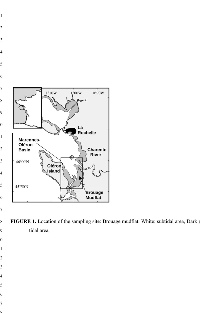
The area selected for collecting foraminifera and Hydrobia ulvae was the upper part of
6
the intertidal Brouage mudflat on the French Atlantic coast at latitude 45° 54’ N and longitude
7
1° 7’ W (Fig. 1). This area was selected owing to the high density of living foraminifera (110
8
individuals per cm3, Armynot du Chatelêt, per. comm.) and because Hydrobia ulvae, a 9
common gastropod from European intertidal mudflats, is the most abundant species among
10
the macrofauna (Haubois and others, 2004). This snail is a deposit-feeder that inhabits muddy
11
sand- and mudflats (Hayward and others, 1998). It ingests sediment and egests inorganic
12
particles compacted into small cylinders (feces).
13
Fig. 1 here 14
The sediment sample was collected at low tide by scraping off the first centimeter of
15
sediment in an area where microphytobenthos was abundant (brown film on surface
16
sediment). Seawater was collected in the same area. If the mud snails and foraminifera cannot
17
be sampled at the same time, then it is necessary to maintain a ready stock of mud snails in the
18
laboratory. This is quite easy since they can be kept living for several weeks if placed in
19
sediment in a cold room (5°C). In the laboratory, 1 g of sediment was sieved with seawater
20
through a nylon mesh of 50-µm openings to eliminate clay- and finer silt-size particles. To
21
eliminate additional fine grains, the remaining material was gently stirred in filtered (0.2 µm)
22
seawater and then allowed to stand for several seconds to let the foraminifera settle to the
23
bottom. Supernatant seawater with fine suspended particles was then eliminated, and seawater
24
re-added. This operation was repeated until the supernatant water was clear. Finally, the
processed sediment and foraminifera were distributed in a glass Petri dish (16 cm diameter).
1
Owing to the fragility of foraminiferal tests, it was impossible to stir the sediment strongly.
2
Consequently, the tests remained incased in their fine-particle agglutinated cysts and flocs of
3
fine sediments remained together with rare coarser grain.
4
To test the efficiency of the treatment with Hydrobia, triplicate Petri dishes were
5
prepared, by addition of 40 and 80 specimens of Hydrobia ulvae, respectively. They were
6
placed in a constant-temperature room (18°C) and kept at a light/dark cycle of 12h/12h for 2
7
days. The dishes were observed every day under a dissecting microscope. After 2 days, the
8
number of hard-shelled foraminifera was counted in all dishes and the species were identified.
9
In addition, experiments were carried out to address the question of whether or not the
10
snails are ingesting any foraminifera. Twenty Hydrobia were placed for 48 hours in triplicate
11
Petri dishes, together with sediment collected in an area rich in living foraminifera and sieved
12
like above. At the same time, six other Petri dishes were prepared with the same sediment but
13
without gastropods. Three of these dishes without Hydrobia were used to count the number of
14
foraminifera at the beginning of the experiment, the three others were used as controls. At the
15
end of the experiment, 48 hours later, the number of foraminifera was counted in the six
16
remaining dishes (3 with Hydrobia and 3 controls without). Moreover, 50 Hydrobia were
17
collected in an area rich in living foraminifera and sediment was collected at the same place in
18
order to evaluate the density of living foraminifera. The shells of the Hydrobia collected in the
19
field as well as those of the experiments (triplicate Petri dishes with 20 Hydrobia after 48 h)
20
were broken and their living material was extracted. Because the guts of Hydrobia are very
21
small and difficult to open without potentially breaking foraminiferal tests (if present), we
22
used a process previously devised by one of us (Debenay, pers. comm.) for studying the gut
23
contents of other gastropods: The living material of the gastropods was immersed for 4 days
24
into a sodium hypochlorite solution with available chlorine of ~ 3% in order to remove
organics. Control experiments, which consisted in the immersion of foraminifera with
1
cytoplasm (potentially living) in the same solution, had shown that the tests, even agglutinated
2
ones, were very well preserved after 4 days in the solution (Debenay, pers. comm.).
3
To complete this experiment, six foraminifera were left with 2 H. ulvae for 24 hours in
4
a Petri dish, and then observed to determine if they were still alive (based on pseudopodial
5
activity).
6
RESULTS 7
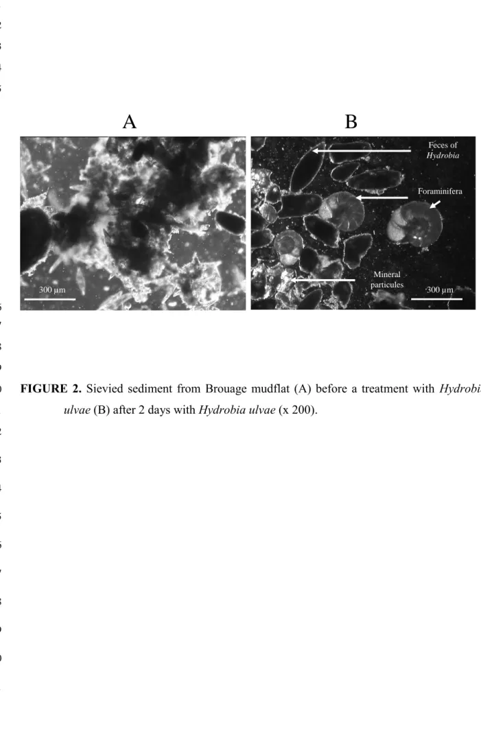
EFFECTOF HYDROBIA ULVAE GRAZINGON THE SEDIMENT 8
During all the experiments with 40 and 80 gastropods, H. ulvae fed on the bottom of the
9
dishes and on the food aggregates surrounding living foraminifera. This activity led to the
10
sorting of the sediment and foraminiferal tests into three components: (1) feces of H. ulvae
11
made up of small cylinders of compacted sediment; (2) clean foraminiferal tests; (3) a few
12
remaining mineral sediment particles.
13
During the first day, feces production began, but the tests were still covered with a layer of
14
fine sediment and/or food and were not easy to distinguish. After two days, snails had cleaned
15
all the foraminiferal tests, which had become very easy to locate and pick (5 seconds per
16
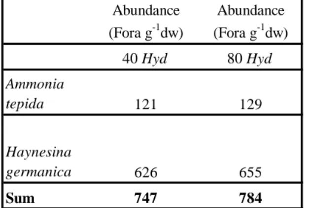
specimen). It was easy to count them (Table 1). No improvement of the cleaning could be
17
observed for experiments lasting more than two days. An additional benefit became evident.
18
The foraminifera were unable to reconstruct their agglutinated cysts after cleaning because all
19
the fine particles of sediment were aggregated. It appeared that the foraminifera had been
20
cleaned but not ingested by the mud snail. In the dishes without H. ulvae, the tests kept their
21
cysts of organic and mineral particles and could hardly be distinguished (Fig. 2). Furthermore,
22
the compaction of the sediment into feces was more efficient with 80 gastropods than with 40.
23
Table 1 here 24
Fig. 2 here 25
EFFECTOF HYDROBIA ULVAE ON THE FORAMINIFERA 1
The experiments were carried out with the same amount of sediment (1 g) in all the
2
Petri dishes. After two days, the number of foraminifera (12 2 Ammonia tepida and 4 2
3
Haynesina germanica, proportions comparable to those found in the natural tidal flat at
4
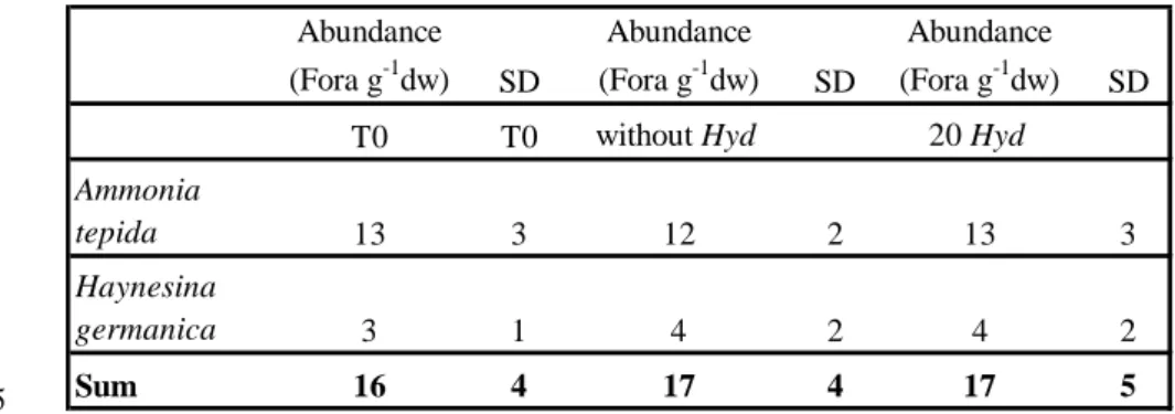
Brouage; Armynot du Chatelêt, per. comm.) was not significantly different between 5
treatments with or without 20 Hydrobia ulvae (Student t test: p> 0.05) and not significantly
6
different from numbers at the start of the treatments (Table 2). The fact that the number of
7
individuals, including juveniles, was not lower in the dishes with Hydrobia suggests that the
8
gastropods did not ingest any foraminifera. This inference is corroborated by the absence of
9
foraminifera in the guts of the 60 Hydrobia from the dishes.
10
Table 2 here 11
The stomach contents of 50 gastropods collected from Brouage tidal flat contained in
12
total only one small test (Ammonia) although foraminifera were abundant and available
13
(~1200 living foraminifera in 50 cm3 - 89% Ammonia tepida, 8% Haynesina germanica, 3% 14
other species). We assume that this lone foraminifera was attached on the shell or snared in
15
the aperture of a snail but was not actually part of the gut contents. These observations
16
suggest that Hydrobia does not ingest foraminifera in the natural environment.
17
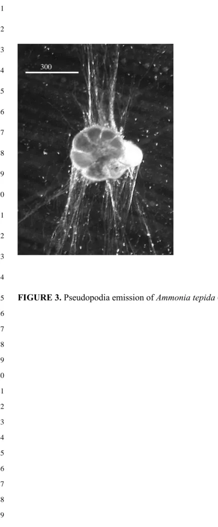
In a further test, six foraminifera were placed together with two H. ulvae and observed
18
at the start and after 24 hours. The foraminifera maintained pseudopodial activity and
19
appeared unharmed by the gastropods (Fig. 3), even though the gastropods had scraped them
20
clean of the agglutinated cysts that incased the tests.
21
Fig. 3 here 22
Several foraminifera extracted with this method were used for a bacterial grazing
23
experiment. They fed normally on bacteria, showing that their health was not perceptibly
affected by the cleaning process. They recovered, returned to their normal behaviors, and,
1
when placed in sediment, they reconstruct their agglutinated cyst.
2 3
DISCUSSION 4
Several of methods for extraction and culture of foraminifera have been published
5
(review in Anderson and others, 1991) but all are highly time-consuming. In this study, we
6
describe a new method for extracting quickly and inexpensively a large number of living
7
benthic shelled foraminifera for culture in the laboratory. This method requires only two hours
8
of actual work to extract 1000 tests (30 minutes for preparation of Petri dishes with sediment
9
containing foraminifera and H. ulvae and 1 h 30 min for picking 1000 tests after the sediment
10
treatment by H. ulvae). The rest of the work is carried out by H. ulvae. In comparison, the
11
picking of 1000 tests from untreated sediment required 20 hours. The grazing activity of
12
Hydrobia ulvae results in the formation of small cylindrical feces of compacted sediment and
13
cleaning of agglutinated cysts from tests of living benthic foraminifera. Most of the organic
14
and inorganic particles, even those in the cysts around foraminiferal tests, are grazed by the
15
mud snails and digested or packed into feces. Small isolated particles are no longer available
16
to the foraminifera for construction of its cyst. The cleaning is harmless to the foraminifera
17
since the gastropod does not have any feeding activity towards the foraminifera. This behavior
18
is different from other gastropods, such as Olivella, that may selectively ingest living
19
foraminifera, as reported by Hickman and Lipps (1983). However, Olivella is much bigger
20
than Hydrobia (2 cm instead of 5 mm). Moreover, unpublished studies have been carried out
21
in the île d’Yeu laboratory on Littorina littorea, Littorina saxatilis, Gibbula umbilicalis and
22
Monodonta lineata, temperate gastropods much bigger than Hydrobia. These studies have
23
shown that very few foraminifera are ingested accidentally by these gastropods when they
24
feed on algae, but that there is no selective ingestion.
After testing the abundance of cleaned tests and the incubation times on a sediment
1
sample of 1 g, we concluded that the process is completed after 2 days with 40 H. ulvae.
2
About the same number of tests was cleaned in the same amount of time by either 40 or 80 H.
3
ulvae. The only benefit of using 80 snails rather than 40 was production of feces that were
4
more compacted, which facilitated the picking of living foraminifera. After this treatment,
5
recovery of living specimens with a fine brush was much easier and faster. Moreover, even
6
smaller specimens were clearly discernable, which is not the case when they are hidden in
7
cysts of sediment particles. For the same treatment time (2 days), it is possible to increase the
8
quantity of sediment treated by increasing the abundance of H. ulvae in bigger Petri dishes.
9
The active feeding of H. ulvae in Petri dishes suggest that this method could be applied to
10
muddy samples in which the mud snail does not occur, such as fine sediments from deeper
11
subtidal habitats or those of the shelf or slope. We suggest too that this method might be
12
adaptable in other coastal and brackish environments where H. ulvae does not live, by using
13
other small deposit feeders, such as Hydrobia salsa and Hydrobia totteni in USA, Hydrobia
14
knysnaensis in South Africa, and Hydrobia buccinoides in Australia. However, these
15
applications require testing.
16 17
ACKNOWLEDGMENTS 18
This work was supported by the CNRS, contract PEVS and supported by research
19
national program ECCO. We thank Jennifer Guarini for reviewing the English. We are
20
grateful to C.A. Brunner and the Associated Editor of JFR for their help in improving the last
21
version of the manuscript.
22 23
REFERENCES 1
ANDERSON, O. R., LEE, J. J., and FABER, W. W. JR., 1991, Collection, maintenance and 2
culture methods for the study of living foraminifera, in Lee, J. J., and Anderson, O.
3
R. (eds.), Biology of Foraminifera,: Academic Press, p. 335-357.
4
ARNOLD, Z. M., 1954, Culture methods in the study of living Foraminifera: Paleontology, v. 5
28, p. 404-416.
6
ARNOLD, Z. M., 1974., Field and laboratory techniques for the study of living foraminifera, in 7
Hedley, R.H., and Adams, C.G. (eds.), Foraminifera: Academic Press, London., v. 1,
8
p. 153-206.
9
BENDER, H., and HEMLEBEN, C. H., 1988, Calcitic cement secreted by agglutinated 10
foraminifers grown in laboratory culture: Journal of Foraminiferal Research, v. 18, p.
11
12-45.
12
BERNHARD, J. M., 2000, Distinguishing live from dead foraminifera: Methods review and 13
proper applications: Micropaleontology, v. 46, p. 38-46.
14
BIJMA, J., FABER, W. W. Jr., and HEMLEBEN, C., 1990, Temperature and salinity limits for 15
growth and survival of some planktonic foraminifers in laboratory cultures: Journal
16
of Foraminiferal Research , v. 20, p. 95-116.
17
BOWSER, S. S., ALEXANDER, S. P., STOCKTON, W. L., and DELACA, T. E., 1992, Extracellular 18
matrix augments mechanical properties of pseudopodia in the carnivorous
19
foraminiferan Astrammina rara: role in prey capture: Journal of Protozoology, v. 39,
20
p. 724-732.
21
BRADSHAW, J. S., 1961, Laboratory experiments on the ecology of foraminifera: 22
Contributions from the Cushman Foundation for Foraminiferal Research v. 12, p.
23
87-106.
BUZAS, M. A., 1978, Foraminifera as prey for benthic deposit feeders: results of predator 1
exclusion experiments: Journal of Marine Research, v. 36, p. 617-625.
2
CAREY, P. G., 1993, Long-Term Culture of Marine Benthic Protists: in Kemp, P.F., Sherr, 3
B.F., Sherr, E.B., and Cole J.J. (eds.), Handbook of Methods in Aquatic Microbial
4
Ecology, Lewis Publishers, CRC Press, Florida, USA, p. 213-227.
5
HAUBOIS, A. G., GUARINI, J. M., RICHARD, P., HEMON, A., AROTCHAREN, E., and 6
BLANCHARD, G., 2004, Differences in spatial structures between juveniles and adults 7
of the gasteropods Hydrobia ulvae on an intertidal mudflat (Marennes-Oléron Bay,
8
France) potentially effect estimates of local demographic processes: Journal of Sea
9
Research, v. 51, p. 63-68.
10
HAYWARD, P., NELSON SMITH, T., and SHIELDS, C., 1996, Guide des bords de mer: Ed 11
Delachaux et Niestlé, Genève, p. 1-351.
12
HEINZ, P., HEMLEBEN, C., and KITAZATO, H., 2002, Time-response of cultured deep-sea 13
benthic foraminifera to different algal diets: Deep-Sea Research, v. 49, p. 517-537.
14
HICKMAN, C.S., and LIPPS, J.H. 1983, Foraminiferivory: selective ingestion of Foraminifera 15
and test alterations produced by the neogastropod Olivella: Journal of Foraminiferal
16
Research, v. 13, p. 108-114.
17
Jepps, M. W., 1942, Studies on Polystomella Lamarck (Foraminifera): Journal of Marine
18
Biology Association U. K., v. 25, p. 612-665.
19
KHARE, N., and NIGAM, R., 2000, Laboratory experiment to record rate of movement of 20
cultured benthic foraminifera: Oil and Natural Gas Corporation Bulletin, v. 37, p.
21
53-61.
22
LE CALVEZ, J., 1938, Recherche sur les foraminifères I: Développement et reproduction: 23
Archives de Zoologie Expérimentale et Générale, v. 80, p. 163-333.
LINKE, P., ALTENBACH, A.V., GRAF, G., and HEEGER, T., 1995, Response of deep-sea 1
benthic foraminifera to a simulated sediment event: Journal of Foraminiferal
2
Research, v. 25, p. 75-82.
3
MOODLEY, L., BOSCHKER, H. T. S., MIDDELBURG, J. J., PEL R., HERMAN, P. M., DE 4
DECKERE, E., and HEIP, C. H. R., 2000, Ecological significance of benthic 5
foraminifera: 13C labelling experiments: Marine Ecology Progress Series, v. 202, p.
6
289-295.
7
MURRAY, J. W., and BOWSER, S. S., 2000, Mortality, protoplasm decay rate, and reliability 8
of staining techniques to recognize 'living' foraminifera: a review: Journal of
9
Foraminiferal Research, v. 30, p. 66-70.
10
MYERS, E. H., 1935, Culture methods for the marine foraminifera of the littoral zone: 11
Transactions of the American Microscopical Society v. 54, p. 264-267.
12
STOUFF, V., DEBENAY, J. P., and LESOURD, M., 1999, Origin of double and multiple tests in 13
benthic foraminifera: observations in laboratory cultures: Marine Micropaleontology,
14 v. 36, p. 189-204. 15 16 17 18 19 20 21 22 23 24 25
1 2 3 4 5 6 7 8 9 10 11 12 13 14 15 16 17
FIGURE 1. Location of the sampling site: Brouage mudflat. White: subtidal area, Dark gray: 18 tidal area. 19 20 21 22 23 24 25 26 27 28 Marennes -Oléron Basin La Rochelle Brouage Mudflat Charente River Oléron Island 46°00'N 1°10W 1°00W 0°90W 45°50'N
1 2 3 4 5
A
B
Foraminifera Feces of Hydrobia Mineral particules 300 µm 300 µm 6 7 8 9FIGURE 2. Sievied sediment from Brouage mudflat (A) before a treatment with Hydrobia 10
ulvae (B) after 2 days with Hydrobia ulvae (x 200).
11 12 13 14 15 16 17 18 19 20 21
1 2 3 4 5 6 7 8 9 10 11 12 13 14
FIGURE 3. Pseudopodia emission of Ammonia tepida (x 200). 15 16 17 18 19 20 21 22 23 24 25 26 27 28 29 300
TABLE 1. Abundance of foraminifera (Fora g-1dw) (dw: dry weight) in the sediment after 1
treatment with different densities of Hydrobia (Hyd).
2 3 4 5 Abundance (Fora g-1dw) Abundance (Fora g-1dw) 40 Hyd 80 Hyd Ammonia tepida 121 129 Haynesina germanica 626 655 Sum 747 784 6 7 8 9 10 11 12 13 14 15 16 17 18 19 20 21
TABLE 2. Abundance of foraminifera (Fora g-1dw) (dw: dry weight) with standard deviation 1
(SD) in the sediment before (T0) and after 48 h treatment without and with Hydrobia (Hyd).
2 3 4 Abundance (Fora g-1dw) SD Abundance (Fora g-1dw) SD Abundance (Fora g-1dw) SD
T0 T0 without Hyd 20 Hyd
Ammonia tepida 13 3 12 2 13 3 Haynesina germanica 3 1 4 2 4 2 Sum 16 4 17 4 17 5 5 6