HAL Id: hal-02113018
https://hal.archives-ouvertes.fr/hal-02113018
Submitted on 27 Apr 2019
HAL is a multi-disciplinary open access
archive for the deposit and dissemination of
sci-entific research documents, whether they are
pub-lished or not. The documents may come from
teaching and research institutions in France or
abroad, or from public or private research centers.
L’archive ouverte pluridisciplinaire HAL, est
destinée au dépôt et à la diffusion de documents
scientifiques de niveau recherche, publiés ou non,
émanant des établissements d’enseignement et de
recherche français ou étrangers, des laboratoires
publics ou privés.
The Notch pathway in the annelid Platynereis : insights
into chaetogenesis and neurogenesis processes
Eve Gazave, Quentin Lemaître, Guillaume Balavoine
To cite this version:
Eve Gazave, Quentin Lemaître, Guillaume Balavoine. The Notch pathway in the annelid Platynereis :
insights into chaetogenesis and neurogenesis processes. Open Biology, Royal Society, 2017, 7 (2),
pp.160242. �10.1098/rsob.160242�. �hal-02113018�
rsob.royalsocietypublishing.org
Research
Cite this article: Gazave E, Lemaıˆtre QIB,
Balavoine G. 2017 The Notch pathway in the
annelid Platynereis: insights into chaetogenesis
and neurogenesis processes. Open Biol. 7:
160242.
http://dx.doi.org/10.1098/rsob.160242
Received: 19 August 2016
Accepted: 3 January 2017
Subject Area:
developmental biology/molecular biology/
cellular biology/genomics
Keywords:
Notch, Platynereis, neurogenesis,
chaetogenesis, lateral inhibition
Author for correspondence:
Guillaume Balavoine
e-mail: guillaume.balavoine@ijm.fr
Electronic supplementary material is available
online at
http://dx.doi.org/10.6084/m9.fig-share.c.3667960.
The Notch pathway in the annelid
Platynereis: insights into chaetogenesis
and neurogenesis processes
Eve Gazave, Quentin I. B. Lemaıˆtre and Guillaume Balavoine
Institut Jacques Monod, CNRS, UMR 7592, Univ Paris Diderot, Sorbonne Paris Cite´, 75205 Paris, France EG, 0000-0002-4647-6640; GB, 0000-0003-0880-1331
Notch is a key signalling pathway playing multiple and varied functions during development. Notch regulates the selection of cells with a neurogenic fate and maintains a pool of yet uncommitted precursors through lateral inhi-bition, both in insects and in vertebrates. Here, we explore the functions of Notch in the annelid Platynereis dumerilii (Lophotrochozoa). Conserved com-ponents of the pathway are identified and a scenario for their evolution in metazoans is proposed. Unexpectedly, neither Notch nor its ligands are expressed in the neurogenic epithelia of the larva at the time when massive neurogenesis begins. Using chemical inhibitors and neural markers, we demonstrate that Notch plays no major role in the general neurogenesis of larvae. Instead, we find Notch components expressed in nascent chaetal sacs, the organs that produce the annelid bristles. Impairing Notch signalling induces defects in chaetal sac formation, abnormalities in chaetae producing cells and a change of identity of chaeta growth accessory cells. This is the first bilaterian species in which the early neurogenesis processes appear to occur without a major involvement of the Notch pathway. Instead, Notch is co-opted to pattern annelid-specific organs, likely through a lateral inhibition process. These features reinforce the view that Notch signalling has been recruited multiple times in evolution due to its remarkable ‘toolkit’ nature.
1. Background
Since developmental biology met molecular and cellular biology, considerable efforts have been deployed in understanding signalling pathways function, modularity, architecture and later evolution. Among them, the Notch pathway has been especially investigated in these frameworks. As a direct juxtacrine sig-nalling system, it provides a mechanism for short-range, localized sigsig-nalling between directly apposing cells in a process called ‘trans-activation’ [1– 5]. Upon binding of a ligand (Delta or Jagged) displayed by a neighbouring cell, the Notch transmembrane receptor is cleaved by the g-secretase complex ( presenilin-nicastrin-APH1-PEN2) resulting in the release of the Notch intra-cellular domain (NICD) into the cytoplasm (additional cleavage of Delta by the g-secretase complex has been also described in vertebrates [6]). After its translocation to the nucleus of the receiving cell, NICD interacts with the CSL (CBF1, suppressor of hairless (Su(H)), Lag-1)/Ncor/SMRT/histone deace-tylase transcriptional complex and activates the transcription of target genes. The same ligands can also interact with the receptor Notch within the same cell (in cis). The Notch pathway functions with a number of ‘core component’ proteins, whose evolutionary emergence has been studied in detail [7]: it was probably already functional as early as in the last common ancestor of all metazoans, but has its roots anchored deep in the eukaryote tree as some non-metazoan unikonts display members of these components.
Expression and functional studies of Notch pathway components during development have been performed in few metazoan species, mostly in
&
2017 The Authors. Published by the Royal Society under the terms of the Creative Commons Attribution License http://creativecommons.org/licenses/by/4.0/, which permits unrestricted use, provided the original author and source are credited.deuterostomes and ecdysozoans. In these two lineages, the remarkably pleiotropic Notch pathway affects cell fate choices associated with lineage segregation, cell differentiation, cell proliferation or apoptosis in many tissues and developmental stages [8–11]. The extreme versatility and pleiotropy of Notch signalling seems at first glance to make the task of recon-stituting an evolutionary history of its involvement in metazoan development rather daunting. In many ways, the Notch pathway could be described as a developmental ‘Swiss army knife’, a generic tool that has been co-opted multiple times in evolution, in various animal lineages, for unrelated or sometimes convergent functions in development. However, comparisons of the roles of Notch between insect and vertebrate have brought to light interesting similarities that have led to speculations about an ancestral involvement of Notch signalling in neurogenesis [12].
Notch is crucial in the early patterning of neurogenesis both in the fruit fly and in vertebrates. The study of Drosophila neurogenesis in particular has led to uncovering a mechanism called ‘lateral inhibition’. In the fly embryo, the central ner-vous system originates from delaminating neural progenitors [13,14], called neuroblasts. These divide asymmetrically to give birth to further dividing neuron precursors (the ganglion mother cells). Neuroblasts are active during both embryogen-esis and larval development, playing the role of neural stem cells. Delaminating neuroblasts are initially specified in clus-ters of ectodermal epithelial cells, called ‘proneural clusclus-ters’. These cells are initially bipotent, giving either a neuroblast or, for the majority of them, epidermal cells. The cell that will eventually become a neuroblast starts to express higher levels of the ligand transmembrane protein Delta, which binds to the Notch transmembrane protein present at the sur-face of neighbouring cells. In these neighbouring cells, the activation of the Notch pathway results in the downregulation of both Delta and neurogenic gene expressions, thus inhibiting the neural fate. In vertebrate embryos, early neurogenesis pro-cesses take place mostly in the neural tube after it has completed its closure. In the thick pseudostratified epithelium of the neural tube, a gradient of Notch1 expression, maxi-mal apically, keeps neurogenic specification on the apical/ ventricular side [15], while differentiating neuron bodies migrate to the basal side. On the apical side, where both sym-metrical and asymsym-metrical cell divisions take place, lateral inhibition by Notch/Delta regulates the pace of neuronal specification, maintaining a pool of neural progenitors [16,17]. This mechanism of lateral inhibition by Notch/Delta has been evidenced in a number of contexts, such as hair cell formation in the inner ear [18]. Interestingly, the inner ear development is also mediated by another mechanism of the Notch pathway called lateral induction, through the alternate ligand Jagged1. Such mechanisms, finely modulated by cis-interactions, exemplify the notion of the Notch pathway as a general and versatile developmental tool that could poss-ibly have been co-opted for playing similar functions. However, there is now considerable evidence that both neurogenesis [19] and nervous system centralization [20] in protostomes and vertebrates share a common origin. In this context, Notch is often seen as a pathway ancestrally involved in neurogenesis patterning in bilaterians. This hypothesis receives support also from cnidarians that have a dispersed nervous system organization [21]. In the sea anemone Nema-tostella, the Notch pathway appears to play a role in regulating the abundance of differentiating nerve cells, even
if the exact roles of specific pathway components remain dis-puted [22 –24]. This pushes the involvement of Notch in nervous system formation back to ancestors of eumetazoans (cnidarians þ bilaterians), at the very evolutionary origin of the nervous system.
While plenty of studies focused on the various aspects of Notch pathway mechanisms and regulations among deuter-ostomes and ecdysozoans, the third large group of bilaterians, i.e. the lophotrochozoans, has been barely studied. The Notch pathway architecture and functions have been investigated so far in two annelid species only. In the leech Helobdella robusta, the Notch pathway has been proposed to be involved in posterior elongation and segment formation [25,26]. In Capitella teleta, the expression patterns of the Notch pathway compo-nents (discussed later in the article) in brain, foregut, chaetal sacs and posterior growth zone suggest a possible involvement of this pathway in brain development, chaetogenesis and segmentation processes [27].
In this study, we analysed the functions of the Notch path-way in the marine annelid Platynereis dumerilii, a leading lophotrochozoan model species for evolution and develop-mental biology. Platynereis dumerilii is considered to be a ‘short branch’ organism that has kept a number of ancestral-looking characters at the genomic [28], anatomical and developmental levels [20,29,30], making it a highly attractive model system for understanding the origin and diversification of these traits. P. dumerilii displays an indirect form of develop-ment (for a review, see [31]): embryogenesis first gives rise to a minute spherical trochophore larva with a small number of dif-ferentiated larval structures, including a precise arrangement of a few neuronal and sensory cells, such as the apical organ or the posterior pioneer neurons [20,32,33]. Later than this larval nervous system, a wave of massive ‘adult’ neurogenesis takes place in the mid-trochophore stage, at two different locations: the episphere at the animal pole, which will give rise to the anterior brain of the worm, and the ventral neural ectoderm, which will give rise to the ganglia of the ventral nerve cord (VNC) [20]. Ventral neurogenesis takes place in thickening stratified or pseudo-stratified epithe-lia where mitoses happen on the apical side and neuron differentiations occur on the basal side, similar to what has been described for the vertebrate neuroepithelium [20,34]. In addition, Platynereis neurogenesis involves a number of the same transcription factors that have already been identified in insect and vertebrate models such as the bHLH NeuroD, neurogenin and achaete-scute-related genes [33].
The aim of this study is to explore the roles of the Notch pathway in Platynereis and to shed light on the ancestral role(s) of Notch in lophotrochozoans and bilaterians (we do not describe here a potential role in posterior elongation and segment formation that require further study in the juvenile worm). Taking advantage of small molecule inhibi-tors, we show here that the Notch signalling pathway does not seem to play a major generic role in the early processes of larval or ‘adult’ neurogenesis in the annelid, even though roles in the formation of a few specific head and trunk neurons cannot be excluded. Instead, at the time when neurogenesis happens, we find a role of Notch in the correct patterning of the cells of chaetal sacs, likely through a lateral inhibition mechanism. Chaetal sacs are the larval and adult organs that are responsible for the formation of chaetae, i.e. locomotory and mechanosensory bristles of the worm. Our results thus challenge the view that the Notch
rsob.r
oy
alsocietypublishing.org
Open
Biol.
7:
160242
2pathway is necessarily involved in nervous system patterning in the same way at a eumetazoan scale. Our data rather sup-port the view of co-options in regulating cell fate specification in multiple contexts.
2. Results
2.1. Notch pathway core components identification: a
lophotrochozoan focus
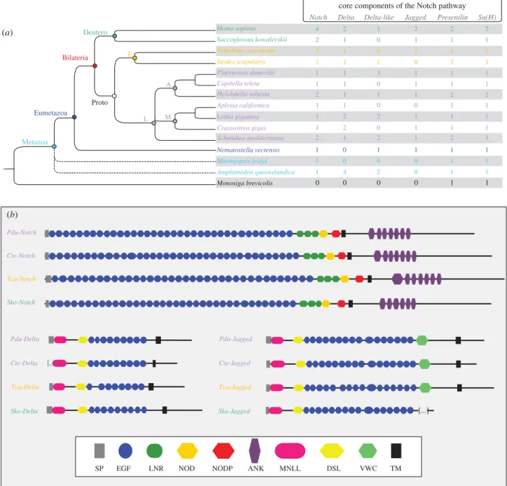
Exhaustive searches on the genome and several transcrip-tomes of P. dumerilii led us to identify core components of the Notch pathway i.e. the receptor Notch (Pdu-Notch,
already identified [28]), the two ‘classical’ ligands Pdu-Delta and Pdu-Jagged, Pdu-Presenilin (component of the g-secretase complex) and the transcription factor suppressor of hairless (Pdu-SuH; figure 1a; electronic supplementary material, figure S1A). In addition, we found three Delta-like genes (i.e. lacking the minimal domain arrangement defined below). We also identified the intracellular regulator Nrarp (Pdu-Nrarp), a gene encoding a small ankyrin (ANK)-repeat protein that is part of a negative feedback loop that attenuates Notch activity in vertebrates [35], the antagonist Numb (Pdu-Numb) and the post-translational modifier Fringe (Pdu-Fringe) (electronic supplementary material, figure S1A). Platynereis Delta, Jagged and Notch possess conserved domain arrange-ments that are very similar to other bilaterian species and
core components of the Notch pathway
Notch Delta Jagged Presenilin Su(H)
Pdu-Notch Pdu-Delta Cte-Notch Cte-Jagged Cte-Delta Tca-Notch Tca-Jagged Tca-Delta Sko-Notch Sko-Jagged Sko-Delta Pdu-Jagged
EGF LNR NOD NODP ANK MNLL DSL VWC
SP TM [...] [...] Delta-like Homo sapiens Tribolium castaneum Saccoglossus kowalevskii Platynereis dumerilii Helobdella robusta Capitella teleta Aplysia californica Nematostella vectensis Crassostrea gigas Lottia gigantea Ixodes scapularis Amphimedon queenslandica Mnemiopsis leidyi Monosiga brevicolis Schmidtea mediterranea M Proto E Deutero L Metazoa Bilateria Eumetazoazoa azoa E A 4 2 1 2 0 2 2 2 2 2 2 2 2 2 1 2 2 4 1 1 1 1 1 1 1 1 1 1 1 1 1 1 1 1 1 1 1 1 1 1 1 1 1 1 1 1 1 1 1 1 1 1 1 1 1 1 1 1 1 1 1 1 1 1 1 1 1 1 1 0 2 0 2 0 0 0 0 0 0 0 0 1 0 0 1 0 0 0 3 3 (b) (a)
Figure 1. Core components of the Notch pathway in metazoans. (a) The presence/absence and number of gene copies at a metazoan scale for the Notch pathway
main components. Fifteen species representatives of the metazoans, with a lophotrochozoan focus, are included. Dashed lines show the unresolved phylogenetic
positions for sponges and ctenophores. (b) Domain arrangement of Notch, Delta and Jagged proteins in four bilaterian species are schematized. See figure inset for
the domain legends. Pdu-Notch presents 36 EGF repeats, three LNR, one NOD domain, one NODP domain and seven ANK repeats. For Pdu-Delta and Pdu-Jagged, we
detected the MNLL region, a Delta/Serrate/Lag (DSL) domain (which mediates binding to Notch receptors in bilaterians) and a series of EGF repeats (nine for
Pdu-Delta and 16 for Pdu-Jagged). In addition to these domains, Pdu-Jagged also contains a Von Willebrand factor C domain (VWC) characteristic of Serrate/Jagged
proteins. The choanoflagellate is in black, the sponge and ctenophore are in light blue, the cnidarian is in blue, lophotrochozoans are in purple, ecdysozoans are in
orange and deuterostomians are in green. Deutero, deuterostomes; Proto, protostomes; E, ecdysozoans; L, lophotrochozoans; A, annelids; M, molluscs.
rsob.r
oy
alsocietypublishing.org
Open
Biol.
7:
160242
3likely to be ancestral in the bilaterian lineage (figure 1b). Interest-ingly, we identified two splice variants for Pdu-Delta (Pdu-Deltatv1 and Pdu-Deltatv2; electronic supplementary material,
figure S1B). In Pdu-Deltatv2, retention of the last intron led to a
shorter sequence lacking the final short ATEV peptide. Despite extensive and specific searches, no other typical Notch, Jagged or Delta proteins have been evidenced.
The origin and evolution of the components and auxiliary factors of the Notch pathway have previously been investigated at a large scale [7]. We performed here a detailed search of Notch pathway core components in metazoan species repre-sentative of all main metazoan lineages (Deuterostomia, Ecdysozoa, Lophotrochozoa, Ctenophora, Cnidaria and Pori-fera), but surveyed more extensively lophotrochozoan species as they have been undersampled in earlier studies due to a lack of available genomes. Data comparisons among lophotro-chozoan, ecdysozoan and deuterostomian species are of special interest in order to reconstruct bilaterian ancestral states (figure 1a). We identified a single unambiguous Notch ortholo-gue in all metazoans species investigated, except in vertebrates, in the enteropneust Saccoglossus kowalevskii, in the annelid H. robusta and in the planarian Schmidtea mediterranea. A more complicated situation emerged for the evolution of Delta and Jagged proteins. In all species but the ctenophore, several Delta-related proteins with various domain compositions are identified. One particular domain composition (MNLL-DSL-(9)xEGF-TM-ATEV), in which not only epidermal growth factor (EGF) motif numbers but also their specific spacings are conserved, presumably corresponds to an ancestral Delta protein that was already present in the bilaterian ancestor [36]. Our study supports this interpretation; indeed genes corre-sponding to this proposed ancestral organization are present in several bilaterian species we sampled. In addition, the EGF domains generally follow the pattern of repeat spacings and cysteine residue spacings noticed previously [36] (electronic supplementary material, figure S1C). As noted above, a con-served alternative splicing upstream of the ATEV peptide is found in both Platynereis and vertebrate proteins, which suggests that this particular splicing is as ancient as the bilater-ian ancestor. In addition to these ancestral Delta proteins, a number of bilaterian genomes code for ‘Delta-like’ proteins (three in Platynereis, two in H. robusta, S. mediterranea and L. gigantea, one in I. scapularis, and the human Dll3), displaying domain variations, the most common being the loss of the ATEV motif and of a number of EGF repeats. There is no indi-cation that orthology relationships can be traced back to the bilaterian ancestors for any of these Delta-like proteins. Jagged proteins are absent outside eumetazoans (alternative scenarios for their emergence have been already discussed [7]) and have been lost or diverged beyond recognition in some bilaterians, such I. scapularis and Aplysia californica (figure 1a).
Phylogenetic analyses of metazoan Delta and Jagged ligands (electronic supplementary material, figure S1A) are based on a very limited length of sequences: only the domains correspond-ing to the MNLL and Delta/Serrate/Lag (DSL). Yet, the limited resolution these trees show is compatible with the interpret-ations given above. Briefly, phylogenetic analyses show that Pdu-Delta and Pdu-Jagged are included in robust Delta and Jagged clades, respectively. Sponge Deltas are clustered and form the sister group of the eumetazoan Delta þ Jagged groups (approximate likelihood-ratio test (aLRT) . 0.90). Inside the Delta and Jagged clades, relationships are not fully
resolved but Platynereis and Capitella sequences always group together. We thus tried unsuccessfully to infer the evolutionary origin of the three Delta-like genes found in Platynereis (elec-tronic supplementary material, figure S1A). The phylogenetic relationships between Platynereis Notch receptor and other metazoan receptors are provided in the electronic supplemen-tary material, figure S1A. As previously noticed [7], the relationships among Notch receptors are not perfectly well resolved and do not follow the metazoan species tree. However the orthology of the Platynereis Notch is not questionable (elec-tronic supplementary material, figure S1A).
To sum up, core components of the Notch pathway in the lophotrochozoan Platynereis appear to be very similar to the ancestral situation of bilaterians. Therefore, investigating its functions in Platynereis is of special relevance to draw evolutionary conclusions.
2.2. The components of the Platynereis Notch pathway
are not generally expressed in neurogenic tissues
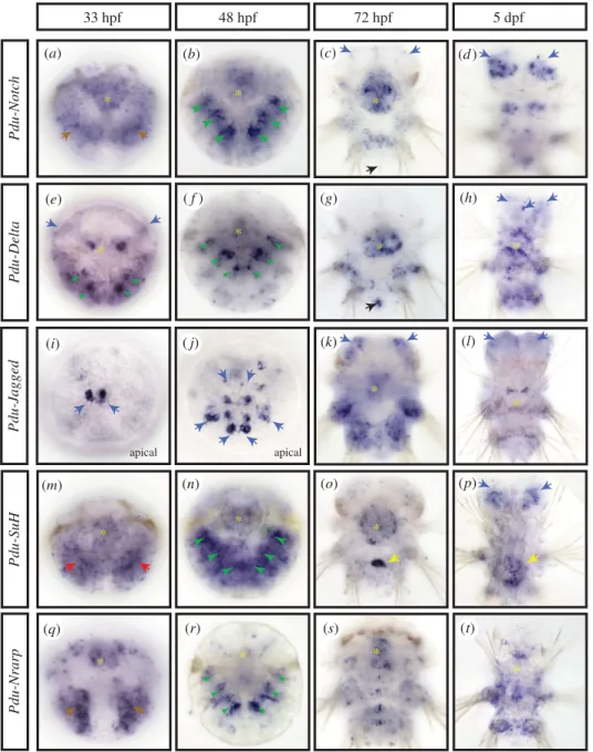
but they are expressed in forming chaetal sacs
We analysed by whole-mount in situ hybridization (WMISH) the expression patterns of five genes, Pdu-Notch, Pdu-Delta, Pdu-Jagged, Pdu-SuH and Pdu-Nrarp (figure 2). We targeted developmental stages when mass adult neurogenesis is taking place: at 33 hpf (hours post-fertilization; mid trocho-phore), the anterior episphere (mostly the future brain of the worm) and the ventral ectoderm (the future nerve cord) start to thicken and numerous mitoses occur superficially; at 48 hpf (late trochophore), neuronal differentiation has started and cell proliferation becomes more localized along the midline; at 72 hpf (three segment nectochaete larva), many brain and nerve cord neurons have differentiated but cell proliferation and thus neurogenesis continue locally and superficially; at 5 dpf (days post-fertilization; late nectochaete larva), cell proliferation and differentiation continue in the brain [20,34,37,38]. The Notch core component expression pat-terns can be categorized in three main structures of the larva: the chaetal sacs, some brain cells including the apical organ and the stomodeum.All genes but Pdu-Jagged are expressed in specific struc-tures called chaetal sacs. The chaetal sacs are responsible for producing chaetae, the retractile chitinous bristles dis-played by the annelid leg-like appendages ( parapodia) that help in locomotion. Chaetal sacs appear by invagination of ectodermal pockets [39]. In the late trochophore stage (48 hpf), when the chaetal sacs are fully formed and produ-cing the first chaetae, Pdu-Notch, Pdu-Delta and Pdu-Nrarp are found in two bilateral ensembles of six patches of expression (figure 2b,f,n,r; green arrowheads). These lateral patches coincide with the 12 chaetal sacs of the larvae: two per future hemi-segment, six in the future neuropodia and six in the future notopodia (the ventral and dorsal moieties of the parapodia). All three gene expressions appear a little stronger in the ventral sacs (figure 2b,f,n,r; green arrow-heads). Pdu-Delta expression is more restricted compared with Pdu-Notch and Pdu-Nrarp (figure 2f, green arrowheads) in a few deep cells of the chaetal sac and is observable early (at 33 hpf ) in four to six bilateral and internalized patches of cells (figure 2e, green arrowheads). Pdu-Su(H) expression while being widespread is more intense in the chaetal sac areas at 48 hpf (figure 2n, green arrowheads).
rsob.r
oy
alsocietypublishing.org
Open
Biol.
7:
160242
4Most of the genes studied (all but Pdu-Nrarp) are also expressed in different populations of brain cells and/or apical organ cells (a sensory structure composed of neurons and cili-ary tuft [40] and present in many marine invertebrate larvae [32]), at several stages. Indeed, at 33 hpf, Pdu-Delta is expressed in few lateral brains cells (figure 2e, blue arrows), while Pdu-Jagged is found in two small groups of different brain cells (figure 2i, blue arrows, apical view). At 48 hpf, the Pdu-Jagged expression pattern expands in the brain, and forms several rows of cells inside and surrounding the apical organ (figure 2j, blue arrows, apical view; electronic supplementary material, figure S2A, internal dotted circle). Co-localization experiments reveal that some of those Pdu-Jagged þ cells in the apical organ are serotoninergic, FMRFAmidergic and RYa peptidergic neurons (electronic supplementary material, figure S2A (v to x), internal dotted circle, purple arrow). Other brain cells expressing Pdu-Jagged are cholinergic neurons (electronic supplementary material, figure S2A (y), orange
arrows). Later, at 72 hpf, the Notch receptor and the two ligands are expressed in several populations of brain cells (figure 2c,k, blue arrows; electronic supplementary material, figure S2A (b to d)). Co-localization with the c-amidated dipeptide RYa neuro-peptide antibody [41] allows us to identify a small number of peptidergic neurons among the Pdu-Notchþ brain cells (elec-tronic supplementary material, figure S2A (g to h), orange arrow). Similarly, Pdu-Delta þ cells are most probably but not exclusively FMRFAmidergic, FLAmidergic and cholinergic neurons (electronic supplementary material, figure S2A (i to o), internal dotted circle, purple arrows). Pdu-Delta and Pdu-Notch are also co-expressed in two bilateral patches of brain cells of unknown identity (electronic supplementary material, figure S2A (e), orange arrows). At this stage, Pdu-Jagged expression is wider in the brain, where it is broadly co-expressed with Pdu-Notch but not Pdu-Delta (figure 2k, blue arrows; electronic supplementary material, figure S2A (d and f)). In addition, Pdu-Delta is found in pyramidal cells of the
Pdu-Delta Pdu-Notch Pdu-Nrarp Pdu-SuH 33 hpf 48 hpf 72 hpf * * * * * * * * * * * Pdu-Jagged apical apical 5 dpf * * * * * (k) (n) (l) (a) (b) (d ) (e) ( f ) (g) (h) (i) ( j) (m) (o) (p) (r) (q) (s) (t) (c)
Figure 2. Expression patterns of the main components of the Notch pathway during Platynereis larval development. WMISH for Pdu-Notch (a – d), Pdu-Delta (e – h),
Pdu-Jagged (i – l), Pdu-Su(H) (m – p) and Pdu-Nrarp (q – t), are shown at four larval stages (33, 48, 72 hpf and 5 dpf ). All panels are ventral views (anterior is up)
except (i and j ) that are apical (dorsal is up). Yellow asterisks mark expression in the stomodeum/forming pharynx. Brown arrows indicate an expression in the
ectoderm, green arrowheads in the chaetal sacs, blue arrows in brain cells, red arrows in the mesoderm, black arrows in the putative mesoteloblasts and yellow
arrows in a cluster of midgut cells.
rsob.r
oy
alsocietypublishing.org
Open
Biol.
7:
160242
5apical organ also expressing Pdu-Notch (electronic supplemen-tary material, figure S2A (e), internal dotted circle, purple arrow). At 5 dpf, a long time after the main wave of neuronal differentiation occurred, widespread expression of Pdu-Notch appears in the brain (figure 2d, blue arrows). At this stage, Pdu-Delta, Pdu-Jagged and Pdu-Su(H) have also an expression in a few brain cells (figure 2h,l,p blue arrows).
Remarkably, all genes are expressed in the stomodeum and/ or pharynx at several stages, either broadly (Pdu-Notch) or in few specific cells (Pdu-Delta; figure 2, yellow asterisks). Finally, at 72 hpf, Pdu-Notch and Pdu-Delta expressions are found in pos-terior internal cells that correspond to the mesoteloblasts (figure 2c,g black arrows), as evidenced by the coexpression with Vasa and Smb, two stem cell markers [42] (electronic supplementary material, figure S2B (b to j, yellow arrows)).
We thus find no general expression of Notch core com-ponents in tissues that are undergoing the main wave of neurogenesis between 33 and 55 hpf. Strikingly, we find no evidence of expression patterns resembling the grid-like expression of Delta genes in the ventral neural ectoderm of arthropods [43–46] or of the ‘salt and pepper’ expression in the neural tube of vertebrates [47]. Instead, Delta expression is limited at these stages and later ones to a few putative neuronal precursor cells, indicating a restricted role in the specification of a limited number of specific neurons. This surprisingly suggests that the Notch pathway may not be generally involved in Platynereis neurogenesis. Conversely, the prominent expression of Notch core components in chaetal sacs suggests a function in Platynereis chaetal sac patterning.
2.3. Notch pathway chemical disruption in Platynereis:
initial characterization of effects
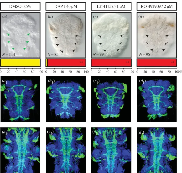
Although the core components of the Notch pathway appar-ently do not show general expression patterns associated with Platynereis larval neurogenesis, we cannot exclude that their expression levels are below the detection level of our WMISH experiments. To test the role of Notch in central nervous system (CNS) formation and chaetogenesis, therefore, we used three pharmacological agents: DAPT, LY-411575 and RO-4929097, all of which are inhibitors of the g-secretase complex [48]. These drugs act by preventing the third cleavage of the Notch receptor, thereby blocking the nuclear relocalization and the activity of NICD as a transcriptional activator in the receiving cell. A large number of larvae (150 at least) were treated in each experiment. As adult neurogenesis and chaetogenesis overlap broadly in time, we applied drugs during a time window encompassing both processes: 24–48 hpf (Time window 1, table 1). During this time period, massive neural ecto-derm proliferation occurs, the neural epithelium thickens considerably and neuronal differentiation starts, with the first elements of the adult neurite scaffold being put in place [34]. The chaetal sacs also start to form from 32 hpf onwards and the production of chaetae starts around 40 hpf [39].
We first tested different concentrations of drugs to determine whether they cause characteristic defects. We con-sidered the lowest concentrations (40 mM DAPT, 2 mM RO-4929097 and 1 mM LY-411575) causing these defects to minimize the potential off-target effects and toxicity of the drugs. Importantly, LY-411575 precipitates above 2 mM, RO-4929097 and DAPT above 40–50 mM in seawater (as already noticed for DAPT [49]). Only RO-4929097 thus
causes defects at a concentration well below solubility pro-blems. Morphological defects were then assessed at 72 hpf, when the three segments, the brain and the VNC are fully formed and all chaetae are externally visible [31], and at 6 days (figure 3).
Treated larvae showed limited but completely similar defects as far as gross morphology and behaviour are con-cerned. At 3 and 6 days, they are well elongated and segmented (figure 3) and show normal muscular contractions, swimming behaviour and phototactism. The swimming speed is faster than normal because treated larvae display very few or completely absent protruding chaetae (in a highly significant way, Student’s test p , 0.01; figure 3a–d). Labellings of the neurite scaffold with an anti-acetylated alpha-tubulin antibody show no gross defects in the head and trunk nervous systems as connectives and commissures appear to form normally at 72 hpf and 6 days (figure 3a1–d2). Consistent results for treated
embryos obtained with all three drugs (with different molecu-lar structures but targeting the same molecule) clearly suggest that their action is specific and that limited off-target effects appear at the chosen concentrations. Nevertheless, to deter-mine whether drug-induced cell death occurred, we used the terminal deoxynucleotidyl transferase dUTP nick end labelling (TUNEL) method and found just a few apoptotic cells in the treated embryos, as for the control (electronic supplementary material, figure S2C (a to a4)).
As Notch plays a role in vertebrates in maintaining stocks of undifferentiated progenitors, it could have also a general role in Platynereis in regulating cell proliferation. To test that, we assessed proliferation at 48 hpf using 5-ethynyl-20 -deoxyuri-dine (EdU) labelling [34] and quantified EdU-labelled cells in structures related to the nervous system (i.e. in the ventral neurectoderm and the episphere), but also in the whole embryo, stomodeum and chaetal sacs (n ¼ 8 for each condition; electronic supplementary material, figure S2C (b to d)). We found few significantly different percentages of EdU þ cells (Student’s test p , 0.05), in treated embryos compared to the control, when looking at the whole embryo, the ventral neurec-toderm or the episphere (electronic supplementary material, figure S2C (d)). Only three comparisons (between dimethyl sulfoxide (DMSO) and DAPT and LY-411575 for the ventral neurectoderm and between DMSO and LY-411575 for the epi-sphere) appeared to give slightly more mitoses in the treated larvae, in a significant way. However, we observed that the total numbers of cells in the whole embryo, the ventral neurec-toderm and the episphere (defined by quantifying DAPIþ cells) are similar in treated embryos and controls (electronic supplementary material, figure S2C (d)).
Together those results tend to indicate that the Notch path-way disruption does not affect considerably either the general VNC or brain formation, nor does it affect in a major way cell proliferation profiles in the ventral and anterior neurectoderm. On the contrary, the disappearance of chaetae rather supports a role in chaetal sac patterning. Those two aspects are extensively studied using specific time windows for treatments (table 1) depending on the process in the following sections.
2.4. Chaetoblast addition is progressive and their
number is finely regulated
To unravel the role of the Notch signalling pathway in the chae-togenesis process, we decided to investigate in depth the
rsob.r
oy
alsocietypublishing.org
Open
Biol.
7:
160242
6Table
1.
Types
of
tr
ea
tment
performed
to
study
specifically
chaetogenesis
and
neur
ogenesis
pr
ocesses.
Four
time
windo
ws
w
er
e
chosen.
Time
windo
w
1
encompass
es
both
pr
ocesses
and
giv
es
a
gener
al
vie
w
of
the
potential
functions
of
the
Notch
pa
thw
ay
in
the
annelid
Pla
tyner
eis
.
Time
windo
w
2
allo
ws
targeting
the
cell
fa
tes
of
chaetal
sa
cs.
Time
windo
w
3
encompasses
the
larval
neur
on
forma
tion.
Time
windo
w
4
encompasses
‘adul
t’
neur
ogenesis.
Fo
r
ea
ch
time
windo
w
and
drug
concentr
ation,
the
phenotype
and
results
observ
ed
at
sev
er
al
stages
ar
e
mentioned.
VNC,
ventr
al
nerv
e
cord;
conc.,
concentr
ation.
drug conc. Time windo w 1: 24 – 48 hpf Time windo w 2: 20 – 48 hpf Time windo w 3: 12/16 – 24 hpf Time windo w 4: 30 – 48 hpf stages phenotypes stages phenotypes stages phenotypes stages phenotypes LY-411575 1 m M 48 hpf follicle cells mark ers expr ession de cr eased; neur ogenesis mark ers expr ession maintained 48 hpf ov er expr ession of chaetoblas t mark er; pr esence of abortiv e chaetoblas ts 24 hpf Ela v expr ession in maintained in 9 neur ons 72 hpf normal number of br ain cells/ number of VNC neur ons slightly enhanced 72 hpf absence of chaetae/normal chaetoblas ts; normal VNC 72 hpf absence of chaetae; dev elopment slightly dela ye d 72 hpf absence of chaetae 6 dpf absence of chaetae/normal VNC RO-4929097 2 m M 48 hpf follicle cells mark ers expr ession de cr eased; neur ogenesis mark ers expr ession maintained 48 hpf absence of chaetae/v ery fe w abortiv e chaetoblas ts 72 hpf absence of chaetae/normal chaetoblas ts; normal VNC 72 hpf absence of chaetae 6 dpf absence of chaetae/normal VNC RO-4929097 30 m M 48 hpf absence of chaetae/dev elopment dela ye d 48 hpf abnormal expr ession of chaetoblas t mark er; pr esence of abortiv e supernumer ary chaetoblas ts 24 hpf Ela v expr ession is maintained in 9 neur ons 72 hpf normal number of br ain cells; dev elopment dela ye d 72 hpf absence of chaetae/dev elopment dela ye d 72 hpf absence of chaetae; dev elopment dela ye d 72 hpf absence of chaetae; dev elopment dela ye d la ter dead DAPT 40 m M 48 hpf follicle cells mark ers expr ession de cr eased; neur ogenesis mark ers expr ession maintained 72 hpf absence of chaetae/normal chaetoblas ts; normal VNC 6 dpf absence of chaetae/normal VNCrsob.r
oy
alsocietypublishing.org
Open
Biol.
7:
160242
7development of the chaetal sacs of Platynereis. Chaetal sacs are composed of several pockets called follicles, each of which pro-duces a single bristle [50]. Each follicle consists of one chaetoblast that builds the bristle by proximal addition of material, mostly chitin and four surrounding follicle cells arranged on top of each other along the chaetae [44] (electronic supplementary material, figure S3A) that possibly add more material along the bristle length. The bristle grows along the canal formed by follicle cells and finally outside of the parapo-dium surface [51]. Morphological and ultrastructural studies revealed that new follicles emerge by internalization of epider-mal surface cells. A central cell, the future chaetoblast, sinks down into the epithelium, surrounded by several cells that will become follicle cells (electronic supplementary material, figure S3A).
We used the properties of the wheat germ agglutinin (WGA), which binds strongly to the b-chitin [39], the main struc-tural component of chitinous chaetae. We thus identified during larval development the position and number of chaetae and, at the proximal end of the bristle, chaetoblasts (figure 4a–e). We also investigated the expression pattern dynamic of Chitin synthase 1 (CS1), a conserved gene encoding an enzyme crucial
for chitin polymerization [52] and expressed specifically in Platynereis chaetoblasts [44] (figure 4f–j). Both stainings give similar results: chaetoblasts and their corresponding chaetae are present in increasing numbers from 33 to 42 hpf (figure 4; electronic supplementary material, figure S3B), first in segments 1 and 2, and then in segment 3. Chaetoblasts first appear near the surface of the larval ectoderm and then sink deeper in the larval body as the chaetal sacs themselves internalize. Chaetoblast numbers are specific for each chaetal sac, depend-ing on segment and dorsal–ventral position (figure 4; electronic supplementary material, figure S3B).
When are chaetal sac cell identities defined? As some chaetoblasts start being functional as early as 33 hpf, their spe-cification and initial internalization must have taken place quite early in larval development. We complemented these stainings with live imaging experiments using the plasma membrane vital dye FM-464 to observe early chaetoblast formation (electronic supplementary material, figure S3C). We detected the presence of ‘rosettes’ of ectodermal cells at the level of the future parapodia, made of one or two central bottleneck-shaped cells that appears to be in the course of internalization, surrounded by six or seven petal-shaped LY-411575 1 µM DAPT 40 µM DMSO 0.5% RO-4929097 2 µM N = 114 N = 85 N = 99 N = 95 0 20 40 60 80 100 0 20 40 60 80 100 0 20 40 60 80 100 0 20 40 60 80 100% ** ** ** (a) (b) (c) (d ) (a2) (a1) (b1) (c1) (d1) (c2) (d2) (b2)
Figure 3. Inhibition of
g-secretase induces defects in bristle formation but no major nervous system phenotype. Ventral views of whole nectochaete larvae (72 hpf )
and 6 dpf worms are shown (anterior is up). Larvae were incubated with DAPT (40
mM, b to b
2), or LY-411575 (1
mM, c to c
2), or RO-4929097 (2
mM, d to d
2) in
DMSO or in DMSO only (control group, a to a
2) from 24 to 48 hpf. Larvae treated with the three drugs targeting the Notch pathway display a clear reduction of the
bristles (a – d). Phenotypic classes were scored as ‘presence of chaetae’ (yellow) and ‘absence or abnormal chaetae pattern’ (red). Bars at the base of each image
represent the percentages of larvae in each phenotypic class. Double asterisks indicate the highly significant differences (Student’s test p
, 0.01) between the mean
numbers of affected versus unaffected larvae in the control and treated groups (sample sizes are indicated on the figure). Antibody labelling against acetylated
tubulin (green) show the axon scaffold of the VNC at 72 hpf (a
1to d
1) and 6 dpf (a
2to d
2). Hoechst nuclear staining is in blue. Larvae treated with the three drugs
display no gross defect in axon guidance, commissural projections or connectives in both stages.
rsob.r
oy
alsocietypublishing.org
Open
Biol.
7:
160242
8cells, already present at the surface of larvae at 27 hpf (elec-tronic supplementary material, figure S3C (a, a0 and a00)). Later in the development deeper roundish structures appear revealing the outlines of the maturing chaetal sacs (electronic supplementary material, figure S3C (b to d00)). We investigated the very early expressions of potential chaetoblast precursor markers (i.e. Pdu-Delta and Pdu-Hes12; figure 5) and follicle cells markers (not shown). We found that both types of markers are expressed as early as 20 hpf. Pdu-Delta and Pdu-Hes12 are expressed in scattered lateral superficial ectodermal cells (figure 5a,f, green arrowheads) in the parapodial field. Those cells are possibly the future chaetoblast cells, as suggested by
their position and arrangement. Later, from 24 to 33 hpf, more cells express both genes in a ‘salt and pepper’ fashion in the whole parapodial field (figure 5b,c,g,e green arrow-heads). While most of these cells are in a superficial position, some are located more internally, suggesting a possible mech-anism of internalization. At 36–39 hpf, superficial expression decreases considerably while a few more internal cells continue to express both genes (figure 5d,e,i,j). As the fully formed chae-tal sacs appear from 42 hpf on, Pdu-Delta is first restricted to a few deep cells in the chaetal sacs of segments 1 and 2 and later appears in segment 3 while Pdu-Hes12 expression is found in all chaetal sacs.
Pdu-CS WGA 33 hpf 36 hpf 39 hpf 42 hpf 48 hpf * * * * * * * * (a) (b) (c) (d ) (e) ( f ) (g) (h) (i) ( j)
Figure 4. Chaetogenesis dynamics during Platynereis embryonic development. WGA staining reveals the arrangement and number of chaetoblasts during the course
of development (33, 36, 39, 42 and 48 hpf ) (a – e). Expression patterns of the chaetoblast marker Pdu-CS1 show similar results ( f – j). From 33 to 36 hpf, active
chaetoblasts are present in only two segments (1 and 2) and then appears in segment 3 at 39 hpf. Around 42 hpf the chaetal sacs show a fixed number of follicles.
All panels are ventral views (anterior is up). Red asterisks mark the artificial staining of glands at 42 and 48 hpf. Large dotted circles indicate the outline of the
embryos, while the small dotted circles indicate the position of the stomodeum.
20 hpf 24 hpf Pdu-Hes12 Pdu-Delta 33 hpf 36 hpf 39 hpf lateral lateral (a) (b) (c) (d ) (e) ( f ) (g) (h) (i) ( j)
Figure 5. Expression patterns of chaetoblast markers during Platynereis early embryonic development. WMISH for Pdu-Delta (a – e) and Pdu-Hes12 (f – j), at five
developmental stages (20, 24, 33, 36 and 39 hpf ) are shown. All panels are ventral views (anterior is up) except (c and h) that are lateral. Blue arrows indicate
an expression in brain cells, green arrowheads in the presumptive chaetal sacs, presumably in chaetoblast cells. From 20 to 33 hpf, scattered superficial cells are
observed (a – c, f – h). Those cells start to get internalized between 33 and 36 hpf and reach their final position after 39 hpf (d,e,i,j). (b) Inset details the ectodermal
nature of Pdu-Deltaþcells and (c) inset shows an apical view of Pdu-Delta þ brain bilateral cells.
rsob.r
oy
alsocietypublishing.org
Open
Biol.
7:
160242
92.5. Molecular signature and arrangement of the
chaetal sac cell types
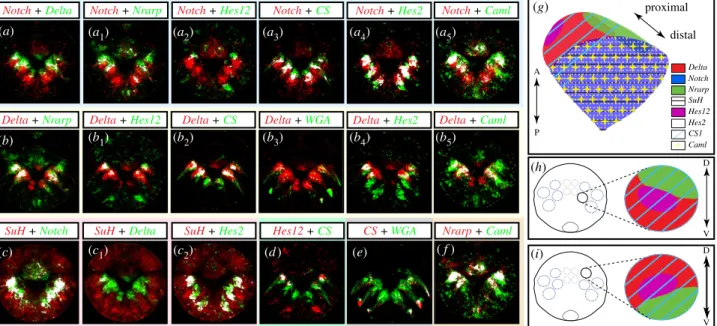
We then used the pattern registration technique recently devel-oped for Platynereis larvae [52] to better unravel the mature follicle cell types’ composition and arrangement. Pattern regis-tration in the case of chaetal sacs does not allow a perfect one-cell resolution analysis because cells do not always display perfectly constant positions within the sacs. It is sufficient however to accurately visualize expression domains in three-dimensional views, to establish gross overlap of expression domains and to corroborate hypotheses of coexpression. Expression patterns of all Notch components expressed in chaetal sac cells (Pdu-Notch, Pdu-Delta, Pdu-SuH and Pdu-Nrarp) were thus scanned, aligned and averaged. We have shown previously [53] the expressions of two Hes (Hairy/enhancer of Split) genes (potential target genes of the Notch pathway in eumetazoans) in the chaetal sacs. Pdu-Hes2 and Pdu-Hes12 that are found in 12 patches corresponding to, respectively, a large proportion and a few internal cells of the chaetal sac, were also included in the database. As we wanted to compare those gene expressions with known markers of chaetal sacs cells, we also add the Pdu-CS1 (a chae-toblast marker) and the Pdu-Caml (a follicle cell marker [39]) genes to this analysis. Finally, to better understand the archi-tecture of chaetal sacs in trochophore larvae, we also used Pdu-Twist, expression of which spans mesodermal territories surrounding the chaetal sacs [54]. We used again the WGA lab-elling to identify the position of the chaetoblasts, located in the proximal-most part of each chaetal sac [39].
In the electronic supplementary material, figure S3D, we provided three different views of three-dimensional surface lab-elling (either expression patterns or WGA staining) rendering possible the understanding of the complex organization of the 12 chaetal sacs (six in the neuropods and six in the notopods) surrounded by mesodermal tissues (Pdu-Twist) in the 48 hpf larvae. Pdu-Delta expression is localized in the internal-most part of the sac (electronic supplementary material, figure S3D (a4, b4, c4, d3, d4)) while Pdu-Notch expression is more
widespread in a large medial area of the sac (electronic supplementary material, figure S3D (a3, b3, c3, d3, d4)).
We next tried to define the identity and molecular signa-ture of the cells composing a chaetal sac by looking at the colocalization patterns of gene expression areas using regis-tration (figure 6). With the WGA staining to localize the chaetoblasts, as well as the Pdu-CS1 expression (figure 6e), we identified Pdu-Delta as a marker of at least some cells in the chaetoblast region (figure 6b2 to b3). Pdu-Hes12 þ cells
fall also in the Pdu-CS1þdomain and are thus chaetoblast cells too (figure 6b1 and d). Pdu-Notch and Pdu-Hes2
expression territories are broadly in the medial part of the sac, and probably correspond to several if not all follicle cells (figure 6a4). Their colocalization with the follicle cell marker
Pdu-Caml supports this interpretation (figure 6a5). Pdu-Nrarp
is co-localized partially with Delta, Notch and Pdu-Caml, and is expressed both in chaetoblasts and some follicle cells (figure 6a1,b and f). Altogether those different
combi-nations of markers allowed us to identify two domains of expression patterns among the sac: one proximal area (being Pdu-Deltaþ, Pdu-Hes12þ, Pdu-CS1þ and Pdu-Nrarpþ) and one distal area (being Pdu-Notchþ, Pdu-Hes2þ, Pdu-Su(H)þ, SuH +Delta
SuH +Notch
Notch + Hes12 Notch + CS Notch +Nrarp
Notch +Delta
Delta + Nrarp Delta +Hes12 Delta +CS Delta +WGA
Hes12 + CS CS + WGA Notch + Hes2
Delta +Hes2
SuH + Hes2 Nrarp + Caml
Notch + Caml Delta +Caml proximal distal A P (g) Delta Notch SuH Hes2 Hes12 Nrarp CS1 Caml V D (h) D V d (a) (c) (b) (a1) (a2) (a3) (a4) (a5) (b1) (b2) (c1) (c2) (b3) (b4) (b5) ( f ) (e) (d ) (i)
Figure 6. Molecular fingerprint of Platynereis chaetal sac cell types at 48 hpf. All images are confocal maximum z-projection of averaged expression patterns
registered on an average larva. Ventral surface views of the whole larvae, anterior side up, are shown. White patterns reveal the colocalization of green and
red pixels. (a to a
5) Pdu-Notch expression slightly overlaps with Pdu-Delta, Pdu-Nrarp and Pdu-CS1, is mutually exclusive with Pdu-Hes12 and is broadly co-expressed
with Pdu-Hes2 and Pdu-Caml. (b to b
5) Pdu-Delta expression overlaps partly with Pdu-Nrarp, broadly with Pdu-Hes12, Pdu-CS1 and WGA, and slightly with Pdu-Hes2
and Pdu-Caml. (c to c
2) Pdu-Su(H) is largely co-expressed with Pdu-Notch and Pdu-Hes2 but not with Pdu-Delta. The chaetoblast marker, Pdu-CS1, is co-expressed
with Pdu-Hes12, Pdu-Delta and WGA staining (d, b
2and e). Pdu-Caml and Pdu-Nrarp are broadly overlapping (f ). The summary of expression patterns territories
relative to a chaetal sac is provided in (g,h,i). (g) Schematic representation of a ventral chaetal sac with the gene expression territories mentioned, in a proximal –
distal axis. See figure inset for the gene colour legends. The cartoons in (h,i) show apical view of a 48 hpf larva with the chaetal sacs represented by blue dashed
circles. The black circles correspond to the location of the stomodeum. (h) Focus on an apical view of ventral chaetal sac gene expression territories. (i) Focus on a
vegetal view of dorsal chaetal sac gene expression territories. Note the mirror-like expression patterns territories between the dorsal and chaetal sacs. A – P,
antero-posterior axis; D – V, dorso-ventral axis.
rsob.r
oy
alsocietypublishing.org
Open
Biol.
7:
160242
10Pdu-Camlþ and Pdu-Nrarpþ) (figure 6g). In addition, Pdu-N-rarp expression pattern is polarized along the dorsal–ventral axis, but in an inverse disposition in notopodial versus neuropodial chaetal sacs (figure 6h,i).
2.6. Treatments with
g-secretase inhibitors suggest
that Notch patterns Platynereis chaetal sac
cell types
The morphological defects obtained through 24–48 hpf (time window 1, table 1) chemical disruptions (figure 3) as well as the continuous expressions of Notch pathway components, especially Pdu-Delta (figures 2 and 5), from the onset of chaetal sac formation to chaetae elongation strongly suggest an exten-sive role in chaetal sac pattern formation and chaetogenesis. We thus investigated the effects of Notch pathway chemical inhibition (Time window 1, table 1) on genes expressed in chae-tal sacs to test whether Notch pathway component expressions themselves are affected (electronic supplementary material, figure S3E). Interestingly, Pdu-Notch is significantly downregu-lated (electronic supplementary material, figure S3E (a to a3); an
expected effect, for example, see [55]), whereas Pdu-Delta seems not affected by any treatments (electronic supplementary material, figure S3E (b to b3)). Similarly, treatments also lead
to drastic decreases of follicle cells marker expression (Pdu-Nrarp, Pdu-Hes2 and Pdu-Caml) while chaetoblast markers (Pdu-Hes12 and Pdu-CS1) are maintained (electronic sup-plementary material, figure S3E (c to g3)). As Notch pathway
disruption does not affect cell proliferation profile and does not induce specific cell death in follicles and chaetal sacs
(electronic supplementary material, figure S2C), the expression losses observed may be related to an abnormal differentiation of follicle cells.
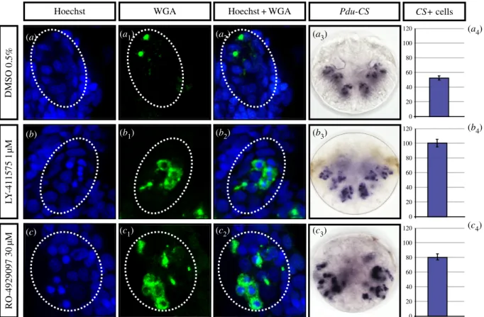
Notch pathway components (Delta, Hes12, Pdu-Nrarp and Pdu-Hes2) are expressed early in surface ectodermal cells, suggesting that they play a role in selecting cells that are going to differentiate into chaetoblasts or follicle cells, in a way that could involve lateral inhibition. We thus also performed early drug treatments (20–48 hpf, Time window 2, table 1) and assessed the resulting phenotypes on chaetoblast differentiation at 48 hpf, using WGA staining as well as the chaetoblast marker Pdu-CS1 (figure 7). LY-411575 was used at 1 mM while RO-4929097 had been raised to a higher concentration (30 mM) to obtain similar results. DAPT used at the limit of solubility (40 mM), while affecting chaetae production as the two other drugs do, gave no phenotype related to chaetoblast differentiation (not shown). In both effi-cient treatments, our experiments revealed the presence of roundish, intensely DAPI-stained cells surrounded by cyto-plasmic WGA-reactive chitin (figure 7b to c2). Many of these
cells do not show however a WGA-stained bristle elongating from it, suggesting that although they have some characteristics of chaetoblasts, they are not capable of producing bristles. We thus call them ‘abortive chaetoblasts’. Chaetoblasts and abortive chaetoblasts are more numerous in treated embryos than in the control (figure 7a to c4), as highlighted by Pdu-CSþ expression
and cell counting (figure 7a4, b4and c4). Abortive chaetoblasts
are located at dispersed, abnormal positions in the sacs, includ-ing close to the surface and presumably at the ‘normal’ locations of follicle cells in a control embryo. No recognizable chaetal sacs form in these early treated larvae, suggesting that
Pdu-CS DMSO 0.5% RO-4929097 30 µM WGA L Y -411575 1 µ M
Hoechst Hoechst + WGA
0 20 40 60 80 100 120 0 20 40 60 80 100 120 0 20 40 60 80 100 120 CS+ cells (a) (a1) (a2) (a3) (b) (b1) (b2) (b3) (c) (c1) (c2) (c3) (a4) (b4) (c4)
Figure 7. Early inhibitions of the Notch pathway induce supernumerary abnormal chaetoblasts within chaetal sacs. Embryos were incubated with LY-411575 (1
mM,
b to b
4), or RO-4929097 (30
mM, c to c
4) in DMSO or in DMSO only (control group, a to a
4) from 20 to 48 hpf. Treated larvae present several packed, intensely
DAPI-stained nuclei surrounded by WGA-DAPI-stained cytoplasm (b to b
2, c to c
2) instead of WGA spots neighbouring the chaetoblast nuclei (a to a
2). The chaetoblast marker
Pdu-CS1 is overexpressed within the whole sacs in treated larvae instead of a packed internal region (b to b
2, c to c
2, and compare to a
3to a
4). All panels are ventral
views (anterior is up).
rsob.r
oy
alsocietypublishing.org
Open
Biol.
7:
160242
11the patterning of these structures is completely compromised, in sharp contrast with later treatments. The internalization pro-cess, linked to the chaetal sac patterning, is consequently presumably affected.
As we are not able to establish unambiguously that the early Delta expressions are exclusively linked to chaetoblast differentiation, we also tested the possible involvement of the Notch pathway in the formation or selection of neural progenitors that will give rise to the few pioneer neurons of the 24 hpf larvae [31]. We thus treated larvae with the drugs (same concentrations as before) from 12 and 16 to 24 hpf (to be sure to encompass the very first neuron for-mation, Time window 3, table 1) and assessed the numbers of neurons (revealed by the postmitotic neuron marker Elav [33]) in both treated and control embryos at 24 hpf. At 24 hpf, the number of Elavþ cells in the trunk is very precise (nine) and is not at all affected by any early treatments (elec-tronic supplementary material, figure S4A).
In conclusion, the inhibition of the Notch pathway in an early time window (Time window 2) results in missformed chaetal sacs and supernumerary chaetoblasts, consistent with a lateral inhibition mechanism, while additional exper-iments should be performed in the future to firmly prove it. By contrast, early larval neurogenesis is not affected. Later treatments (window 1), or with more limited drug doses, result in abnormal gene expressions and possibly abnormal differentiation of follicle cells.
2.7. No major involvement of the Notch pathway
in ventral neurogenesis and brain patterning
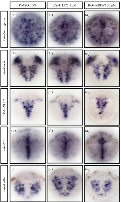
is evidenced
Although none of the components of the Notch pathway are obviously expressed in the forming VNC (figures 2 and 5) and the apparent morphology of the ventral nervous system does not appear much disturbed after g-secretase inhibitor treatments (figure 3), we cannot exclude that Notch neverthe-less could have a role in patterning and neuronal specification. We thus tested further the normal formation of the ventral nervous system by using several markers specific for different steps of neurogenesis and VNC patterning (Neuro-genin, Pax6, Nk2.2, Slit and Collier) on 48 hpf-treated embryos for all drugs (Time window 1, table 1 and figure 8). The proneural gene Neurogenin (Pdu-Ngn) is exclusively expressed in neural progenitor cells (figure 8a) [33]. Pdu-Pax6 and Pdu-Nk2.2 are expressed in neurogenic columns and are key to medio-lateral patterning of neurons in vertebrates (figure 8b,c) [20]. Pdu-Slit is expressed in the ventral midline and is a key axon guidance factor in insects and vertebrates (figure 8d) [34]. Collier (Pdu-Coe), as a marker of neuronal differ-entiation, is expressed in differentiating and/or fully differentiated neurons (figure 8e) [56]. In concordance with the morphology and behaviour, we observed no expression pat-tern alteration in the forming CNS on treated larvae at concentrations (LY-411575 1 mM, RO-4929097, 30 mM, DAPT 40 mM; data not shown) that perturb chaetal sac patterning, either at gross levels or in the precise spatial patterns of these five genes (figure 8a to e2). In addition, no overlapping
expression was observed between either Pdu-Notch or Pdu-Delta and Pdu-SoxB, Pdu-Ngn and Pdu-Pax6, central elements of the neurogenic network at eumetazoan scale [24] (electronic supplementary material, figure S4B).
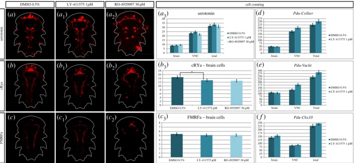
While inhibiting Notch clearly does not disrupt patterning of the VNC at 48 hpf, we then tested the possibility that a Notch activity reduction may enhance neurogenesis in older embryos. We thus treated larvae (LY-411575 (1 mM) and RO-4929097 (30 mM)) from 30 to 48 hpf (Time window 4, table 1), a time window that encompasses the massive neuro-genesis process in Platynereis [34] but prevents too drastic phenotypes. We assessed the phenotype at 72 hpf, at the cellu-lar level (figure 9). We used several markers and antibodies of differentiated neurons or specific types of neurons (serotoni-nergic, FMRFAmidergic, RYa peptidergic, cholinergic and interneurons), and investigated the number of cells that are positive for each of them, in both control and treated larvae. Serotonin, Crya [41] and FMRFa antibody stainings reveal no gross defaults between the control and LY-411575 treated larvae, while the RO-4929097 treated larvae appear to be delayed in their development (a to c2). Manual counting of
ser-otoninergic (a3), RYa peptidergic (b3) and FMRFAmidergic (c3)
neurons shows no significant differences in both brain and VNC between the control and treated larvae (even for the delayed ones), except for one case (RYa brain cells are signifi-cantly reduced in the RO-4929097 condition compared to control). We then used three other markers of neurogenesis: Pdu-Collier for all the differentiated neurons, Pdu-VAchT for the cholinergic neurons [20] and Pdu-Chx10 for a subset of interneurons [20]. Using registration, we obtained an averaged expression pattern for these genes in both control and LY-411575-treated larvae (not for RO-4929097 as the concentration used led to delayed larvae, preventing from a correct align-ment during registration; electronic supplealign-mentary material, figure S4C). Using the IMARISsoftware, we counted on these averages the number of positive cells for each gene in both the VNC and brain. This automatized approach for cell count-ing is highly linked to the in situ hybridization experiment efficiency and gives only a rough estimate of number of neur-ons. For all three genes, the number of neurons in the brain is almost equivalent between the control and treated larvae, while a few more interneurons are found after Notch inhi-bition (figure 9d–f). The number of interneurons is nevertheless constant in the VNC, while the cholinergic neur-ons and the overall differentiated neurneur-ons in general appear to be slightly more numerous in treated larvae. However, these numbers may be biased by the quality of the in situ hybridiz-ation and are in no way comparable to the drastic phenotypes observed in other models (in different contexts, see [57–60]).
3. Discussion
3.1. Evolution of the Notch pathway architecture: a
lophotrochozoan focus
We investigated the evolution of the Notch pathway core components in metazoan genomes with a strong focus on lophotrochozoan species, and updated a previous analysis done at the eukaryotic level [7]. In a majority of species, a single Notch gene was found. The presence of supplementary copies of genes in H. sapiens is most probably due to the two whole-genome duplication events (2R) at the origin of ver-tebrates [61] while some lineage-specific duplications are also evidenced (for example, in H. robusta and S. mediterranea). The receptor Notch structure (domain composition and num-bers of domain copies) is conserved in a large majority of
rsob.r
oy
alsocietypublishing.org
Open
Biol.
7:
160242
12cases, suggesting that the last common ancestor of bilaterians probably possessed a single Notch protein composed of 36 EGF repeats, three LNR repeats, NOD and NODP domains and seven ANK repeats. A more complex evolutionary history of the Delta/Delta-like proteins is suspected, but a single Delta gene was probably present in the urbilaterian ancestor with a structure (MNLL/DSL/9 EGF repeats/ATEV) that has been mostly conserved in extant bilaterians. The ATEV motif, which binds to PDZ domain proteins [62], is coded in most bilaterian Delta genes, while we identified in Platynereis a splice variant lacking this motif. Additional lophotrochozoan Delta-like proteins are found but their origin is uncertain.
Drawing strong conclusions on the early metazoan history of the Delta-like ligand is difficult as non-bilaterian data are rather limited. At least one Delta-like gene was present in the last metazoan ancestor (with presumably multiple dupli-cations events in the sponge lineages) but the structure of this ancestral gene cannot be firmly reconstructed from the available data (figure 1a; electronic supplementary material, figure S1D) [7]. In conclusion, the urbilaterian genome har-boured well-defined Notch, Delta and Jagged genes. This situation is remarkably conserved in Platynereis, thus making it legitimate to discuss their functions in the worm in a bilater-ian evolutionary context.
LY-411575 1 µM DMSO 0.5% Pdu-Nk2.2 Pdu-Collier Pdu-Neur ogenin Pdu-Slit Pdu-Pax 6 RO-4929097 30 µM (a) (a1) (a2) (b1) (b2) (c1) (c2) (d1) (d2) (e1) (e2) (c) (d ) (b) (e)
Figure 8. The Notch pathway does not regulate general ventral nerve chord patterning in 48 hpf Platynereis larvae. Ventral views of whole trochophore larvae
(48 hpf ) are shown (anterior is up). Larvae were incubated with LY-411575 (1
mM, a
1to e
1), in DMSO or in DMSO only (control group, a – e) from 24 to 48 hpf, or
RO-4929097 (30
mM, a
2to e
2) from 30 to 48 hpf. WMISH, in control and treated larvae, reveals no gross defects in the expression of several genes involved in the
process of neurogenesis after inhibition of the
g-secretase.
rsob.r
oy
alsocietypublishing.org
Open
Biol.
7:
160242
133.2. Is the Notch pathway ancestrally linked to central
neurogenesis?
We think we have here gathered convergent arguments to rule out a major involvement of the Notch pathway in the early events of central neurogenesis in Platynereis, thereby contrasting with the situation known from insects and vertebrates:
— There is no major expression of the receptor Pdu-Notch or the ligand Pdu-Delta at the time when mass adult neu-rogenesis is taking place in the episphere (future brain) and the ventral neuroectoderm. Both genes are found expressed in sub-populations of cells in the brain (notably in the apical organ) but only at very late larval stages (72 hpf onward) when the bulk of brain neurons have already differentiated.
— Previously, we have shown the expression of two of the 13 Hes genes present in Platynereis (Hes12 and Hes13) in the presumptive VNC during larval neurogenesis [53]. As Hes genes are sometimes considered as ‘canonical Notch effector genes’, their expression could indicate a limited involvement of Notch in the events of central neurogen-esis. Nevertheless, none of them is found in a very broad pattern that would suggest a role in general neuro-genesis [53]. Finally, larvae treated with g-secretase inhibitors do not show any Hes12 expression increase (electronic supplementary material, figure S3E). We thus feel confident that Notch does not act through any Hes gene (alone or in combination with another Hes) to pat-tern Platynereis VNC, while we cannot exclude that some Hes may play a role in more limited aspects such as the differentiation of specific neuron types. We also would like to stress the fact that the status of Hes genes as ‘core’ Notch pathway targets is the result of a historical over-simplification, based mostly on Drosophila studies.
The Hes superfamily was shaped by many independent lineage-specific tandem duplication events leading to a high diversity of Hes members [53]. Consequently, a regu-lation of Hes by Notch should not be systematically expected.
— Treatments with three drugs preventing the normal clea-vage of the Notch receptor do not result in major defects in the nervous system, when applied during the two time windows of neurogenesis (i.e. during the formation of the specific larval and pioneer neurons, and during the later mass adult neurogenesis events in the VNC and the brain). The normal patterning of the CNS is preserved as indicated by gene expressions. Near normal proliferation of both anterior and ventral neural ectoderm is maintained. Near normal numbers of neurons are differentiating and form nerve connections that seem unperturbed. We never-theless observed a slight increase of cholinergic neurons in the VNC after drug treatments. Pdu-VAchT (the cholinergic marker used) is known to be also expressed in the periph-eral nervous system (PNS) of Platynereis (J Be´hague and P Kerner 2017, unpublished data). We thus cannot exclude a limited role of the Notch pathway in peripheral neuron differentiation. However, the persistence of normal peri-pheral nerves in treated larvae does not suggest a predominant role in PNS formation either.
— By contrast, the same three drugs are causing specific defects in organs where the core components of the Notch pathway are specifically expressed, the chaetal sacs. These defects seem to be compatible with the Notch pathway being involved in a process of lateral inhibition in the chaetal sacs.
The minor involvement of the Notch pathway in Platynereis generic neurogenesis comes as an important surprise and two alternative evolutionary scenarios can be proposed to interpret this striking fact:
brain VNC total
Pdu-Chx10
DMSO 0.5% LY-411575 1 mM
DMSO 0.5% LY-411575 1 µM RO-4929097 30 µM
0 2 4 6 8 10 12 14 16 18
DMSO 0.5% LY-411575 mM RO-4929097 30 mM
cRYa – brain cells
0 1 2 3 4 5 6 7 8
DMSO 0.5% LY-411575 mM RO-4929097 30 mM
FMRFa – brain cells 0 5 10 15 20 25 30 35 40 brain VNC total serotonin 0 25 50 75 100 125 150 175 200 225 250 275 brain VNC Total Pdu-Collier 0 25 50 75 100 125 150 175 200 225 250 275 300 brain VNC Total Pdu-Vacht * serotonin cRya FMRFa cell counting 250 0 25 50 75 100 125 150 175 200 225 DMSO 0.5% LY-411575 1 mM DMSO 0.5% DMSO 0.5% LY-411575 1 mM LY-411575 1 mM (a) (b) (a1) (a2) (b1) (b2) (c) (c1) (c2) (a3) (b3) (c3) (e) ( f ) (d ) RO-4929097 30 mM