Publisher’s version / Version de l'éditeur:
Vous avez des questions? Nous pouvons vous aider. Pour communiquer directement avec un auteur, consultez la première page de la revue dans laquelle son article a été publié afin de trouver ses coordonnées. Si vous n’arrivez pas à les repérer, communiquez avec nous à [email protected].
Questions? Contact the NRC Publications Archive team at
[email protected]. If you wish to email the authors directly, please see the first page of the publication for their contact information.
https://publications-cnrc.canada.ca/fra/droits
L’accès à ce site Web et l’utilisation de son contenu sont assujettis aux conditions présentées dans le site LISEZ CES CONDITIONS ATTENTIVEMENT AVANT D’UTILISER CE SITE WEB.
Pure and Applied Chemistry, 90, 8, pp. 1283-1324, 2018-07-12
READ THESE TERMS AND CONDITIONS CAREFULLY BEFORE USING THIS WEBSITE. https://nrc-publications.canada.ca/eng/copyright
NRC Publications Archive Record / Notice des Archives des publications du CNRC : https://nrc-publications.canada.ca/eng/view/object/?id=46979c4a-fe2b-44b7-b566-400c6b8cc013 https://publications-cnrc.canada.ca/fra/voir/objet/?id=46979c4a-fe2b-44b7-b566-400c6b8cc013
Archives des publications du CNRC
This publication could be one of several versions: author’s original, accepted manuscript or the publisher’s version. / La version de cette publication peut être l’une des suivantes : la version prépublication de l’auteur, la version acceptée du manuscrit ou la version de l’éditeur.
For the publisher’s version, please access the DOI link below./ Pour consulter la version de l’éditeur, utilisez le lien DOI ci-dessous.
https://doi.org/10.1515/pac-2017-0101
Access and use of this website and the material on it are subject to the Terms and Conditions set forth at
Engineered nanomaterials and human health: Part 1. Preparation,
functionalization and characterization (IUPAC Technical Report)
Gubala, Vladimir; Johnston, Linda J.; Liu, Ziwei; Krug, Harald; Moore, Colin
J.; Ober, Christopher K.; Schwenk, Michael; Vert, Michel
IUPAC Technical Report
Vladimir Gubala*, Linda J. Johnston, Ziwei Liu, Harald Krug, Colin J. Moore,
Christopher K. Ober, Michael Schwenk and Michel Vert
Engineered nanomaterials and human
health: Part 1. Preparation, functionalization
and characterization (IUPAC Technical Report)
https://doi.org/10.1515/pac-2017-0101
Received January 4, 2017; accepted April 19, 2018
Abstract: Nanotechnology is a rapidly evolving field, as evidenced by the large number of publications on the synthesis, characterization, and biological/environmental effects of new nano-sized materials. The unique, size-dependent properties of nanomaterials have been exploited in a diverse range of applications and in many examples of nano-enabled consumer products. In this account we focus on Engineered Nanomateri-als (ENM), a class of deliberately designed and constructed nano-sized materiNanomateri-als. Due to the large volume of publications, we separated the preparation and characterisation of ENM from applications and toxicity into two interconnected documents. Part 1 summarizes nanomaterial terminology and provides an overview of the best practices for their preparation, surface functionalization, and analytical characterization. Part 2 (this issue, Pure Appl. Chem. 2018; 90(8): 1325–1356) focuses on ENM that are used in products that are expected to come in close contact with consumers. It reviews nanomaterials used in therapeutics, diagnostics, and consumer goods and summarizes current nanotoxicology challenges and the current state of nanomaterial regulation, providing insight on the growing public debate on whether the environmental and social costs of nanotechnology outweigh its potential benefits.
Keywords: analysis; nanomaterial; nanoparticle; nanoscale; nano-toxicology.
CONTENTS
1 Introduction ... 1284
2 Terminology for nanomaterials ... 1286
3 Preparation of engineered nanomaterials ...1287
3.1 Silica nanoparticles ... 1288
3.2 Titanium dioxide nanoparticles ... 1289
Article note: This document was prepared in the frame of IUPAC Project 2013-007-1-700. Sponsoring bodies: The Chemistry and
Human Health Division and the Polymer Division: see more details on p. 1317.
*Corresponding author: Vladimir Gubala, University of Kent, Medway School of Pharmacy, Central Avenue, Anson Building,
Chatham ME44TB, UK, e-mail: [email protected]. http://orcid.org/0000-0001-6301-3632
Linda J. Johnston: Measurement Science and Standards, National Research Council Canada, Ottawa ONK1A0R6, Canada Ziwei Liu and Christopher K. Ober: Cornell University, Department of Materials Science and Engineering, 310 Bard Hall, Ithaca,
NY 14853-1501, USA
Harald Krug: Empa – Materials Science and Technology, Department of Materials Meet Life, Lerchenfeldstrasse 5, 9014 St.,
Gallen, Switzerland
Colin J. Moore: FOCAS Research Institute, Dublin Institute of Technology, Kevin St., Dublin 8, Ireland
Michael Schwenk: In den Kreuz̈ckern 16/1, D 72072 Tuebingen, Germany (formerly Medical School Hannover) Michel Vert: University Montpellier 1, 15 Avenue Charles Flahault, BP 14491, 34093 Montpellier Cedex 5, France
3.3 Iron oxide nanoparticles ... 1290 3.4 Gold nanoparticles ... 1290 3.5 Silver nanoparticles ...1291 3.6 Quantum dots (QD) ...1292 3.7 Polymeric nanoparticles ... 1293 3.8 Carbon nanotubes ... 1294 3.9 Liposomes ... 1295 3.10 Bioinspired nanomaterials ... 1295
4 Surface functionalization of nanomaterials ... 1296
4.1 Silica coatings ... 1297
4.2 Ligand exchange ... 1298
4.3 Polymer coating ... 1298
4.4 Surface coatings of carbon nanotubes ... 1299
4.5 Biomolecule recognition ...1300
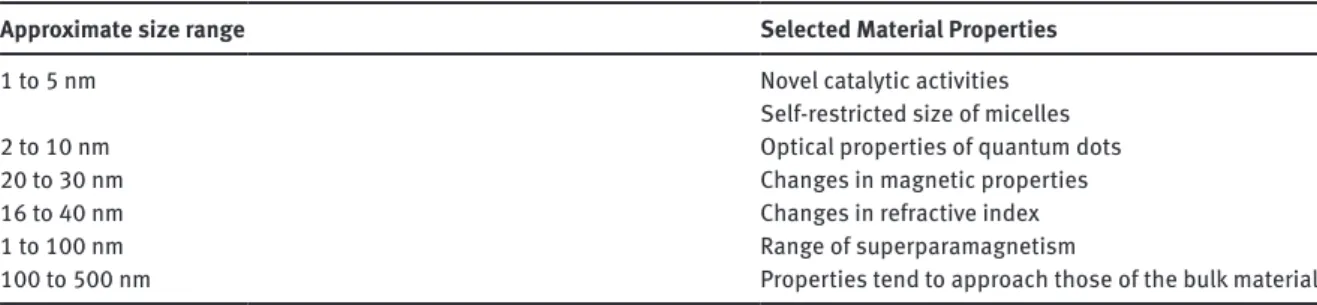
5 Size-dependent features of nanomaterials ... 1303
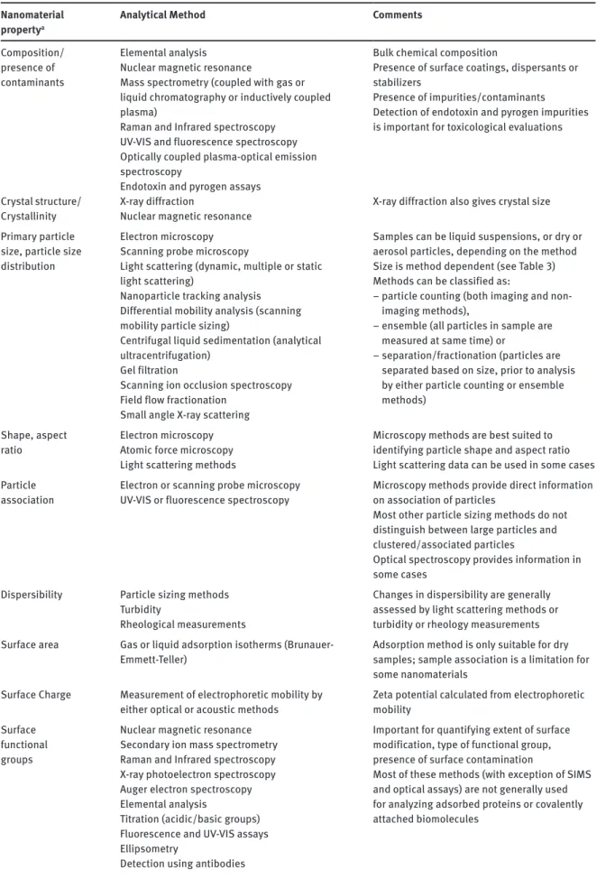
6 Characterization methods for nanomaterials ... 1304
6.1 Pre-analytics and sample preparation ...1304
6.2 Composition/purity/structure ...1306
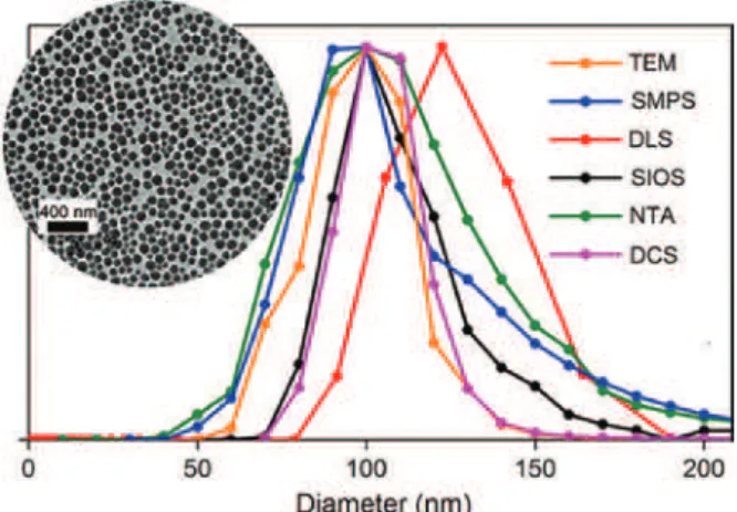
6.3 Shape, size and size distribution, aspect ratio ... 1307
6.3.1 Microscopy particle counting methods ...1309
6.3.2 Particle counting by non-imaging methods ... 1310
6.3.3 Ensemble methods ...1311
6.3.4 Fractionation or classifying methods ...1312
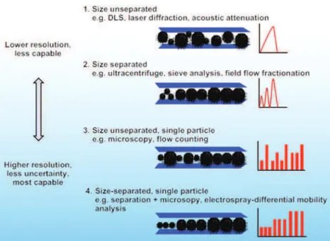
6.4 Comparison of particle sizing methods ...1313
6.5 Dispersion and particle association ... 1314
6.6 Surface properties ... 1314
6.6.1 Surface charge ...1315
6.6.2 Surface chemistry ...1315
6.7 Matrix-related restrictions ...1315
7 Discussion and closing remarks ... 1316
Membership of sponsoring bodies ... 1317
References ... 1318
1 Introduction
Nanotechnology promises to be at the forefront of many 21st century technological breakthroughs. Engineered
nanomaterials (ENM) are a class of deliberately designed and prepared materials with nanoscale dimensions that have a rapidly expanding range of applications in our everyday life. The immense interest in nanotech-nology and the new opportunities to improve the performance of traditional products that ENM present has led to the development of a ‘nano-particle ZOO’ [1]. Until recently, many efforts have focused on the use of simple ENM (e.g. metal oxides and carbon-based nanomaterials [2]) in applications such as cosmetics [3, 4], energy storage [5, 6], surface coatings [7], and textiles [8]. However, there is now significant interest in more complex nanomaterials, which already have, or are expected to find, an important role as advanced drug delivery systems in nanomedicine, ultra-sensitive reporters in biomedical diagnostics, and multifunctional probes in environmental management and in advanced electronics, transport, information and communica-tion technology, defense, and manufacturing.
The utility of ENM can be attributed to the occurrence of unique size-dependent characteristics that are not exhibited by the bulk material and that lead to useful mechanical, optical, magnetic, and
biological properties. There are many important characteristics to consider, including size distribu-tion, association state, shape, porosity, crystal structure, chemical composidistribu-tion, surface chemistry, charge, stability, degradation, and recycling. Unfortunately, none of these can be singled out as the most important for aspects related to human health. A large number of review articles have summa-rized the unique structural features and physicochemical properties of nanomaterials that elicit their biological response [9–12].
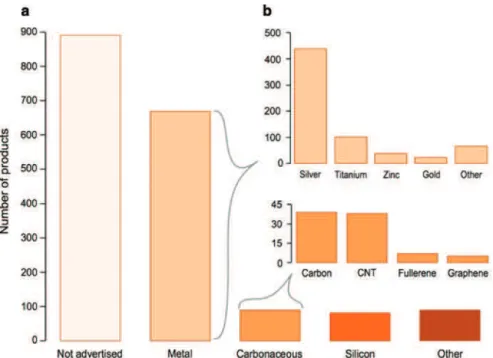
Nanomaterials are now widely used in commercial products, as illustrated by an article by Vance et al. [13] which found that, according to the Nanotechnology Consumer Products Inventory, 1814 nano-enabled products were available in the market (from 622 companies in 32 countries) in the year 2014 (see Fig. 1). Interestingly, 49 % of these products did not declare the composition of the nanomaterial they contained. The majority of nanomaterials used in consumer products were metallic (37 %), with 24 % of products containing silver nanoparticles (NP) (presumably due to their antimicrobial properties). The largest sector (42 %) of the market was attributed to nanomaterials used in Health and Fitness and clearly shows that the impact of nanomaterials on human health will be a key issue for the nanotechnology industry [14].
The objective of this work is to present an overview of ENM with a special focus on those where physical contact with consumers can be expected. Due to the complexity of the topic and the large volume of pub-lished material, the account is divided into two separate, albeit interconnected, documents. Part 1 focuses on: i) the preparation of various types of ENM, ii) methods for nanomaterial functionalization, and iii) the ana-lytical tools and methods used for the characterization of their physicochemical properties. Part 2 provides a detailed account of: i) nanomaterials used in therapeutics and diagnostics, ii) nanomaterials in consumer goods, and importantly, iii) the difficulties associated with the study of nanomaterial toxicity. This includes toxicological targets, pharmaco- and toxico-kinetics of nanomaterials, the four nanotoxicology principles, and the current state of nanomaterial regulation.
Fig. 1: Nanomaterials in commercially available products in 2014 according to the Nanotechnology Consumer Products
Inven-tory. (a) 49 % of products did not declare the composition of the nanomaterial used. (b) Of those that report the nanomaterial, metallic nanomaterials (particularly silver nanoparticles) were the most heavily used in commercial products (CNT = carbon nanotubes). Reproduced with permission from ref. [13].
2 Terminology for nanomaterials
The broad applicability of nanotechnology has led to considerable variation in the terms and definitions used by various scientific communities and regulatory authorities [15]. Despite this, it is generally accepted that nanomaterials have two defining characteristics: a size on the nanoscale (typically between 1 nm to 100 nm) and unique size-dependent properties that are not exhibited by the bulk material. In an attempt to develop a common approach for describing nanomaterials, a CODATA-VAMAS working group recently released the second version of a Uniform Descriptor System (UDS) for Materials on the Nanoscale [16]. This project aims to develop a system that provides a unique description for nanomaterials and allows one to determine if two materials are equivalent to whatever degree is desired. Nanomaterials cannot be uniquely specified by simple or even complex names, since characteristics such as size, shape, and surface functionality will determine their properties. The UDS is being developed with extensive international consultation, including discus-sions with the International Organization for Standardization (ISO) and American Society for Testing and Materials technical committees for Nanotechnology, as well as the Organization for Economic Co-operation and Development Working Party on Manufactured Nanomaterials. It is designed to be compatible with the international definition of nanomaterials from ISO technical committee 229 [17] – Nanotechnologies and the European Commission guidelines for regulation of consumer products containing nanomaterials [18]. ISO defines a nanomaterial as a material with any external dimension in the nanoscale or having internal struc-ture or surface strucstruc-ture in the nanoscale; nanoscale is defined as the size range from approximately 1 nm to 100 nm and it is noted that properties that are not extrapolations from a larger size are predominantly exhib-ited in this size range [19]. Engineered nanomaterials are deliberately designed and prepared materials with nanoscale dimensions, a similar definition to manufactured nanomaterials, which are defined as “intention-ally produced nano-sized materials for commercial purposes to have specific properties or compositions”. Incidental nanomaterials are generated as an unintentional by-product of a manufacturing, biotechnology, or other process [19].
Both the UDS and ISO TC/229 have adopted “nano-object” as the generic term to describe a material with one, two, or three dimensions on the nanoscale. The UDS has further sub-divided nanomaterials into 4 cat-egories: individual nano-objects, collections of nano-objects, bulk materials that contain nano-objects, and bulk materials with nanoscale features. Although much initial work on nanomaterials focused on approxi-mately spherical particles, there is now significant interest in high aspect ratio particles and one-dimensional materials. A wide range of terms have been used to describe these non-spherical materials. The ISO definition restricts the use of the term nanoparticle to a nano-object with all 3 dimensions on the nanoscale where the lengths of the longest and shortest axes do not differ significantly (see Fig. 2) [20]. This is similar (although not identical) to a definition adopted by IUPAC [21]. The term nanofibre is used for nano-objects with two external dimensions in the nanoscale and the third dimension significantly larger and not necessarily on the nanoscale; the terms nanorod, nanotube, and nanowire are recommended for solid, hollow, and electrically conducting nanofibres, respectively. Finally, the term nano-plate is used for nano-objects with one external dimension in the nanoscale and the other two external dimensions significantly larger. The various terms have not been employed systematically in the literature and a variety of other terms have been used. Where practical, we have adopted the ISO terminology for dealing with nano-objects with one, two, or three dimen-sions in the nanoscale.
The association of individual nano-objects into clusters is frequently a limitation in preparing suspen-sions, incorporating nanomaterials in composites, and maintaining their activity in a biological environ-ment. Much of the literature (and ISO definitions) distinguish between the strength of the interactions that lead to the clustering of nano-objects, with the term agglomeration used to describe clusters that are held together by weak forces and that can be disrupted with modest energy input. Aggregates are considered to be clusters of primary particles that are held together by strong forces and are difficult or impossible to disrupt. These terms are generally applied to solid nanomaterials, but are problematic, as there is a continuum of possibilities for the strength of forces leading to the association of individual particles and because the terms are not applied consistently in the literature. There are a number of self-assembled nano-objects (liposomes,
micelles, mesoporous silica, etc.). Some are held together by non-covalent molecular interactions and are particularly important nanomaterials in the context of health and biomedical applications. Self-assembled nano-objects are typically soft materials and frequently have a highly organized structure that is dictated by the intermolecular interactions (π-stacking, hydrogen bonds, hydrophobic, electrostatic, hydrophilic) that lead to their self-assembly (e.g. assembly of phospholipid to liposomes). This is in contrast to the cluster-ing or association of solid nano-objects, which is driven by surface interactions. It has been recommended that the terms aggregate and agglomerate be used interchangeably for these soft materials (see IUPAC report [21]). Based on the difference in terminology between various scientific communities, we believe that it may be necessary to distinguish between the association of solid nano-objects and nanosized self-assemblies of molecules. Since in most cases it is not clear which type of association is occurring for specific nano-objects, we avoid the use of aggregation/agglomeration here and instead use the term association.
3 Preparation of engineered nanomaterials
Two general concepts are used to produce nanomaterials: top-down and bottom-up. The top-down method starts with a bulk material from which particles are detached (e.g. by ablation or etching) or from which material is erased (e.g. lithography). The bottom-up method starts with atoms or molecules that react chemi-cally or are self-assembled to give nanosized particles. The synthetic procedure is followed by a purification step, which usually employs dialysis, chromatography, centrifugation, or ultrafiltration to purify the pristine nanomaterial from the reagents or undesired by-products.
Key quality control factors for the synthesis of ENM include homogeneous composition, narrow size range, stability of the material, and high reproducibility of the method. Engineered nanomaterials with a wide range of compositions, sizes, and shapes are now readily available (see Figs. 1 and 3). Commercial materials include homopolymers, copolymers or biopolymers, self-assembled organic molecules, nanocar-bon materials, inorganic oxides such as silica and titanium dioxide, metals, metalloids, quantum dots, and constructs consisting of several of these materials. The various materials are designed to exhibit properties, such as specific band gap energies, superparamagnetism, size-tunable emission, antibacterial activity, textile protection, biocompatibility, and biodegradability. Typically, the size, composition, pore size, and surface Fig. 2: Classification of nanomaterials. Nanostructured materials (boxes with dashed lines) are not discussed in this review.
functionalization of the ENM are the main factors that govern its suitability for a particular application. In this section we provide a short overview of the synthetic methods that are used to synthesize common classes of nanomaterials. For each class of material, we also briefly describe the main applications that are likely to have direct impact on the human body and the specific properties that enable them. The application areas include biomedical uses (biosensing and diagnosis, drug delivery, imaging, cancer therapy) and consumer products such as cosmetics, food, and textiles.
3.1 Silica nanoparticles
Silica nanoparticles can be divided into nanoporous (less than 2 nm diameter) and mesoporous (2 to 50 nm diameter) categories depending on their structure. They have significant potential for medical and non-medi-cal applications due to their large surface area, high cheminon-medi-cal stability, biocompatibility, and straightforward functionalization. Example applications include filler or reinforcement for advanced composite materials such as dental nanocomposites, food additives (E551: silicium dioxide) in dietary products to reduce the fat content or as anti-caking agents, controls for texture and shelf-life of various cosmetic products, and, recently, new platforms for biomedical applications such as drug delivery and diagnostic testing employed for selecting targeted therapies [23, 24].
Synthetic methods for the production of silica NP include reverse microemulsion, flame synthesis, and sol-gel processing; the sol-gel process is the most widely used, since it provides control of particle size distri-bution and morphology through the systematic monitoring of reaction parameters [25]. The sol-gel process is based on the hydrolysis and condensation of silicon alkoxides such as tetraethoxysilane (TEOS), commonly known as tetraethyl orthosilicate, in a mixed solution of water and alcohol with either an acid (e.g. HCl) or a base (e.g. NH3) as catalyst. In 1968, Stöber developed a reaction system that enabled the controlled growth of spherical silica particles of uniform size ranging from 50 to 2000 nm in diameter [26]. This initial work was followed by the modification of the method to prevent association and produce smaller, homogeneous, and uniformly sized silica NP [27–30]. It has been proven that particle size can be controlled by adjusting reaction parameters, such as solvent, temperature, and concentration of silicon alkoxides and ammonia. Both tem-perature and concentration affect the rates of hydrolysis and condensation, and hence control the balance between the nucleation and growth processes [27]. The presence of electrolytes in the reaction medium can Fig. 3: Schematic illustration of different architectures of Engineered Nanomaterials.
also assist in controlling particle size and structure. For example, the addition of small amounts of anionic electrolytes into the reaction mixture produced uniformly sized silica NP with diameters ranging from 20.5 to 34.1 nm, decreasing particle size by more than 70 % [31] when compared to the medium without electrolytes. This occurs because electrostatic repulsion due to anions minimizes particle association and simultaneously inhibits particle growth.
Mesoporous silica NP have been widely explored as drug delivery systems, because their high specific surface area and large pore volume provide unique advantages for the encapsulation of a variety of ther-apeutic agents [31]. The synthesis is controllable and simple to perform and is based on the formation of liquid-crystalline mesophases of amphiphilic molecules (surfactants) that serve as templates or structure directing agents around which precursors condense to form silica. Various sizes and morphologies with highly ordered mesoporous channels [32] can be obtained by tailoring the molar ratio of silica precursors and surfactants [33], manipulating pH [34], and by adding co-solvent [35]. Ultrasmall sub-10 nm silica particles have been synthesized using hexadecyltrimethylammonium bromide (CTAB) as a structure directing agent and poly(ethylene oxide) (PEO) modified silane to quench particle formation [36]. CTAB acts as a template to generate silica particles with a single pore size and the introduction of PEO generates a modified surface that is suitable for biomedical applications. The same group have since advanced their method of generating ultrasmall silica NP by employing water as a solvent [37].
3.2 Titanium dioxide nanoparticles
Nano-sized titanium dioxide (TiO2) is used across a wide range of applications, such as paint pigments, glazes, enamels, plastics, paper, fibers, foods, pharmaceuticals, cosmetics, toothpastes, antimicrobial appli-cations, and catalysts for air and water purification or for energy storage [38]. However, nanoscale TiO2 is predominantly used in personal care products, such as topical sunscreens that block ultraviolet radiation and other skin care products [39]. TiO2 NP have a greater protective ability than micron sized particles and provide a more transparent product with less residue. Other applications of TiO2 would also benefit from smaller primary particle sizes. Therefore, the amount of nanoscale TiO2 used in consumer goods is expected to increase significantly. Food-grade TiO2 has also been shown to contain up to 35 % primary particles that are less than 100 nm in diameter [40].
The sol-gel method used to make silica NP can also be applied to TiO2 NP synthesis. The reaction involves the hydrolysis of a titanium precursor, usually titanium alkoxides [41] or titanium halides [42], followed by condensation. Different particle sizes and shapes can be obtained by varying reaction parameters, such as concentration, pH, pore size, and temperature [43]. Surface stabilizers are used to form an insulating organic layer on the surface, preventing particle association. Although sol-gel processes usually result in amorphous materials, different methods have been reported to improve the crystallinity of TiO2 NP, including calcination [44], ultrasound irradiation [45], and the addition of a base [46], or of salts and acids [47].
Micelles[48] and reverse micelles [49, 50] are also employed for the synthesis of TiO2 NP. Restricting the reaction to the interior of micelles or reverse micelles provides control of particle size in either aqueous or non-aqueous solution. The ratio of titanium precursor, surfactant, and water; the ammonia concentration; and the reaction temperature are all significant in controlling particle size and size distribution. TiO2 particles synthesized via this method have an amorphous structure. In order to increase crystallinity while avoiding association, further annealing in the presence of micelles at temperatures considerably lower than those required for the traditional calcination treatment [51] can be applied.
Other popular synthetic approaches include hydrothermal and solvo-thermal methods, which are almost identical except for the use of water and organic solvents, respectively. These methods are normally con-ducted under high pressure in autoclaves made of steel with PTFE liners, enabling the use of temperatures above the boiling point of a given solvent. Usually, the solvo-thermal method yields better control of size, size distribution, and crystallinity of TiO2 NP than that achieved with the hydrothermal method, due to the higher boiling point of some organic solvents [44].
3.3 Iron oxide nanoparticles
Iron oxide nanoparticles are the most commonly employed magnetic nanoparticles (MNP). Engineered with a hydrophilic surface coating, they have demonstrated great promise in the biomedical field. Aside from their unique magnetic properties, injectability and high accumulation in the target tissue or organ make them suit-able for diagnostic and therapeutic applications, such as magnetic resonance imaging (MRI) contrast agents [52], targeted drug delivery [53], and magnetic field assisted radionuclide therapy [54].
Various methods have been developed to synthesize uniform iron oxide-based MNP with homogene-ous composition and narrow size distribution [55]; these include microemulsions [56], sol-gel synthesis [57], sonochemical reaction [58], hydrothermal reaction [59], thermal decomposition [60], and electrospray syn-thesis [61]. However, the co-precipitation technique is probably the simplest and most efficient method [55]. Fe3O4 particles can be synthesized via co-precipitation of iron(II) and iron(III) ions in aqueous solution [62]. Although large amounts of NP can be synthesized in this way, the control of particle size distribution remains a challenge and the nucleation and growth steps must be separated, as explained by LaMer’s theory, in order to produce uniform NP [63]. This can be achieved by burst nucleation with hot injection of reagents into the reaction system, which leads to faster nucleation and a concomitant decrease in particle size. Surfactants are necessary to coat the particle surface and prevent the association of smaller particles.
Thermal decomposition is another important method and is now routinely used to prepare iron oxide-based MNP with controlled size and morphology [64]. Metal precursors, usually organometallic complexes, undergo decomposition at high temperature in an organic solvent with surfactant added for NP stabilization and size control. For example, uniform MNP of 20 nm in diameter are synthesized by the decomposition of iron(III) acetylacetonate, Fe(acac)3 at high-temperature (265 °C), in phenyl ether and in the presence of alcohol, with oleic acid and oleylamine as surfactants [65]. FeNP with sizes ranging from 5 to 19 nm can be produced via a similar method by the decomposition of iron(0) pentacarbonyl [Fe(CO)5] in dioctyl ether with oleic acid and oleylamine [66]. Further controlled oxidation transforms them to uniform γ-Fe2O3 NP. The size and morphology of NP can be controlled by varying the reaction conditions, including time, temperature, concentration, and ratios of reactants. This method can be extended to alloy particles, such as CoFe and CoFe2O4, by simultaneous decomposition of Fe(CO)5 and Co2(CO)8 in 1,2-dichlorobenzene [67], or Fe(acac)3 and Co(acac)2 in hexadecane-1,2-diol [68].
Iron-based MNP can be synthesized through the reduction of metal salts in the presence of surfactant, which prevents particle association [69]. A wide range of common reducing agents, including hydrazine [70], sodium borohydride [71], lithium borohydride [72], and polyols [73], can be used for metal salt reduction. The synthesis of CoFeNP is also achieved by the simultaneous co-reduction of iron(II) sulfate and cobalt chloride salts with sodium borohydride [74]. By using potassium borohydride, hollow CoFe is produced [75]. Reactions carried out in polyols, such as ethylene glycol or propane-1,2-diol, tend to yield more uniform products. Such polyols effectively act as bidentate chelating agents for the solvated metal cations and, in some cases, also serve as reducing and/or stabilizing agents for the metal nanoparticles [76].
3.4 Gold nanoparticles
Gold nanomaterials with novel properties, biocompatibility, and relatively low toxicity, have attracted considerable interest in applications such as diagnostics and therapeutics [77]. More recently, their use in cosmetics and toiletries as anti-wrinkling agents to inhibit the formation of advanced glycation end products has also been explored. Gold nanomaterials can now be divided into two distinct categories: gold nanoparticles (Au NP) and gold nanoclusters. The most interesting property of gold nanoparticles is their tunable surface plasmon resonance, which induces a dramatic enhancement in their absorption and scat-tering. The surface plasmon band is tunable from the UV to the NIR region by changing the particle size (1 to 100 nm length scale) and shape [78]. Although the term nanoparticle defines an object with all three dimensions below 100 nm, such as spheres, cubes or prisms, the term gold nanoparticle is widely used in
the literature to describe most gold nano-objects, independent of whether they fit this definition (e.g. gold rods, plates).
Gold nanoclusters are usually smaller than 3 nm, composed of several to roughly one hundred atoms, and are characterized by the strong quantum confinement effect of free electrons in the particles. Recent synthetic advances enabled the preparation of water-soluble gold nanoclusters with tunable size or emission colors, which accelerated their use as novel labels in biosensing and bioimaging [79].
Significant effort has been devoted to the synthesis of gold nanoparticles, focusing on control over size, shape, solubility, stability, and functionality [80]. Developed by Turkevich et al. [81] in 1951, the citrate reduction of HAuCl4 is still one of the most commonly used synthetic methods for Au NP production. In this method, [AuCl4]− is reduced by sodium citrate, which acts as both reducing agent and stabilizer, in boiling
water. Particles with sizes ranging from 12 to 150 nm can be obtained by varying the nucleation rate and through changing the sodium citrate concentration.
Another breakthrough in gold nanoparticle synthesis is the Brust-Schiffrin method. In this method, [AuCl4]− is transferred from an aqueous phase to toluene using tetraoctylammonium bromide as the
phase-transfer reagent and reduced with aqueous sodium borohydride in the presence of dodecanethiol. The nanoparticles synthesized are covered with strongly bound thiol-ligands which prevent association. Gold nanoparticles with sizes ranging from 1.5 to 5.2 nm were obtained by adjusting reaction conditions, including the gold to dodecanethiol ratio, temperature, and the reduction rate [82].
A water-based synthetic method is often preferred over the two-phase method, since it uses less toxic solvents and the NP are hydrophilic, and thus more biocompatible. Functional block polymers can also be introduced into the system, where they simultaneously serve as reductants, structure-directing agents, and colloidal stabilizers. For example, in a one-step synthesis of Au NP with an average diameter of about 10 nm, the (PEO-PPO-PEO) block copolymers act as efficient reductants and stabilizers. The reaction takes place in air-saturated aqueous solution at room temperature, and no other reducing agents are needed [83]. When water and commercially available polymers are used, this synthetic method is environmentally benign and economical.
3.5 Silver nanoparticles
Silver has many attractive characteristics that make it applicable to a number of fields, including opto-electronics, catalysis, chemical/biological sensing, in vivo imaging, and medicine [84]. Silver nanoparticles (Ag NP) have proven to be particularly attractive as antibacterial agents [85] and are regularly incorporated into apparel to prevent the formation of odour-causing bacteria [86]. They provide an excellent platform for Surface Enhanced Raman Spectroscopy detection and enable single molecule detection. The utility of Ag NP in a variety of applications has led to the investigation of different approaches for nanoparticle preparation using chemical and physical approaches.
Chemical reduction is the most common approach for the production of Ag NP and typically involves three components: (i) metal precursor, (ii) reducing agents, and (iii) stabilizers/capping agents. The most common metallic precursors are silver nitrate, silver acetate, silver citrate, and silver chlorate, while the most frequently used reducers are sodium borohydride and sodium citrate. The main drawback of chemical syn-thesis is the inability to produce NP of well-defined shape and size. For example, the Creighton method is limited to producing Ag NP of around 10 nm. The Lee-Meisel method, which employs AgNO3 in a variation on the Turkevich method used for Au NP synthesis, produces 30 to 150 nm NP with various morphologies (spheres, polyhedrons, plates) [87]. Reproducible Ag NP synthesis can be related to the initially formed nuclei that can develop into different crystal sizes and morphologies, which are dependent on reaction conditions [88]. Indeed, it has been highlighted that even small impurities in silver nitrate can influence the shape and crystal structure of the resultant NP, as reviewed by Xia [89]. The association of Ag NP is typically prevented through the introduction of stabilizing agents, such as poly(N-vinylpyrrolidone), on the particle surface. Bastú et al. reported the controlled synthesis of highly uniform citrate-stabilized Ag NP by reducing AgNO3
using a combination of sodium citrate and tannic acid [87]. Small, uniform Ag NP (10 to 20 nm) could be produced by varying the molar ratio of tannic acid to sodium citrate (0.005:1 up to 1:1). Particles of defined size (e.g. 15 nm) were then used as ‘seeds’ for the subsequent production of larger uniform NP by sequentially adding more sodium citrate, tannic and AgNO3 in a defined ratio. The new generation of well-defined larger NP was then used as new ‘seeds’ to create even larger NP with a narrow size distribution. Overall, the authors reported the ability to produce citrate-stabilized Ag NP over a range of 10 to 200 nm with standard deviation less than 10 %.
Prominent physical methods of Ag NP production are laser ablation and evaporation/condensation. Laser ablation involves the use of a silver plate immersed in a liquid phase. A focused, high-energy laser irradiates the metal to generate a heated plasma containing a high concentration of silver atoms and ions that cool in the solvent, facilitating Ag NP growth [90, 91]. A number of factors govern the size, shape, growth rate, and uniformity of the resultant NP. These factors include the laser wavelength, pulse duration (femto-, pico- or nanosecond regime), laser fluence, ablation time, and the solvent properties [92]. Another physical preparative method is the evaporation/condensation technique, which uses a tube furnace. One advantage of physical synthesis methods is that they do not require chemical reagents and can therefore generate high purity colloids.
Alternative ‘green synthesis’ methods of generating Ag NP are becoming increasingly popular. Green synthesis uses biological sources, such as bacteria [93], fungi [94] and plants [95], which have naturally occurring biocompatible reducing agents and stabilizers for NP generation. These methods circumvent the need to use potentially harsh chemical agents and aim to minimize the environmental impact of NP produc-tion. However, the scalability of such approaches remains unresolved. A recent example of this was demon-strated by Giessen and Silver when they engineered 25 nm diameter encapsulins from Thermotoga maritima to produce 13.5 nm Ag NP. Encapsulins are a new class of microbial nanocompartments and were exploited to confine particle growth [93]. This was enabled by genetically modifying the encapsulin to display the silver-binding AG4 peptide, first reported by Naik et al. [96], on their compartmental interior. The authors high-lighted that the exact mechanism of silver precipitation by AG4 remains to be fully elucidated. However, the hydroxyl groups of tyrosine residues have been shown to be active in silver ion reduction [97] and NMR studies of |AG4 by Lee et al. [98] reported shifts in the hydrophobic side chains of leucine, phenylanaline, and arginine during Ag NP formation, indicating that they may serve as NP formation sites.
3.6 Quantum dots (QD)
Quantum dots (QD) are nanoscale semiconductor crystals that are generally composed of elements from groups 2–4, 3–5, or 4–6 and have sizes ranging from 2 to 10 nm [99, 100]. They have size dependent optical properties that are due to quantum confinement effects on electronic states. QD have unique advantages over traditional fluorescent dyes, including a broad excitation range, narrow and size-tunable emission, high luminescence yields, and good photochemical stability [101], making them appealing in vivo and in vitro fluo-rophores in biological applications [102].
Since Brus et al. first reported colloidal QD in 1983 [103], researchers have been developing synthetic methods for these nanomaterials. The current methods can be divided into two classes: organometallic syn-thesis based on high-temperature thermolysis and aqueous synsyn-thesis with thiols as stabilizers [100]. In 1993, Bawendi et al. reported a simple route for the synthesis of high-quality near-uniform 2–6 QD by injecting orga-nometallic reagents, such as dimethylcadmium, into hot coordinating solvent, which provided temporally discrete nucleation, as well as the controlled growth of macroscopic quantities of QD with sizes from about 1.2 nm to about 11.5 nm [104]. This method was later optimized to a one-pot synthesis by using cadmium oxide as the precursor instead of dimethylcadmium, which is difficult to handle and unstable [105]. Core-shell QD, in which the higher-band-gap shell protects the inner core from oxidation and improves the photolumines-cence quantum yields, provides materials with novel properties that are more attractive for applications [106]. Examples of core-shell QD include CdS/CdSe [107], CdS/ZnS [108], and CdSe/ZnS [106, 109]. In the synthesis
of CdSe/ZnS QD, uniform CdSe quantum dots are first synthesized through pyrolysis of Cd and Se precursors and then injected into a solution of trioctylphosphine oxide to which Zn and S precursors are added dropwise under a N2 atmosphere to form a ZnS layer. When prepared in organic solvent, the poor aqueous dispersibil-ity resulting from hydrophobic surface ligands makes QD unsuitable for biomedical applications. Therefore, ligand exchange with hydrophilic molecules and polymers or silica coatings is necessary, as discussed below.
Compared to thermolysis in organic solvents, aqueous methods are preferable, since they are cheaper and more reproducible, use less toxic materials, and produce biocompatible QD with high aqueous stability and biocompatibility [110]. This makes them more promising for biomedical use [111], even though they have lower photoluminescence quantum yields and therefore significant effort has been directed towards aqueous synthesis methods [110]. In the synthesis of CdTe, cadmium perchlorate is dissolved in water in the presence of NaHTe followed by addition of a thiol stabilizer such as 2-sulfanylethanol [112], 2-sulfanylacetic acid [113] or cysteine [114], which are soluble or miscible in water at appropriate pH. CdTe nanocrystals with excellent aqueous dispersibility are produced after refluxing under aerated conditions [115]. By taking advantage of high temperature hydrothermal synthesis techniques, the photoluminescence quantum yield and size dis-tribution of CdTe QD are significantly improved [110]. In order to modify this time-consuming procedure, microwave radiation is often introduced [116]. Studies have shown that microwave assisted aqueous synthe-sis is generally faster, simpler, and more energy efficient compared to conventional hydrothermal synthesynthe-sis [117, 118]. It is believed that fast and uniform heating from microwave radiation results in good crystallinity and high quality QD.
3.7 Polymeric nanoparticles
Polymeric materials have been used in a range of pharmaceutical and biotechnology products for more than 70 years. Polymer-based multifunctional nanoparticles capable of targeting, diagnosis, and drug delivery were developed more recently [119]. Polymeric NP can be loaded or coated with drugs and can be used to administer, carry, and deliver drugs [120]. Ever since Langer and Folkman reported sustained release of biochemically active macromolecules from biodegradable polymers in 1976 [121], polymer-based NP have impacted virtually every branch of medicine [122]. Today, the most commonly used polymers for drug release applications include poly(D,L-lactic acid)-co-(glycolic acid) (PLAGA), poly(lactic acid) (PLA) and poly(glutamic acid) (PGA), due to their favorable bioavailability, biocompatibility, biodegradability, and low toxicity [123]. Some polymer-based NP have received regulatory approval as drug delivery systems in humans [124, 125].
A number of bottom-up and top-down methods are available for the preparation of polymeric NP. The bottom-up techniques – emulsion, interfacial and precipitation polymerization – employ a monomer as the starting point [126]. The top-down techniques include solvent emulsion evaporation [127–130], solvent dis-placement [131, 132], and salting out methods [133, 134] and start with a polymer that is preferably bio-resorb-able and ideally bio-degradbio-resorb-able in vivo [135]. The presence of surfactant is essential to prevent nanoparticle association and ensure the stability of nanodispersions. Solvent emulsion evaporation is the most common technique and requires the emulsification of the polymer solution as the first step, followed by homogeniza-tion of the resulting mixture [136]. The emulsificahomogeniza-tion method can be divided into two categories, oil-in-water (O/W) emulsification (single emulsion), and water-in-oil-in-water emulsification, W/O/W (double emulsion). The O/W emulsification technique is suitable for entrapping hydrophobic drugs, while W/O/W is generally used for the encapsulation of hydrophilic drugs and proteins [128, 137]. The size of polymeric NP can be con-trolled by adjusting reaction parameters, such as stirring rate, temperature, and the type and concentration of dispersing agent [138].
Solvent displacement, also called nanoprecipitation [132], involves the use of an organic solvent that is miscible with an aqueous phase in the presence or absence of surfactant [131, 139–141]. Polymers and drugs are dissolved in the organic solvent and are transferred to an aqueous solution, where the hydrophobic polymers
and drug self-assemble into core-shell nanospheres that precipitate [142]. This technique is amenable to scale-up to an industrial scale and requires only gentle stirring with no high shear stress [126].
Non-degradable polymers, such as polystyrene, also play an important role in this field. In medicine, polystyrene microspheres have been used in the treatment of hepatocellular carcinoma, based on the principle that the vascular bed of many solid tumors is hyperpermeable to macromolecules and nano-particles, allowing their accumulation in tumors [143]. The synthesis of polystyrene particles is typically accomplished by bottom-up polymerization, although this method cannot independently control particle size and molecular weight [144]. Top-down techniques such as nanoprecipitation were also employed [145, 146].
“Intelligent” nanoparticles composed of stimulus-responsive polymers, for example pH-responsive polymers (e.g. poly(acrylic acid)) and thermal-responsive polymers (e.g. poly(N-isopropylacrylamide)), have been extensively studied. When subjected to external signals they produce physical or chemical changes that include variations of macromolecular structures, solubility, surface properties, swelling, and dissocia-tion [147]. Most stimulus responsive polymers are synthesized via bottom-up techniques. Conjugated poly-mers also show potential for biomedical applications. For example, cyanovinylene–backbone conjugated polymers can be used as in vivo nanoprobes due to their high chromophore density and high fluorescence efficiency [148]. The nanoparticle synthesis method is quite similar. In this case, hydrophobic monomers are dissolved in a neat liquid surfactant and then micellized in water, allowing polymerization to occur in the nanoscopic non-polar interior of the micelles.
3.8 Carbon nanotubes
Carbon nanotubes (CNT) are hollow nanofibers (see Fig. 2) whose walls are composed of sheets of graphene that exhibit a hexagonal arrangement of sp2-hybridized carbon atoms and were first observed and described
by Radushkevich and Lukyanovich in 1952 [149]. Their unique structures result in impressive mechanical, thermal, magnetic, optical, electrical, surface, and chemical properties that have potential for extensive biomedical applications in areas such as in vivo imaging, biosensing, and drug delivery [150, 151]. CNT are divided into two basic forms, single walled carbon nanotubes (SWCNT) consisting of a single tube of gra-phene, and multiwalled carbon nanotubes (MWCNT) composed of several concentric tubes of graphene.
Arc discharge, laser ablation, and chemical vapor deposition are the three main production methods for CNT. The simplest method to achieve large-scale synthesis applies a DC arc discharge between two graph-ite electrodes in the presence of a metal catalyst at low pressure in a controlled atmosphere composed of inert/or reactant gas [152]. The catalyst used and the type and pressure of the gas affect the CNT morphol-ogy, purity, and yield. Researchers concluded that arc discharge in an organic atmosphere produced more MWCNT because organics could be ionized and decomposed into hydrogen and carbon atoms, which con-tribute to MWCNT synthesis.
Laser ablation, another synthetic method for CNT first demonstrated in 1995 [153], favors the growth of SWCNT with controlled diameter depending on reaction temperature. The principles and mechanisms are similar to arc discharge with the difference that the energy is provided by a laser beam focused onto a metal-graphite composite target, which is placed in a high-temperature furnace. A number of factors including laser intensity, furnace temperature and metal catalyst affect the size, morphology, yield, and physical character-istics of SWCNT [154].
Chemical vapor deposition is now the standard method for synthesis of CNT. It is not only controllable, but also economically viable for large scale CNT production [155]. This method involves decomposition of various hydrocarbons over a transition metal supported catalyst, which can be carried out at atmospheric pressure [152] in a temperature range of 300 to 1200 °C [156]. A variety of gaseous and liquid hydrocarbon feedstocks can be used as precursors. Besides transition metals and rare earth elements, catalysts formed from metals combined with natural minerals, such as forsterite, diopside, quartz, and magnesite, have also been used [157].
3.9 Liposomes
Although liposomes are not typically considered to be a class of ENM, they are often functionalized to have novel properties for biomedical applications. Liposomes have significant potential for drug delivery and cur-rently represent the majority of the clinically approved nano-enabled therapeutics. Drug delivery applica-tions typically require the functionalization of the liposomes to improve stability and incorporate targeting moieties. Liposomes came to prominence in 1965 following work by Bangham [158] in which the amphiphilic nature of their structure was revealed through the identification of the phospholipid bilayer. They are used to carry hydrophilic drugs in their interior and hydrophobic drugs inside the membrane, which has led to liposomes becoming the most widely studied drug delivery nanomaterial for cancer treatment.
Several techniques, such as Bangham’s method, detergent-depletion, reverse phase evaporation, ether/ ethanol injection and emulsion methods, as well as alternative methods including dense gas and supercriti-cal fluid techniques have been reported and reviewed elsewhere [159, 160]. Due to the differences in prepa-ration methods and lipid compositions, liposomes can be classified according to their lamellarity (uni- and multilamellar vesicles), size (small [diameter below 100 nm], intermediate [100 to 250 nm], or large [above 250 nm]), and surface charge (anionic, cationic, or neutral). The ‘First generation’ liposomes used for drug delivery suffered from problems, such as rapid accumulation in mononuclear phagocyte system organs (e.g. liver and spleen). Their circulation half-life has been dramatically improved by grafting biocompatible poly-mers, such as polyethylene oxide, to the liposome surface. This class of liposome has since been dubbed ‘stealth liposomes’ [161]. In the case of the commercially available stealth liposome Doxil®, a liposome
plat-form encapsulating the chemotherapy drug doxorubicin, a circulation half-life 100 times that of free doxo-rubicin is observed [162]. Doxil® has also been shown to lower cardiotoxicity 7-fold in comparison to free
doxorubicin. Liposomes are widely available commercially, with global sales in the hundreds of millions of dollars annually. Doxil® and Myocet® encapsulate the chemotherapy drug doxorubicin, with the latter being
a non-PEGylated liposomal system. AmBisome® is a liposomal formulation of the broad-spectrum polyene
antifungal agent amphotericin B [163].
3.10 Bioinspired nanomaterials
Biomacromolecules are typically nanosized, more or less bio-tolerable, and intrinsically biodegradable, provided suitable enzymes are present and the macromolecules have not been chemically modified. These properties make them attractive starting points for producing engineered nanomaterials for biomedical appli-cations. Proteins, such as serum albumin and ferritin, and nucleic acids are among the more commonly used biomacromolecules in NP synthesis. Serum albumin, the predominant protein in blood, has binding sites for amphiphilic molecules and can be cross-linked, for example using glutaraldehyde, to form nanoparticles. Cross-linking in the presence of a drug generates a nanoparticle that can be used for slow drug release [164]. However, excessive use of glutaraldehyde as the cross-linker can inhibit the degradability of the nanomate-rial and, as the particle degrades, the released glutaraldehyde may cause undesirable interactions with other molecules in the body. Ferritin is an 8-nm diameter iron-storage protein that consists of 24 identical subunits that form a cavity containing approximately 4000 iron(III) atoms. The high iron content makes ferritin a useful contrast agent for imaging techniques [165]. In addition, disassembly of the protein scaffold provides a source of iron nanoparticles and its reassembly in the presence of a drug provides a novel drug delivery system. In a third example, viral structural proteins can self-assemble in vitro to give virus-like particles that lack genetic material. Incorporation of antigens provides a powerful vaccine and virus-like particles can also be used to encapsulate drugs [166]. Similarly, encapsulins, which are spherical bacterial organelles with a diameter of about 30 nm, have been used to engineer nanosized materials.
DNA nanostructures can be engineered by programmable self-assembly to give well-defined architec-tures (DNA origami). These nanostrucarchitec-tures can be equipped with functional groups at preselected sites and are promising substrates for vaccination, drug delivery, and other applications [167]. Similarly, RNA
nanoparticles can be assembled from suitable precursor RNA strands [168]. When engineered from RNA strands that influence gene-expression (interference RNA) or carry catalytic activity (catalytic NA), they are a promising approach for various biotechnology applications. Moreover, carbohydrates of various chain length and composition (e.g. dextran, chitosan) have been used either with or without further modification as the basis for ENM. Carbohydrate-based nanoparticles have been highlighted for their medical applications for diagnostics and therapy, molecular imaging, and drug delivery, particularly in cancer chemotherapy.
4 Surface functionalization of nanomaterials
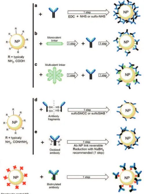
Most nanomaterials require modification or functionalization, either during the initial synthesis or for post-synthesis applications (see Fig. 4). In some cases, modifications are necessary to improve the stability of the nanomaterial (towards dissolution or association), to change its hydrophobicity or hydrophilicity for disper-sion in appropriate solvents, or to enhance biocompatibility. In other cases, surface functionalization may protect or improve the performance of the core material or reduce the overall cost of the material. Water-soluble functional nanomaterials are indispensable for various biomedical applications, but most of the good synthetic methods for noble metals, quantum dots, and magnetic metal oxides produce nanomaterials with a hydrophobic surface [78]. Therefore, water solubilization and functionalization to convert hydrophobic NP into hydrophilic ones and the introduction of additional chemical functionality are key steps for applications [169]. Different strategies have been used to functionalize nanoparticles with a variety of ligands, such as small molecules, surfactants, polymers, and biomolecules [170]. For therapeutic applications, it is common to introduce a polymer coating that is biocompatible (for example PEO, known by its trivial name PEG) and also to include targeting moieties. These include a variety of bio-recognition elements, such as antibodies, nucleic acids, peptides, and ligands for specific receptors. In many cases, the functionalization of the nano-material can be used to produce a multifunctional platform that incorporates both drug delivery and diag-nostic imaging capabilities. The procedures used for surface modification and stabilization include many chemicals, some of which can have a significant effect on NP properties. Precise information is required on the qualitative and quantitative presence of the ligands and stabilizing moieties attached to the surface,
as well as residual reagents that may not have been removed during purification. In the case of targeting ligands, it is particularly important to control their surface orientation and verify that they retain their native binding affinity for the receptor of interest.
Here we review five of the most common strategies for nanomaterial functionalization: silica coating, ligand exchange, polymer coating, surface modification of carbon nanotubes, and bioconjugation to intro-duce targeting moieties. The general challenges and approaches used for surface modification and function-alization are summarized in several recent reviews [172–179].
4.1 Silica coatings
The introduction of a silica coating is one of the most widely used methods for the surface functionalization of NP [180, 181]. Silica serves as a robust layer that prevents the degradation of optical properties, imparts water solubility, and protects the core NP from the external environment [182, 183]. Classic silica coating methods include the use of silane coupling agents [184], the Stöber method [185, 186], and reverse micro-emulsion [187]. Silica coatings can be grafted on most types of nanomaterials that have a chemical or elec-trostatic affinity for silica. Silica coating is usually formed as a one to tens of nanometer-thick porous silica shell on the surface of the nanomaterial core [188]. This process is frequently referred to as silanization in the literature [189–191].
The concept of silane coupling agents was reported by Plueddemann and his coworkers in 1962 [184]. Since then researchers have developed silane coupling agents with different functional groups for NP surface modification. As examples, (3-methacryloxy)propyl]trimethoxysilane [192], (3-aminopropy)ltriethoxysilane (APTES) [193], (3-isocyanatopropy)ltrimethoxysilane [194] and 3-(trimethoxysilyl)propyl methacrylate [195] have been used to functionalize nanoparticles with surface hydroxyl groups, such as silica, TiO2, and Fe3O4 NP. The methoxy/ethoxy groups of these silanes condense on the NP surface, forming a silica layer around the particle. As a result, functional groups are imparted on to the NP surface and are available for future reactions.
A very common approach for the preparation of silica shells is the Stöber method, in which as-prepared NP act as seeds for silica growth in an ethanol/water mixture and yield core-shell spheres. The prerequisite for this method is that the NP surface must have a significant chemical or electrostatic affinity for silica. Silanes can directly polymerize on oxide NP, such as Fe3O4 and TiO2, that have hydroxide-terminated sur-faces; silane hydrolysis and condensation take place under basic conditions. However, for NP that do not have a passivating oxide layer in solution, such as noble metal and quantum dots, a ligand exchange using 3-(trimethoxysilyl)propyl methacrylate and APTES [196] is necessary to render the particles hydrophilic before the Stöber method is applied to grow thicker silica shells. The ligand exchange mechanism will be discussed in the next section. In 2003, Graf et al. developed a simpler and faster method based on the use of poly(vinylpyrrolidone) (PVP), which has been shown to adsorb onto a wide variety of colloidal particles [197, 198]. Upon PVP adsorption, the particles are transferred into a solution of ammonia in ethanol and directly coated with a silica layer of variable thickness by the addition of TEOS in a seeded growth process [197]. The particle stability and homogeneity of the coating shell are significantly affected by the molecular weight of PVP. Longer PVP chains improve the stability of the particles but lead to a rough surface coating. Thus, the optimal PVP lengths for coating different-sized NP usually have to be determined empirically.
The reverse microemulsion method is an alternative approach for silica shell formation. This method provides nanoreactors for the confined synthesis and silica coating of nanoparticles [187]. It can be applied to both hydrophilic QD stabilized with 3-sulfanlpropanoic acid or sulfanylacetic acid and hydrophobic QD prepared by the trioctylphosphine/trioctylphosphine oxide method [199]. In a reverse microemulsion, small water droplets are stabilized by a surfactant in a hydrophobic continuous phase. Hydrolysis and condensa-tion of the silica precursor (e.g. TEOS) take place at the water-oil interface or in the water phase, resulting in highly uniform silica particles [200]. Hydrophilic QD coated with 3-sulfanylpropanoic acid or 2- sulfanylacetic acid transfer easily into the small water droplets, where silica growth occurs. For hydrophobic QD, hydrolyzed
TEOS molecules replace the original hydrophobic ligands, facilitating the transfer of the QD into the hydro-philic interior of the micelles. The reverse microemulsion method has better control over particle size and size distribution than the Stöber method [201].
4.2 Ligand exchange
The use of small ligands that can replace the initial as-synthesized surface coating is a popular strategy for NP surface modification [202]. Highly crystalline and uniform NP are prepared in organic solvents at high temperatures using a stabilizer-modified surface that controls growth and prevents association during syn-thesis. This normally yields hydrophobic NP; ligand exchange is then used to render the NP hydrophilic and suitable for biomedical applications [203].
Ligand molecules usually have two parts: the anchoring groups and the functional groups. The former bind the ligand onto the NP surface with high affinity, while the latter provide colloidal stability and function-ality for further applications [202]. The choice of ligand depends on the core materials, particle size, and the solvent used. For example, QD and noble metal NP have surfaces that can easily bind thiols, disulfides, phos-phines, and amines [102]. Metal oxide NP surfaces typically bind phosphonic acids and carboxylic acids [204]. Trioctylphosphine/trioctylphosphine oxide-coated quantum dots are usually transferred to aqueous solution by replacing the original hydrophobic ligands with hydrophilic thiol-based ligands, such as 2-sul-fanylacetic acid, 3-sulfanylpropanoic acid, 11-sulfanylundecanoic acid, or 2-sulfanylsuccinic acid. Bidentate and multidentate molecules (for example, 6,8-disulfanyloctanoic acid [205, 206], and carbodithioate ligands [207]) have also been developed, as they are more strongly anchored to the NP surface and provide longer-term colloidal stability than monodentate ligands. The carboxylic acid groups on these ligands also increase NP colloidal stability by charge repulsion and have flexible conjugation sites [208]. At the appropriate pH, other charged ligands with quaternary amine, tertiary amine, or sulfonic acid groups can also endow NP with excellent colloidal stability [209].
A second strategy for ligand modification is surfactant addition, during which the hydrophobic moiety of the surfactant associates with the hydrocarbon chain of the initial surface ligands, while the hydrophilic groups are exposed on the surface of the NP, rendering them water soluble [64]. CTAB is a widely-used sur-factant and a known phase transfer agent. When dodecylamine-capped gold nanoparticles in chloroform are mixed with water containing CTAB, the hydrocarbon chain of CTAB assembles on the dodecylamine layer by hydrophobic interaction and the positively charged ammonium moieties extend into the solution, resulting in transfer of the hydrophobic nanoparticles into the aqueous phase [210].
4.3 Polymer coating
ENM that are designed for biomedical applications may be recognized as foreign objects and therefore elimi-nated from the blood stream by the reticuloendothelial system [211, 212]. To avoid premature ENM clearance by the immune system and prolong their half-life in circulation, it is common to introduce “stealth” proper-ties [213] by surface modification with biocompatible polymers [214, 215]. The preferred method is the adsorp-tion or grafting of poly(ethylene oxide) (PEO), a linear polymer consisting of repeat units of –CH2-CH2-O-. A version of this polymer, terminated on both ends with –OH groups, is very well known as poly(ethylene glycol) (PEG). The two terms are used in the literature interchangeably. The systematic term PEO is used more in chemical and polymer sciences, while the trivial term PEG is very common in biological and material sci-ences. In fact, the popularity of the trivial term PEG gave rise to the word ‘PEGylation’ and its analogues to describe the process of attaching segments of PEO to virtually any object or molecule. The term PEGylation is now extensively used across many scientific disciplines (including polymer science) and is also used in this document. We will however, refrain from using PEG, replacing it with the IUPAC-recommended, systematic term PEO.
PEGylation of NP can be accomplished by covalently grafting, entrapping, or physically adsorbing PEO segments to the NP surface [216–219]. There are two synthetic approaches: a one-pot method where in situ formation of NP and polymer coating take place concurrently, and a two-step method where the NP are first prepared and then coated with polymer [212]. Physisorption of PEO onto gold NP surfaces can be achieved through the one-pot method. An aqueous mixture of gold precursor and PEO is bombarded with synchrotron X-rays and PEO-gold NP are formed [220]. Covalent bonding is more complicated, since a thiol-functional-ized PEO is used to passivate the gold surface with thiol groups and form a poly(ethylene oxide) layer on the NP surface. Both the PEO molecular weight and the type of anchoring ligand affect the stability of the PEO-coated NP [221]. Gold NP coated with high-molecular-weight PEO are more stable than those with low-molecular-weight PEO, while NP coated with thioctic acid-anchored PEO exhibit higher colloidal stability than NP coated with thiol-anchored PEO [222]. The PEO coating can be further functionalized with additional moieties, such as antibodies, proteins, dextran, and DNA.
PEGylation of metal oxide NP usually involves introducing a silane between the surface and the PEO layer. This approach can be achieved either by reacting the NP with silanes and then functionalizing with PEO, or by reacting NP with commercially available PEO-siloxane. In the latter case, oleic acid-capped Fe3O4 NP undergo a ligand exchange reaction with PEO-siloxane in toluene, and the PEO coated NP are soluble and stable in aqueous solution [223]. One-pot PEGylation of metal oxide NP is usually achieved in one of two ways: co-precipitation of salts or thermal decomposition of metallorganics in the presence of polymer. In the case of co-precipitation, PEO-coated Fe3O4 NP with various sizes have been synthesized by treating iron(II) and iron(III) solution with a base in the presence of PEO-containing graft copolymer, poly(glycerol monoacrylate)-g-poly(PEO methyl ether acrylate). The polymer block is chemisorbed to the particle surface by the coordination of 1,2-diols to the Fe atoms while the PEO block extends into the water [224]. Similarly, PEO-gallol can be coated onto Fe3O4 NP. The high binding affinity of the gallol and trihydroxybenzene units toward iron oxide allow a successful re-dispersion of freeze-dried particles [225]. Another one-pot synthesis of Fe3O4 NP functionalized with monocarboxyl-terminated PEO is achieved by the thermal decomposition of ferric triacetylacetonate (Fe(acac)3) in the presence of carboxy-PEO via coordination between carboxylic acid and Fe on the NP surface. Particle size is determined by the molecular weight of carboxy-PEO and the concen-tration of Fe(acac)3, as well as by the ratio between them [226].
Block polymers are even more powerful NP stabilizers than homopolymers because they undergo phase separation and exhibit unique properties, such as surface reactivity, elasticity, selectivity, and resistance [227]. For example, degradable PLAGA NP can be coated with an amphiphilic PEO-PLA diblock copolymer, in which the hydrophobic PLA block adsorbs on the PLAGA surface and the hydrophilic PEG block extends from the NP surface and forms a protective coating [228]. However, such physically adsorbed polymers may not be stable and may undergo desorption in vivo due to replacement by blood components with higher affinity to the NP surface. Therefore, covalent binding of polymer is preferred in order to increase coating stability. A series of amphiphilic di-block copolymers of biodegradable PLAGA or PLA with PEO has been produced through ring opening polymerization of monomers in the presence of monomethoxy PEO [229]. Different solubility of the two blocks results in phase separation and a core-shell structure can be formed by an O/W emulsification procedure [230]. Although it is generally accepted that PEO-based coatings repel proteins, some poly(ethylene oxides) seem to be compatible with proteins. For example, a surface modified with PLA-PEO was reported to be able to adsorb proteins [231], provided that these proteins were present at their physiological concentrations [232].
4.4 Surface coatings of carbon nanotubes
Despite the promising biomedical applications of CNT, the high cytotoxicity attributed to their highly hydro-phobic surface and their lack of biodegradability [233] limits their use in many biological systems. There-fore, surface modification and functionalization of CNT to improve their solubility and biocompatibility has become a prerequisite for their applications. Many studies have shown that increased solubility of function-alized CNT improves their performance and lowers their toxicity [234, 235], as they have low association and
![Fig. 4: Surface-modifications of ENM. Adapted from ‘Coatings for nanomaterials’, ref [171].](https://thumb-eu.123doks.com/thumbv2/123doknet/14041968.459060/15.892.157.710.698.1060/fig-surface-modifications-enm-adapted-coatings-nanomaterials-ref.webp)