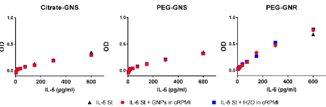
Polymer-coated gold nanospheres do not impair the innate immune function of human b lymphocytes in vitro
Texte intégral
Figure


Documents relatifs
There are only two classical class II B (β or light chain) genes for which the exon 2 sequence (which contains the polymorphic positions corresponding to the peptide-binding region)
i) a-PVA-SPION had no effect on the survival of human immune cells in the whole blood analysis but ii) the cytokine secretion was dose-dependently increased in this
This new model allows us to estimate the amount of water contributed by each individual lobe to any particular measurement made by Rosetta (Fig. 2 shows how the outgassing model
tubulin and DAPI. a) Representative images show single Z-sections at the height of the largest diameter of the nucleus (DAPI, blue), actin (green) and tubulin (red) in fixed
Nous obtenons une contradiction, donc X est infini : il existe une infinité de nombre premier de la forme 4k
MHC class II molecules induction after infection of bovine brain endothelial cells by Cowdria ruminantium.. Bovine brain endothelial cells
Pour avoir accès au module de formation continue et répondre aux questions, vous devez vous rendre sur le site du Pharmactuel www.pharmactuel.com et cliquer sur « Module de
[r]
