Algorithms and circuits for motor control and learning in the songbird
by
Michael E. Stetner
Submitted to the Department of Brain and Cognitive Sciences
in partial fulfillment of the requirements for the degree of
Doctor of Philosophy
at the
MASSACHUSETTS INSTITUTE OF TECHNOLOGY
February 2019
Massachusetts Institute of Technology 2019. All rights reserved.
Signature redacted
A uthor ...
. . .
Brain and Cognitive Sciences Department
January 11, 2019
Cetfidby.*Signature
redacted
Certified by
...
10Sg
a u e r d
c d
...
Michale S. Fee
Glen V. and Phyllis F. Dorflinger Professor and Professor of Neuroscience
Thesis Supervisor
Signature redacted
Accepted by
...
- mMatthew A. Wilson
e'
afairchild Professor of Neuroscience and Picower Scholar
Director of Graduate Education for Brain and Cognitive Sciences
M SACHUSETTSINSTITUTE OF TECHNOLOGY
APR 2 5 Z019
L
R1
Algorithms and circuits for motor control and learning in the songbird
byMichael E. Stetner
Submitted to the Department of Brain and Cognitive Sciences on January 11, 2019
in Partial Fulfillment of the requirements for the Degree of Doctor of Philosophy in Neuroscience
ABSTRACT
From riding a bike to brushing our teeth, we learn many of our motor skills through trial and error. Many biologically based trial and error learning models depend on a teaching signal from dopamine neurons. Dopamine neurons increase their firing rates to signal outcomes that are better than expected and decrease their firing rates to signal outcomes that are worse than expected. This dopamine signal is thought to control learning by triggering synaptic changes in the basal ganglia. What are the origins of this
dopaminergic teaching signal? How do synaptic changes in the basal ganglia lead to changes in behavior? In this thesis, I study these questions in a model of skill learning - the songbird.
In the first part of my thesis, I develop a computational model of song learning. This model incorporates a dopaminergic reinforcement signal in VTA and dopamine-dependent synaptic plasticity in the singing-related part of the basal ganglia. I demonstrate that this model can provide explanations for a variety of experimental results from the literature.
In the second part of my thesis, I investigate a potential source of the dopaminergic error signal in VTA. I performed the first recordings from one cortical input to VTA: the dorsal intermediate arcopallium (Ald). Previous studies disagree on the role of Ald in behavior. Some studies argue that AId contributes vocal error information to VTA. Other studies suggest that AId is not involved in the computation of error signals, but is instead responsible for controlling head and body movements. I directly tested these hypotheses by recording single neurons in AId during singing and during natural movements. My results support a motor role for Ald - Ald neurons had highly significant changes in activity during head and body movements. Meanwhile, following vocal errors Aid neurons had small but marginally significant decrease in firing rate. In a more detailed analysis, I developed an automated behavior classification algorithm to categorize zebra finch behavior and related these behavior classes to the activity of single units in Aid. My results support the hypothesis that Ald is part of a general-purpose motor control network in the songbird brain.
Thesis Supervisor: Michale S. Fee
Acknowledgments
I would not have been able to complete this thesis without help from my coworkers, friends, and family. My committee members, Matthew Wilson, Ann Graybiel, and Bence Olveczky for their thoughtful questions, practical advice, and positive attitude. Most of all, I am grateful for the guidance from my advisor, Michale Fee. His energy, clarity of thought, and precise explanations were all things that tried so hard to imitate.
Michale also assembled a great group of graduate students and post docs that overlapped with me during my time in the lab: Tatsuo Okubo, Emily Mackevicius, Galen Lynch, Dmitriy Aronov, Aaron Andalman, Michael Long, Liora Las, Jesse Goldberg, Anusha Narayan, Maya Bronfeld, Yael Mandelblat-Cerf, Natasha Denisenko, Lena Veit, Husain Danish, Andrew Bahle, Michael Happ, Nader Nikbakht, and Joe Scherrer. I am thankful for their enthusiasm and at our marathon lab meetings and their camaraderie at our daily lunches together. I am also thankful for the behind-the-scenes work of the lab staff. Dan Rubin, Tim Currier, Shelbi Ferber, Anton Konovchenko, Collyn Messier, and Kailey Miller cared for our research subjects and performed excellent histology, and Margo Oleary helped keep us all organized. I am especially appreciative of the lab members who contributed directly to the work in this thesis. Galen Lynch was always the first person I turned to with questions about anything technical, from electronics to statistics. His advice shaped pieces of every chapter. Shelbi Ferber helped with the video analysis in Chapter 3. Abe Akkas and Adrian Cho contributed code and ideas to the analyses in Chapter 4. Outside of the lab, I had the kind support of my friends and family. Thank you to Megan Krench, Phil Isola, and Kean Jaime-Bustamante for the years of living together and sharing food, stories, and a cat. Thanks to Jon Malmaud for all the games we played together, and to Jessi and Mike Sanders for all the time we spent outdoors together. Finally, I would like to thank my family for their patience and
encouragement, especially my wife Katie for being my greatest cheerleader, editor, audience, and partner.
Table of Contents
A cknowledgm ents... 5
Table of Contents... 7
Attributions and publications... 8
1 Introduction ... 10
2 Com putational m odel of reinforcement learning... 33
3 Activity of neurons in AId during vocal errors and natural movements ... 78
4 Neural coding of head and body movements in motor cortex of freely behaving zebra finches 112 5 Discussion... 146
Appendix A Recording system for neural activity and movements ... 156
Attributions and publications
The concept for the model in Chapter 2, including the structure of the network and the learning rule, is taken from (Fee and Goldberg, 2011). The code is all my own. Chapters 3 and 4 are entirely my work.
None of this work has been published in a peer reviewed journal yet. I plan to submit Chapter 2 as a paper to PLOS ONE and combine Chapters 3 and 4 into a second paper to be submitted to PLOS ONE.
1
Introduction
We perform hundreds of complex motor sequences every day - from coordinating our
fingers to tie our shoes to moving our lips and tongue and vocal cords to speak. These behaviors require extreme precision; our brains must bring the right body part to the right place at precisely the right time. These behaviors are not innate but learned through practice. With continued practice, these behaviors are maintained over a lifetime despite changes to our body. The mechanisms underlying the control and learning of these precise motor behaviors remain a topic of active research in neuroscience.
Neuroscientists study songbirds as a model of motor performance and learning, most commonly the zebra finch. Songbirds are natural vocal learners. Juvenile songbirds are born not knowing how to sing, but over development, they learn to imitate an adult tutor. While they are young, they hear an adult tutor sing and memorize this song. If juveniles are raised in isolation without an adult tutor, their song never converges to a stable adult song typical of juveniles raised naturally (Eales, 1985, 1987; Marler, 1981; Williams et al., 1993). Through several months of independent practice, they learn to produce an imitation of the song from their tutor (Immelmann, 1969; Tchernichovski et al., 2001). Their practice can occur alone, without an instructor to tell them when they make a mistake. Rather, they must be able to hear themselves sing; deafening juvenile birds prevents them from learning an accurate imitation of their tutor (Konishi, 1965; Marler and Waser, 1977). This implies they are judging the sound of their own song and producing a teaching signal internally.
In this thesis, I explore several ideas related to this internal teaching signal. In Chapter 2, I construct a computational model of the learning process and demonstrate how a simple teaching
signal can account for some surprising features of song learning. Then, I search for the source of the teaching signal by recording from a cortical brain area thought to carry auditory information during singing. Surprisingly, I discover that this brain area does not contain error related signals, but instead contains signals related to head and body movements. In Chapter 4, 1 further investigate these movement related signals. The remainder of this introduction reviews the literature on vocal production and learning in songbirds and how it relates to theories of learning in general.
1.1 Adult song production
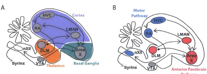
Adult zebra finches sing a complex song, which is composed of repetitions of a single motif. The adult song is highly stereotyped because all renditions of the motif are nearly identical. The motif itself is about one second long and is composed of a fixed sequence of two to ten syllable types, separated by periods of silence. Each syllable type has distinct acoustic features, which are sometimes further subdivided into notes. Multiple renditions of the motif are sung in sequence to form a bout. The songbird is an attractive model for skill learning and performance because the entire process of song production and learning is confined to a small number of discrete brain nuclei that are wholly dedicated to singing. A simplified diagram of some of these brain areas is
shown in Figure 1.1.
A
B
rtex
RA LMAN
nXil ALMre
Syrinx Basal Ganglia
Figure 1.1: Diagram of nuclei of the song system projected
Motor Pathway
LMAN
X DLM Area
Syrinx Anterior Forebrain Pathway
Production of the adult song requires a discrete set of brain nuclei called the motor pathway. At the top of the motor pathway sits the cortical nucleus HVC (used as proper name), which projects to downstream cortical nucleus RA (robust nucleus of the arcopallium) (Nottebohm et al., 1976). RA is similar to primary motor cortex in mammals (Dugas-Ford et al., 2012; Jarvis et al., 2005; Pfenning et al., 2014; Reiner et al., 2004), and it projects directly to the brainstem where it synapses with various motor control structures. These structures include the twelfth cranial nerve (nXII), which contains the motor neurons that innervate the syrinx. RA also projects to several midbrain nuclei not shown in Figure 1.1. It projects to midbrain respiratory centers nucleus retroambigualis and nucleus paraambigualis, as well as to the midbrain vocal center, the dorsomedial nucleus of the intercollicular complex (Vicario, 1991; Wild, 1993). In the following paragraphs, I review a series of studies of the motor pathway, and I describe a simple model for adult song production that focuses on the role of the descending pathway from HVC to RA to the brainstem.
In reality, HVC has additional connections that complicate this simple model. For the sake of completeness, I include a brief description of these extra connections here, but the function of these connections is outside the scope of this thesis. HVC receives input from multiple brain areas in the thalamus and cortex. In the thalamus, nucleus Uvaeformis projects to HVC (Nottebohm et al., 1982) and is thought to play a vital role in the production of a stereotyped, adult song (Danish et al., 2017; Hamaguchi et al., 2016). HVC is also reciprocally connected with auditory regions in the caudal mesopallium (Akutagawa and Konishi, 2010; Bauer et al., 2008), which may play a role in song learning (Roberts et al., 2017). Finally, HVC also receives input from the cortical nucleus interface (Foster and Bottjer, 1998; Nottebohm et al., 1982). The discussion of the motor pathway in rest of the thesis will only focus on HVC, RA, and the brainstem.
Multiple lines of evidence have implicated HVC in generating the stereotyped structure that is characteristic of adult song. In birds of any age, lesions of HVC cause the song to revert to ajuvenile-like song consisting only of highly variable babbling (Aronov et al., 2008). As expected from a brain area driving stereotyped vocalizations, the neural activity in HVC is stereotyped and locked to the motif (Yu and Margoliash, 1996). Furthermore, the projection neurons in HVC have a pattern of activity that is not just highly stereotyped but is also highly sparse. During singing, the projection neurons are silent, except when emitting a high frequency burst of action potentials, which each last about six milliseconds and occur at the same time on each rendition of the motif with less than one millisecond jitter (Kozhevnikov and Fee, 2007). The bursting pattern is slightly different depending on the target of the projection neuron. All projection neurons project to one of two targets, RA in the motor pathway, or the basal ganglia nucleus Area X. HVC neurons that project to RA emit at most one burst per motif, while HVC neurons that project to Area X emit up to four bursts during the motif (Hahnloser et al., 2002; Kozhevnikov and Fee, 2007). As a population, the projection neurons produce bursts that are distributed roughly uniformly over the
duration of the motif (Lynch et al., 2016; Picardo et al., 2016).
The sparse, stereotyped firing patterns in HVC have led to the hypothesis that HVC projection neurons provide a clock-like signal that controls the timing of the song. A variety of manipulations to HVC during singing specifically affect the temporal structure of the song. Perturbing the ongoing activity in HVC with electrical stimulation causes the song to terminate prematurely (Vu et al., 1994; Wang et al., 2008). Local cooling, which slows down the dynamics in the target brain region, slows the temporal structure of the song uniformly when applied to HVC, but not to RA (Long and Fee, 2008), but see (Hamaguchi et al., 2016). Since the song stretching is uniform, the dynamics in HVC are presumed to be controlling the song at all timescales. The
exact form of these dynamics is still an open question, but there are several hypotheses. In one hypothesis, HVC projection neurons form a synfire chain (Abeles, 1991; Jin et al., 2007; Li and Greenside, 2006), where the population of projection neurons active at one time directly activate the population active at the next time. Consistent with this model, intracellular recordings from HVC projection neurons found that their membrane potential is relatively constant until just before the beginning of a burst (Long et al., 2010). Alternative hypotheses incorporate a role for inhibitory interneurons in controlling the timing of HVC bursts (Gibb et al., 2009; Kosche et al., 2015; Yildiz and Kiebel, 2011). These results are all consistent with a hypothesis in which HVC generates the timing signals that control the song.
How do timing signals in HVC evoke stereotyped patterns of activity in the muscles of the vocal organ? In one model of song production, HVC drives stereotyped patterns of muscle activity through RA. Like HVC, the firing patterns in RA during singing are bursty, but the RA neurons are much less sparse (Leonardo and Fee, 2005; Yu and Margoliash, 1996). However, RA does not contribute to the temporal dynamics of the song. Manipulations such as electrical stimulation or mild cooling of RA do not alter the temporal structure of the song (Aronov et al., 2011; Long and Fee, 2008; Vu et al., 1994; Wang et al., 2008). Instead, RA is thought to contribute directly to muscle activation. The projection from RA to the motor neurons of the vocal organ is arranged myotopically (Vicario, 1991), and there is massive reduction in the number of cells from about 8000 RA neurons (Gurney, 1981) to only seven muscles (DUring et al., 2013). Within the population of RA neurons that project to each muscle, the sum of many converging RA neurons could create a smooth motor command for the downstream muscle (Leonardo and Fee, 2005). One hypothesis for vocal learning is that the process requires setting the proper synaptic weights from
the timing signal in HVC to the correct motor output in RA (Doya and Sejnowski, 1995; Fiete et al., 2007; Leonardo and Fee, 2005; Yu and Margoliash, 1996).
1.2 Song learning
Song learning proceeds through several stages. The earliest vocalizations are highly variable babbling, similar to the babbling of human infants (Doupe and Kuhl, 1999). This stage, also known as subsong, is characterized by syllables with a broad distribution of durations that cannot be separated into distinct syllable types (Aronov et al., 2008; Marler, 1981). Next, the first stereotyped elements of song appear in the form of syllables with a stereotyped duration, or "protosyllables" (Aronov et al., 2011). As the protosyllable gains stereotyped acoustic structure, it also splits into multiple distinct syllable types (Liu et al., 2004; Okubo et al., 2015). These syllables make up a motif that is further refined as the acoustic structure becomes more and more similar to the tutor song (Tchernichovski et al., 2001).
Throughout learning, the song gradually shifts from variable to stereotyped. The simultaneous reduction in variability and increase in similarity to the tutor song suggest that vocal exploration early in development is shaped by auditory feedback to match the desired goal. This gradual learning from exploration is shared by many other models that fall under the umbrella of reinforcement learning.
1.3 Reinforcement learning in mammals
Reinforcement learning (RL) is a conceptual framework for learning by trial and error. The origins of reinforcement learning date back to early animal behavior experiments and Thorndike's law of effect: "Actions that produce a satisfying effect in a particular situation become more likely to occur again in that situation" (Thorndike, 1898). Taking inspiration from the way animals learn,
computer scientists developed a mathematical framework for learning using the three elements from Thorndike's law: actions, situations or "states", and satisfying effects or "rewards" (Sutton and Barto, 1998). A learner discovers associations between actions, states, and rewards using the final essential ingredient of a reinforcement learning system: exploration. By trying different actions in each state, the learner accumulates knowledge of which paths lead to the most reward. RL-based artificial intelligence systems have been very successful in learning to play a wide variety of games including backgammon (Tesauro, 1995), go (Silver et al., 2016, 2017), and a variety of video games (Mnih et al., 2013).
There are several practical problems in implementing reinforcement learning systems. First, the number of possible state-action combinations may be so large that it cannot be fully explored in a reasonable amount of time. This problem is called the curse of dimensionality. This problem is typically addressed by using a function to approximate the value of each state or state-action combination. When the system encounters a new situation, it estimates the value of the new situation based on similar situations from its past experience.
Another practical problem for RL systems is linking states and actions with subsequent rewards that may arrive after a long delay. For example, in chess, only the final move will give the immediate reward of a checkmate, but every previous move may have contributed to the victory. More generally, when a reward is received, the credit must be distributed among all past states and actions that caused this reward. A naive solution is to remember the complete history of states and actions, and when a reward arrives, distribute it accordingly. A more common solution is to learn without waiting for the rewards to arrive. As a substitute for future rewards, use the estimated future rewards, perhaps from an approximated value function. Then, the teaching signal is the difference between the reward expected in the previous state and the actual and expected rewards
from the new state. This reward prediction error or "temporal difference error" is a central concept in RL.
Biologically based models of reinforcement learning following discovery of reward prediction error signals in dopamine neurons. Dopamine neurons encode a wide variety information about rewards and other behavioral events. Dopamine neurons increase their firing rates after an animal receives an unexpected reward such as a morsel of food or a drop of juice (Ljungberg et al., 1992). They are also correlated with other behavioral events, such as salient or aversive events (Matsumoto and Hikosaka, 2009) and movement onsets (Howe and Dombeck, 2016; Jin and Costa, 2010). Within the diversity of dopamine signals, the signal that is relevant to reinforcement learning is reward prediction error, the difference between the reward and the expected reward. When an animal receives an unexpected reward, the reward prediction error is positive. If the reward is fully expected, for example due to the presentation of a conditioned stimulus, then the reward prediction error is zero. Finally, if an expected reward is omitted, the reward prediction error is negative at the time when the animal expected the reward. The reward prediction error was originally conceived by Rescorla and Wagner as a teaching signal for classical conditioning (Rescorla and Wagner, 1972). Reinforcement learning algorithms rely on a closely related quantity, the "temporal difference error" (Sutton and Barto, 1998). The signals in dopamine neurons correspond to this teaching signal (Bayer and Glimcher, 2005; Schultz, 2002; Schultz et al., 1997; Waelti et al., 2001), and the dopaminergic midbrain projects widely throughout the brain (Beckstead et al., 1979; Oades and Halliday, 1987; Swanson, 1982). Thus, the dopamine signal is positioned to be a global teaching signal for reward-related behaviors. Accordingly, manipulation of the activity of dopamine neurons can drive powerful changes in behavior. Electrical stimulation of the dopaminergic midbrain and surrounding areas is so reinforcing that animals that are given a
lever that triggers stimulation in VTA and surrounding brain areas will self-stimulate for hours, often until exhaustion (Corbett and Wise, 1980; Olds and Milner, 1954). Subsequent experiments demonstrated behavioral reinforcement could also be achieved by stimulating only the dopaminergic neurons in VTA (Tsai et al., 2009). To cause such a dramatic change in behavior, dopamine must cause profound changes in the brain.
Where does dopamine change the brain during learning? In many hypotheses, the site of learning is in the basal ganglia and more specifically in the striatum. The basal ganglia receive a massive input from the dopaminergic midbrain, and damage to the basal ganglia disrupts skill learning. For these reasons and many others, the basal ganglia are implicated in action selection and habit learning (Graybiel, 2008). The architecture of the basal ganglia supports the possibility of action selection based on a wide range of sensory and cognitive information about state. Thousands of synapses from a wide range of brain areas converge onto striatal medium spiny neurons (MSNs), the most prevalent cell type in the striatum (Kemp and Powell, 1971). These inputs from cortex have the potential to relay state information that could be used in selecting the best action for a given situation (Graybiel et al., 1994; Wickens and Arbuthnott, 2010). Changing the strengths of these corticostriatal synapses may be an important part of learning. MSNs change their activity during trial and error learning (Barnes et al., 2005; Jog et al., 1999), and such changes may be triggered by dopamine activity. In brain slices, glutamatergic inputs to MSNs undergo plasticity based on spike timing and dopamine rules (Shen et al., 2008), and this sort of learning rule is sufficient to solve the credit assignment problem (Izhikevich, 2007). Indeed, corticostriatal synapses are strengthened after learning to press a lever to trigger electrical stimulation of the dopaminergic midbrain (Reynolds et al., 2001), as well as other tasks (Xiong et al., 2015). But which corticostriatal synapses are strengthened? Answering this question is impossible without
knowing the representation of the information in the corticostriatal connections. Unfortunately, the cortical inputs to have not been extensively characterized electrophysiologically (Turner and DeLong, 2000). In songbirds, however, there are only two cortical inputs to the basal ganglia in the song system, and both of them have been recorded during behavior.
1.4 The Anterior Forebrain Pathway: Brain areas for vocal learning in songbirds
Like skill learning in mammals, vocal learning in songbirds requires a basal ganglia circuit. In songbirds, this circuit is called the Anterior Forebrain Pathway (AFP, Figure 1.1B, and it includes brain areas in the cortex, basal ganglia, and thalamus (Figure 1.1A). Area X forms a cortico-basal ganglia-thalamo-cortical loop, similar to basal ganglia circuits in mammals. Area X receives input from two cortical areas, HVC and LMAN, and sends output to the thalamic nucleus DLM (medial portion of the dorsolateral thalamus). To complete the loop, DLM projects back to LMAN. Lesions to any of the nuclei in the AFP in juvenile birds prevent learning an accurate imitation, but lesions to the AFP in adult birds have minimal effects on their song. Since the AFP is not necessary for song production in adulthood, hypotheses about the function of the AFP focus on its role in programming the motor system to perform the adult song.
Many early studies of the role of the AFP in learning observed the changes in behavior after lesions to the output nucleus of the AFP, LMAN. Lesions of LMAN in juvenile birds prevent further learning and cause vocalizations to immediately become highly stereotyped, sometimes just repeating a single syllable. These prematurely crystalized songs are not good imitations of the tutor song (Bottjer et al., 1984; Scharff and Nottebohm, 1991). Even outside the context of natural song learning, LMAN lesions prevent the song from changing in other ways. Typically, deafening an adult bird with a mature, stereotyped song will cause the song to gradually degrade and become move variable (Nordeen and Nordeen, 1992). However, if LMAN is lesioned, the song remains
stereotyped even after deafening (Brainard and Doupe, 2000; Horita et al., 2008). The song also typically changes after damage to the tracheosyringeal nerve, which innervates the syrinx. With LMAN intact, birds add and remove syllables from their songs in the weeks following the nerve injury (Williams and McKibben, 1992), but if LMAN is lesioned before the nerve injury, the number of added and dropped syllables is reduced (Williams and Mehta, 1999). The common theme across all of these situations is that the song can only be variable and malleable when LMAN
is intact.
Area X is the singing-related part of the basal ganglia, and early studies suggested that it played a complimentary role to LMAN in the song learning process. Lesions of Area X in young birds prevent them from learning an accurate imitation of their tutor song (Sohrabji et al., 1990). However, unlike LMAN lesions which immediately reduce variability, Area X lesions cause the song to remain in a permanent state of variable syllable structure and ordering (Scharff and Nottebohm, 1991). One interpretation of these results is that Area X reduces or shapes the variability generated by LMAN. I will return to this hypothesis in more detail in Section 1.6.
Area X shares many features with the mammalian basal ganglia. Area X contains elements from both striatum and pallidum. It contains cell types that match cells mammalian striatum and pallidum in both anatomical and physiological features (Farries and Perkel, 2000, 2002; Goldberg and Fee, 2010; Goldberg et al., 2010). One of these cell types, the medium spiny neuron (MSN) bursts once per motif (Goldberg and Fee, 2010). Also like mammals, these MSNs have dopamine-dependent synaptic plasticity rules (Ding and Perkel, 2004).
1.5 Contributions of brain nuclei in the AFP to generating vocal variability
Perhaps the most salient feature of the AFP is its contribution to vocal variability. The lesions to the AFP discussed in the previous section all have some effect on variability, either
reducing it or prolonging it. Which nuclei are involved in generating this variability and how do they do it? LMAN plays a central role in generating vocal variability. Lesion. or pharmacological
inhibition of LMAN reduces variability in vocalizations of birds of all ages, but the stereotyped structure of the song is preserved (Bottjer et al., 1984; Olveczky et al., 2005; Scharff and Nottebohm, 1991). In support of its role in driving variability, LMAN neurons have firing patterns that are highly variable, with some motif-locked activity (Aronov et al., 2008; Olveczky et al., 2005). While these data do not rule out the participation of other brain areas like Area X or RA in the generation of variability, at least some of the dynamics underlying this variability occur within. Mild cooling of LMAN in juvenile birds lengthens the duration of non-stereotyped syllables
(Aronov et al., 2011).
Multiple lines of evidence support the idea that LMAN affects vocal output by directly activating neurons downstream in RA. LMAN projection neurons are glutamatergic and send collaterals to both Area X and RA (Nixdorf-Bergweiler et al., 1995; Vates et al., 1997). In RA, the variable activity from LMAN may add to stereotyped activity driven by HVC to cause random fluctuations in vocal output. Electrical stimulation of LMAN can cause instantaneous changes in acoustic features of the song (Kao et al., 2005), and pharmacologically inactivating LMAN reduces the variability of firing patterns in RA (Olveczky et al., 2011).
Does Area X contribute to the variability generated by the AFP? Multiple forms of variability persist after Area X lesions, suggesting that variability is generated elsewhere. Both babbling in juveniles (Goldberg and Fee, 2011) and motif-to-motif variability in pitch in adults (Ali et al., 2013) are unaffected by Area X lesions. However, an alternative view suggests that some forms of vocal variability depend on Area X (Kojima et al., 2018). This variability may originate with the variable spiking patterns of pallidal neurons inside Area X (Woolley and Kao,
2015; Woolley et al., 2014) or its glutamatergic interneurons (Budzillo et al., 2017). The preponderance of evidence supports the idea that Area X is not necessary for generating most forms of variability. For this thesis we will adopt the hypothesis that variability is generated in LMAN.
1.6 Circuit level model of reinforcement learning in the AFP
Further study of the contribution of the AFP to song learning has been challenging because natural song learning takes weeks to months and proceeds unpredictably (Deregnaucourt et al., 2004; Tchernichovski et al., 2001). Using a new operant conditioning paradigm, changes at a predictable target in the song can be learned in just hours. The song is monitored in real time by a computer, and the pitch of the song is computed at one target time in the motif. This pitch varies from rendition to rendition due to the influence of LMAN. When the pitch on the current rendition is below a threshold, a loud burst of white noise is played 50 milliseconds or less after the target time through a loudspeaker next to the cage. Over several hours, the bird learns to sing at a higher pitch to avoid triggering the noise (Andalman and Fee, 2009; Charlesworth et al., 2011; Turner and Brainard, 2007; Warren et al., 2011). This learning by conditional auditory feedback requires the same brain areas as natural song learning. These pitch changes cannot be learned after lesions of Area X (Ali et al., 2013). Just as in natural song learning, the actively generated variability from LMAN is required too (Warren et al., 2011), but subsequent experiments demonstrated that LMAN
contributes even more to the learning process.
Since the discovery that learning ceases after LMAN lesions, LMAN was hypothesized to send the motor pathway an instructional signal. It was unclear how LMAN could drive vocal variability while also giving an instructive signal. This instructional signal was thought to be auditory (Bottjer et al., 1984), the result of a comparison of the what the bird hears himself sing
against a stored memory of his tutor song. Initially this theory found support from recordings LMAN neurons in anesthetized birds, which revealed auditory responses to playback of the bird's own song or the song of his tutor (Doupe, 1997; Doupe and Konishi, 1991; Lewicki, 1996; Margoliash, 1983; Volman, 1996). However, subsequent studies ruled out an auditory role for LMAN by recording LMAN neurons in awake birds and finding no response to noise bursts played during singing (Leonardo, 2004).
Surprisingly, LMAN does relay an instructional signal to the motor pathway, but not an auditory one. Instead, the instructional signal comes from a bias embedded in the LMAN-driven variability. The variability is biased towards variants that reduce vocal errors. In conditional auditory feedback experiments, where a bird learns to change the pitch of his song at a targeted time to escape bursts of white noise, pharmacological inactivation of LMAN removes the variability as expected, but also removes the recently learned shift in pitch (Andalman and Fee, 2009; Warren et al., 2011). The learned shift in pitch returned after the pharmacological agent has washed out. Of course, this bias must be temporary because LMAN is not necessary for song production in adults. Indeed, if the pitch shift is maintained for multiple days, LMAN inactivation has a progressively smaller effect (Warren et al., 2011).
The transient role of LMAN in learning new song changes has led to a hypothesis in which song learning occurs in two stages proceeding in tandem. First, learning within the AFP allows LMAN to generate biased variability that biases the song towards variants that sound more similar
to the tutor song. Second, the bias is consolidated into the HVC -> RA pathway. For the remainder
of the thesis, I will focus on the first learning process that occurs within the AFP.
Learning in the AFP is driven by reward prediction error signals from VTA. During conditional auditory feedback, neurons in VTA that project to Area X decrease their activity
immediately following a noise burst. Inversely, when a noise burst is omitted, these neurons increase their activity immediately after the target time (Gadagkar et al., 2016). This pattern of activity is a reward prediction error, very similar to the reward prediction error in mammalian VTA neurons. In this case, a noise burst is played at the target time on about 50% of renditions, so the expected reward is midway between the low reward of a noise burst and a high reward of an escape. Therefore, a noise burst is lower than the expected reward and an escape is higher.
Manipulating Area X-projecting VTA neurons directly is sufficient to drive learning, substituting for the noise burst. Just like noise bursts, inhibition of VTA neurons that project to Area X is aversive. For example, inhibiting VTA neurons when the bird sings at a lower pitch causes the bird to shift his song towards a higher pitch. Inversely, stimulating VTA neurons is reinforcing and causes birds to sing in the stimulated pitch range more often (Xiao et al., 2018). Several computational models of song learning have been proposed using this type of error signal (Doya and Sejnowski, 1995; Fee and Goldberg, 2011; Fiete et al., 2007).
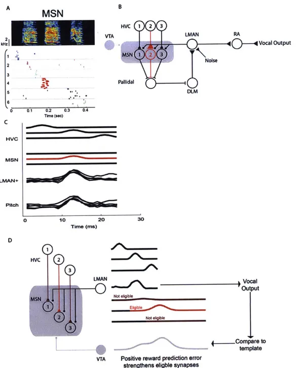
This thesis focuses on the framework laid out by Fee and Goldberg (2011), in which a reinforcement signal from VTA strengthens corticostriatal synapses in Area X to drive bias in LMAN (Fee and Goldberg, 2011). Area X receives an image of the vocal variability via the axon collaterals from LMAN. When a reinforcement signal arrives from VTA, Area X can associate the reinforcement with the pattern of variability that caused it, in order to reproduce that pattern of LMAN activity again on future renditions. This computation must take place independently at every moment in the song, since the variant that makes the song "better" at one time may not make the song "better" at a different time in the motif. Therefore, the reinforcement signal must strengthen HVC synapses such that the timing signals from HVC drive the appropriate pattern of bias at each moment. This model hypothesizes a three-part learning rule that depends on the inputs
from HVC, LMAN, and VTA: dopamine input from VTA strengthens HVC synapses onto MSNs that recently received a signal from LMAN. In Chapter 2, I implement this model and show that it reproduces a wide variety of experimental results.
1.7 Origins of the reward prediction error signal
Like many biological models of reinforcement learning, my computational model depends on a reward prediction error (RPE), which experimental evidence suggests is carried by VTA neurons. A reward prediction error is just one of many signals that have been described in VTA neurons in birds and mammals; VTA neurons also carry a wide range of information relating to rewards, salient events, and motivation. To understand the learning process, it is crucial to understand how the brain computes the RPE signal in VTA neurons. In Chapter 3 of my thesis, I investigate the contribution of one area of the songbird brain to the RPE signal in VTA neurons.
The source of the reward prediction error signal in VTA is an area of active research. The output of VTA dopamine neurons is consistent with a simple subtraction of the actual reward minus the expected reward (Eshel et al., 2015), so it is tempting to imagine that just two inputs converge on VTA, one carrying reward information and the other carrying prediction information. Unfortunately, the truth is much more complicated.
1.7.1 Reward-related activity in the inputs to VTA in mammals
In mammals, VTA receives input from a wide range of brain areas, both cortical and subcortical (Geisler et al., 2007; Sesack and Grace, 2010; Watabe-Uchida et al., 2012), including from the lateral hypothalamus, ventral striatum, ventral pallidum, frontal cortex, and lateral habenula. Untangling the roles of brain areas is especially difficult because the areas that project to VTA are also interconnected with each other (Geisler and Zahm, 2005). No consensus has
emerged on how the signals are combined to form a reward prediction error (Watabe-Uchida et al., 2017). Nevertheless, I review a few elements of this circuit from studies in rodents and primates in the next few paragraphs, and then I will return to the possible sources of reward prediction error in birds.
One pathway for reward related signals comes from the lateral habenula (LHb). LHb projects directly to VTA, as well as indirectly though the rostromedial tegmental nucleus (RMTg). Neurons in both LHb and RMTg have responses that are the inverse of reward prediction error responses in VTA (Hong et al., 2011; Jhou et al., 2009; Matsumoto and Hikosaka, 2007). Their neurons decrease their firing rates upon presentation of a reward or a reward-predicting cue and increase their firing rates in response to an aversive stimulus or an omitted reward. LHb and RMTg send a GABAergic projection to VTA, and if this signal inhibits VTA dopamine neurons, it can reproduce the reward prediction error observed in those neurons. Even though it seems like all the information necessary to compute a reward prediction error is present in these neurons, they are not necessary for many of the response properties of VTA neurons. Even after lesions of LHb, VTA neurons still increase their firing rates in response to rewards and reward predicting cues. LHb lesions only eliminate the phasic decrease in VTA neuron activity when a reward is omitted (Tian and Uchida 2015). For its response to reward to persist after LHb lesions, VTA must receive reward-related signals from other parts of the brain.
Another source of reward signals comes from the orbitofrontal cortex (OFC). Single neurons in the OFC increase their firing rates when a mammal is presented with a rewarding stimulus or when a reward is expected (Feierstein et al., 2006; Padoa-Schioppa and Assad, 2006; Rolls, 2000; Schoenbaum and Eichenbaum, 1995; Tremblay and Schultz, 1999; Wallis and Miller, 2003). Lesions of OFC disrupt value-guided choice behavior, especially when the options change
in value (Butter et al., 1969; Izquierdo et al., 2004; Pickens et al., 2003). These lesions also change the activity of VTA dopamine neurons, but some reward information remains in their activity patterns (Takahashi et al. 2011). Further research will be necessary to finish untangling the roles of the LHb, OFC, and the many other brain areas that are connected to VTA.
1.7.2 Inputs to VTA in songbirds
A similar search is ongoing for the sources of auditory error signals in the VTA of songbirds. In zebra finches, Area X-projecting VTA neurons decrease their activity following a burst of white noise played during singing. When an expected noise burst is omitted, these neurons increase their firing rates (Gadagkar et al., 2016). This error signal is widely believed to be the result of a comparison between what a bird hears during singing and a stored memory of the tutor song. The components of this computation may be carried by the inputs to VTA, and several studies have explored the roles of these inputs in vocal learning.
One input to VTA, which is not the focus of my thesis, is from the ventral pallidum (VP), a part of the basal ganglia that is outside of Area X (Chen et al., 2018; Gale et al., 2008). Chen et al. discovered vocal error related information in VP in recent, unpublished work. In their recordings, VTA-projecting VP neurons carry reward-related information, including predicted reward and even reward prediction error (Chen et al., 2018). These signals may play an important role in computing the reward prediction error signals in VTA.
In my thesis, I have focused on inputs to VTA from a second set of inputs to VTA from cortical areas. In zebra finches, the only cortical input to VTA comes from the intermediate arcopallium (Gale et al., 2008). The intermediate arcopallium has been subdivided into two parts, ventral (AIv) and dorsal (Ald), and these two divisions may have different contributions to VTA.
AIv is required for vocal learning and sends a vocal error signal to VTA. Lesions of AIv in juvenile birds prevents them from learning an accurate imitation of the song of their tutor (Mandelblat-Cerf et al., 2014). In addition to lesion studies, neurophysiological evidence also supports a role for AIv in song learning. Av receives input from auditory pathways (Kelley and Nottebohm, 1979; Mello et al., 1998; Vates et al., 1996), some of which have a phasic increase in their activity immediately following a noise burst during singing (Keller and Hahnloser, 2009). Similar to these auditory brain areas, neurons in AIv that project to VTA also have a phasic increase in activity after a noise burst is delivered during singing (Mandelblat-Cerf et al., 2014). If these AIv neurons inhibited the neurons in VTA that project to Area X, it could be responsible for the decrease in activity in those neurons following a noise burst.
In contrast, the role of AId in behavior is controversial, and a major focus of my thesis is to help resolve this controversy. Separate lines of research from different labs have focused on two different hypotheses for the role of AId. One hypothesis is that AId contributes to vocal learning by processing vocal errors, perhaps similar to AIv. The second hypothesis is that AId is not related to singing or vocalization at all, but is instead related to controlling head and body movements. Before explaining these two hypotheses in more detail, I will first review the anatomy of AId.
Aid is part of a separate network of brain areas that parallel the song system in their anatomical positions and connectivity. This anatomy is depicted schematically in Figure 1.2. In the arcopallium, AId is immediately lateral to RA (Bottjer et al., 1989). These areas both send descending projections out of the telencephalon. They also project to adjacent regions in the dorsal thalamic zone (DTZ) (Bottjer et al., 2000), and these portions of the thalamus project to neighboring regions of nidopallium. In the song system, DLM projects to LMAN, while in the parallel network DTZ projects to LMANshell, a region of parvocellular cell bodies surrounding
LMAN (Johnson and Bottjer, 1992). LMANshell completes the loop by projecting back to AId directly and indirectly via dNCL (dorsal part of the caudolateral nidopallium) (Bottjer et al., 1989, 2000). The similarities between the anatomy and the song system and the anatomy of this parallel pathway implies that there may be some shared function between these two pathways.
One hypothesis is that the parallel pathway, including AId, is involved in vocal learning. Even though there are no known connections between this parallel pathway and the ascending auditory system, two of the nuclei in this pathway, dNCL and LMANshell, show physiological evidence of processing auditory information. In dNCL, immediate early gene expression is high after singing as well as after hearing playback of a tutor or conspecific song, compared to birds who did not sing or hear song playback (Bottjer et al., 2010). In LMANshell, recordings under anesthesia found neurons that respond to playback of the bird's own song or the tutor song (Achiro and Bottjer, 2013). During singing, about 10% of units in LMANshell either increase or decrease their activity during the production of syllables that are more similar to syllables in the tutor song (Achiro et al., 2017). It is not known if these signals are relayed to VTA because no recordings have been performed in AId.
dNCL Aid Ctx LMANshell DTZ Motor Mst Centers Basal Ganglia Figure 1.2: Brain areas of the parallel pathway
The vocal learning hypothesis has also been tested with lesions of AId, but the results of these studies are difficult to interpret due to the anatomical relationship between AId and AIv. In one study, lesions of AId in juvenile birds was reported to impair imitation (Bottjer and Altenau, 2010). However, a subsequent study contradicted these results and found that AId lesions had no impact on learning. Instead, only lesions of Av impacted learning (Mandelblat-Cerf et al., 2014). One possible explanation is that the earlier study lesioned both AId and AIv, since they are adjacent. Anatomically, not only are AIv and AId adjacent, they may also be partially overlapping. This overlap is possible because AIv is defined by its outputs: AIv is the portion of intermediate arcopallium that projects to VTA (Mandelblat-Cerf et al., 2014; Mello et al., 1998). On the other hand, AId is defined by its inputs: AId is the portion of intermediate arcopallium that receives a projection from LMANshell, part of the nidopallium surrounding the song nucleus LMAN (Bottjer et al., 2000; Johnson et al., 1995). By these definitions, there is a small portion on the medial edge of AId that overlaps with AIv. However, AId is largely distinct from the VTA projecting region AIv (Mandelblat-Cerf et al., 2014). Given that only a small portion of AId projects to VTA, it may seem unlikely that AId as a whole is contributing to the vocal error signals in VTA. Rather than focus on this small overlapping region, in this thesis, I investigate the role of AId as a whole by recording from single neurons throughout AId.
An alternative hypothesis for AId is that it not involved in singing or song learning at all, but instead controls head and body movements. The most striking evidence in favor of this hypothesis comes from a study of immediate early gene expression by Feenders et al. (2008). They examined immediate early gene expression throughout the zebra finch brain during two different activities, singing and hopping. During singing, immediate early gene expression is high in the nuclei of the song system, including HVC, RA, LMAN, and Area X, but low in the surrounding
areas of the brain. During hopping, the expression pattern is reversed, with high expression in the areas surrounding the song system, including AId, LMANshell, and other areas in its parallel pathway. The juxtaposition of the song system with a network of hopping-related brain areas led Feenders et al. to hypothesize that the song system evolved from a more general motor control circuit (Chakraborty and Jarvis, 2015; Feenders et al., 2008). By this hypothesis, AId should be involved in motor control, not vocal learning.
Additional evidence supports the hypothesis that AId is involved in motor control. Anatomically, AId sends output to multiple subcortical motor centers including the midbrain reticular formation and the deep layers of the tectum (Bottjer et al., 2000). Consistent with a role in controlling movements, large lesions of AId cause akinesia (Mandelblat-Cerf et al., 2014). However, no recordings of AId have ever been performed in an awake bird.
Chapters 3 and 4 of my thesis test two hypotheses of Aid function that have emerged from the literature. In Chapter 3, I test the hypotheses that AId is involved in processing vocal errors or controlling movements. I perform the first recordings of AId in an awake bird and record the same neurons during both singing and natural movements. I find evidence that AId predominantly encodes movement-related information with large modulations in activity around the times of head rotations and whole-body movements such as pecking. As a population, these neurons have a very small reduction in firing rate following noise bursts, so I cannot rule out that they are involved in processing vocal errors.
In Chapter 4, I further investigate the role of AId in movements. I design a microdrive for recording simultaneous neural activity and movement signals. To analyze the movement signals, I develop an algorithm to automatically classify natural zebra finch behavior and relate these behavior classes to the neural activity in AId.
1.1 Summary
Trial and error learning is ubiquitous in animal behavior. Among animal models of trial and error learning, the songbird has several advantages. Song learning involves a relatively small number of brain areas with known connectivity, and neural activity has been recorded from many of these brain areas during the same learning behavior. However, there are several notable gaps in understanding. First, there is no computational model that capture the recently discovered role of a basal ganglia circuit in driving biased variability during song learning. Second, there is some uncertainty about which brain areas are involved in computing an error signal. This thesis seeks to fill those gaps.
Chapter 2 constructs a computational model of song learning based on RL that provides a unified explanation for many behavioral results that could not be explained by previous models.
Chapter 3 contains the first recordings from AId in an awake bird. Each neuron was recorded both during singing and during natural movements. These recordings lend strong support to the idea that AId neurons are related to movements and not to processing vocal errors.
Chapter 4 further quantifies the head and body movements of freely moving birds and explores the relationship of neural activity in AId to these movements.
Chapter 5 discusses the relationship between the song circuit and the surrounding brain
2 Computational model of reinforcement learning
2.1 IntroductionThe song learning process has inspired a series of computational models based on reinforcement learning. In the earliest reinforcement learning model of song learning, Doya and Sejnowski proposed that LMAN drives stochastic changes in HVC-RA weights on each rendition of the song. In the model, the vocal output is compared to a stored memory of the tutor song by Area X. Area X sends a reinforcement signal to LMAN that indicates if the perturbation in this trial made the song sound better than average. If so, the HVC-RA weights are modified to keep the perturbation (Doya and Sejnowski, 1995, 1998). This paper established an important framework that is adopted by future models of song learning, including in this paper. Variability from LMAN is correlated with a reinforcement signal to bias the song towards variants that give high reinforcement. Following this procedure, the HVC-RA weights undergo stochastic gradient ascent to maximize the reinforcement signal. Unfortunately, there is no known biophysical mechanism that can perturb synaptic strengths to a new value on every motif, as the Doya and Sejnowski model requires.
Subsequent experiments demonstrated that LMAN has a rapid, glutamatergic influence on the song, which inspired the next generation of song learning models. Electrical stimulation of LMAN causes rapid and transient fluctuations in the song (Kao et al., 2005). In light of this, Fiete et al. proposed a new model in which LMAN caused random fluctuations in RA activity. A reinforcement signal to RA strengthened HVC-RA synapses if an LMAN fluctuation caused the song to sound better than average (Fiete et al., 2007). Fiete et al. made an explicit distinction
between the two classes of synapses onto RA neurons. Each RA neuron receives a single "empiric" synapse from LMAN that drives random fluctuations in RA activity but does not change in strength during learning. Each RA neuron receives many plastic synapses from HVC that do change with learning. A plastic synapse is eligible for strengthening when it is coactive with the empiric synapse, and eligible synapses are strengthened when a reinforcement signal is received. Eligible synapses maintain an eligibility trace to allow for a delayed reward signal. One prediction of this model is that removing LMAN after learning will leave the learned song intact and only remove vocal variability.
Detailed studies of learning revealed that LMAN contributes more than just variability
-the variability can be biased in a direction to reduce errors. In -these studies, rapid learning was induced with a short, loud burst of noise which is played while the bird is singing (Andalman and
Fee, 2009; Turner and Brainard, 2007). The noise is contingent on the pitch of the song at a single
moment in the motif - for example, pitches above a threshold are hit with noise while pitches
below the threshold escape punishment (Figure 2.1E). Over just a few hours, birds rapidly learn to sing at a lower pitch at the targeted time to avoid the noise. Surprisingly, this learned shift in pitch is driven by activity in LMAN. When LMAN is pharmacologically inactivated or its output to RA blocked after learning induced by conditional auditory feedback, the mean pitch returns to its pre-learning levels (Figure 2.lF) (Andalman and Fee, 2009; Warren et al., 2011). LMAN is carrying a signal that biases vocal output towards variants that escape the noise. There must be a separate
learning process in the LMAN circuit that rapidly learns this bias.
The discovery of biased variability led to a conceptual model in which Area X learns to bias ongoing variability in LMAN (Fee and Goldberg, 2011). This is the model we implement in this chapter. In this model, Area X associates variations in LMAN activity with a dopaminergic
reinforcement signal from VTA. Then on subsequent renditions, Area X feeds back to reactivate the same LMAN neurons that were associated with reward. Of course, this computation must be
performed locally at each time in the song - the increase in pitch that makes the song sound better
at one time may not make the song sound better at another time. Therefore, the model proposes that plastic changes in the timing signals from HVC drive a bias that is specific to one time in the song. Fee and Goldberg proposed the following three-part learning rule for MSNs in Area X: an HVC-MSN synapse is strengthened when the HVC and LMAN inputs are co-active, followed by reward.
The Fee and Goldberg model retains the plastic/empiric synapse distinction from the Fiete et al. model. Each model MSN receives a single empiric synapse from LMAN and a single plastic synapse from HVC representing the neurons that are active at a single moment in the song.
Borrowing from the mammalian reinforcement learning literature, Fee and Goldberg hypothesized that the reinforcement signal to Area X comes from dopaminergic neurons in VTA and SNc. This prediction was confirmed a few years later with the discovery of a vocal error signal in dopamine neurons that project to Area X (Gadagkar et al., 2016). The activity in these dopamine neurons decreased following a noise burst during singing, but increased when an expected noise burst was omitted.
Alternative models for song learning combine vocal exploration and auditory feedback in different ways. For example, auditory feedback can be used to build a forward model of the vocal production system that predicts the auditory feedback that would be created by a given motor command. This prediction could be used in place of a real-time reinforcement signal to train the song production circuitry (Troyer and Doupe, 2000). Another alternative is to learn an inverse model, rather than a forward model. An inverse model can translate a desired auditory stimulus
into the motor commands necessary to recreate that stimulus. Once learned through exploration and feedback, such an inverse model could produce an imitation by replaying the auditory memory of the tutor song through the inverse model (Hahnloser and Ganguli, 2013).
In this chapter, we implement the Goldberg and Fee model as a firing rate-based neural network and discover that it quantitatively reproduces several features of song learning. I begin by demonstrating that the model can learn a simple song. Next, I explore some of the potential weakness of this reinforcement learning model. In general, reinforcement learning models scale poorly with the number of degrees of freedom and with the delay and precision of the reinforcement signal. I demonstrate that the model is robust to the number of degrees of freedom in the song and the shape of the reward signal.
I also demonstrate that the model can explain several surprising behavioral results from conditional auditory feedback experiments. The model reproduces the precise temporal structure of learning during conditional auditory feedback. In a careful analysis of learning during conditional auditory feedback (CAF), Charlesworth et al (2011) found that the pitch changes in a small window about 20ms around the target time. This observation was especially interesting because the exact temporal structure of this learning can be predicted from the average of the trials that escape punishment (Charlesworth et al., 2011). The precision of this learning is especially surprising considering that the reinforcement signal may be delayed and imprecise in time. Our model reproduces this feature of learning, but only when the reward signal is indeed broad.
In another set of experiments based on CAF, Charlesworth et al. demonstrated that LMAN is capable of latent learning (Charlesworth et al., 2012). In Bengalese finches, blocking the projection of LMAN to RA leaves about 70% of the vocal variability intact (Chariesworth et al., 2012; Warren et al., 2011). Performing CAF does not result in a pitch change as long as the block
on LMAN->RA is in place. However, as soon as the blockade is removed, the song immediately has a bias that allows it to escape the noise, as if this bias was learned covertly but was only able to be expressed through the connection from LMAN to RA. Crucially, no bias is learned at all if LMAN activity is inhibited using muscimol. We demonstrate that our model is capable of covert learning in this fashion by receiving a copy of the non-LMAN variability through the RA->DLM->LMAN projections, as suggested by Charlesworth et al.
In the final sections of this chapter, we extend the model in more speculative directions. First, we explore how MSNs in Area X might develop their characteristic sparse firing patterns. In our original model, each MSN was active once per motif because it received input from a single HVC neuron. In reality, there is massive convergence from cortical neurons onto MSNs (Kincaid
et al., 1998), yet individual MSNs are active at only a single moment during singing (Goldberg
and Fee, 2010). We demonstrate that adding long-term depression (LTD) and lateral inhibition allows each MSN to develop this selectivity from initial all-to-all connectivity during the learning process.
Finally, we extend the model to include more of the internal circuitry of Area X. Like mammalian basal ganglia, Area X contains two pathways: the direct and indirect pathways (Farries et al., 2005). In models of mammalian basal ganglia function, the direct pathway activates movements while the indirect pathway inhibits movements (Albin et al., 1989; Alexander and Crutcher, 1990a; DeLong, 1990; Mink, 1996). Our original model only contains the direct pathway, so in the final section we extend these ideas to our learning model in which the indirect pathway biases song output away from variants that sound worse. This compliments the direct pathway, which biases the song towards variants that sound better.
2.2 Methods
Our implementation of the Fee and Goldberg model uses units with smoothly varying activities representing the aggregated firing rates of groups of hundreds to thousands of neurons in Area X and connected brain regions. The model includes HVC, LMAN, Area X, DLM, and VTA. The roles of each of these brain areas are described in the following sections.
2.2.1 HVC
HVC produces stereotyped timing signals that uniformly cover the entire song. Each HVC projection neuron bursts highly selectively and reliably at 1-3 moments in the song (Figure 2.1 B) (Kozhevnikov and Fee, 2007). For simplicity, HVC units in our model burst once per motif. Each
unit Hi emits a single burst at a unique time ti with a burst that lasts TH milliseconds in the motif
and is otherwise silent. Each burst is shaped like one period of a sine squared curve and sequential bursts are staggered by 1 ms and scaled so that the sum of all HVC activity is always one:
Hi(t) = sinz
(
(t - ti)) ti !; t < ti + T H(TJ
I
otherwise0
2.2.2 LMAN
LMAN produces biased variability in the output of the model. The output of each LMAN unit is a combination of a constant baseline level of activity (LO), intrinsic variability (Z), and excitatory input from DLM that drives bias (D):
Ln (t) = [Lo + Zn(t) + Dn (t)]0
Both Z and D can be positive or negative and cause fluctuations around baseline LMAN activity Lo. The random noise process Z was constructed so that the pitch fluctuations from the