K. Kinkel R. Forstner F. M. Danza L. Oleaga T. M. Cunha A. Bergman J. O. Barentsz C. Balleyguier B. Brkljacic J. A. Spencer Received: 11 September 2008 Revised: 4 December 2008 Accepted: 11 January 2009 Published online: 5 February 2009
# European Society of Radiology 2009
Staging of endometrial cancer with MRI:
Guidelines of the European Society
of Urogenital Imaging
Abstract The purpose of this study was to define guidelines for endome-trial cancer staging with MRI. The technique included critical review and expert consensus of MRI protocols by
the female imaging subcommittee of the European Society of Urogenital Radiology, from ten European insti-tutions, and published literature be-tween 1999 and 2008. The results indicated that high field MRI should include at least two T2-weighted sequences in sagittal, axial oblique or coronal oblique orientation (short and long axis of the uterine body) of the pelvic content. High-resolution post-contrast images acquired at 2 min ± 30 s after intravenous contrast injection are suggested to be optimal for the diagnosis of myometrial invasion. If cervical invasion is suspected, addi-tional slice orientation perpendicular to the axis of the endocervical channel is recommended. Due to the limited sensitivity of MRI to detect lymph node metastasis without lymph node-specific contrast agents, retroperito-neal lymph node screening with pre-contrast sequences up to the level of the kidneys is optional. The like-lihood of lymph node invasion and the need for staging lymphadenectomy are also indicated by high-grade his-tology at endometrial tissue sampling and by deep myometrial or cervical invasion detected by MRI. In conclu-sion, expert consensus and literature review lead to an optimized MRI protocol to stage endometrial cancer.
Keywords Uterus, endometrium . Uterine neoplasms, staging, uterine neoplasms . Magnetic resonance (MR), gynecologic oncology K. Kinkel
Clinique des Grangettes,
Geneva University Hospital and Institut de Radiologie,
7, Chemin des Grangettes, CH1224 Chêne-Bougeries/Geneva, Switzerland R. Forstner Zentralröntgeninstitut, LandesklinikenSalzburg, Mullner Hauptstr. 48, A-5020 Salzburg, Austria F. M. Danza
Dipartimento di Bioimmagini e scienze radiologiche,
Università Cattolica del S. Cuore, 8, largo A. Gemelli,
00168 Rome, Italy L. Oleaga
Radiology Department, Hospital Clinic, C/Villaroel 170,
Barcelona, 08036, Spain T. M. Cunha
Department of Radiology, Instituto Português de Oncologia de Lisboa Francisco Gentil, R. Prof. Lima Basto,
1099-023 Lisboa Codex, Portugal A. Bergman
Department of Radiology, Uppsala University Hospital, SE-751 85 Uppsala, Sweden
J. O. Barentsz
Department of Radiology, Radboud University Nijmegen Medical Center, Nijmegen, The Netherlands
C. Balleyguier
Department of Radiology, Institut de Cancérologie Gustave Roussy, 39, rue Camille Desmoulins, 94805 Villejuif Cedex, France B. Brkljacic
Department of Diagnostic and Interventional Radiology, University Hospital“Dubrava”, Avenija G.Suska 6,
Zagreb, Croatia B. Brkljacic
Medical School, University of Zagreb, 10000 Zagreb, Croatia
J. A. Spencer
Department of Clinical Radiology, St James’s Institute of Oncology, Leeds, LS9 7TF, UK
K. Kinkel (*) Institut de radiologie, Clinique des Grangettes, 7, chemin des Grangettes, CH 1223
Chêne-Bougerie/Geneva, Switzerland e-mail: [email protected] Tel.: +41-22-3050379
Introduction
Endometrial cancer is the fourth most frequent cancer in women and now the most common gynecological cancer in many developed countries [1]. Surgical treatment options depend on the local extent of disease [2, 3]. In low-risk patients only hysterectomy with bilateral oophorectomy is performed, whereas in selected high-risk patients lym-phadenectomy, omental and peritoneal biopsies are in-cluded in the surgical treatment [4]. The most recent surgical trend consists in performing lymphadenectomy through laparoscopy [5]. The disadvantage of systematic lymphadenectomy is a 7–10% risk of lymph cyst formation after surgery [5, 6], increased anesthesia and operating time, and the need for a specialized oncologic surgeon. However, when lymphadenectomy is not performed, most patients will undergo either a second surgery with pelvic lymphadenectomy when pathology of the uterus reports deep myometrial or cervical invasion, or a systematic pelvic or intravaginal postoperative radiation therapy. Pelvic radiation therapy might not be necessary if prior lymphadenectomy has not shown lymph node invasion [7]. Patient selection for primary lymphadenectomy at the time of hysterectomy is therefore a current debate in oncological gynecology [4].
In patients with endometrial cancer, histological tumor grade and depth of myometrial invasion strongly correlate with lymph node metastases and patient survival [8]. Therefore, patients are considered at high risk for lymph node invasion when high tumor grade is diagnosed at endometrial biopsy or when deep myometrial invasion is identified before surgery. A meta-analysis of reported MRI studies suggests that preoperative identification of deep myometrial invasion in patients with endometrial cancer is best performed with contrast-enhanced MRI of the pelvis with an accuracy of about 91% [9]. Without the use of contrast-enhanced MRI and using only the knowledge of preoperative tumor grade from the endometrial biopsy, the probability of deep endometrial invasion has been estimated at respectively 13% for tumor grade 1, 35% for tumor grade 2 and 54% for tumor grade 3 [10]. After a positive MRI for deep myometrial invasion, those probabilities change to respectively 60, 84 and 92% for tumor grade 1, 2 and 3. When MRI of the pelvis does not identify deep myometrial invasion, the probability of such a condition decreases to respectively 1, 5 and 10% for tumor grade 1, 2 and 3 [10]. However pitfalls in diagnosing the extent of endometrial cancer have been reported with MRI when associated adenomyosis or large leiomyoma impairs exact assessment of the depth of myometrial invasion [11, 12]. To reduce potential pitfalls or mis-interpretation of false-negative or false-positive findings of myometrial or cervical invasion, MRI images should be acquired with enough technical quality to allow adequate image interpretation. Many of the studies analyzed in the meta-analysis [9] were small series reported from the
proving stage of MRI, and some revealed low image quality and a lack of technical standards. Thus, the female pelvis subcommittee of the European Society of Urogenital Imaging (ESUR) formed a working group to establish technical guidelines for endometrial cancer staging with MRI based on extended clinical practice.
Material and methods
MRI protocols for staging endometrial cancer were collected from ten European institutions. Inclusion criteria to participate in the guidelines topic“Staging endometrial cancer with MRI” were: to be a member of the European Society of Urogenital Imaging (ESUR) and to perform at least ten MR imaging examinations per year for staging biopsy-proven endometrial carcinoma. The questionnaire included the following details: field strength and type of coil, patient preparation, type of sequence with detailed geometry and contrast information, such as FOV, matrix scan and reconstruction, slice thickness, gap, orientation, saturation bands, 2D versus 3D sequence, TR/TE, number of acquisitions, number and lengths of dynamic sequences, bolus IV injection and use of subtraction techniques. In addition, published literature between 1999 and 2008 was reviewed through a Medline literature search of abstracts in the English language on studies in human subjects, including the following key words: “Uterine neoplasm(s) AND MR imaging” or “Endometrial carcinoma AND MR imaging.” Articles that did not include technical details following the information of the questionnaire were excluded. The results were presented in an excel sheet including descriptive statistics, and were discussed and divided in topics with agreement and disagreement. Topics with disagreement were conpared to the literature. Experts in favor of one technical option were asked to document their point of view with the corresponding literature, which was discussed by an expert panel. Literature work was compared taking into consideration year of publication, number of cases and performance of the technique. When technical details varied and no published work on performance was found, the subject was discussed and resolved in consensus based on the majority of participat-ing members. Members of the group met twice a year during a 2-year period and were asked to apply technical recommendations issued after the first two meetings during year 1 before the meetings in year 2 to allow further discussion and making final conclusions in consensus.
Results
Technical consideration with agreement
Among the ten ESUR members, seven worked at 1.5 T, two at 1.0 T and one institution at 0.5 T. Agreement concerned
fasting (3–6 h) and intramuscular injection of peristaltic inhibitors (1 mg glucagon or 20 mg butyl-scopolamine) unless contraindicated (e.g., diabetes or pheochromocyto-ma) to decrease peristalsis artifacts. Furthermore, there was consensus to apply anterior and superior saturation bands and to use a combination of at least two T2-weighted unenhanced (TR/TE 4,000/90 ms) and one T1-weighted enhanced sequence for the assessment of myometrial invasion. Oblique coronal and axial slice orientation according to the long and short axis of the endometrial cavity should be applied under medical supervision as technicians might not have sufficient anatomical knowl-edge to clearly identify the long axis of the uterine body (although with adequate training this should become possible). For optimal image quality, T2-weighted images covering the pelvis should be acquired with a small FOV (20–25 cm) and ideally with a 512×512 matrix. Slice sections of 4 mm in T2-weighted imaging should avoid difficulties in analyzing small postmenopausal uteri or underestimation of myometrial depth invasion because of
thin overstretched myometrial walls by large tumors. Disruption of the junctional zone at T2-weighted images or discontinuity on early subendometrial contrast enhance-ment on dynamic imaging has been described as a landmark for the diagnosis of myometrial invasion [13– 16] (Fig.1). However, this finding is typically not seen in the menstrual and first part of the cycle. Furthermore, in women at postmenopausal age where such an enhancement pattern is reported as typical, its visualization may be impaired by the low spatial resolution of dynamic contrast-enhanced imaging [17, 18]. Slice section of 2 mm with enough spatial resolution is only possible in 3D gradient echo sequences, which may be preferable to standard enhanced 2D sequences.
As lymph node metastases may skip the pelvis and occur primarily in the para-aortic region, the retroperitoneum should be included in the imaging protocol. This protocol option has the advantage of allowing simultaneous detection of hydronephrosis. Table1shows the results of the literature search, including technical protocols.
Fig. 1 a: Figure1: MRI of the pelvis in a 75-year-old patient with biopsy-proven endometrial carcinoma grade 2. (a) Sagittal T2-weighted image of the uterus demonstrates an enlarged hyperintense endometrium with an interruption of the low signal intensity of the posterior junctional zone (arrow), suggesting myometrial invasion. (b) The axial oblique T2-weighted image of the uterus, perpendi-cular to the direction of the cavity seen in (a) and passing through the interrupted juntional zone, demonstrates heterogeneous signal intensity of the upper junctional zone (arrow) without evidence of deep endometrial invasion. (c) The 3D contrast-enhanced gradient echo image with fat suppression acquired 2 min after contrast injection confirms a heterogeneous interface between the less enhancing endometrial cancer and the strongly enhancing normal myometrium, suggesting superficial mymometrial invasion. Stage IB was confirmed at histopathology; 50×50 mm (300×300 DPI). b: Figure1: MRI of the pelvis in a 75-year-old patient with biopsy-proven endometrial carcinoma grade 2. (a) Sagittal T2-weighted image of the uterus demonstrates an enlarged hyperintense endo-metrium with an interruption of the low signal intensity of the posterior junctional zone (arrow), suggesting myometrial invasion. (b) The axial oblique T2-weighted image of the uterus, perpendi-cular to the direction of the cavity seen in (a) and passing through the interrupted juntional zone, demonstrates heterogeneous signal
intensity of the upper junctional zone (arrow) without evidence of deep endometrial invasion. (c) The 3D contrast-enhanced gradient echo image with fat suppression acquired 2 min after contrast injection confirms a heterogeneous interface between the less enhancing endometrial cancer and the strongly enhancing normal myometrium, suggesting superficial mymometrial invasion. Stage IB was confirmed at histopathology; 50×50 mm (300×300 DPI). (c): Figure1: MRI of the pelvis in a 75-year-old patient with biopsy-proven endometrial carcinoma grade 2. (a) Sagittal T2-weighted image of the uterus demonstrates an enlarged hyperintense endo-metrium with an interruption of the low signal intensity of the posterior junctional zone (arrow), suggesting myometrial invasion. (b) The axial oblique T2-weighted image of the uterus, perpendi-cular to the direction of the cavity seen in (a) and passing through the interrupted juntional zone, demonstrates heterogeneous signal intensity of the upper junctional zone (arrow) without evidence of deep endometrial invasion. (c) The 3D contrast-enhanced gradient echo image with fat suppression acquired 2 min after contrast injection confirms a heterogeneous interface between the less enhancing endometrial cancer and the strongly enhancing normal myometrium, suggesting superficial mymometrial invasion. Stage IB was confirmed at histopathology; 50×50 mm (300×300 DPI)
Discordance concerning dynamic post-contrast sequences
A single discordant topic concerned the type of contrast-enhanced T1-weighted sequences: 2D versus 3D sequence, a single or up to eight dynamic acquisitions after intrave-nous contrast medium, as well as the length of a single dynamic acquisition that varied between 18 s and 3 min 21 s. Dynamic image acquisition has been recommended since Yamashita et al. in 1993, observing less contrast between the tumor and the normal myometrium at 5 min after injection of contrast agent compared to 2 min after injection [13]. The authors demonstrated that at dynamic imaging acquired every 30 s, normal myometrium showed strong early enhancement at 90 s after bolus enhancement, particularly at the junctional zone. The tumor invading the myometrium demonstrated decreased enhancement com-pared to the strong enhancement of normal myometrium. The greatest contrast between invaded and non-invaded myometrium was visible at 2 min after injection. Most publications after 1993 therefore presented imaging protocols with dynamic enhancement. In 2004, Manfredi et al. demonstrated an optimal contrast-to-noise ratio between the endometrial tumor and the normal myometri-um at 2 min 30 s compared to 30 s and 1 min 30 s after
intravenous injection of contrast agent [28]. The imaging protocol used a dynamic 3D gradient echo sequence (FMSPGR) with one unenhanced and three enhanced acquisitions of 1-min acquisition time each. A quantitative analysis of the contrast-to-noise ratio between the endo-metrial tumor and the normal myometrium was performed through a region-of-interest analysis at a workstation.
A discussion was raised about the utility of dynamic acquisition due to the newly proposed optimal time point of 2 min 30 s that could be obtained with a single enhanced acquisition with higher spatial resolution. Reported false-negative findings of deep myometrial invasion in small uteri could be due to a technical protocol that chose a decreased spatial resolution of dynamic imaging in favor of higher temporal resolution to allow dectection of the optimal time point of strong myometrial enhancement. To avoid understaging of endometrial carcinoma due to an overstretched myometrium by tumor distention of the cavity, smaller slice thickness, field of view and higher matrix are technical solutions to decrease the occurrence of these pitfalls [12]. Associated benign uterine disease can be responsible for overstaging of myometrial invasion due to hypovascular myomas or adenomyosis mimicking deep endometrial invasion (Figs.2and3). If endometrial polyps are present in the endocervical channel, cervical invasion Table 1 Results of the literature
search according to year of publication
First author Journal Year Literature reference
Cabrita Eur J Gynaecol Oncol 2008 [19]
Shen AJR 2008 [20]
Park Gyn Oncol 2008 [21]
Ortashi EJOG 2008 [22]
Sala AJR 2007 [23]
Chung Gyn Oncol 2007 [24]
Nakao Gyn Oncol 2006 [25]
Messiou Clinical Radiology 2006 [18]
Barwick Clinical Radiology 2006 [26]
Nasi Radiol Med 2005 [27]
Manfredi Abdom Imaging 2005 [14]
Manfredi Radiology 2004 [28]
Utsunomiya AJR 2004 [29]
Tanaka Eur Radiol 2003 [30]
Ohguri Eur Radiol 2002 [15]
Cunha Int J Gynecol Cancer 2001 [31]
Ueda Eur Radiol 2001 [32]
Grasel Radiology 2000 [33]
Kinkel Radiology 1999 [9]
Shibutani Abdom Imaging 1999 [34]
Joja Radiat Med 1999 [16]
Lee Radiographics 1999 [35]
can be overdiagnosed. A single high spatial resolution 3D gradient echo sequence was suggested as the optimal enhanced sequence for the diagnosis of myometrial invasion by endometrial cancer. The increased length of those sequences usually allows selective fat suppression during image acquisition and a matrix of 512×512 that should increase detection of deep myometrial invasion and allow the diagnosis of transmyometrial invasion (invasion up to the uterine serosa, stage IIIA) with higher confidence. Because of rectilinear k-space sampling in the y direction during 3D sequences, the signal intensity of the first commercially available 3D sequences corresponded to the contrast acquisition at the middle of the acquisition time (for example, 2 min 30 s for a single 3D sequence with an acquisition time of 5 min for GE). This sampling time might be earlier and vary according the magnet constructor and the magnet version. In more recently developed 3D fat-suppressed angiographic sequences, such as the THRIVE (Philips), VIBE (Siemens) or VIBRANT (GE) sequence, the time point used to acquire sequence information relative to contrast corresponds to 20% of the acquisition time, depending for the same sequence also on the magnet and the software version. Let’s check the timing of intravenous contrast injection in two different centers in which the radiologist wants to acquire post-contrast images at 2 min after contrast injection with two different 3D sequences of the same acquisition time: For a 3D sequence of 4 min 10 s acquisition time (250 s) and an image contrast acquisition at 20% of acquisition time (at 50 s), the
intravenous contrast agent should be injected 70 s prior to acquisition start to allow collection of contrast information at 120 s. If a 3D sequence of 4 min 10 s has a time point that acquires sequence information relative to contrast at mid-sequence acquisition time (2 min 5 s), the intravenous contrast can be injected 5 s after acquisition start. This example demonstrates the importance of communication with the application specialist of the magnet to know when information relative to the contrast is acquired during acquisition time for a specific 3D sequence, magnet and software version, therefore allowing adequate timing of intravenous injection of contrast agents. A summary of the technical protocol recommendation is presented in Table2.
Criteria for image interpretation according to tumor stage The MR staging criteria for endometrial cancer follows the guidelines of the surgically-pathologically based FIGO classification [26,37]. Stage O tumors (carcinoma in situ) are not visualized on MRI. In stage I endometrial cancer, the tumor is limited to the uterine corpus. It encompasses tumor without myometrial invasion (stage IA), super-ficially (IB) and deeply (IC) invasive cancers of the myometrium. In stage IA normal, diffusely or focally thickened endometrium larger than 3 mm in postmeno-pausal age may be seen. The key feature in imaging is identification of the intact low signal intensity junctional zone at T2-weighted imaging. At T1-weighted dynamic Fig. 2 (a): Figure2: MRI of the pelvis in a 70-year-old patient with
grade 3 endometrial carcinoma. (a) Sagittal T2-weighted image of the uterus shows a thin endometrial cavity and a thickened junctional zone containing hyperintense lines (arrow), suggesting adenomyosis. Identification of the endometrial tumor is difficult. (b) The sagittal contrast-enhanced 3D gradient echo image with fat suppression acquired at 2 min after contrast injection demonstrates a hypointense area extending from the uterine cavity up to the outer myometrium (arrow), diagnosing deep endometrial invasion beyond the area of associated adenomyosis. Stage 1C with adenomyosis was confirmed at histopathology; 50×50 mm (300×300 DPI). (b):
Figure2: MRI of the pelvis in a 70-year-old patient with grade 3 endometrial carcinoma. (a) Sagittal T2-weighted image of the uterus shows a thin endometrial cavity and a thickened junctional zone containing hyperintense lines (arrow), suggesting adenomyosis. Identification of the endometrial tumor is difficult. (b) The sagittal contrast-enhanced 3D gradient echo image with fat suppression acquired at 2 min after contrast injection demonstrates a hypointense area extending from the uterine cavity up to the outer myometrium (arrow), diagnosing deep endometrial invasion beyond the area of associated adenomyosis. Stage 1C with adenomyosis was confirmed at histopathology; 50×50 mm (300×300 DPI)
Table 2 Suggested MR imaging protocol for staging endometrial cancer Patient preparation and positioning:
Fasting: 3–6 h
Antiperistaltic agent if not contraindicated Supine position
Pelvic phased-array coil T2-weighted imaging:
Image orientation: Sagittal, axial oblique (perpendicular) and coronal oblique (parallel) to the uterine cavity. If cervical involvement is suspected, additional axial oblique image orientation perpendicular to the long axis of the endocervical channel
Slice thickness for the pelvis:≤4 mm, FOV 20–25 cm. High-resolution matrix (512 × 512)
Extended FOV for assessing the retroperitoneum with coronal T1-weighted or axial T2-weighted sequence with fat suppression Contrast-enhanced imaging
Suggested for higher accuracy of the diagnosis of deep myometrial invasion. Recommended for atrophic uteri, associate adenomyosis or fibroids, or in suspected advanced tumors (suspicious bladder or rectal wall invasion)
2D or 3D techniques may be performed with the optimal tumor/myometrial contrast timing between 90 and 150 s. A single 3D acquisition technique seems a good alternative to dynamic imaging as it combines a high tumor-myometrium contrast with multiplanar reformations and thin slice sections
Fig. 3 (a): Figure3: MRI of the pelvis in a 56-year-old patient with grade 1 endometrial cancer and an enlarged uterine size at pelvic sonography. (a) Sagittal T2-weighted image of the uterus demon-strates multiple submucosal and interstitial leiomyoma associated with an enlarged uterine cavity and a small interruption of the anterior junctional zone (arrow), suggesting superficial myometrial invasion. (b) An axial oblique image of the upper uterine cavity shows no junctional zone because of multiple submucosal leiomy-oma protruding into the cavity. (c) Contrast-enhanced 3D gradient echo image with fat suppression in the same orientation as in image (b) shows an enhancing line (arrow) between the submucosal leiomyomas and the endometrial tumor associated with a slight decrease in visibility of the line at the right side. Histopathology of the hysterectomy specimen demonstrated superficial myometrial invasion of the uterus (stage IB); 50×50 mm (300×300 DPI). (b): Figure3: MRI of the pelvis in a 56-year-old patient with grade 1 endometrial cancer and an enlarged uterine size at pelvic sonogra-phy. (a) Sagittal T2-weighted image of the uterus demonstrates multiple submucosal and interstitial leiomyoma associated with an enlarged uterine cavity and a small interruption of the anterior junctional zone (arrow), suggesting superficial myometrial invasion. (b) An axial oblique image of the upper uterine cavity shows no junctional zone because of multiple submucosal leiomyoma
protruding into the cavity. (c) Contrast-enhanced 3D gradient echo image with fat suppression in the same orientation as in image (b) shows an enhancing line (arrow) between the submucosal leiomy-oma and the endometrial tumor associated with a slight decrease in visibility of the line at the right side. Histopathology of the hysterectomy specimen demonstrated superficial myometrial invasion of the uterus (stage IB); 50×50 mm (300×300 DPI). (c): Figure3: MRI of the pelvis in a 56-year-old patient with grade 1 endometrial cancer and an enlarged uterine size at pelvic sonography. (a) Sagittal T2-weighted image of the uterus demonstrates multiple submucosal and interstitial leiomyoma associated with an enlarged uterine cavity and a small interruption of the anterior junctional zone (arrow), suggesting superficial myometrial invasion. (b) An axial oblique image of the upper uterine cavity shows no junctional zone because of multiple submucosal leiomyoma protruding into the cavity. (c) Contrast-enhanced 3D gradient echo image with fat suppression in the same orientation as in image (b) shows an enhancing line (arrow) between the submucosal leiomyoma and the endometrial tumor associated with a slight decrease in visibility of the line at the right side. Histopathology of the hysterectomy specimen demonstrated superficial myometrial invasion of the uterus (stage IB); 50×50 mm (300×300 DPI)
imaging complete subendometrial enhancement may be visible. If the junctional zone is not visible, a sharp tumor-myometrial interface at T2-weighted imaging and at contrast-enhanced images suggests an intact myometrium. It is not unusual to see minimal abnormality, such as a small amount of fluid within the endometrial cavity after diagnostic resection.
Conversely, in stage IB and IC cancer myometrial invasion is found. In stage IB, which refers to myometrial invasion of less than half of the myometrial thickness, disruption or irregularity of the low signal intensity junctional zone by intermediate signal intensity mass is identified at T2-weighted imaging (Figs. 1 and3). If the junctional zone is not visible, as in up to half of postmenopausal women or in associated findings, such as fibroids and adenomyosis, contrast-enhanced MRI helps in the assessment of the depth of myometrial invasion [9]. In stage IB at approximately 2 min or 90 to 150 s at contrast-enhanced imaging, an irregular endometrium/myometrium interface is best identified [28]. In stage IC the endometrial cancer extends into the outer half of the myometrium, but a small stripe of deep normal myometrium is preserved (Fig.2).
Stage II endometrial cancer is characterized by tumor extension into the cervix, but not beyond the uterus. In stage IIA the internal os and the endocervical canal are widened by the hyperintense or inhomogeneous tumor signal at T2-weighted imaging or the hypointense tumor at contrast-enhanced imaging. If the low signal intensity ring of the normal cervical stroma is disrupted, stage IIB can be diagnosed [38,39] (Fig.4). Axial oblique image orienta-tion perpendicular to the cervical canal with thin secorienta-tion
thickness will improve the assessment of cervical invasion in endometrial cancer [34]. Direct cervical stroma invasion may occur without invasion of the endocervical mucosa because of adjacent myometrial invasion. Due to reduced contrast uptake of the cervical stroma compared to the myometrium, the contrast resolution between the tumor and the cervical stroma may be reduced on contrast-enhanced images.
In stage III tumor extends beyond the uterus, but is confined to the true pelvis. In stage IIIA transmyometrial involvement and disruption of the T2 hypointense uterine serosa and/or irregular uterine contour is noted. Other findings may include adnexal or parametrial involvement. Metastases to or direct extension to the upper vagina with focal loss of the low signal intensity vaginal wall is typical for stage IIIB. Pelvic and/or para-aortic lympadenopathy indicates stage IIIC. Imaging signs of lymph node involvement include a diameter equal to or greater than 8 mm in the pelvis or in round nodes and of greater than 10-mm short axis in retroperitoneal or oval nodes.
Stage IV endometrial cancer is defined by tumor extension beyond the true pelvis or involvement of the bladder or rectal mucosa. MRI findings include disruption of the low signal intensity of the bladder or rectal wall, a mucosal or intra-luminal mass (stage IVA) best identified with contrast-enhanced sequences. Distant metastases (IVB) are rare at presentation and better seen at CT or at an extended MR imaging protocol to the abdomen. Abdominal lymph node metastases with the exception of retroperitoneal and inguinal lymph nodes, signs of perito-neal spread and metastases to the lungs and liver are found in stage IVB.
Indications for MRI in endometrial cancer
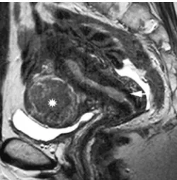
In comparison to ovarian and cervical cancer, there is a debate about the usefulness of pretreatment MRI [40]. The main reason is that endometrial cancer is treated in the majority of cases by surgery. However, therapeutic options may vary and depend on tumor stage, tumor grade and medical condition of the patient. The growing integration of radiologists in multidisciplinary teams is central in defining tailored treatment options, and imaging findings may modify the therapeutic approach [40]. Indications for MRI with proven or suspected endometrial cancer include: high grade, serous or clear cell adenocarcinomas; suspicion of advanced disease, including cervical stroma extension and confirmation of stage III and IV disease; screening for lymph node enlargement as a roadmap for lymph node sampling; medical contraindication for surgical staging; suspected endometrial cancer with inability of curettage (e.g., cervical stenosis). The key advantage of MRI is in demonstrating stage IC disease, outer myometrial invasion or stage II disease, cervical involvement, in cases that would otherwise have been understaged or undertreated. Fig. 4 Sagittal T2-weighted image of the uterus in a 60-year-old
patient with grade 2 endometrial carcinoma. The thickened hyperintense endometrium invades the mucosa of the endocervical channel and the adjacent hypointense cervical stroma (arrow). A large subserosal leiomyoma is identified in the anterior uterine wall (asterix). Stage IIB was confirmed at histopathology; 50×50 mm (300×300 DPI)
Open questions
Preoperative lymph node imaging has been limited by the low performance of all imaging techniques, including MRI. Size criteria often do not correlate with histological findings because of the high frequency of micrometastases [19]. This is why MRI serves rather as a roadmap for guiding lymph node sampling than for detecting stage IIIC endometrial cancer [41]. Assessment of lymph node invasion in patients with endometrial or cervical cancer using a lymph node-specific contrast agent, ferumoxtran-10, composed of ultrasmall particles of iron oxide (USPIO), increases the sensitivity of lymph node metas-tasis from 29% to 93% when nanoparticle-enhanced MRI is performed [42]. The product is evaluated in research protocols and requires injection 24 h prior to the MRI examination. Other studies in patients with prostate cancer have shown that there is no need to perform a MRI study before and after injection of nanoparticles [43]. Further consensus is required about how to proceed for staging MRI for patients with endometrial cancer, including nanoparticle-enhanced MRI as a one-stop staging exami-nation. According to Rockall and co-authors, the high negative predictive value of MR lymphography could potentially allow avoiding surgical lymph node dissection, whereas a positive result of MR lymphography could indicate the site of metastatic nodes to direct surgical planning or radiation therapy according to the surgical risk profile of the patient. The commercial availability of USPIO in the near future is however uncertain.
F18- FDG PET/CT is useful in detecting distant metastases and in the surveillance of recurrent endometrial cancer. Its value for staging endometrial cancer is not yet established. First data show no superiority of PET/CT
compared to MRI in the detection and lymph node assessment of endometrial cancer [21].
There are some patients for whom lymphadenectomy will occur on the basis of factors from endometrial sampling, e.g., high-grade histology, or from basic MRI, e.g., outer myometrial or cervical invasion. Thus, newer MR technologies should be used selectively and after multidisciplinary discussion to avoid unnecessary expense.
Diffusion- weighted imaging is emerging as a promising tool in assessing gynecologic cancers. As mean ADC values of endometrial cancer are lower than that of myometrium, this technique may aid in assessing myo-metrial invasion [20]. Further research is required to define if it should be included as an adjunct sequence in the preoperative assessment of endometrial cancer.
In conclusion, in patients sent for MR staging of an endometrial carcinoma, the female imaging subcommittee of the European Society of Urogenital Radiology sug-gested the use of at least two T2-weighted sequences followed by a single enhanced acquisition at optimal image orientation according to the results of sagittal, oblique short or long axis T2-weighted images. Selection of imaging planes requires close medical supervision during image acquisition. Further research should test the accuracy of a single high-resolution 3D gradient echo sequence in patients with endometrial cancer to diagnose myometrial and stromal invasion and evaluate the clinical utility of emerging technologies, such as nanoparticle and functional imaging. Indications for MRI include: high grade, serous or clear cell adenocarcinomas; suspicion of disease greater than stage 1B; screening for lymphadenopathy; medical contraindication for surgical staging; suspected inability of curettage.
References
1. Jemal A, Siegel R, Ward E, Murray T, Xu J, Thun MJ (2007) Cancer statistics, 2007. CA Cancer J Clin 57:43–66 2. Bakkum-Gamez JN, Gonzalez-Bosquet
J, Laack NN, Mariani A, Dowdy SC (2008) Current issues in the manage-ment of endometrial cancer. Mayo Clin Proc 83:97–112
3. Sorosky JI (2008) Endometrial cancer. Obstet Gynecol 111:436–447
4. Zuurendonk LD, Smit RA, Mol BW, Feijen HW, de Graaff J, Sykora D, de Winter KA, vd Wurff A, Snijders MP, Kruitwagen RF (2006) Routine pelvic lymphadenectomy in apparently early stage endometrial cancer. Eur J Surg Oncol 32:450–454 Epub 2006 Mar 2020 5. Querleu D, Leblanc E, Cartron G,
Narducci F, Ferron G, Martel P (2006) Audit of preoperative and early com-plications of laparoscopic lymph node dissection in 1,000 gynecologic cancer patients. Am J Obstet Gynecol 195:1287–1292 Epub 2006 May 1283 6. Savino L, Borruto F, Comparetto C,
Massi GB (2001) Radical vaginal hys-terectomy with extraperitoneal pelvic lymphadenectomy in cervical cancer. Eur J Gynaecol Oncol 22:31–35
7. Bottke D, Wiegel T, Kreienberg R, Kurzeder C, Sauer G (2007) Stage IB endometrial cancer. Does lymphade-nectomy replace adjuvant radiothera-py? Strahlenther Onkol 183:600–604 8. Boronow RC, Morrow CP, Creasman
WT, Disaia PJ, Silverberg SG, Miller A, Blessing JA (1984) Surgical staging in endometrial cancer: clinical-patho-logic findings of a prospective study. Obstet Gynecol 63:825–832
9. Kinkel K, Kaji Y, Yu KK, Segal MR, Lu Y, Powell CB, Hricak H (1999) Radiologic staging in patients with endometrial cancer: a meta-analysis. Radiology 212:711–718
10. Frei KA, Kinkel K, Bonel HM, Lu Y, Zaloudek C, Hricak H (2000) Predic-tion of deep myometrial invasion in patients with endometrial cancer: clin-ical utility of contrast-enhanced MR imaging—a meta-analysis and Baye-sian analysis. Radiology 216:444–449 11. Scoutt LM, McCarthy SM, Flynn SD,
Lange RC, Long F, Smith RC, Chambers SK, Kohorn E, Schwartz P, Chambers JT (1995) Clinical stage I endometrial carcinoma: pitfalls in preoperative as-sessment with MR imaging. Work in progress. Radiology 194:567–572 12. Kinkel K (2005) Pitfalls in staging
uterine neoplasm with imaging: a re-view. Abdom Imaging 5:5
13. Yamashita Y, Harada M, Sawada T, Takahashi M, Miyazaki K, Okamura H (1993) Normal uterus and FIGO stage I endometrial carcinoma: dynamic gado-linium-enhanced MR imaging. Radiol-ogy 186:495–501
14. Manfredi R, Gui B, Maresca G, Fanfani F, Bonomo L (2005) Endometrial can-cer: magnetic resonance imaging. Abdom Imaging 6:6
15. Ohguri T, Aoki T, Watanabe H, Nakamura K, Nakata H, Matsuura Y, Kashimura M (2002) MRI findings including gadolinium-enhanced dy-namic studies of malignant, mixed mesodermal tumors of the uterus: dif-ferentiation from endometrial carcino-mas. Eur Radiol 12:2737–2742 Epub 2002 May 2709
16. Joja I, Asakawa T, Shiraiwa M, Shibutani O, Okuno K, Akaki S, Togami I, Kudo T, Hiraki Y (1999) Endometrial carcinoma: multisection dynamic MR imaging using a three-dimensional FLASH technique during breath holding. Radiat Med 17:211– 218
17. Yamashita Y, Torashima M, Takahashi M, Tanaka N, Katabuchi H, Miyazaki K, Ito M, Okamura H (1993) Hyper-intense uterine leiomyoma at T2-weighted MR imaging: differentiation with dynamic enhanced MR imaging and clinical implications. Radiology 189:721–725
18. Messiou C, Spencer JA, Swift SE (2006) MR staging of endometrial carcinoma. Clin Radiol 61:822–832
19. Cabrita S, Rodrigues H, Abreu R, Martins M, Teixeira L, Marques C, Mota F, de Oliveira CF (2008) Magnetic resonance imaging in the preoperative staging of endometrial carcinoma. Eur J Gynaecol Oncol 29:135–137
20. Shen SH, Chiou YY, Wang JH, Yen MS, Lee RC, Lai CR, Chang CY (2008) Diffusion—weighted single-shot echo-planar imaging with parallel technique in assessment of endometrial cancer. AJR Am J Roentgenol 190:481–488
21. Park JY, Kim EN, Kim DY, Suh DS, Kim JH, Kim YM, Kim YT, Nam JH (2008) Comparison of the validity of magnetic resonance imaging and posi-tron emission tomography/computed tomography in the preoperative eva-luation of patients with uterine corpus cancer. Gynecol Oncol 108:486–492 Epub 2008 Jan 2016
22. Ortashi O, Jain S, Emannuel O, Henry R, Wood A, Evans J (2008) Evaluation of the sensitivity, specificity, positive and negative predictive values of pre-operative magnetic resonance imaging for staging endometrial cancer. A pro-spective study of 100 cases at the Dorset Cancer Centre. Eur J Obstet Gynecol Reprod Biol 137:232–235 23. Sala E, Wakely S, Senior E, Lomas D
(2007) MRI of malignant neoplasms of the uterine corpus and cervix. AJR Am J Roentgenol 188:1577–1587
24. Chung HH, Kang SB, Cho JY, Kim JW, Park NH, Song YS, Kim SH, Lee HP (2007) Accuracy of MR imaging for the prediction of myometrial invasion of endometrial carcinoma. Gynecol Oncol 104:654–659
25. Nakao Y, Yokoyama M, Hara K, Koyamatsu Y, Yasunaga M, Araki Y, Watanabe Y, Iwasaka T (2006) MR imaging in endometrial carcinoma as a diagnostic tool for the absence of myometrial invasion. Gynecol Oncol 102:343–347
26. Barwick TD, Rockall AG, Barton DP, Sohaib SA (2006) Imaging of endo-metrial adenocarcinoma. Clin Radiol 61:545–555
27. Nasi F, Fiocchi F, Pecchi A, Rivasi F, Torricelli P (2005) MRI evaluation of myometrial invasion by endometrial carcinoma. Comparison between fast-spin-echo T2w and coronal-FMPSPGR Gadolinium-Dota-enhanced sequences. Radiol Med (Torino) 110:199–210 28. Manfredi R, Mirk P, Maresca G,
Margariti PA, Testa A, Zannoni GF, Giordano D, Scambia G, Marano P (2004) Local-regional staging of endo-metrial carcinoma: role of MR imaging in surgical planning. Radiology 231:372–378
29. Utsunomiya D, Notsute S, Hayashida Y, Lwakatare F, Katabuchi H, Okamura H, Awai K, Yamashita Y (2004) En-dometrial carcinoma in adenomyosis: assessment of myometrial invasion on T2-weighted spin-echo and gadolini-um-enhanced T1-weighted images. AJR Am J Roentgenol 182:399–404 30. Tanaka YO, Nishida M, Tsunoda H, Ichikawa Y, Saida Y, Itai Y (2003) A thickened or indistinct junctional zone on T2-weighted MR images in patients with endometrial carcinoma: pathologic consideration based on microcircula-tion. Eur Radiol 13:2038–2045 31. Cunha TM, Felix A, Cabral I (2001)
Preoperative assessment of deep myo-metrial and cervical invasion in endo-metrial carcinoma: comparison of magnetic resonance imaging and gross visual inspection. Int J Gynecol Cancer 11:130–136
32. Ueda M, Otsuka M, Hatakenaka M, Sakai S, Ono M, Yoshimitsu K, Honda H, Torii Y (2001) MR imaging findings of uterine endometrial stromal sarcoma: differentiation from endometrial carci-noma. Eur Radiol 11:28–33
33. Grasel RP, Outwater EK, Siegelman ES, Capuzzi D, Parker L, Hussain SM (2000) Endometrial polyps: MR imag-ing features and distinction from en-dometrial carcinoma. Radiology 214:47–52
34. Shibutani O, Joja I, Shiraiwa M, Asakawa T, Miyagi Y, Kudo T, Hiraki Y (1999) Endometrial carcinoma: effi-cacy of thin-section oblique axial MR images for evaluating cervical invasion. Abdom Imaging 24:520–526
35. Lee EJ, Byun JY, Kim BS, Koong SE, Shinn KS (1999) Staging of early endometrial carcinoma: assessment with T2-weighted and gadolinium-en-hanced T1-weighted MR imaging. Radiographics 19:937–945 discussion 946–937
36. Koyama T, Togashi K, Konishi I, Kobayashi H, Ueda H, Kataoka ML, Itoh T, Higuchi T, Fujii S, Konishi J (1999) MR imaging of endometrial stromal sarcoma: correlation with pathologic findings. AJR Am J Roent-genol 173:767–772
37. Benedet JL, Bender H, Jones H 3rd, Ngan HY, Pecorelli S (2000) FIGO staging classifications and clinical practice guidelines in the management of gynecologic cancers. FIGO Com-mittee on Gynecologic Oncology. Int J Gynaecol Obstet 70:209–262
38. Hricak H, Stern JL, Fisher MR, Shapeero LG, Winkler ML, Lacey CG (1987) Endometrial carcinoma staging by MR imaging. Radiology 162:297– 305
39. Matsushita H, Kodama S, Kase H, Kurata H, Tanaka K (1996) [Usefulness of magnetic resonance imaging in the determination of cervical involvement in endometrial cancer]. Nippon Sanka Fujinka Gakkai Zasshi 48:821–826
40. Spencer JA, Messiou C, Swift SE (2008) MR staging of endometrial cancer: needed or wanted? Cancer Imaging 8:1–5
41. Koyama T, Tamai K, Togashi K (2007) Staging of carcinoma of the uterine cervix and endometrium. Eur Radiol 17:2009–2019
42. Rockall AG, Sohaib SA, Harisinghani MG, Babar SA, Singh N, Jeyarajah AR, Oram DH, Jacobs IJ, Shepherd JH, Reznek RH (2005) Diagnostic perfor-mance of nanoparticle-enhanced magnetic resonance imaging in the diag-nosis of lymph node metastases in patients with endometrial and cervical cancer. J Clin Oncol 23:2813–2821
43. Harisinghani MG, Saksena MA, Hahn PF, King B, Kim J, Torabi MT, Weissleder R (2006) Ferumoxtran-10-enhanced MR lymphangiography: does contrast-enhanced imaging alone suf-fice for accurate lymph node charac-terization? AJR Am J Roentgenol 186:144–148