BASIC SCIENCE
Phosphate concentration in ophthalmic
corticoid preparations
W. Bernauer&M. A. Thiel&K. M. Rentsch
Received: 3 December 2007 / Revised: 1 February 2008 / Accepted: 6 February 2008 / Published online: 21 March 2008
# Springer-Verlag 2008
Abstract
Background Topical preparations, high in phosphate, may cause calcification when used on a damaged corneal surface. The knowledge of the phosphate concentration in medications helps to prevent corneal calcifications. Our study gives an overview of the amount of phosphate contained in ophthalmic corticoid preparations.
Methods Samples of 38 commercially available corticoid preparations were tested. The quantification of phosphate was performed using the molybdate method on a Modular P autoanalyzer.
Results 18 of 38 preparations (47%) had a phosphate concentration above physiological levels (>1.45 mmol/l). It varied greatly, and ranged from less than 0.1 mmol/l (18 preparations) to 62.6 mmol/l. The corticoids that were tested included betamethasone sodium phosphate (18.3–35.5 mmol/l), dexamethasone (0.1–17.6 mmol/l),
dexamethasone sodium phosphate (<0.1–62.6 mmol/l), fluorometholone (<0.1–22.5 mmol/l), and prednisolone acetate (<0.1–0.5 mmol/l).
Conclusions The phosphate concentration in corticoid-phosphate formulations varies greatly, and is mainly determined by the chosen buffer. The prednisolone acetate preparations showed physiological phosphate concentra-tions. For a treatment on a damaged corneal surface, preparations with physiological phosphate concentrations should be used.
Keywords Buffer . Cornea . Corneal calcification . Phosphate concentration . Steroids
Introduction
Eye-drop preparations, high in phosphate, may cause severe corneal calcification when used on a damaged ocular surface [1–4]. Daly et al. reported rapid corneal calcification in chemically injured eyes after irrigation with phosphate-buffered saline [2]. Similar irreversible deposits were described after ocular surface disease and frequent use of phosphate-buffered hyaluronic acid artificial tears [3], and after amniotic membrane transplantation with phosphate-rich lubrication [4].
Different corticoid compounds are presently used for the topical treatment of chemical burns, postoperative tissue injury, ocular surface disease and inflammatory conditions [5]. Affected eyes may develop corneal epithelial defects, and thus become susceptible to corneal calcification when exposed to phosphate-rich preparations.
There is only a small amount of information on the phosphate content of topical preparations [6] since buffers are regarded as additives. In preparations containing corticoid-Graefes Arch Clin Exp Ophthalmol (2008) 246:975–978
DOI 10.1007/s00417-008-0788-5
This work has not previously been presented. Proprietary interest: none.
W. Bernauer
:
M. A. ThielDepartment of Ophthalmology, University of Zürich, Zürich, Switzerland
W. Bernauer (*)
OMMA Eye Center and University of Zürich, Theaterstrasse 2, CH-8001 Zürich, Switzerland e-mail: [email protected] M. A. Thiel Cantonal Hospital, Lucerne, Switzerland K. M. Rentsch
Institute of Clinical Chemistry, University of Zürich, Zürich, Switzerland
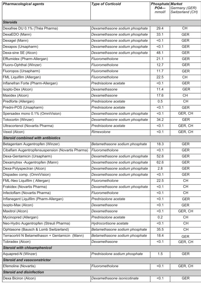
Table 1 Phosphate concentration in commercially available preparations that contain corticoids (German and Swiss market)
Pharmacological agents Type of Corticoid Phosphate PO4--- mmol/l Market Germany (GER) Switzerland (CH) Steroids
Dexafree DU 0.1% (Théa Pharma) Dexamethasone sodium phosphate 29.4
DexaEDO (Mann) Dexamethasone sodium phosphate 33.1
Dexagel (Mann) Dexamethasone sodium phosphate <0.1
Dexapos (Ursapharm) Dexamethasone sodium phosphate <0.1 Dexa-sine SE (Alcon) Dexamethasone sodium phosphate 48.1
Efflumidex (Pharm-Allergan) Fluorometholone 21.1
Fluoro-Ophthal (Winzer) Fluorometholone 12.7
Fluoropos (Ursapharm) Fluorometholone 11.7
FML Liquifilm (Allergan) Fluorometholone 22.5
Inflanefran Forte (Pharm-Allergan) Prednisolone acetate <0.1
Isopto-Dex (Alcon) Dexamethasone 11.4
Maxidex (Alcon) Dexamethasone 17.6 CH
CH CH GER GER GER GER GER GER GER GER GER
Predforte (Allergan) Prednisolone acetate 0.5
Predni-POS (Ursapharm) Prednisolone acetate <0.1
Spersadex mono 0.1% (OmniVision) Dexamethasone sodium phosphate <0.1
Totocortin (Winzer) Dexamethasone sodium phosphate 34.2
Ultracortenol (Novartis Pharma) Prednisolone acetate <0.1 GER, CH GER, CH
Vexol (Alcon) Rimexolone <0.1 GER, CH
Steroid combined with antibiotics
Betagentam Augentropfen (Winzer) Betamethasone sodium phosphate 18.3 GER GER
GER
Cibaflam Augentropfensuspension (Novartis Pharma) Fluorometholone <0.1 GER Dexa-Gentamicin (Ursapharm) Dexamethasone sodium phosphate 52.6 GER Dexamytrex -Augentropfen (Mann) Dexamethasone sodium phosphate 62.6 GER Dexa-Polyspectran (Alcon) Dexamethasone sodium phosphate 2.8 GER Dispadex comp. (OmniVision) Dexamethasone sodium phosphate <0.1 GER
GER GER
GER
FML-Neo Liquifilm ( Allergan) Fluorometholone 22.5 CH
CH CH CH
Frakidex (Novartis Pharma) Dexamethasone sodium phosphate <0.1
Infectoflam (Novartis Pharma) Fluorometholone <0.1
Inflanegent Liquifilm (Pharm-Allergan) Prednisolone acetate <0.1
Isopto-Max (Alcon) Dexamethasone <0.1
Maxitrol (Alcon) Dexamethasone <0.1 GER, CH
GER, CH
Mycinopred (Allergan) Prednisolone acetate 0.2 CH
CH CH Neo-Hydro Augentropfen (Streuli Pharma) Hydrocortisone acetate <0.1
Ophtasone (Bausch & Lomb Switzerland) Betamethasone sodium phosphate 35.5 Terracortril N Betamethason + Gentamicin (Mann) Betamethasone sodium phosphate 18.4
Tobradex (Alcon) Dexamethasone <0.1
Steroid with chloamphenicol
Aquapred-N (Winzer) Prednisolone sodium phosphate 1.5 GER
Steroid and vasoconstrictor
Efemoline (Novartis) Fluorometholone <0.1 GER, CH
Steroid and disinfection
Dexa Biciron (Alcon) Dexamethasone isonicotinate <0.1 GER
phosphates, small amounts (<0.1 mmol/l) of free phosphate anions may be due to ester hydrolysis. Phosphorylated corticoids are popular with manufacturers, since they allow preparation of a clear solution [5].
Our study investigates the amount of phosphate contained in ophthalmic corticoid preparations. This information should help to prevent sight-threatening corneal calcification.
Materials and methods
All the ophthalmic drop preparations that contain corticoids and that are listed in the“Rote Liste 2007” (Rote Liste Service GmbH, Frankfurt/Main, Editio Cantor Verlag, Aulendorf, Germany) and the “Arzneimittelkompendium der Schweiz 2007” (Documed AG, Basel, Switzerland) were included in this study. When the preparations were available in multidose and unit dose containers, the samples were taken from the preservative-free formulation. For technical reasons, gel preparations were not included. The quantification of the phosphate was performed with the molybdate method on a Modular P autoanalyzer (Roche Diagnostics, Basel, Switzer-land) [7]. The precision of these measurements was guaranteed by the inclusion of standardised phosphate solutions as controls. The day-to-day coefficient of variation was 1.7%. The results were compared to the physiological concentrations published elsewhere [7,8].
Results
The phosphate concentrations of the ocular therapeutics that were studied are given in Table1. The preparations are listed in alphabetical order and in groups of active drugs. The steroid compound of the therapeutic is shown in the second column. Eighteen of 38 corticoid preparations (47%) had a phosphate concentration above physiological levels (>1.45 mmol/l). The concentration varied greatly and ranged from less than 0.1 to 62.6 mmol/l. Medications with betamethasone sodium phos-phate showed phosphos-phate concentrations from 18.3 to 35.5 mmol/l, dexamethasone from less than 0.1 to 17.6 mmol/l, dexamethasone sodium phosphate from <0.1 to 62.6 mmol/l, fluorometholone from <0.1 to 22.5 mmol/l, and prednisolone acetate from <0.1 to 0.5 mmol/l.
Discussion
Ophthalmic preparations with corticoids may contain high concentrations of free phosphate. Several medications with phosphorylated corticoids were, however, very low in free phosphate ion (Table1). This indicates that in corticoid eye
drops, as in other ophthalmic preparations [9,10], the total amount of free phosphate ion is primarily determined by the choice of the buffers, and that phosphate ester bounds resist hydrolysis.
In ophthalmic preparations, the vehicle is an agent other than the active drug or preservative. It is added to a formulation to provide proper tonicity, buffering, and viscosity to complement drug action [11]. Its buffering system may consist of acetic, boric and hydrochloric acid, and of potassium or sodium bicarbonate, borate, phosphate and citrate [11]. Phosphates are presently widely used in ophthalmic preparations due to their high buffering capacity around pH 7.4.
Phosphate buffers, however, play a role in the process of inadvertent corneal calcification [1–4, 12]. Calcification occurs when calcium cations and phosphate anions interact within the tissue to form insoluble crystals. In the cornea, deposition typically occurs as hydroxylapatite Ca5(PO4)3OH
[2,3]. Deposition can be observed as a spectrum of clinical findings, ranging from subtle superficial changes to massive calcification of the entire cornea with visual loss.
“Boundary conditions” for the onset of corneal calcifi-cation, particularly a critical concentration of phosphate anions, cannot be defined at present. In an animal model, rapid corneal calcification developed after chemical injury, combined with a large epithelial defect, an alkaline pH, when the eyes were irrigated with Isogutt (Dr. Winzer Pharma GmbH, Germany) [1]. In this formulation, a phosphate concentration of 148 mol/l was measured [6].
The amount of phosphate ion delivered to the ocular surface by ophthalmic corticoid formulations may appear minor when compared to the treatment of chemical injuries or epithelial defects with artificial tears [3,9]. It has to be borne in mind, however, that the ion product of calcium cations and phosphate anions in aqueous humour, tears and interstitial fluids is, even under physiological conditions, close to their solubility product. A minor increase of one of the components may therefore push this system towards precipitation. Furthermore, corticoids are often used in severely damaged eyes that are prone to the complication of calcification. The high phosphate concentration of some steroid preparations may therefore trigger the process of crystallisation. We do recommend that steroid preparations with physiological phosphate concentrations are used in order to reduce the risk of corneal calcification, particularly when frequent application is required.
References
1. Schrage NF, Schloßmacher B, Aschenberner W et al (2001) Phosphate buffer in alkali eye burns as an inducer of experimental corneal calcification. Burns 27:459–464
2. Daly M, Tuft SJ, Munro PM (2005) Acute corneal calcification following chemical injury. Cornea 24:761–765
3. Bernauer W, Thiel MA, Kurrer M, Heiligenhaus A, Rentsch KM, Schmitt A, Heinz C, Yanar A (2006) Corneal calcification following intensified treatment with sodium hyaluronate artificial tears. Br J Ophthalmol 90:285–288
4. Anderson SB, de Souza RF, Hofmann-Rummelt C, Seitz B (2003) Corneal calcification after amniotic membrane transplantation. Br J Ophthalmol 87:587–591
5. Polansky JR, Weinreb RN (1984) Anti-inflammatory agents– steroids as anti-inflammatory agents. In: Sears MI (ed) Handbook of Experimental Pharmacology. Springer, Berlin Heidelberg New York, pp 459–538
6. Bernauer W, Thiel MA, Rentsch KM (2006) Phosphate in ophthalmologischen Präparaten. Ophthalmologe 103:416–417 7. Knedel M, Haeckel R, Seidel D, Thiery J, Vonderschmitt DJ,
Hänseler E (1986) Analytical performance of the random access
analyser Hitachi 737. A multicentre evaluation. J Clin Chem Clin Biochem 31:409–432
8. Nevyas AS, Raber IM, Eagle RC Jr et al (1987) Acute band keratopathy following intracameral Viscoat. Arch Ophthalmol 105:958–964
9. Bernauer W, Thiel MA, Langenauer UM, Rentsch KM (2006) Phosphate concentration in artificial tears. Graefes Arch Clin Exp Ophthalmol 244:1010–1014
10. Bernauer W, Thiel MA, Rentsch KM (2007) Phosphate concen-tration in antiglaucoma medications. Klin Mbl Augenheilk 224:249–251
11. Burstein NL (1995) Ophthalmic drug formulations. In: Bartlett DJ, Jaanus SD (eds) Clinical ocular pharmacology. Butterworth-Heinemann, Boston, pp 21–45
12. Schrage NF, Kompa S, Ballmann B, Reim M, Langefeld S (2005) Relationship of eye burns with calcifications of the cornea. Graefes Arch Clin Exp Ophthalmol 243:780–784