CASE REPORT
Heart Vessels (2005) 20:116–119 © Springer-Verlag 2005
DOI 10.1007/s00380-004-0785-5
Bilgehan Karadag · Lukas E. Spieker · Simon Wildermuth Thomas Boehm · Roberto Corti
Cardiac arrest in a soccer player: a unique case of anomalous
coronary origin detected by 16-row multislice computed tomography
coronary angiography
Received: March 11, 2004 / Accepted: May 28, 2004
His ventriculogram showed a preserved left ventricular systolic function with discrete anterolateral and diaph-ragmal hypokinesia. The dominant right coronary artery (RCA) showed a localized dissection in its proximal seg-ment without any impact on flow (Fig. 1). The left anterior descending artery (LAD) describing an “anterior dot sign,” the circumflex (Cx), and a small septal branch arose with separate ostia from the right coronary sinus.
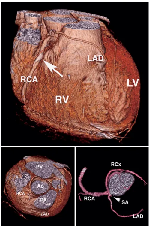
Sixteen-row multislice computed tomography (MSCT) confirmed the course of the anomalous coronary arteries originating with separate ostia from the right coronary sinus (Fig. 2). The LAD coursed anteriorly to the pulmo-nary artery, a small septal branch coursed between the aorta and the pulmonary artery, and the Cx coursed be-tween the aorta and the pulmonary vein. The localized non-flow-limiting dissection in the proximal RCA was also clearly demonstrated by MSCT (Fig. 2, arrow).
An ICD (cardioverter defibrillator) was implanted. Following an uncomplicated hospital course, the patient was discharged and remained asymptomatic.
Discussion
Coronary artery anomalies are infrequent findings which are usually incidentally detected by cardiac catheterization (prevalence 0.3%–1.0%) or at postmortem examination (prevalence 0.3%).1
An anomaly characterized by all three coronary ostia originating separately from the right coro-nary cusp is far less commonly reported.
Although most anomalies are minor variations in coro-nary anatomy without any clinical significance, some anomalies are associated with morbidity and mortality. Therefore, clinicians should be aware of such anomalies as well as their significance and consequences. Several anomalies of the left main coronary artery arising from the right coronary sinus and some anomalies of three sepa-rate coronary ostia arising from the right coronary cusp have been reported in the literature,2,3
since they particu-larly increase the risk for severe complications such as Abstract Anomalous origin of the coronary arteries may
be present in otherwise normal subjects without clinical significance, but can also be the cause of myocardial ischemia and sudden death in both adults and teenagers. In particular, the origin of the left main coronary artery or left anterior descending artery from the right sinus of Val-salva or right coronary artery may result in compression of the vessel during or immediately after exercise. We present a unique case of coronary anomaly with four separate coronary ostia originating from the right coronary sinus in a soccer player with sudden cardiac arrest. Multislice contrast-enhanced computed tomography has emerged as a valid noninvasive method for the diagnosis of coronary artery anomaly.
Key words Cardiac arrest · Multislice computed tomogra-phy · Coronary angiogratomogra-phy · Exercise
Case report
A 51-year-old man with a history or recurrent exercise-induced syncope was resuscitated during a soccer game af-ter cardiac arrest due to documented ventricular fibrillation. On admission, the conscious patient was asymptomatic with an unremarkable physical examination. ECG revealed sinus tachycardia with nonspecific intraventricular con-duction delay, ST-depression on anteroseptal leads, and without prolonged corrected QT time. Chest X-ray and biochemical examinations showed no pathological findings and negative troponin T.
B. Karadag · L.E. Spieker · R. Corti (*)
Department of Cardiology, University Hospital Zurich, Rämistrasse 100, CH-8091 Zurich, Switzerland
Tel. ⫹41-1-255-2121; Fax ⫹41-1-255-4401 e-mail: [email protected]
S. Wildermuth · T. Boehm
Department of Radiology, University Hospital Zurich, Zurich, Switzerland
117
angina pectoris, acute myocardial infarction, or sudden death.
In the present case, although the left system arose entirely from the right cusp, all four vessels had separate ostia. To our knowledge, this is the first report describing four separate coronary artery ostia arising from the right coronary cusp. A specific approach to these patients has not been described in terms of clinical significance and therapy.
Anatomical features
Coronary anomalies are rare incidents in the general popu-lation and although most anomalies are without clinical significance, some may be associated with angina pectoris, acute myocardial infarction, heart failure, and sudden car-diac death even in absence of atherosclerosis.1,4,5
The most common anomaly is the absence of the left main trunk with separate LAD and LCx ostia (incidence 0.47%). The next most common anomalies are the LCx originating from the right sinus of Valsalva (0.45%), the RCA originating from the ascending aorta above the sinus of Valsalva (0.18%), and the RCA originating from the left sinus of Valsalva (0.13%). The incidence of the LAD originating from the right sinus of Valsalva is 0.02%.6
Clinical features
Coronary anomalies can simply be classified as ischemia-producing and non-ischemic. Nonischemic anomalies
with-out a clinical risk include a single coronary anomaly, RCx arising from the right coronary sinus, and separate ostia of all three arteries.7
The clinical significance is determined by the type of anomaly, the anatomical course of the artery in relation to the great vessels, and the extent of myocardium supplied by the anomalous artery.
The high-risk ischemia-producing anomalies consist of (i) anomalous origin of one or more coronary arteries arising from the pulmonary trunk, (ii) left main and right coronary artery from the opposite aortic sinus, (iii) single coronary artery, and (iv) hypoplastic coronary arteries.
Several potential mechanisms have been proposed to be associated with ischemia and sudden cardiac death in pa-tients with anomalous coronary arteries from the contra-lateral sinus of Valsalva. The anatomical course of the anomalous arteries in relation to the great vessels of the aorta and pulmonary trunk (anterior, posterior, and interarterial course) is suggested as one of the possible mechanisms. The anterior and posterior course is thought to be benign. An interarterial course may result in compres-sion of the anomalous coronary artery between the aorta and the pulmonary trunk secondary to dilatation of these great vessels during exercise.8
This type of coronary anomaly appears to be the most common cause of sudden death among the congenital coronary malformations in young trained athletes.9
The oblique take-off of the anomalous artery producing a slit-like orifice in the aortic wall can collapse like a valve during exericse.1,10,11
In a histopathological study,8–10 the proximal portions of the anomalous coronaries were docu-mented to be intramural (i.e., within the aortic tunica media), which could further aggravate the coronary obstru-ction, particularly with expansion of the aorta during exercise.
Vasospasm of the anomalous coronary may also cause ischemia, possibly as a result of endothelial injury.12 Additionally, the repetitive attacks of ischemia caused by anomalous coronaries may result in myocardial necrosis and fibrosis, which creates an electrically unstable focus predisposing to lethal ventricular arrhythmias.
Incidence of cardiac death
A review of 242 patients with isolated coronary anomalies demonstrated cardiac death in 59%, in which 39% were sudden cardiac death.13
The incidence of sudden cardiac death related to coronary artery anomalies is reported as 11% in exercising individuals aged 8–66 years, 0.6% in the general population aged under 40 years, and 23% in com-petitive athletes with a mean age of 17 years.14
According to the Sudden Death Committee of the American Heart Asso-ciation, coronary anomalies cause 19% of sudden cardiac deaths in athletes.
Noninvasive diagnostics
Cardiac magnetic resonance imaging (MRI) is a reliable noninvasive imaging and screening modality which avoids
Fig. 1. Coronary angiography demonstrating coronary vessels
origi-nating from the right coronary cusp: the left anterior descending coronary (LAD), the circumflex (Cx), and a small septal branch (SA). The right coronary artery (RCA) shows a limited, non-flow-limiting dissection (arrow)
118
radiation and contrast agents. It may be superior to con-ventional angiography in determining the correct origin of anomalous coronaries and their spatial relationships with the great vessels. Its greatest limitation is determining the distal course of the anomalous coronaries; therefore, com-bination of MRI with angiography greatly improves vessel definition.15,16
Transesophageal echocardiography may also detect coronary anomalies, but it is not entirely noninvasive and the predictive value is low.
As described in the present case, contrast-enhanced computed tomography with three-dimensional reconstruc-tion offers excellent determinareconstruc-tion of the origin and course of anomalous coronaries and their relationships with the
great vessels. The disadvantages of this technique in com-parison to other noninvasive techniques are the require-ment of ionizing radiation and potentially nephrotoxic and allergenic contrast agents.
Conclusions
We report a unique case of coronary anomaly associated with sudden cardiac arrest. We documented four separate coronary artery ostia originating from the right coronary cusp. To our knowledge, this is the first report describing this entity.
Fig. 2. Three-dimensional reconstructions
(volume rendering) based on the isotropic multislice computed tomography angiography data set clarify the relation between the abnormal origin and the course of the coronary arteries and the surrounding structures such as the aorta (Ao), the pulmonary artery (PA), and pulmonary vein (PV). Right (RV) and left ventricle (LV) are indicated. The arrow (upper panel) indicates the non-flow-limiting dissection of the right coronary artery (RCA). RCx, right circumflex artery; LAD, left anterior descending artery; SA, small septal branch
119 An interarterial course, possibly resulting in
compres-sion of the anomalous coronary artery between the aorta and pulmonary trunk secondary to dilatation of these great vessels during exercise, appears to be a significant cause of sudden arrest among the congenital coronary malfor-mations, as demonstrated by our unique case. Based on our case, the clinical profile of this type of anomaly is malignant, requiring aggressive treatment including ICD implantation and close follow-up.
References
1. Taylor AJ, Rogan KM, Viramani R (1992) Sudden cardiac death associated with isolated congenital coronary artery anomalies. J Am Coll Cardiol 20:640–647
2. Lauer JE, Ritchie ME (2001) Three separate coronary artery ostia arising from the right coronary cusp: a case repot. Cathet Cardivasc Intervent 52:496–499
3. Roberts WC, Shirani J (1992) The four subtypes of anomalous origin of the left main coronary artery from the right aortic sinus (or from the right coronary artery). Am J Cardiol 70:119–121 4. Kimbiris D, Iskandrian AS, Segal BL, Bemis CE (1978)
Anoma-lous aortic origin of coronary arteries. Circulation 58:606–615 5. Angelini P, Villason S, Chan AV Jr, Diez JG (1999) Normal
and anomalous coronary arteries in humans. In: Angelini P (ed) Coronary artery anomalies: a comprehensive approach. Lippincott Williams & Wilkins, Philadelphia, pp 27–150
6. Bhatt DL (2000) Left heart catheterization. In: Marso SP, Griffin BP, Topol EJ (eds) Manual of cardiovascular medicine, 2000 edn. Lippincott Williams & Wilkins, Philadelphia
7. Bittl JA, Levin DC (1997) Coronary arteriography. In: Braunwald E (ed) Heart disease: a textbook of cardiovascular medicine, 5th edn. Saunders, Philadelphia, pp 259–262
8. Taylor AJ, Byers JP, Cheitlin MD, Virmani R (1997) Anomalous right or left coronary artery from the contralateral coronary sinus: “high-risk” abnormalities in the initial coronary artery course and heterogeneous clinical outcomes. Am Heart J 133:428–435 9. Basso C, Maron BJ, Corrado D, Thiene G (2000) Clinical profile of
congenital coronary artery anomalies with origin from the wrong aortic sinus leading to sudden death in young competitive athletes. J Am Coll Cardiol 35:1493–1501
10. Cheitlin MD, De Castro CM, McAllister HA (1974) Sudden death as a complication of anomalous left coronary origin from the ante-rior sinus of Valsalva. A not-so-minor congenital anomaly. Circu-lation 50:780–787
11. Virmani R, Chun PKC, Goldstein R, Robimowitz M, McAllister HA (1984) Acute takeoffs of the coronary arteries along the aortic wall and congenital coronary ostial valve-like ridge: association with sudden death. J Am Coll Cardiol 3:766–771
12. Maddoux GL, Goss JE, Ramo BW, Raff GL, Heuser RR, Shadoff N, Leatherman GF, Blake K, Wilson JN, Deane WM (1989) Angina and vasospasm at rest in a patient with an anomalous left coronary stem. Cathet Cardiovasc Diagn 16:95–98
13. Virmani R, Burke AP, Farb A (2001) Sudden cardiac death. Cadiovasc Pathol 10:211–218
14. Angelini P, Velasco JA, Flamm S (2002) Coronary anomalies: incidence, pathophysiology, and clinical relevance. Circulation 105:2449–2454
15. McConnell MV, Ganz R, Selwyn AP, Li W, Edelman RR, Manning WJ (1995) Identification of anomalous coronary arteries and their anatomic course by magnetic resonance coronary angio-graphy. Circulation 92:3158–3162
16. Post JC, van Rossum AC, Bronzwaer JG, de Cock CC, Hofman MB, Valk J, Visser CA (1995) Magnetic resonance angiography of anomalous coronary arteries: a new gold standard for delineating the proximal course? Circulation 92:3163–3171