ORIGINAL PAPER
Sagittal craniosynostosis combined with ossified
cephalhematoma
—a tricky and demanding puzzle
Georges Louis Kaiser&Valérie Oesch
Received: 5 September 2008 / Published online: 23 October 2008
# Springer-Verlag 2008
Abstract
Introduction Four cases of sagittal synostosis combined with ossified cephalhematoma prompted the authors to present the data and to discuss the implications. Large cephalhematoma of the vertex at birth with subsequent ossification occurred in all with final sizes of 6.5–10 by 4.0–5.5 by 0.8–1.8 cm. At surgery with 2–6 months, the mean skull index was 64.75, sagittal suture completely closed, and a disfiguring bony mass present in all.
Discussion In three of the original cohort of 106 sagittal synostoses, ossified cephalhematoma was removed in one piece together with the suture. In contrast to pathogenesis of common parietal cephalhematomas, cephalhematoma in sagittal synostosis is rather induced by periosteal detachment of the midline by strong shearing forces because molding is hindered in the lateral direction. Frequent and fast complete ossification is possibly directed by the same local factors (e.g., Noggin) which lead to premature fusion of cranial sutures. Some treatment principles of ossified cephalhema-toma in sagittal synostosis may be applied to surgery of common types.
Keywords Ossified cephalhematoma . Sagittal synostosis . Pathogenesis cephalhematoma . Ossification
cephalhematoma . Treatment
Introduction
The clinical findings and the early and intermediate natural history of cephalhematoma [22] and of craniosynostoses [17,26] are well known. The observation of four cases of sagittal synostosis combined with an unusual and nearly identical clinical presentation of an ossified cephalhema-toma on the vertex prompted the authors to present their data and to discuss the implications, particularly the following topics: (1) possible factors leading to ossification of cephalhematoma, (2) pathogenesis of cephalhematoma in general and specifically in sagittal synostosis, and (3) surgical treatment of ossified cephalhematoma in sagittal synostosis and its application to those ossified cephalhe-matomas with classic localization.
Materials and methods
From 1991 to 2007, 106 infants with sagittal synostosis have been operated by a modified extended vertex craniectomy [7] and cranioplasty. Three of them (=2.8%) had a neonatal cephalhematoma which was followed by ossification in any case. A further case of ossified cephalhematoma in sagittal synostosis of a partner university hospital was added to the three cases. A confirmed consent was obtained from the parents to use the data of their children. The clinical and radiological evaluation included the peri- and postnatal history, the local findings and their evolution, data of surgery and histological examination, and follow-up findings. In general, all craniosynostosis patients have prospective follow-ups in regular intervals increasing with age until puberty.
Presented as poster at the XXIst ESPN Meeting. Montreux, Switzerland—May 11th–15th 2008. Oesch V, Schobinger S, Bielek J, Bittel M, Kummer M, Kaiser G. Ossified cephalhematoma in sagittal synostosis.
G. L. Kaiser
:
V. Oesch (*)Department of Pediatric Surgery, Children’s Hospital, Inselspital, University of Berne,
3010 Bern, Switzerland e-mail: valerie.oesch@insel.ch
Results
In Table 1, the obstetric history and the condition of the four patients at birth are listed. All are born spontaneously at term by primipara, but in all, instrumental deliveries were needed.
Table2shows the precise findings of ongoing ossification on the plain X-ray of the skull at 2 to 2.5 months of age. The area of ossification has always an oblong–oval shape which is lined up in the direction of the closed sagittal suture and distributed symmetrically on both sides. The mean area is 7.3 cm long, 4.8 cm wide, and 1.3 cm deep. Three patients have one osseous prominence (Fig.1). Case 4 shows two prominences (Fig. 2); in contrast to cases 1 and 2 with a homogeneous osseous structure, in case 3 and 4, one and two central luminescences, respectively, are visible corresponding to two small cavities. All patients had radiological signs of premature closure of the hole sagittal suture.
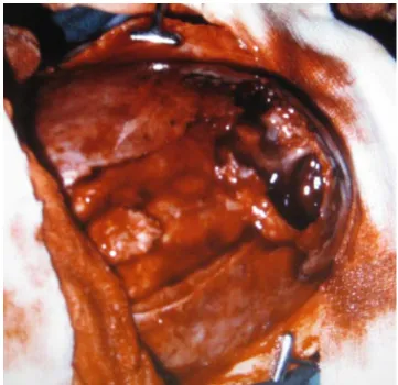
Table3 summarizes the natural history of cephalhema-toma and the findings of ossified cephalhemacephalhema-toma as well as the head measures at surgery. A distinctly recognizable cephalhematoma has been present in all shortly after birth. It was always localized in the midline over the sagittal suture. The same is true for the disfiguring ossified cephalhematoma with a largest diameter of >5 to 10 cm at surgery which corresponded clinically (Fig.3) as well as radiologically to a stage 4. Except for case 4, all patients had cavities within the ossified cephalhematoma which were more or less distinctly visible by the naked eye (Fig.4). In case 4, such cavities were seen histologically (Fig. 5a–c). All patients had a distinct scaphocephaly with a mean skull index of 64.75 at a mean time of 3 months and 1 week.
In Table4, age at surgery, type of surgery, and follow-up time of the individual patients are quoted. Except for the added case from the partner university hospital, all had a vertex craniectomy in which the ossified cephalhematoma was removed together with the midline bone strip over the sagittal sinus in one piece including the fused sagittal suture (Figs. 6 and 7). For cranioplasty, small pieces of the removed bone parts were used to cover the whole defect like a mosaic except for a 1–2 cm wide zone along the
midline (a method which we are using since many years with success). The follow-up time was in every case long enough to prove a regular and complete reossification and permanent correction of the former deformity caused by the sagittal synostosis and the ossified cephalhematoma.
Discussion
In the presented cohort of 106 infants with sagittal synostosis, cephalhematoma has been observed in three during the neonatal period. The percentage of 2.8 cephal-hematomas corresponds to some of the figures quoted in the literature in a general population [16,27], but the fact that ossification occurred in all and was already completely developed to stage 3–4 at the age of 2 to 2.5 months is remarkable. It is different from the common course of ossification which is much less frequent and displays chronologically variable periods of ossification. Besides the possibility of a biased phenomenon, the occurrence of factors leading to frequent and fast complete ossification must be discussed, as for instance, local factors of the adjacent dura and suture cells as Noggin under- and Runx-2 overexpression [8] among other mechanisms [10] which are possibly involved in premature fusion and reossification of the surgically opened sagittal suture. In contrast to the common parietal cephalhematoma which is somewhat apart from the normal sutures, the presented vertex hematomas lie directly over a pathological sagittal suture and by it close to the dura.
In the recent Anglo-American literature, the occurrence of ossified cephalhematoma is quoted as occasional, rare, or unknown and restricted to few case reports [4, 6, 12]. Already in the seventies and much earlier, it has been claimed by some that a persistent osseous prominence is an extremely rare sequel of cephalhematoma because there is even, in case of fully established ossified cephalhematoma, a slow resolution of the mass which merges over months into the general contour of the growing calvaria [22,31]. At the beginning of the fifties and sixties, Bloch [1] and Vernon [30] quoted studies which, currently took place 10 years ago with an incidence of 3.2% to 10% persistent
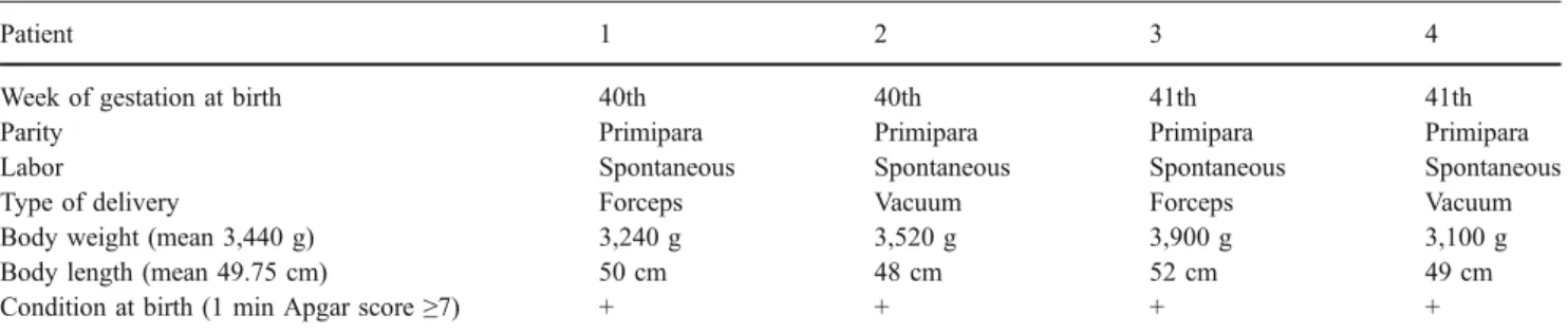
Table 1 Obstetric history and condition of the patients at birth (n=4)
Patient 1 2 3 4
Week of gestation at birth 40th 40th 41th 41th
Parity Primipara Primipara Primipara Primipara
Labor Spontaneous Spontaneous Spontaneous Spontaneous
Type of delivery Forceps Vacuum Forceps Vacuum
Body weight (mean 3,440 g) 3,240 g 3,520 g 3,900 g 3,100 g
Body length (mean 49.75 cm) 50 cm 48 cm 52 cm 49 cm
ossified cephalhematomas, still visible after several years. Figures of the early seventies are 1.6% to 10% [25,31,34] although less reliable due to much smaller number of patients.
Some of the cephalhematomas may go clinically and radiologically through different stages of subperiosteal ossification [3,9,22,25,31]. In stage 1, a ridge is palpable around the edge of cephalhematoma. In stage 2, the formed bone at the edges grows subperiosteally step by step over the dome of the bulging hematoma. A thin pliable layer of bone becomes palpable which has been described as comparable with a rustling parchment [31] or celluloid ball [2]. In stage 3, the bony covering becomes thicker and solid, and in stage 4, a prominent firm mass or a shell-like thickening of the skull is palpable. According to histological
examination, all these stages are processes of ossifications and not of calcifications [3] and should be named ossifying cephalhematomas.
Although ossification may start at the end of the first postnatal week and reaches stage 2 already at the end of the second week, permanent ossification only occurs at the end of the sixth week if resorption of cephalhematoma is not completed by this time [31]. The observation that resorption of cephalhematomas may occur by 1 to 6 weeks with a median time of three to four weeks [31] and that different lengths are quoted in the literature for the four stages of ossification [1,9,25,31] points to a large variation of the time course of resorption and ossification of cephalhematoma, and complete resolution is still possible in the early stages of ossification. This conclusion is strengthened by the frequent occurrence of resolution during the early ongoing ossification which can be observed in one third of the patients [31] in comparison to the relatively low incidence of resolution in permanently ossified cephalhematoma.
A relation between the initial size of cephalhematoma and the type of resolution has been rejected by some
Table 2 Radiological findings of ongoing ossification of cephalhematoma (n=4)
Patient 1 2 3 4
Age at X-ray (months) 2.5 2 2.5 2
Area cephalhematoma 10.0×5.0×1.0 7.5×4.0×1.8a 8.0×5.5×1.3 6.5×5.0×0.8
Structure cephalhematoma right–left symmetry + + + +
Number of prominences 1 1 1 2
Homogeneity + + –b –b
Signs of sagittal synostosis + + + +
aRadiological size of cephalhematoma at age 5 days 7.0×4.75×0.45 bOne and two luminescences
Fig. 1 Anteroposterior X-ray of the skull in case 2 at age 2 months. The ossified cephalhematoma with homogeneous osseous structure lies symmetrically over the middle third of the fused sagittal suture. A distinct impression of the superior sagittal sinus is recognizable without any signs of sagittal suture
Fig. 2 Lateral X-ray of the skull in case 4 at age 2 months. Instead of one, two prominences of ossified cephalhematoma are visible which lies over the middle and posterior third of the fused sagittal suture. In addition, two distinct luminescences are visible inside of the ossified cephalhematoma
authors [31]. On the other hand, all case reports including ours with disfiguring permanent ossified cephalhematoma had initially very large cephalhematomas of minimally the same size. According to Chung et al. [3] and Wechselberg and Sanders [31], the individual time course of the two ongoing processes—“resorption” and “ossification”—are crucial to the origin of a permanent ossified cephalhematoma and its possible persistence over years, which means resorption of the clot does not proceed rapidly and is not completed in the early stages of ossification. This hypothesis is confirmed by the observation that in almost all radiological work-ups with computed tomography (CT) or magnetic resonance imaging (MRI) [3, 4, 12] and in all operative
specimens, residual cavities with remnants of consolidated hematoma and/or with abnormal vascularization are present as in ours.
The vertical site of cephalhematoma was in all four cases of proven sagittal synostosis identical and unusual in comparison to the typical localization of cephalhematomas. The frequency of neonatal cephalhematomas is reported to be 0.2% to 2.5% of life births [16,27] or even higher, as for instance following vacuum extraction with a mean inci-dence of 6% [6], and depends for the most part on the composition of the evaluated women and newborns as well
Table 3 Natural history of cephalhematoma
Patients 1 2 3 4
Clinical recognition of cephalhematoma
Day 1–2 Day 1–2 Day 1–2 Day–4
Time of surgery
(mean 3 months 1 week)
2.5 months 2.0 months 6.0 months 3.0 months
Site of ossified cephalhematoma Midline anterior half of sagittal suture
Midline middle third of sagittal suture
Midline middle third of sagittal suture
Midline middle and posterior third Size of ossified cephalhematoma
(largest diameter)
10 cm >5 cm >5 cm >5 cm
Gross and histological morphology Subtotal ossification With residual cavity (ies) With abnormal vascularization
In all patientsa Head circumference/skull index
(mean 64.75)
>97% >97% >97% >75%
56 67 68 68
Findings of ossified cephalhematoma and head measures at surgery (n=4)
aIn case 4, cavity (ies) were not visible by the naked eye but were seen histologically
Fig. 3 Case 1 at age 2.5 months. The disfiguring ossified cepha-lhematoma is located over the anterior half of the fused sagittal suture and has an oblong–oval shape in the direction of the former suture
Fig. 4 Operative findings in case 1 at age 2.5 months in whom the sagittal suture (on the left side) and the ossified cephalhematoma (on the right side) have been removed step by step. A large irregular cavity is visible within the ossified cephalhematoma with a dark-red vascularization
as on a careful and repeated clinical examination in the newborn period. Independently of frequency, 85% to 91% of cephalhematomas have a unilateral parietal or, less frequently, a bilateral parietal localization [19, 31]. The occipital site is with about 10% the second most frequent site; in addition, rarely frontal and multiple localizations occur. Midline vertical site is quoted only once in the literature [20].
For the most frequent types of vaginal delivery, the preferred localization of cephalhematoma over the parietal bone does not permit a detailed statement about the pathogenesis of cephalhematoma in the individual case because there is no correlation between right or left occipital presentation at birth and the laterality of cephal-hematoma [5, 31]. On the other hand, cephalhematoma must develop on the entrance of the head in the osseous pelvis or shortly thereafter because only in this moment equally exists the chance of involvement of the right or left side of the prominent upper posterior angle of the parietal bones. Likewise, cephalhematoma in caesarean section was only present if labor had already started and was interrupted during the first stage [5]. According to Churchill et al. [5], cephalhematomas occur significantly more frequently in occiput posterior and occiput transverse presentations than in the common occiput anterior presentations. The former types of presentations are therefore a risk factor for development of cephalhematoma as primiparity, high-term birth weight, prolonged labor, and instrumental deliveries in general [31, 34]. Although in the latter group, forceps deliveries are prone to development of cephalhematoma specifically in midforceps and forceps rotation [5, 19], vacuum extractions increase the risk of cephalhematoma and certain types of intracranial hemorrhage more than low forceps deliveries [32].
The observation of cephalhematomas by ultrasound during pregnancy [11, 24, 33] on the one hand and after cesarean section [5, 16, 31] and in vertex-born babies on the other hand points to the possibility of different pathophysiological mechanisms which lead to circumscript detachment of periosteum from the underlying bone combined with rupture of transversing blood vessels [16]. Hartley and Burnett [13] suggested a trauma of dragging nature as precipitating factor. Out of the two following mechanisms described in the literature [3,25]—(1) parallel
forces are set up between periosteum and skull which result in a shearing mechanism and (2) sudden or prolonged compression and inward movement of the skull with displacement of the bone from the periosteum—the former one fits better the hypothesis of a movement with effort as shown by the following observations: Smaller parietal cephalhematomas are usually localized in the upper posterior parietal bone and larger types take on an oblong–oval shape. Therefore, in the common vaginal
Fig. 5 a–c Histological work-up of ossified cephalhematoma and sagittal suture in case 4 at age 3 months. Hematoxylin and eosin stain. a Site of the transition of the parietal bone (on the left side) to the ossified cephalhematoma (on the right side). A distinct difference is recognizable between them concerning thickness and structure of the bone. b Central part of ossified cephalhematoma. It consists mostly of trabeculated bone with innumerable vascularized cavities which are different from those in the parietal bone and the former sagittal suture. c Site of the former sagittal suture. There is a normal bone structure without any suture tissue
delivery, there is shearing of the periosteum in a parallel and longitudinal manner at the level of the promontory and/ or symphysis due to the normal rotation movement of the head. In the already mentioned occiput posterior and transverse presentations, or the in midforceps and forceps rotation deliveries, the same mechanism takes place though to a more severe degree. The above mentioned uniform distribution pattern of cephalhematoma with preference for parietal bone shows that it results from and is due to the anatomical, and physiological characteristics of the birth channel, e.g., impact of the parietal area against the pelvis [13], and its frequency is triggered by the known risk factors. In case of the observed cephalhematomas in sagittal synostosis, the already mentioned pathogenic mechanisms seem not very likely because the head should be presented in such a way that instead of the parietal bones as in normal delivery, the vertex with the sagittal suture gets into close contact with the promontory and/or symphysis as for instance in increased posterior or anterior parietal presen-tation (asynclitism). A possible pathogenic mechanism is a bilateral detachment of periosteum including the midline by strong shearing vertical forces because molding of the head is hindered in the lateral direction by premature fusion of the sagittal suture. During surgery of the fused sagittal suture, it can be observed that the site of periosteal insertion to the closed suture and of the latter one to the dura is very
small and less adherent in contrast to a normal suture. Therefore, a bilaterally symmetrical cephalhematoma is developing which comes together, takes on an oblong–oval configuration, and occupies a large distance of the midline. The hypothesis that synostosis of the sagittal suture is secondary to the ossified cephalhematoma claimed by Martinez-Lage et al. [20] in their case report on sagittal synostosis combined with ossified cephalhematoma must be discarded in spite of the cited experimental models of craniosynostosis due to the contradictory statements between the extreme scapho-cephaly on X-ray and the clinically seemingly open sagittal suture on both sides of the cephalhematoma. Our cases had already scaphocephaly at birth and clinical and/or radiological signs of premature closure of sagittal suture before ossification of the cephalhematoma.
In our last three cases, the disconfiguring ossified cephalhematoma was removed in one piece together with a bone strip adjacent to the fused sagittal suture. This method is much easier than local excision which might be dangerous and combined with considerable blood loss. The same proceeding could be applied to the common parietal ossified cephalhematoma.
Table 4 Time and type of surgery for sagittal synostosis and cephalhematoma and follow-up time (n =4)
Patient 1 2 3 4
Age at surgery (months) 2.5 2.0 6.0 3.0
Type of surgery Stepwise excision cephalhematoma and linear craniectomy Vertex craniectomy including cephalhematoma and cranioplasty Vertex craniectomy including cephalhematoma and cranioplasty Vertex craniectomy including cephalhematoma and cranioplasty
Follow-up time (years) 16.0 15.10 9.6 0.6
Fig. 6 View to the outer side of the bone strip of 10 cm length in case 3 which includes the site of the former sagittal suture as well as the ossified cephalhematoma. The oblong–oval bony prominence lies in the middle third of the former suture site and is still disfiguring at age 6 months
Fig. 7 View to the inner side of bone strip of 9 cm length in case 4 which includes the site of the former sagittal sutures and the ossified cephalhematoma (not visible in the picture). Except for few millimeters of a recognizable suture line adjacent to the coronal suture, the former sagittal suture is replaced by a bony ridge on the left and by an impression of the superior sagittal sinus on the right side
The operative techniques for the removal of common ossified cephalhematomas have been described by Kaufman et al. [15] and recently by Chung et al., Chung et al., and Gupta et al. [3,4,12]. The latter authors propose excision of the outer bulging bone including the remnants of the consolidated hematoma over the inner calvarial bone which is left behind. Remodeling of the operative field with multiple small pieces of the outer bulging bone is only performed if there is a depression of the involved parietal bone or if there is a defect or diminished thickness of it. Excision of the whole ossified cephalhematoma beyond the junctional area of the involved parietal bone as it is proposed above and mentioned in Gupta’s case history [12] has several advantages in spite of major surgery. The whole ossified cephalhematoma can be handled outside of the patient, a possible injury of the underlying structures by the osteotome can be avoided, and a depression of the whole parietal bone as it is observed frequently in and after large cephalhema-tomas can be corrected at the same time. As pointed out by Chung et al. and Gupta et al. [3,12], CT and/or MRI is not only useful if a cephalhematoma has not resolved within 6 weeks after birth for definition of the stage of ossification but also in every case preoperatively for differential diagnosis and to describe the expansion of the lesion in all three dimensions. Recently, Jang et al. [14] have proposed simple excision and periosteal reattachment in ossified cephalhematoma with good cosmetic appearance instead of additional cranioplasty for the correction of a depressed area. Evacuation of native cephalhematoma by puncture in the second week of the neonatal period or thereafter is still not yet the usual practice [18,28] after a controversial debate many years ago [1,2,16,22,23,27,29–31], but there are exceptions of it as suspicion of infected cephalhematoma in which diagnostic puncture is needed [18, 21] or as large cephalhematomas. The latter one may be combined with or lead to depression of the parietal bone which can be temporary if resorption occurs [25] and permanent if ossification develops as shown by the X-rays of several case reports. Depression and/or ossification can be corrected or avoided by proper puncture in time [2,29], for instance in selected cases with a size of a walnut to an apple [23,30].
Acknowledgement The assistance of S. Schobinger at Institute of Pathology, University of Berne for performance and interpretation of the histological work-up in case 4 is greatly appreciated.
References
1. Bloch P (1950) Sur la persistance du céphalmatome (persistence of cephalhematoma). Bull Fed Soc Gynecol Obstet Lang Fr 2:384–385 2. Bruniquel MG (1967) Le céphalématome doit-il rester sacro-saint? (Must the cephalhematoma remain sacrosanct forever?). Bull Fed Soc Gynecol Obstet Lang Fr 19:480–481
3. Chung HY, Chung JY, Lee DG, Yang JD, Baik BS, Hwang SG, Cho BC (2004) Surgical treatment of ossified cephalhematoma. J Craniofac Surg 15:774–779
4. Chung KC, Buchman SR, Maher HA, Dauser RC (2005) Surgical management of calcified cephalhematoma and associated skull defect in infancy. Ann Plast Surg 34:99–102
5. Churchill JA, Stevenson L, Habhab G (1966) Cephalhematoma and natal brain injury. Obstet Gynecol 27:580–584
6. Doumouchtsis SK, Arulkumaran S (2006) Head injury after instrumental vaginal deliveries. Curr Opin Obstet Gynecol 18:129–134
7. Epstein N, Epstein F, Newman G (1982) Total vertex craniectomy for the treatment of scaphocephaly. Child’s Brain 9:309–316 8. Gabbay JS, Heller J, Spoon DB, Mooney M, Acarturk O, Askari
M, Wasson KL, Bradley JP (2006) Noggin underexpression and Runx-2 overexpression in a craniosynostosis rabbit model. Ann Plast Surg 56:306–311
9. Gianelli A (1951) Tre casi di cefaloematoma esterno die neonati a decorso non comune (Three cases of external cephalhematoma in the newborn; atypical evolution). Minerva Pediatr 3:591–598
10. Gosain AK, Recinos RF, Agresti M, Khanna AK (2004) TGF-β1, FGF-2, and receptor mRNA expression in suture mesenchyme and dura versus underlying brain in fusing and nonfusing mouse cranial sutures. Plast Reconstr Surg 113:1675–1684
11. Grylack L (1982) Prenatal sonographic diagnosis of cephalhematoma due to prelabor trauma. Pediatr Radiol 12:145–147
12. Gupta PK, Mathew GS, Malik AK, Al Derazi T (2007) Ossified cephalhematoma. Pediatr Neurosurg 43:492–497
13. Hartley JB, Burnett CWF (1944) An enquiry into the causation and characteristics of cephalhematoma. Br J Radiol 17:33–41 14. Jang DG, Kang SG, Lee SB, Yoo DS, Huh PW, Cho KS, Kim DS,
Park CK, Kang JK (2007) Simple excision and periosteal reattachment for the treatment of calcified cephalhematoma. Technical note. J Neurosurg 106(2 Suppl):162–164
15. Kaufman HH, Hochberg J, Anderson RP, Schochet SS Jr, Simmons GM Jr (1993) Treatment of calcified cephalhematoma. Neurosurgery 32:1037–1040
16. Kendall N, Woloshin H (1952) Cephalhematoma associated with fracture of the skull. J Pediatr 41:125–132
17. Laitinen I (1956) Craniosynostosis: premature fusion of the cranial sutures. Ann Paediatr Fenn 2(suppl):1–130
18. LeBlanc CM, Allen UD, Ventureyra E (1995) Cephalhematomas revisited. When should a diagnostic tap be performed. Clin Pediatr (Phila) 34:86–89
19. Levin H, Bacci OJ (1955) Cephalhematoma in the newborn. Pa Med J 58:782–783
20. Martinez-Lage JF, Esteban JA, Martinez Perez M, Poza M (1984) Craniostenosis secondary to calcified subperiosteal hematoma: case report. Neurosurg 15:703–704
21. Mohon RT, Mehalic TF, Grimes CK, Philip AG (1986) Infected cephalhematoma and neonatal osteomyelitis of the skull. Pediatr Infect Dis 5:253–256
22. Morgan JE (1944) Calcifications in cephalhematomas of newborn infants. Am J Obstet Gynecol 48:702–705
23. Pauli M (1958) Zur Behandlung des Kephalhaematoma externum (Therapy of external cephalhematoma). Kinderärztl Prax 26:1–6
24. Petrikovsky BM, Schneider E, Smith-Levitin M, Gross B (1998) Cephalhematoma and caput succedaneum: do they always occur in labor? Am J Obstet Gynecol 179:906–908
25. Seifert UD (1973) Das Kephalhämatom—Entstehung und Rückbildung (The cephalhematoma—pathogenesis and regression). Pädiatr Grenzgeb 12:97–108
26. Shillito J Jr (1982) Craniosynostosis. In: Youmans JR (ed) Neurological surgery. Saunders, New York, pp 1447–1466
27. Tan KL (1970) Cephalhematoma. Aust N Z J Obstet Gynaecol 10:101–106
28. Uhing MR (2005) Management of birth injuries. Clin Perinatol 32:19–38
29. Unrein HD (1970) Konservative oder chirurgische Behandlung des Kephalhämatoms (Conservative or surgical treatment of cephalhematoma). Kinderärztl Prax 38:552–556
30. Vernon S (1961) Aspiration for cephalhematoma. Conn Med 25:634–635
31. Wechselberg K, Sanders U (1971) Untersuchungen zur Diagnose und Prognose der geburtstraumatischen Kephalhämatome (Diagnosis and
prognosis of cephalhematoma due to birth injury). Med Monatsschr 25:202–207
32. Wen SW, Liu S, Kramer MS, Marcoux S, Ohlsson A, Sauvé R, Liston R (2001) Comparison of maternal and infant outcomes between vacuum extraction and forceps deliveries. Am J Epidemiol 153:103–107
33. Winter TC, Mack LA, Cyr DR (1993) Prenatal sonographic diagnosis of scalp edema/cephalhematoma mimicking an ence-phalocele. Am J Roentgenol 161:1247–1248
34. Yasunaga S, Rivera R (1974) Cephalhematoma in the newborn. Clin Pediatr (Phila) 13:256–260