J. Hyg., Comb. (1981), 86, 139 1 3 9 Printed in Great Britain
Antibody capture radioimmunoassay for anti-rubella IgM
P. P. MORTIMER
Virus Reference Laboratory, Central Public Health Laboratory, Colindale Avenue, London NW9 5HT
R. S. TEDDER
Department of Virology, Middlesex Hospital, Medical School, London WIN SAA
M. H. HAMBLING
Public Health Laboratory, Bridle Path, Leeds L815 1TR
M. S. SHAFI
Public Health Laboratory, Central Middlesex Hospital, Park Royal, London NW\Q INS
F. BURKHARDT, U. SCHILT
Institutfiir Hygiene und Medizinische Microbiologie, Friedbuhlstrasse 51, Bern, Switzerland
(Received 17 October 1980)
SUMMARY
An M-antibody capture radioimmunoassay (MACRIA) for anti-rubella IgM was developed. Under optimum conditions positive serum specimens bound up to 20 times as much radioactivity as negative specimens. Positive reactions were expressed in arbitrary units/ml by comparison with a calibration curve derived from results obtained with dilutions of a standard serum.
The specificity of the assay was confirmed by testing IgM and IgG rich fractions of positive sera. One hundred and forty specimens from blood donors, patients whose sera contained rheumatoid factor and patients with acute, non-rubella, virus infections were tested by MACRIA. No significant non-specific reactions were detected.
Paired sera from acute rubella (25 patients) and individual sera from suspected rubella (69 patients) were tested for anti-rubella IgM by MAGRIA and by haemag-glutination inhibition following sucrose-density-gradient fractionation. There was close agreement between the two methods. The capture assay was more sensitive and could be used to detect the weak IgM response in women given RA 27/3 vaccine. After the natural infection, the MACRIA was strongly positive for two months and remained weakly so for a further two months. Repeat testing of sera demonstrated good reproducibility of the assay.
MACRIA proved a simple, sensitive and specific test for anti-rubella IgM and compared favourably with currently used techniques.
INTRODUCTION
Immunoglobulin8 (Ig) of various classes can be extracted from serum by incu-bation with a solid phase to which class specific antiglobulins have been attached. This principle of antibody capture can be applied to the diagnosis of recent viral infection by coating a solid phase with antibody to the Fc fragment of human IgM fanti-/*') (Diment and Pepys, 1978). In successive stages the serum to be tested, the virus antigen and a radio- or enzyme-labelled antibody to the antigen are added to the solid phase. Increased binding of the label indicates that virus specific IgM, a recognized marker of recent infection, is present in the test serum. Experience with M-antibody capture radio-immunoassays (MACRIA) for acute viral hepatitis (Flehmig et al. 1979, Mortimer et al. 1981) has already shown that they are a sensitive means of detecting recent infection.
All the methods in routine use for the detection of rubella-specific IgM involve a preliminary physical separation of IgM from serum (Vesikari & Vaheri 1968, Pattison & Dane 1975, Cradock-Watson et al. 1979) and there is a need for a simpler test. This paper reports the development of an M-antibody capture assay for use in the diagnosis of rubella which does not entail preliminary treatment of the specimens. The specificity of the assay is demonstrated and the results of tests on specimens from known and suspected cases and from vaccinees are compared with those obtained by other tests for rubella antibody.
MATERIALS AND METHODS
(a) Specimens and controls
The sera examined for anti-rubella IgM included acute-phase, convalescent and follow-up specimens from patients with rubella-like illnesses, and post-immuniza-tion specimens from women given RA 27/3 vaccine (Almevax, Wellcome Labor-atories). Other specimens were taken from random blood donors and from patients with measles, infectious mononucleosis or hepatitis and tested, as appropriate, for measles complement-fixing antibody, for the presence of heterophile agglutinins, or for anti-hepatitis A virus (anti-HAV) or anti-hepatitis B virus (anti-HBc) specific IgM. Sera with high titres of rheumatoid factor (RF), obtained from the Department of Immunology, Middlesex Hospital Medical School, were also examined.
An anti-rubella IgM positive standard, prepared from a pool of five sera known to contain high levels of anti-rubella IgM, was divided into aliquots and stored at
- 30 °C. Serum from the re-calcified plasma of a rubella antibody-negative blood donor was used as the negative control.
Antibody capture tadioimmunoassay 141
(6) MACRIA material*
The following materials were used in the MACRIA for anti-rubella IgM. Poly-styrene beads, 6-4 mm diameter, were obtained from Northumbria Biologicals, Cramlington, U.K. Coating buffer (0-01 M sodium carbonate buffer, pH 9-6) and the reagent diluent and washing fluid (phosphate buffered saline with 0-05% Tween 20, pH 7-4, PBST) were purchased as concentrates from Don Whitley Scientific, Shipley, U.K. Antisera to the fi chain of human IgM were purchased from Seward Laboratory, London SE1 and from Dako, Copenhagen, Denmark. Human cord blood collections were made at the Institute of Obstetrics, Queen Mary's Hospital, London SW15. Rabbit and sheep serum', and 25% kaolin sus-pension were purchased from Flow Laboratories, Irving, U.K. Rubella antigen (a Tween and ether-treated alkaline extract of rubella-infected BHK cells, batches 26/79 and 2/80, HAI titre 1 in 128) was prepared by the Division of Microbiological Reagents and Quality Control of the Public Health Laboratory
Service (PHLS).
Anti-rubella IgG was prepared from an immune human serum, HAI titre 1 in 5120, or a rabbit serum, HAI titre 1 in 2560, by fractionation of the whole serum on DE 52 gel (Whatman Ltd., Maids tone, U.K.) as described by Tedder and Wilson-Croome (1981). To prepare the rabbit antiserum the Judith strain of rubella was grown in RK13 cells which were then frozen and thawed, suspended in phosphate buffered saline and sonicated. This material, mixed with an equal volume of Freund's incomplete adjuvant, was injected intramuscularly into rabbits. After one and four months further doses without adjuvant were given intra-peritoneally, and the rabbits bled ten days after the last injection.
The human and rabbit anti-rubella IgG fractions were radio-iodinated by the method of Bolton & Hunter (1973), absorbed over human serum proteins linked to sepharose 4B (Pharmacia Ltd., Hounslow, U.K.) and stored for up to six months at 4 °C in 20 mM Tris saline azide, pH 7-4, with 4 % bovine serum albumin.
The iodinated IgG fractions were diluted for use to an activity of 105 counts per
minute in 175 /i\. The diluent for the radio-labels was PBST containing rubella antibody negative sera, inactivated at 56 °C for 4 hours, from the same species as the sera used as reagents in the assay, namely sheep (5%), human (5%) and rabbit (20%). Radioactivity was measured in a 16 channel y-counter (NE 1600, Nuclear Enterprises, Edinburgh, U.K.).
(c) MACRIA method
The assay was a three stage procedure. The first stage was an incubation of anti-/t-coated beads with a dilution of the test serum. The second stage was a prolonged incubation of the beads in a dilution of rubella antigen, and the final
stage was the application to the beads of mI-labelled anti-rubella IgG. Between
each stage and at the end of the procedure the beads were washed four times in PBST using the pentaivash system (Abbott Laboratories, Basingstoke, U.K.).
In developing a satisfactory assay the aim was to find conditions that gave strong specific reactivity in terms of the ratio of the counts of the anti-rubella
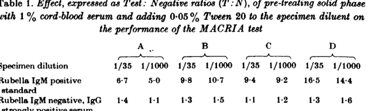
Table 1. Effect, expressed as Test: Negative ratios (T:N), of pre-treating solid phase
with 1 % cord-blood serum and adding 0*05 % Tween 20 to the specimen diluent on the performance of the MACRIA test
A B C D
* •
A A A A _ Specimen dilution 1/35 1/1000 1/35 1/1000 1/35 1/1000 1/35 1/1000 Rubella IgM positive 6-7 5 0 9-8 10-7 9-4 9-2 16-5 14-4
standard
Rubella IgM negative, IgG 1-4 11 1-3 1-5 11 1-2 1 3 1-6 strongly positive serum
A, No Tween 20, no cord serum: B, Cord serum treated solid phase: C, Tween 20 in specimen diluent: D, Cord serum treatment and Tween 20 in diluent.
IgM positive standard or other serum under test (T) to the rubella antibody-negative control (N) and to specific IgM antibody-negative but IgG positive sera. Variations in the reagents, dilutions, and incubation conditions were assessed to this end.
Preparation of beads for the first stage of the test
Polystyrene beads were coated by immersion in one of three dilutions in coating buffer (1 in 100, 1 in 500 and 1 in 2500) of the sheep (Seward) and the rabbit (Dako) anti-/t serum. Two specimen dilutions, 1 in 35 and 1 in 1000, and an antigen dilution of 1 in 20 were used. At both specimen dilutions the beads coated with the
1 in 500 dilution of the anti-/* sera performed best, and slightly better T:N ratios were obtained with the sheep anti-/* coated beads. Further experiments showed that the presence of 0-05 % Tween in the specimen diluent and pre-soaking of the anti-/t coated beads in 1 % kaolin-treated anti-rubella negative human cord serum improved the T:N ratios obtained with the IgM positive standard and control negative sera (Table 1).
Choice of specimen dilution for the first stage
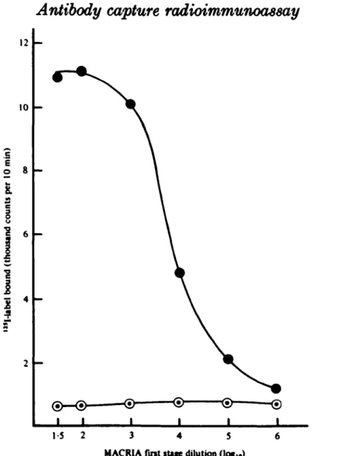
The effect of varying the dilution of the specimen applied to the bead in the first stage of the test was investigated (Fig. 1). The standard anti-rubella IgM positive serum and the rubella antibody negative control serum were applied at dilutions
from 1 in 35 to 1 in 10s to beads coated in a 1 in 500 dilution of anti-/t serum.
A 1 in 20 dilution of antigen was used in the second stage of the test. The highest specific label binding occurred at specimen dilutions of 1 in 35 and 1 in 100. At a
dilution of 1 in 104 there was half of the maximum label uptake, and at a dilution
of 1 in 10e label uptake was reduced to that of the negative control serum. The
experiment was repeated using specimens with intermediate concentrations of anti-rubella IgM prepared by diluting the positive standard in the negative control serum (Table 2). For all concentrations of anti-rubella IgM, tests at low specimen dilutions gave the best results.
Antibody capture radioimmunoassay
143
12 10 .5 E oI
a 11
2 4 1-5 2 3 4 5 MACRIA first sttfe dilution (log^)Fig. 1. Titration of anti-rubella IgM positive standard serum in PBST. I, anti-rubella IgM positive standard; 0 , anti-rubella antibody negative control.
Table 2. Test: Negative (T:N) ratios obtained when the anti-rubella IgM positive
standard, at concentrations with various MACRIA unitages, was tested at specimen
dilutions of 1/10-1/10000
Specimen Anti-rubella IgM units per ml*
dilution , * > in test 100 17-9 180 12-5 9 1 5 1 30 15 3 15 2 104 6-9 2-6 1/10 1/35 1/100 1/1000 1/10000
• Achieved by diluting positive standard in negative serum. 10 11-7 11-7 7-9 4 4 1-7 6-9 7-2 4-5 2-5 12 1 3-9 3-7 3 0 1-6 11 0-3 2-3 2 0 1-8 1-3 10
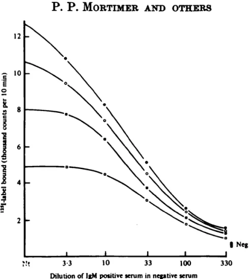
Effect of varying the antigen dilution in the second stage
Various antigen dilutions were used in the second stage of the test. Dilutions from 1 in 10 to 1 in 80 were applied for 20 h to beads which had been incubated with a range of concentrations of the anti-rubella IgM standard. A plot of the results (Fig. 2) showed that there was a straight line relationship between the proportion of IgM in the specimen that was specific and the counts bound when a
I Neg N't 3 3 10 33 100
Dilution of IgM positive serum in negative serum
330
Fig. 2. Effect of antigen dilution on MACRIA test results: titration of rubella IgM positive standard serum in rubella antibody negative serum at four antigen dilutions. Antigen dilutions used were 1 in 10 ( # ) , 1 in 20 (Q), 1 in 40 ( • ) and 1 in 80 ( • ) •
Table 3. Effect on reactivity of various dilutions of the positive standard of adding
20% heat-inactivated rabbit serum to the diluent for the lt6I-labelled rabbit
anti-rubella IgO
Strength of No rabbit serum 20 % rabbit serum
positive f * v * >
standard counts per (units per ml) 10 min.
T:N ratio counts per 10 min. T:N ratio 100 30 10 3 1 neg 11992 10694 7987 5036 2994 1326 8-8 7-9 5-9 3-7 2-2 12136 9482 7193 4625 2640 807 150 11-7 8-9 5-7 3-3
low dilution of antigen was applied. At higher dilutions of antigen insufficient was apparently present to maximize label binding by strong concentrations of anti-rubella IgM.
Comparison of human and rabbit ^I-labelled IgO
A comparison of mI-labelled human and rabbit anti-rubella IgO was made.
The proportion of the labels bound by the anti-rubella IgM positive standard in repeated tests and the T: N ratios obtained were very similar. The effect of adding heat-aggregated homologous serum to the label diluent, which was intended to
Antibody capture r^adioimmunoassay 145
suppress anti-species-mediated uptake of radioactivity to the solid phase, was substantial (Table 3).
Variations in incubation conditions
The above experiments were conducted with the specimens applied for 4 h at 37 °C in the first stage, the antigen for 20 h at 4 °C in the second stage, and the
m
I-labelled IgG for 4 h at 37 °C in the final stage of the assay. Variations of these incubation conditions were investigated. Overnight incubation at room tempera-ture or 4 °C in the first stage gave similar results, but incubation for less than 4 h yielded lower T:N ratios. Uptake of antigen during the second stage continued, in the case of the most strongly positive specimens, for at least 40 h, but an incu-bation period of 20 h was sufficient to obtain satisfactory results. In the final stage,
incubation with 126I-labelled anti-rubella IgG for more than 4 h at 37 °C did not
increase T: N ratios.
As a result of these investigations the following MACRIA procedure was adopted as a diagnostic method.
(i) Polystyrene beads in a conical flask were coated by immersion for 18 h in a 1 in 500 dilution of sheep anti-/e serum in coating buffer and stored at 4 °C in the same solution until used.
(ii) The beads required for each batch of tests were transferred to another flask, washed in PBS (phosphate buffered saline, Dulbecco A), and shaken in a
1 % dilution of cord serum in PBS for 3 h at room temperature.
(iii) Test, standard and other control sera, diluted 1 in 35 or 1 in 1000 in PBST, were prepared in the wells of plastic trays (Abbott Laboratories) and an unwashed bead added to each specimen. The volume of each reactant added, sufficient to cover the bead, was 175 /el. The trays were incubated for 4 h at 37 °C.
(iv) The beads were re-washed and incubated for 20-40 h at 4 °C in a 1 in 20 dilution of rubella antigen in PBST.
(v) The beads were washed again and incubated for 4 h at 37 °C with 115
I-labelled anti-rubella IgG. In the experiments described in the Results section this was a rabbit antiserum unless otherwise indicated.
(vi) The beads were washed once more and the radioactivity bound to each bead counted for 10 min.
The MACRIA tests reported in Sections II and III of the Results were per-formed in one laboratory on sera sent under code from the other collaborating laboratories.
Standardization
For each run of tests a calibration curve was prepared. The anti-rubella IgM positive standard (arbitrary strength 100 units per ml) was diluted in negative control serum to strengths of 30, 10, 3, 1 and 0-3 units per ml and each dilution tested in duplicate. A value in units per ml, based on the counts bound, was assigned to each specimen under test by reference to the curve derived from the results of the tests on the dilutions. The curve for the 1 in 20 dilution of antigen in Fig. 2 is an example of a standard curve. Specimens containing > 1 unit per
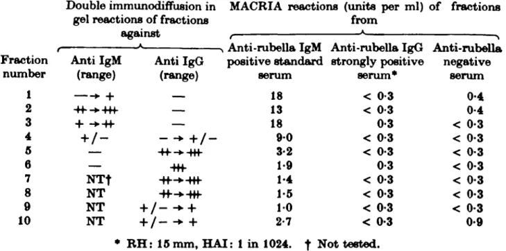
Table 4. MACRIA tests on sucrose density gradient centrifugation
fractionated rubella antibody positive and negative serum
Double immunodiffusion in MACRIA reactions (units per ml) of fractions gel reactions of fractions from
against , * ^ -N Anti-rubella IgM Anti-rubella IgG Anti-rubella Fraction Anti IgM Anti IgG positive standard strongly positive negative
number (range) (range) serum serum* serum 1 • + — 18 < 0-3 0-4 2 4f -+4+f — 13 < 0-3 0-4 3 + -*4f — 18 0-3 < 0-3 4 + / - - -• + / - 9-0 < 0-3 < 0-3 5 — -H--*-Rf 3-2 < 0-3 < 0-3 6 — -Hf 1-9 0-3 < 0-3 7 N T f -ff-*-Hf 1-4 < 0-3 < 0-3 8 NT -ff-^-Hf 1-5 < 0-3 < 0-3 9 NT + / - - • + 10 < 0-3 < 0-3 10 NT + / - -• + 2-7 < 0-3 0-9
* RH: 15 mm, HAI: 1 in 1024. t Not tested.
ml were regarded as positive, and specimens containing < 0-3 units per ml as negative. Specimens in the intermediate range were considered to be equivocal. The T:N ratio of the 1 unit per ml dilution of the standard was usually > 2-5, and of the 0-3 unit per ml dilution of the standard ^ 1*5.
(d) Other methods
Rubella haemagglutination inhibition (HAI: PHLS, 1978), and rubella radial haemolysis (RH) tests (Kurtz et al. 1980) were used for examining whole sera. Serum fractions were prepared by sucrose density gradient centrifugation (SDGC). The fractions were collected through a hole punched in the bottom of the centri-fuge tube. For HAI testing either the sera to be fractionated were pre-treated with heparin (Burkhardt, Schilt & Saner, 1980), or the fractions were tested using 2-mercaptoethanol reduction to check the specificity of the HAI test (Hambling, unpublished). Passive haemagglutination tests for RF were done at serum dilu-tions from 1 in 40 to 1 in 10000 using the RAHA kit (Fujizoki Ltd., Tokyo, Japan).
RESULTS
I. Specificity
To determine the specificity of the MACRIA for anti-rubella IgM, tests were made on fractions of the rubella IgM positive standard serum, on an anti-rubella positive serum from a patient without a history of recent infection, and on the anti-rubella negative control serum (Table 4). Only the fractions from the first serum were MACRIA positive. The IgM-rich fractions reacted strongly, and there was a trail of activity through the IgG-rich fractions which was probably an artefact of the method used for collecting the gradients.
Antibody capture radioimmwnoassay 147
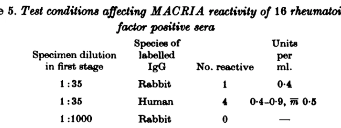
Table 5. Test conditions affecting MACRIA reactivity of 16 rheumatoid
factor positive sera
Specimen dilution in first stage 1:35 1:35 1:1000 Species of labelled IgG Rabbit Human Rabbit No. reactive 1 4 0-0 Units per ml. 0-4 4-0-9, m 0-5 _
Various categories of specimen not expected to contain anti-rubella IgM were tested in order to investigate possible non-specific reactivity in whole sera. Sera from 24 patients with acute viral infections (twelve with measles, six with infec-tious mononucleosis, three with hepatitis A, three with hepatitis B), and 100 blood donors were tested at a specimen dilution of 1 in 35. All but six had < 0-3 units per ml anti-rubella IgM. Four had 0*4 units, one 1*1 units and one 1-4 units per ml. One of the latter two was a 13-year-old girl with infectious mononucleosis who was later found to have been given rubella vaccine five months previously. The other was a 25-year-old female blood donor who had not been exposed to natural rubella or immunized recently. She was re-tested after a later donation. The first and second serum specimens were then found to contain 0-8 and 1*0 units per ml of anti-rubella IgM respectively.
When the donors' specimens were tested by RH it was seen that non-specific binding increased slightly with the amount of non-IgM antibody present. The mean counts of the eight rubella antibody negative donors' sera was 57 % of that of the ' cut-off' value given by duplicate tests on the 0-3 unit per ml dilution of the standard serum, and the mean count of the seven donors' sera that had an RH zone of 14 mm was 76 % of the ' cut-off \ This tendency for antibody positive sera to be more reactive than antibody negative sera was undiminished when they were re-tested at a specimen dilution of 1 in 1000.
Sixteen RF positive (HA titre range 1 in 320 to 1 in 5120, geometric mean 1 in 1612), rubella RH positive (zone diameter range 7-14 mm, mean 10-9 mm)
sera were tested at specimen dilutions of 1 in 35 and 1 in 1000, using 1>sI-labelled
rabbit anti-rubella IgG. The experiment was repeated at the 1 in 35 dilution using both the rabbit and the human IgG labels (Table 5). In no case did an RF positive serum cause a reaction ^ 1 unit per ml and use of the lower specimen dilution did not increase background binding of the label. There was a slight increase in
back-ground binding when the 1S5I-labelled human IgG was used rather than the rabbit
IgG.
II. Reproducibility
The reproducibility of the MACRIA was investigated. Sera from 60 patients thought to have had rubella were tested on separate occasions using the same specimen dilution, 1 in 35, but different batches of antigen. Forty-two sera were MACRIA positive on both occasions, the correlation co-efficient of the counts being 0-04. Four sera were in the range 0-4-1-0 units per ml and 13 were t. 0-3
Table 6. Results on sera from 69 suspected cases of rubella tested for anti-rubella IgM
by HAI after sucrose density gradient centrifugation (SDGC-HAI) and by MACRIA
SDGC-HAI positive 47 0 SDGC-HAI negative 1* 21 MACRIA
Positive (> 1 unit per ml) Negative ( < 1 unit per ml)
* A baby whose mother was known to have had rubella in the fourth month of pregnancy.
units per ml on both occasions. One serum was 0*8 units in the first and 1*5 units in the second test.
The effect of varying the specimen dilution used in the first stage of the test was investigated. The same 60 sera were re-examined at two dilutions, 1 in 35 and 1 in 1000. The same 42 sera as before were positive at both dilutions, and the correlation co-efficient of the counts at the two dilutions was 0-93. Twenty-four out of the 28 collected within 30 days of onset of a rash and 13 out of the 14 collected between 31 and 60 days bound more counts at the 1 in 35 than at the 1 in 1000 dilution. In general about a fifth more counts were bound at the 1 in 35 dilution by sera collected in the first month, and a third more bound by sera collected in the second month. Of the 18 specimens that were not positive the same 13 as before were ^ 0 - 3 units per ml at both dilutions and the same five fell within the equivocal range.
III. Evaluation of MACRIA in diagnostic use
Acute phase and convalescent sera from 25 cases of rubella showing sero-conversion in both HAI and RH tests ( < 1 in 10 to ^ 1 in 20, no zone to a zone
^ 8 mm diameter) were tested by MACRIA. Six of the acute phase sera were MACRIA positive (range 1 -4-5-0 units per ml) and seven slightly reactive (range 0-4-0-9 units per ml). No specimen collected after the sixth day was MACRIA negative. All the convalescent specimens were MACRIA positive (range 3*8-80 units per ml).
Serum specimens from 69 suspected cases of rubella, previously tested by SDGC-HAI, were tested by MACRIA (Table 6). The results by both methods were identical except that SDGC-HAI did not detect specific IgM at birth in a baby whose mother was known to have had rubella in pregnancy.
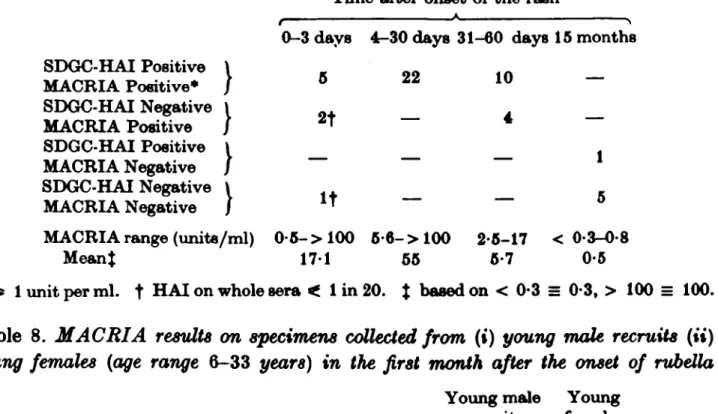
Fifty sera collected at intervals from 18 male army recruits who had rubella confirmed by SDGC-HAI tests (Burkhardt et al. 1980) were also examined (Table 7). Five out of eight specimens collected in the first three days and all 36 specimens collected between four and sixty days after the onset of the rash were MACRIA positive. The six specimens collected fifteen months later were all MACRIA negative. The MACRIA response of the male recruits was stronger than that of a group of young women from whom specimens had been collected over the same period after the appearance of the rash (Table 8).
Antibody capture radioimmunoassay 149
Table 7. Results on fifty specimens taken from eighteen male recruits affected in an
outbreak of rubella and tested for anti-rubella IgM by HAI after sucrose density gradient centrifugation (SDGC-HAI) and by MACRIA
Time after onset of t h e rash
0-3 days 4-30 days 31-60 days 15 months SDGC-HAI Positive \ _ MACRIA Positive* / 5 22 10 SDGC-HAI Negative \ MACRIA Positive / 2* "~ 4 SDGC-HAI Positive MACRIA Negative SDGC-HAI Negative \ MACRIA Negative / IT — — <> MACRIA range (units/ml) 0 - 5 - > 100 6-6-> 100 2-5-17 < 0-3-0-8
M e a n t 1 7 1 55 5-7 0-5
* ^ 1 unit per m l . t H A I on whole sera < 1 in 20. $ based on < 0-3 = 0-3, > 100 = 100.
Table 8. MACRIA results on specimens collected from (») young male recruits (ii)
young females (age range &-33 years) in the first month after the onset of rubella
Number of subjects Number of specimens
Median interval in days, onset to collection Mean MACRIA* (units per ml)
Number of specimens scoring > 100 units per ml
• Based on < 0-3 = 0-3, > 100 = 100.
Table 9. Persistence of the anti-rubella IgM response, measured by MACRIA, in
sera from women with serologicaUy confirmed rubeUa
Month after onset of rash
Young male recruits 18 29 6 50-2 12 Young females 36 58 7 19 2 2 2nd 3rd 4th 5th 6th Number of specimens 9 4 4 4 1 Range of MACRIA results* 3-1-80 0-7-2-4 1-2-5-8 < 0-3-0-9 0-3 Meanf of MACRIA results 3 1 1 6 3-6 0-6 0 3
* Units per ml. f Based on < 0-3 = 0-3
month after rubella verified by SDGC-HAI were tested by MACRIA (Table 9). All four sera collected in the fourth month but none of five collected in the fifth and sixth months were positive.
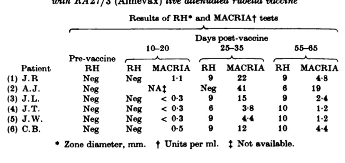
Sera collected at intervals after immunization from six adult women given RA27/3 rubella vaccine were tested by RH and MACRIA (Table 10). None had rubella antibody before immunization. An anti-rubella IgM response was detected in all six, strongest at 25-35 days, but detectable up to 65 days after vaccine was given.
Table 10. Radial haemolysis (RH) and MAORI A responses of tvomen immunized
with 1L427/3 (Almevax) live attenuated rubella vaccine
Results of RH* and MAORIA| tests
(1) (2) (3) (4) (5) (6) Patient J.R A.J. J.L. J.T. J.W. C.B. t x re-vaccine RH Neg Neg Neg Neg Neg Neg RH Neg Neg Neg Neg Neg 10-20 A MACRIA 11 NAJ < 0-3 < 0-3 < 0-3 0-5 Days post-vaccine : RH 9 Neg 9 6 9 9 25-35 A MACRIA 22 41 15 3-8 4 4 12 RH 9 6 9 10 10 10 55-65 A MACRIA 4-8 19 2-4 1-2 1-2 4 4 • Zone diameter, mm. f Units per ml. % Not available.
DISCUSSION
The purpose of this investigation was to establish the best conditions under which to apply the antibody capture principle to the assay of anti-rubella IgM, and to use them to test coded clinical material already tested for rubella antibody by other means. The conditions eventually used in the MACRIA were chosen because they allowed the test to be completed within two days, maintained a straight line relationship between the proportion of anti-rubella to total IgM in the specimens and the counts bound, and effectively suppressed non-specific binding of the label.
Three features of the MACRIA for rubella used in this study were of particular note. Firstly the specimens were applied in the first stage of the test at dilutions (1 in 35 to 1 in 1000) lower than used in published MACRIA methods using other antigens (1 in 1000 to 1 in 10000). In the rubella assay the 1 in 35 dilution was more sensitive to low levels of specific IgM and no more likely to cause non-specific label binding than the 1 in 1000 dilution. The higher dilution was only advantageous if it was desired to confine positivity more narrowly to those specimens collected within a month of natural infection.
The second important feature of the test was the length of the second stage. In MACRIA for anti-HBc IgM the second stage can be brief (Mortimer et al. 1981), but, as in the analogous test for anti-HAV IgM (Flehmig et al. 1979), the complete uptake of rubella antigen in MACRIA was found to depend on pro-longed contact. This stage must therefore last overnight or longer.
A third factor that critically affected the quality of the assay was the method of preparation of the radio-label. Any treatment of the anti-serum to be labelled that is likely to aggregate globulins (e.g. heat inactivation, ammonium sulphate precipitation) is to be avoided. The iodination method of Bolton and Hunter is preferable because it is mild and yields a label with a long shelf life. Better results can also be obtained by adding heat-aggregated homologous serum to the label diluent.
Antibody capture radioimmunoassay 151
calibration curve derived from dilutions of a standard IgM positive serum in negative serum, proved practical and reproducible. By multiplying these arbitrary units by the total serum IgM concentration a measure of the amount of specific IgM could be obtained, but, for diagnostic purposes, the value in uncorrected units, representing the proportion of total IgM that is rubella-specific, is enough. The alternative procedure for quantifying specific IgM by MACRIA, in which each specimen is tested in serial dilution to establish an end-point, is open to dilution error and unnecessarily laborious.
Tests on three categories of non-rubella sera, those from patients with other acute viral infections, from random blood donors, and from rubella antibody positive subjects with RF did not reveal significant non-specific reactivity. Duermeyer, Wielaard & van der Veen (1979) have reported that RF positive sera react with peroxidase-labelled IgG unless the IgG has been pepsin-digested to remove the RF binding site. With radio-labelling methods this has not been found necessary (Roggendorf et al. 1980, Mortimer, Parry & Appleton, 1981), except in special circumstances (Tedder & Wilson-Croome, 1981). We suggest, nevertheless, that MACRIA positive sera be screened for RF until the resistance of these tests to non-specific interference under routine conditions is definitely established.
The development of the MACRIA response during rubella was clearly demon-strated in this investigation. It was already detectable by the time that HAI and RH tests for antibody had become positive and preceded, by several days, reac-tivity in the RH test (which is insensitive to IgM antibody). It rapidly increased in strength in the week after the appearance of the rash, and there was no difficulty in demonstrating a positive MACRIA reaction once the RH test had become positive or the rubella HAI test showed a titre of > 1 in 20. The MACRIA response remained strongly positive for two months and then diminished, so that the reaction was very weak after four months. A similar pattern has been des-cribed in MACRIAs for anti-HAV and anti-HBc (Roggendorf et al. 1980, Mortimer et al. 1981). The rubella MACRIA response was stronger in men than women, perhaps because total serum IgM concentrations are normally lower in men (Stoop et al. 1969). Alternatively this may represent a true sex difference in immune response to rubella virus.
The correlation of the MACRIA with the SDGC-HAI test for specific IgM was very close. Over 100 SDGC-HAI positive sera were examined during these investi-gations, and all but one were MACRIA positive. Two sera collected in the first three days of rubella and four sera collected in the second month were MACRIA positive, SDGC-HAI negative, indicating the greater sensitivity of MACRIA.
Previous attempts to detect the specific IgM response after rubella vaccination have shown that existing methods are too insensitive to be reliable. MACRIA detected specific IgM in all vaocinees sero-negative before immunization. The responses were, however, weaker than after natural infection, in the range 1-40 units per ml compared with 10- > 100 units per ml after natural infection. Patients with pre-existing rubella antibody have been found not to make a MACRIA response. Thus the presence of antibody before immunization appears to
deter-mine whether vaccinees produce specific IgM, and this observation has since been borne out in a study of 100 women vaccinated in a general practice (to be published). Although the rubella MACRIA could probably be improved by using a radio-label containing a higher proportion of specific IgG, it is already apparent that M-antibody capture is the most sensitive and specific method for rubella diagnosis. Other advantages are that it only requires a few microlitres of serum and is applicable both to adults' and infants' sera (a report of its use in the diagnosis of congenital infection is in preparation). The introduction of this method to clinical virology laboratories would improve and simplify testing for recent infection by rubella.
We thank Dr A. H. Tomlinson who prepared the rabbit anti-rubella serum, and Dr A. Hutchinson, Mrs Olive Jones and the staff of the Blood Transfusion Service Laboratory, Edgware who provided materials used in the investigations. We also thank Dr D. M. S. Dane and Dr M. S. Pereira for help and advice.
REFERENCES
BOLTON, A. E., A HUNTER, W. M. (1973). The Labelling of Proteins to High Specific
Radio-activities by Conjugation to a "*I-Containing Acylating Agent. Biochemical Journal 133, 529-39.
BURKHABDT, F., SCHILT, U. A SANER, H. (1980). Virologische Diagnostic der
Rotelninfek-tion, Befunde bei einer Rubella-Epidemie in einer Rekrutensohule. Schweizerische
Medizin-ische Wochenachrijt 110, 555-62.
CBADOCK-WATSON, J. E., RIDEHALOH, M. K. S., PATTISON, J. RM ANDERSON, M. J. A
KANORO, H. O. (1979). Comparison of immunofluoreecence and radioimmunoassay for
detecting IgM antibody in infants with the congenital rubella syndrome. Journal of tfypiene 83,413-23.
DIMENT, J. A. A PEPYS, J. (1978). Immunoeorbent Separation of IgG and IgM for the
Radioimmunoassay of Specific Antibodies. Affinity Chromatography, (ed. O. Hoffmann-Ostenhof et al.), pp. 229-23. Oxford and New York: Pergamon Press.
DUERMEYBR, W., WIELAARD, F. A VAN DEB VEEN, J. (1979). A New Principle for the
Detec-tion of Specific IgM Antibodies applied in an ELISA for Hepatitis A. Journal of Medical
Virology 4, 25-32.
FLBHMIO, BM RANKE, M., BBRTHOLD, H., A GERTH, H-J. (1979). A Solid-Phase
Radio-immunoassay for Detection of IgM Antibodies to Hepatitis A Virus. Journal of Infectious
Diseases 140,169-75.
KURTZ, J. B., MORTIMER, P. P., MORTIMER, P. R., MOROAN-CAPNER, P., SHAFI, M. S. A
WHITE, G. B. B. (1980). Rubella Antibody Measured by Radial Haemolysis.
Charac-teristics and Performance of a Simple Screening Method for Use in Diagnostic Laboratories.
Journal of Hygiene 84, 213-23.
MORTIMER, P. P., PARRY, J. V. A APPLETON, HAZEL (1981). Diagnosis of recent hepatitis A
infection: a comparison of two methods for detecting specific IgM. Journal of Hygiene (in the Press).
MORTIMER, P. P., VANDBBVELDE, E. M., PABBY, J. V., COHEN, B. J. A TEDDER, R. S. (1981).
The anti-HBc IgM Response in the Acute and Convalescent Phases of Acute Hepatitis.
Journal of Infection (in the Press).
PATTI8ON, J. R. A DANE, D. S. (1975). The detection of specific IgM antibodies following
infection with rubella virus. Journal of Clinical Pathology 28, 377-82.
PUBLIC HEALTH LABORATORY SERVICE STANDING ADVISORY COMMITTEE ON VIRAL REAGENTS
(1978). Haemagglutination-inhibition test for the detection of rubella antibody. Journal of
Hygiene, 81,373-82.
Antibody capture radioimmunoajdiy 153
Solid Phase Test Systems for Demonstrating Antibodies Against Hepatitis A Virus (Anti-HAV) of the IgM-Class. Journal of Medical Virology 5, 47-62.
STOOP, J. W., ZEGERS, B. J. M., SANDER, P. C. & BALLIEUX, R. E. (1969). Serum
Immuno-globulin Levels in Healthy Children and Adults. Clinical and Experimental Immunology 4, 101-12.
TEDDER, R. S. & WILSON-CROOME, RUTH (1981). Detection of IgM class antibody to hepatitis
B core antigen: a comparison of two methods. Journal of Medical Virology, (in the Press).
VESIKARI, T. & VAHERI, A. (1968). Rubella: a method for rapid diagnosis of recent infection