The Journal of Heredity 72: 360-362. 1981.
Some rare cases of chimerism
in twin cattle and their
proposed use in determining
germinal cell migration
G. Stranzlnger, G. Dolf, R. Fries, and H. StockerABSTRACT: Three dizygotic, heterosexual twins with
chimerlsms carrying marker chromosomes are de-scribed. Phenotypic and cytogenetlc methods were used to identify these animals. The occurrence of germinal cell migration causing gonad chimerism can be detected by the marker chromosome event under conditions de-scribed in this report.
T H E P H E N O M E N O N of chimerism has been
studied and discussed frequently in recent years1. In embryo transfers for twin
produc-tion in cattle chimerism is a potential problem because females with male twins are likely to become sterile10. Such females are unsuitable
for breeding and have subnormal weight-gain efficiency compared to males. Freemartins among twins are generally identified by rectal palpation of adult animals8, as well as by the
The authors are professor and graduate students, respectively, at the Swiss Federal Institute of Technology (ETH), Institute of Animal Production, CH-8902 Zurich. They are grateful to Mrs. K. Emler, Miss Keeman, Mrs. Jenny, and Mrs. Kirianoff for their assistance in the preparation, translation and typing of the manuscript. These studies have been financed by the AGRO-Fonds ETH and the Swiss Association for Artificial Insemination. The authors thank Professor H. Abplanalp for his critical review of the manuscript.
© 1981, American Genetic Association.
normal cow
^ x
heterozygote centric fusion I t/29© ©
egg cell poll call
sperm population XX chJm. case A sterile •perm population XX * female chromosome* XY= male M : marker chromosome 1/29
7 \
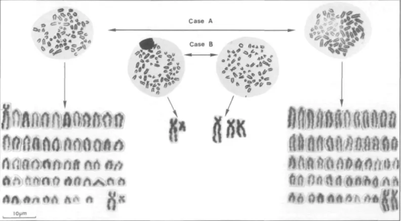
FIGURE 1 Fertilization of a normal egg cell with sperm carrying the centromere fusion chromosome.
Also shown are the potential combinations of various germ cells (see text).
size and form of the clitoris. A blood group analysis is successful only if a sufficient number of factors are tested so as to distin-guish the two erythrocyte populations. Such studies become more reliable if additional biochemical markers13 or an H-Y antigen
test12 are included. A further diagnostic
pos-sibility is a cytogenetic examination as de-scribed by Herschler and Fechheimer4'5.
Given sufficient cell material one can deter-mine the origin of cells on the basis of sex chromosomes. However, the cell proportion
is of little value when considering the degree of virilization.
A high proportion of affected females are freemartins. The sex differentiation of the male partner seems to be quite normal, but according to various reports1 fertility troubles
do occur causing increased rates of culling among affected males. Thus, the presence of XX primordial germ cells in testes9'16 could
lead to disturbances in spermatogenesis and thus alter reproductive fitness as well as sex ratio among offspring. However, the time and
Table I. Metaphases counted from short-term lymphocyte cultures and one long-term flbroblast culture (muscle cells) of chimeric twin pairs A, B, and C and repeat investigations in bulls B and C
Date of Material examination Blood 24-9-79 9-10-79 22-4-80 17-7-80 14-8-80 25-9-80 9-10-80 20-11-80 29-1-81 12-2-81 26-2-81 Fibroblast 15-5-81 cell XYM 27(20) 23(22) Pair A* male female type cell XX XYM 110(80) 28(24) 82(78) 24(24) type XX 90(76) 77(76) male type XX 89(89) 85(85) 84(84) 97(97) 95(95) 98(98) 93(93) 97(97) 92(92) 21(87) Pair type XXM 11(11) 15(15) 16(16) 3(3) 5(5) 2(2) 7(7) 3(3) 8(8) 3(13) B female cell XY 93(93) type XXM 7(7) male cell XY 32(44) 32(39) 34(34) 38(38) 51(51) 28(28) 50(50) 57(57) 54(54) 11(48) PairC type XXM 41(56) 61(61) 66(66) 62(62) 49(49) 72(72) 50(50) 43(43) 46(46) 12(52) female cell type XY XXM 23(35) 42(65)
* X, Y = sex chromosomes; M = marker chromosome; () = percentage
ways of migration of XX cells into an XY male gonad is very uncertain. For one thing, im-plantation of the embryo occurs relatively late, approximately 20 days after fertilization, and the development of placental vessels is even further delayed. According to Jost et al.7,
go-nadal differentiation is nearly complete at that time, thus preventing the entry of female cells into the gonad from a twin.
The positive identification of chimeric cells in the male gonad and their subsequent de-velopment into spermatozoa that fertilize and give rise to offspring can be achieved by coat color marker genes11, or more easily by
marker chromosomes such as the 1/29 centric fusion chromosome found quite often in cattle populations.
Materials and Methods
An experiment was conducted using mixed semen from three bulls of the Angus, Sim-mental, and Bavarian Brown breeds and having diploid numbers of 60, 59, and 58 chromosomes, respectively. The last two bulls are carriers of the 1/29 centric fusion either heterozygously or homozygously. As a first step, twin calves of different sex sired by the heterozygous translocation carrier (59) were produced. Second, a cytogenetic investigation was carried out to find animals with the nec-essary chromosome composition to prove go-nodal chimerism. Only male calves were used, since the female partners were more likely to be sterile. Several heterosexual twin pairs were obtained sired by the heterozygote Simmental bull known to carry the centric fusion 1/29. In all cases, the males appeared
normal, while females showed definite symptoms of freemartinism.
Standard techniques for leucocyte culture and chromosome preparations were used14 to
study the three twin pairs A, B, and C. Re-peated cultures (pair A, two times; the bulls from B and C, nine times) were made to assure accurate results especially as related to the possible influence of cell selection in vitro. Finally, muscle biopsies were taken from bulls B and C and fibroblast cultures established to get indications of chimerism from the chro-mosome preparations.
Results and Discussion
The cytogenetic analysis showed that in twin pair A the male resulted from the fertil-ization of a normal egg cell with sperm carrying the centromere fusion chromosome' (designated as M in Figure 1), while the fe-male developed from fertilization with a normal sperm. In the twin pairs B and C the reverse situation prevailed with females carrying the marker chromosomes. The di-agram in Figure 1 illustrates the initial situa-tion and the potential combinasitua-tions of the various germ cells. For the sake of simplicity, all autosomes that are not essential in this di-agram have been omitted. The didi-agram in-cludes gametes expected from male twins with hypothetically chimeric gonads including those of animals B and C that may eventually lead to conclusive progeny tests. In Table I the results of the number of cell populations from the three twin pairs and the results of repeat analyses of bulls B and C from blood cultures and one fibroblast study are presented. All
three cases show consistent results. The white blood cells from the bull of pair C varies in the male cells from 28 to 57 percent and in the female cells from 41 to 72 percent. The bull in pair B had a very low proportion of female cells (2-16 percent); therefore, the animals represent, to some extent, both possibilities of dominating cells. Figure 2 shows the meta-phases of the four different cell types and two karyograms including the sex chromosomes and marker chromosomes of the chromosome situation in pair B. In the fibroblast cultures the ratio of the two cell types are in the range of the lymphocytes (bull B, 87/13 percent; bull C, 48/52 percent). Therefore, the chimeric cell mixture is comparable in both tissues. The bulls from twin pairs B and C can thus be used in subsequent matings to further investigate gonodal chimerism because if their testes also are chimeric they should transmit the marker chromosome to some female offsprings de-rived from their twin sister.
The important factor in this unusual case of chimerism is the use of translocation chro-mosome 1/29, as first suggested by Herschler and Fechheimer4. Their marker chromosome
in a set of bovine triplets and our case is the 1/29 centric fusion type that was studied ex-tensively by Gustavsson3 and others2.
Since twinning in cattle occurs in less than 5 percent of births6 and the frequency of
centric fusions also is low in most breeds, special matings involving bulls known to be translocation heterozygotes are essential for investigations of chimerism, as proposed in this paper. Numerous biochemical and ge-netical phenomena may be associated with sexual chimerism of cattle twins, but these
9 ^ V
rtfi!flO;fln
Hit fi.Oifi 0
Iv^) wfn fffl
10pmD ft M 0/>
r>OA^vf»i
ft* A Itw
Case A Case B O o «i cmm
FIGURE 2 Metaphases (case A and B) and two karyotypes (case A) and additional significant chromosomes (case B) of the chimeric cattle twins.
may be masked by embryonic mortality. Therefore, the described cases could serve as model examples with which to better under-stand certain events involved in sex differ-entiation or genetic expression. Since chim-erism is commonly found among heterosex-ual, dizygotic twins we believe that chimerism between like-sexed twins is equally frequent and should receive further attention in future studies, especially with animals from embryo transfers.
A single offspring with a centromeric fusion chromosome from the described males B and C would support the theory of germ cell chimerism. Such studies are now in prog-ress.
References
1. D U N N , H. O., K. M C E N T E E , C. E. HALL, R. H. JOHNSON, JR., and W. H. STONE. Cytogenetic and reproductive studies of bulls born co-twin with freemartins. ]. Reprod. Fert. 57:21-30. 1979.
2. ELDRIDGE, F. E. High frequency of a Robertso-nian translocation in a herd of British White cattle. Vet. Rec. 96:71-73.1975.
3. GUSTAVSSON, I. Cytogenetics, distribution and phenotypic effects of a translocation in Swedish cattle. Hereditas 63:68-169.1969.
4. HERSCHLER, M. S. and N. S. FECHHEIMER. Centric fusion of chromosomes in a set of bovine triplets. Cylogenetics 5:307-312.1966. 5. and . The role of sex chromosome
chimerism in alterning sexual development of mammals. Cytogenetics 6:204.1967.
6. JOHANSSON, I., B. LINDHE, and F. PIRCHNER. Causes of variation in the frequency of monoz-ygous and dizmonoz-ygous twinning in various breeds of cattle. Hereditas 78:201-234.1974. 7. |OST, A., B. VIGIER, and J. PREPIN. Freemartins
in cattle: the first step of sexual organogenesis. /. Reprod. Fert. 29:249-379.1972.
8. KAESTLI, F. Neuere Untersuchungen uber die Rinderzwicke—eine Literaturubersicht. Schwei'z. Arch. Tierheilk. 121:425-429.1979. 9. O H N O , S., J. M. TRUIILLO, C. STENIUS, L. C.
CHRISTIAN, and R. L. TEPLITZ. Possible germ cell chimeras among newborn dizygotic twin calves (Bos taurus). Cytogenetics 1:258-265. 1962.
10- . The problem of the bovine freemartin. J. Reprod. Fert. (Suppl.) 7:53-61.1969.
11. ROWSON, L. E. A., and R. NEWCOMB. A method of determining whether germinal cells migrate in bovine chimaeras. VIIIth Intern. Congress of Animal Reprod. and Artificial Insemination. Cracow, p. 266-268.1976.
12. STOCKER, H. and G STRANZINGER. H-Y Anti-gen: Genetische Aspekte und Nachweismeth-oden (ein Literaturuberblick). Zuchthyg. 16: 1-10.1981.
13. STONE, W. H., D. T. BERMAN, W. J. TYLER, and M. R. IRWIN. Blood types of the progeny of a pair of cattle twins showing erythrocyte mosai-cism. /. Hered. 51:136-140.1960.
14. STRANZINGER, G. Die Wirkung verschiedener Belastungseinflusse auf die Mitoseaktivitat der Leukozyten von weiblicheri Kalbern. Z. Tierzuchtg. Zuchtungsbiol. 92:27-34.1975. 15. . New approach of demonstrating
fertil-izing capacity of bulls with centric fusion chro-mosomes. 4th Eur. Colloq. Cytogenet. Domest. Anim. p. 220-224.1980.
16. TEPLITZ, R. L., Y. S. M O O N , and P. k. BASRUR. Further studies of chimerism in heterosexual cattle twins. Chromosoma 22:202-209.1967.
The Journal of Heredity 72:362-363. 1981.
Left ostium straight: an
inherited anomaly of the female
genital tract in the rabbit
D. D. Crary and R. R. Fox
ABSTRACT: An inherited anomaly resulting in the left
oviduct extending lateral to the kidney with the ostium opening upward in an otherwise normal animal is de-scribed. This may be the result of a mechanical problem caused by the persistence of early embryonic mesen-teries. Inheritance appears to be due to an autosomal recessive gene with incomplete penetrance. We pro-pose the symbol tos for the gene responsible lor this condition.
ABNORMALITIES of the female genital tract are relatively rare in wild and domestic ani-mals and most are associated with abnor-malities of the urinary tract. The Mullerian
The authors are, respectively, research associate, and senior staff scientist, The Jackson Laboratory, Bar Harbor, Maine 04609. This investigation was supported in part by NIH research grant RR-00251 from the Division of Research Resources to the Jackson Laboratory, and in part by institutional funds of the Jackson Laboratory. The Jackson Lab-oratory is fully accredited by the American Associ-ation for AccreditAssoci-ation of Laboratory Animal Care.
© 1981, American Genetic Association.
ducts are formed from invaginations of the coelomic epithelium of the urogenital ridges7.
Details of their growth are not well known but early stages of their development suggest a close relationship between Mullerian and Wolffian ducts7. When a portion of the
Wolf-fian duct is absent, the Mullerian duct will grow only as far as the terminal end of the Wolffian duct.
The developing uterine tubes fail to match the elongation of the trunk as a whole and the flaring ostial ends finally lie some 13 segments posterior to the level of their origin1. Normally
the ostium is tied to the ovary by the fimbriae7
and is pulled around mechanically when the ovary migrates posteriorly so that the open end lies over the anterior end of the gonad.
In 1955 when an adult rabbit of strain AX/J was necropsied, it was shown to be normal in every respect except that the anterior end of the left oviduct projected anteriorly and lat-erally to the kidney with the ostium projecting straight up instead of curling around the ovary. In 1958 six more cases were observed. Since that time many more such animals have been seen both in strain AX/J and in its subline AXBU/J. We know of no similar condition in any other animal. In this communication we report the mode of inheritance and describe the pathology of this new condition in the rabbit.
Materials and Methods
Our data were obtained from the necropsy records of the rabbit colony of the Jackson Laboratory. All rabbits that die or are killed for experimental purposes are necropsied and
checked for gross abnormalities of all major organ systems3. The data for this report
con-cern two partially inbred strains, AX/J and AXBU/J5.
Standard genetic and statistical analyses were used to determine the mode of inheri-tance.
Results
Description. Figure 1 shows the relative position of the oviducts and ostia in a rabbit with left ostium straight. The right oviduct and ostium are normal with the ostium tubae curving around its ovary. The left oviduct is straight and projects craniad lateral to the kidney. The ostium tubae opens as a straight extension of the oviduct. In some specimens a thin mesentery tied the ostium tubae to the spleen, but most lacked such a mesentery. It may have existed temporarily in earlier de-velopment. The left ovary is in its normal po-sition, but the left ostium tubae remains closer to its primitive location.
Litter size is significantly reduced in af-fected animals (Table I) of both strains. However, we have evidence that at least some of the eggs from the left ovary of affected fe-males can make the long journey to, and be picked up by, the straight left ostium tubae.
Genetics. From September 1969 through December 1977, 1909 strain AX/J females were examined and 106 (5.6 percent) were found with straight left ostia. Similarly, among 1397 strain AXBU/J females, 99 (7.1 percent) had the left ostium straight. In contrast, of 12,133 females from all other strains examined during the same time period, only 5 (0.0004