British Journal of Rheumatology 1996;35:1091-1095
ASSESSMENT OF URINARY HYDROXYPYRIDINIUM CROSS-LINKS
MEASUREMENT IN OSTEOARTHRITIS
M. P. HELLIO LE GRAVERAND, A. M. TRON, M. ICHOU,* M. C. DALLARD, M. RICHARD,* D. UEBELHARTf and E. VIGNON
Pavilion F and * Biochemistry Laboratory, Claude Bernard University, Edouard Herriot Hospital, place d'Arsonval, 69437 Lyon Cedex 03, France and f University Hospital of Geneva, Switzerland and Rush Presbyterian, St Luke's
Medical Center, Chicago, IL, USA
SUMMARY
The aim of this study is to re-evaluate urinary collagen cross-links, previously proposed as markers of osteoarthritis (OA). The urinary excretion of collagen cross-links, pyridinoline (PYD) and deoxypyridinoline (DPD), was measured using high-performance liquid chromatography (HPLC) in 114 patients with OA, 19 patients with rheumatoid arthritis (RA) and 40 healthy subjects. An increase in PYD and DPD, expressed per millimole of creatinine, was confirmed in RA. However, PYD and DPD in patients with hip OA, knee OA and polyOA were similar, and did not differ from controls. In patients with radiographic end-stage OA, PYD and DPD were significantly higher than in patients with an early OA, but not significantly higher than in controls. The PYD/DPD ratio did not vary with the OA stage. Thus, urinary collagen cross-links are not elevated in OA, but could reflect bone sclerosis and/or erosion in late OA.
KEY WORDS: Pyridinium cross-links, Osteoarthritis, Rheumatoid arthritis.
THE covalent bridges or cross-links between the adjacent molecules of collagen stabilize the extracellu-lar matrix of connective tissues. Owing to their natural fluorescent properties, the pyridinium cross-links, pyridinoline (PYD) and deoxypyridinoline (DPD), have been identified in many connective tissues, essentially in bone and cartilage [1, 2]. DPD is mostly found in bone, while PYD is found both in skeletal and vascular connective tissues [1, 3]. Unlike hydroxy-proline, pyridinium cross-links are released following mature collagen degradation in peptide-free forms and are excreted unchanged in urine. Pyridinium cross-links are measured by high-performance liquid chromatography (HPLC), paper chromatography and enzyme-linked immunoassay (ELISA) [4]. Both paper chromatography and ELISA do not allow PYD and DPD to be distinguished. Quantitative analysis using ion-paired reversed-phase liquid chromato-graphy followed by fluorescent detection has been improved [5].
Pyridinium cross-links have already been proposed as an index of bone resorption in Paget's disease [6], primary hyperparathyroidism [7], bone metastasis [8] and osteoporosis [9]. PYD and DPD were also investigated in osteoarthritis (OA) and rheumatoid arthritis (RA). They were suggested as markers of joint degradation [10-15]. These studies led us to correlate urinary pyridinium cross-links with the measurement of joint space narrowing in patients with hip OA [16]. However, initial results obtained in such patients clearly conflicted with reported studies. The present
Submitted 21 December 1995; revised version accepted 26 April 1996.
Correspondence to: E. Vignon, Claude Bernard University, Edouard Herriot Hospital, Pavilion F, place d'Arsonval, 69437 Lyon Cedex 03, France.
work is a re-evaluation of urinary concentrations of pyridinium cross-links in OA patients.
PATIENTS AND METHODS Patients
The OA group was made up of 114 consecutive patients, referred to the rheumatology clinic for symptomatic OA. There were 47 hip OA (coxOA), 31 knee OA (gonOA) and 36 patients with generalized OA (polyOA). All these patients fulfilled the ACR criteria for OA [17]. A normal control group was composed of 40 healthy volunteers. A joint disease control group was composed of 19 patients with classic RA, according to the ARA criteria. Age, sex, menopausal status, weight and body mass index are given in Table I.
Anteroposterior roentgenograms of lumbar rachis, pelvis and knees taken in the weight-bearing position, a sunrise view of the patellofemoral joints and dorsal X-ray of the hands were performed in OA patients and normal controls. Films were read by a single experienced observer who was blinded to the patient's status. X-ray OA was scored as follows: stage 1, minimal joint space loss and/or osteophytosis; stage 2, substantial but incomplete joint space loss; stage 3, total and focal joint space narrowing; stage 4, nearly complete joint space narrowing and subchondral bone erosion (intraobserver reproducibility: k = 0.76) [18]. In patients with bilateral OA, the most severely affected side was analysed.
Westergren erythrocyte sedimentation rate (ESR) (mm/h), routine blood haematology and biochemistry tests (red and white cell counts, alkaline phosphatase, alanine aminotransferase, calcium, phosphorus, gly-caemia, albumin and creatinine) were performed in OA and RA patients. None of the patients suffered from renal impairment, diabetes or a metabolic bone disease. © 1996 British Society for Rheumatology
1092 Controls OA RA Characteristics JV 40 114 19
BRITISH JOURNAL OF RHEUMATOLOGY VOL- 35 TABLE I
of patients with osteoarthritis (OA), rheumatoid arthritis (RA) and Age 42.9 (12) 62.44(11.5) 58.3 (18) Weight 70.56 (13.4) 73.72 (12.5) 66.67 (15) BMI 24.37 (4.2) 27.74 (4.2) 24.75 (4) NO. 11 controls (median Sex ratio M/F 21/19 49/65 8/11 andiD.) Post-menopausal/ non-post-menopausal 9/10 50/15 7/4
Measurement of urinary pyridinoline and deoxypyridino-line
Morning urine samples were collected from all patients and stored at — 20°C prior to analysis. Urine samples were centrifuged at 1000 g during 10 min and an aliquot of urine was hydrolysed by heating with an equal volume of 12 M HC1 at 116°C during 16 h, to convert all cross-links to the free form. The hydro-lysates were fractionated by partition chromatography on CFl cellulose and the pyridinium compounds were quantified by reverse-phase, isocratic, ion-paired HPLC as described by James et al. [19]. The ion-pairing agent used was the octane sulphonic acid. Concen-trations of urinary pyridinium cross-links were expressed relative to values of urinary creatinine (nmol/mmol Cr). The coefficient of variation of the PYD and DPD assay was 14 and 15%, respectively. Statistical analysis
A comparison between two groups was performed using Student's f-test. Analysis of variance was used when there were more than two groups. Correlations were studied using simple linear regression.
RESULTS
Figure 1 shows the individual values of PYD and DPD for each group. Means and standard deviations according to age and sex are given in Table II. PYD and DPD excretion, as well as the PYD/DPD ratio, were unrelated to sex, age, menopausal status, weight or body mass index (BMI) in each of the control, OA and RA groups. In healthy control individuals, mean values and standard deviations of PYD and DPD/mmol of creatinine were 34.5 ± 4.9 and 6.9 ± 1.1 nmol/mmol, respectively. The PYD/DPD ratio was 8.1 ± 1.9 nmol/mmol. The strong correlation between PYD and DPD (r = 0.76; /> < 0.001) is illustrated in Fig. 2.
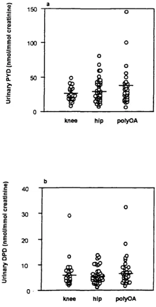
Urinary concentrations of PYD and DPD in OA patients were slightly but not significantly lower than those in the control group (30.2 ±1.7 and 6.2 ± 0.4 nmol/mmol, respectively). The PYD/DPD ratio was unmodified in OA (5.5 ± 0.2 nmol/mmol). PYD was sometimes increased in polyOA, but no significant difference was found between patients with hip OA, knee OA and polyOA (Fig. 3). Urinary concentrations of hydroxypyridinium cross-links ac-cording to radiographic scores were studied in patients with hip or knee OA. They clearly increased with radiographic scores, but the difference between the four groups was not significant. When pooling patients with early OA (X-ray score 1 and 2) and patients with late
OA (X-ray score 3 and 4), PYD and DPD were, respectively, 1.47- and 1.4-fold higher in late OA than in early OA (P = 0.003 and 0.04, respectively) (Fig. 4). The PYD/DPD ratios were 4.4 ± 1.3 and 6.1 ±2.7, respectively. The urinary excretion of PYD and DPD was unrelated to NSAID uptake.
PYD and DPD were increased by 42.5 and 29.7%, respectively, in RA patients, when compared with normal controls. The difference was significant
S 150 £ u "5 E o E c a a. c 3 100 50 -OA RA Controls o c
e
o "5 E o C 40 30 20 10 -OA RA ControlsFIG. I.—Urinary concentrations of pyridinoline (PYD) (a) and deoxypyridinoline (DPD) (b) in patients with osteoarthritis (OA) and rheumatoid arthritis (RA) and controls. All concentrations are expressed relative to urinary creatinine.
HELLIO LE GRAVERAND ET AL.: PYRIDINIUM CROSS-LINKS AND OA 1093 TABLE II
Pyridinium cross-link values (median and S.D.) in patients with osteoarthritis (OA), rheumatoid arthritis (RA) and controls relative to sex and menopausal status. Values are expressed relative to urinary creatinine
Controls OA RA PYD DPD (nmol/mmol) (nmol/mmol) PYD DPD N (nmol/mmol) (nmol/mmol) PYD DPD N (nmol/mmol) (nmol/mmol) Males Non-post-menopausal females Post-menopausal females Total 21 10 9 40 33.77 ±3.9 6.71 ± 1.9 49 27.45 ± 2.2 4.9 ± 0.4 8 47.61 ± 3.9 8.58 ± 1.6 32.86 ± 6.6 37.78 ± 8.6 34.5 ± 4.9 7.89 ± 2.4 6.34 ± 1.9 6.92 ± 1.1 15 50 114 22.9 ±2.1 33.17 ±2.8 30.17 ± 1.7 4.9 ± 0.7 7.42 ± 0.7 6.21 ± 0.4 4 7 19 52.3 ± 10.9 53.2 ± 10.5 50.66 ± 5.9 7.72 ± 1.49 10.4 ± 2.2 8.98 ± 1.04
{P < 0.02) for PYD only. The difference with the OA group was significant for both parameters (P = 0.018 and P = 0.02, respectively). The PYD/DPD ratio was 6 ± 0.5. PYD and DPD were significantly (P < 0.05) higher, by 104.1 and 69.7%, respectively, in RA patients treated with steroids than in RA patients without steroids (Table III). The latter were not significantly different from the normal controls. The PYD/DPD ratio did not vary with the steroid intake. There was no correlation between cross-links and ESR.
DISCUSSION
The present work does not confirm the previously reported increased urinary concentrations of PYD and DPD m OA [13, 15].
The difference between our results and those of previous studies does not seem to be in relation to the controls. Our number of normal controls is rather higher than that of previous studies, in some of which the same control group was used repeatedly [11,12,15]. Means and standard deviations of the present normal control group were also close to those of previous studies in which the same HPLC method was used [10, 19]. There were more women and old patients in our OA group than in the control group. The urinary excretion of pyridinium cross-links is similar in men and pre-menopausal women. It was also found to be unrelated to age. An increased excretion of PYD and DPD in peri- and post-menopausal women was demonstrated [20, 21]. However, increasing the number of menopaused women in our control group would have increased the normal values accordingly.
The selection of OA patients could be an explanation
FIG. 2.—Correlation between urinary pyridinoline (PYD) and urinary deoxypyridinoline (DPD) values in controls. Spearman test:
P< 0.001 and r = 0.76.
for our results. MacDonald et al. [15] found a higher urinary excretion of PYD in gonOA than in coxOA and polyOA. Astbury et al. [14] demonstrated that the urinary excretion of pyridinium cross-links could be related to joint damage. The present work does not support the findings of MacDonald et al. and rather
I
o o E E o n c 3 150 100 " 50 "knee hip potyOA
£ 40 re £ o •5 o 30 20 10
Knee hip potyOA
FIG. 3.—Urinary pyridinoline (PYD) (a) and deoxypyridinoline (DPD) (b) values in osteoarthritis (OA) subgroups. All concen-trations are expressed relative to urinary creatinine.
1094 BRITISH JOURNAL OF RHEUMATOLOGY VOL. 35 NO. 11
s
o PYD • early OA • late OA CO £ uI
I
c DPDFIG. 4.—Urinary pyridinoline (PYD) and deoxypyridinoline (DPD) values relatively to radiographic scores. Patients were divided into early OA (X-ray scores 1 or 2) and late OA (X-ray scores 3 or 4). PYD and DPD increased significantly in late OA: P •= 0.003 and
P •» 0.04, respectively.
suggests an increase in some cases of polyOA. It demonstrates that PYD and DPD excretion increased significantly with the radiological stage of OA. Although there was no significant difference between the late OA subgroup and the normal group, the selection of a larger number of patients with late OA might have made the difference between OA and the control significant.
The tissular topography of hydroxypyridinium
Pyridinium cross-link RA + steroids RA — steroids Total N 9 10 19 TABLE III values (median and s.D.)
intake PYD 78.7 ± 7.2 38.5 ± 2.3 50.6 ± 5.9 relative to steroid DPD 12.9 ± 7.6 ± 8.8 ± 1.8 0.4 1.04 DPD/PYD 7.1 ±0.9 5.3 ± 0.3 6.1 ±0.4
cross-links has been studied in detail [1,2]. DPD could be considered as a sensitive marker of bone degradation since it is mostly found in bone and dentine, and the turnover of the latter tissue is negligible [1]. PYD is a major collagen cross-link in bone, cartilage and other connective tissues such as muscle and intervertebral discs [1, 22]. Although bone resorption is probably the major source of urine hydroxypyridinium cross-links [13, 14], the PYD/DPD ratio may help to define collagen degradation in cartilage relative to other tissues. In agreement with previous studies in OA patients [10, 13], PYD and DPD were highly correlated (r = 0.75, P < 0.001), and the PYD/DPD ratio was unchanged. In patients with advanced OA, both PYD and DPD were significantly increased and the PYD/DPD ratio was also un-changed. Thus, most probably, the observed increased excretion of pyridinium cross-links in patients with late OA reflects the bone erosion and/or the increased sclerotic bone remodelling of joint epiphysis that occurs in the advanced stages of OA. Unfortunately, bone sclerosis was not graded in our score of OA [18]. The unchanged excretion of PYD and DPD in earlier stage OA suggests that the markers certainly do not reflect collagen degradation of the articular cartilage.
An increased urinary excretion of pyridinium cross-links has been reported in RA [12, 13, 14, 23, 24]. A small control group of RA patients was included in this study to test the validity of our measurements. We were able to confirm a dramatic increase in the urinary excretion of PYD and DPD in RA patients. Interestingly, this increase was only found in patients treated with steroids. Since joint destruction in RA patients was not investigated, the results suggest that an increase in urinary PYD and DPD was related to severe forms of RA justifying steroids, and/or with the increased bone resorption induced by steroids.
ACKNOWLEDGEMENT
Part of this work was supported by grant no. 32-35582.92 from the Swiss National Foundation for Scientific Research to DU.
REFERENCES
1. Eyre DR, Koob JJ, Van Ness KB. Quantitation of hydroxypyridinium crosslinks by HPLC. Anal Biochem 1984;137:380-8.
2. Eyre DR, Paz MA, Galop PM. Crosslinking in collagen and elasu'n. Annu Rev Biochem 1984;53:717-48. 3. Robins SP, Duncan A. Pyridinium crosslinks of bone
collagen and their location in pcptides isolated from rat femur. Biochim Biophys Ada 1987^14:233-9.
4. Robins SP. An enzyme-linked immunoassay for the collagen crosslink pyridinoline. Biochem J 1982;207: 617-20.
5. Black D, Duncan A, Robins SP. Quantitative analysis of the pyridinium crosslinks of collagen in urine using ion-paired reversed-phase high-performance liquid chro-matography. Anal Biochem 1988;169:197-203.
6. Uebelhart D, Gineyts EC, Chapuy MC, Delmas PD. Urinary excretion of pyridinium crosslinks: A new
HELLIO LE GRAVERAND ET AL.\ PYRIDIN1UM CROSS-LINKS AND OA 1095
marker of bone rcsorption in metabolic bone disease.
Bone Miner 1990;8:87-96.
7. Seibel MJ, Gartenberg F, Silverberg SJ, Ratcliffe A, Robins SP, Bilizikian JP. Urinary hydroxypyridinium crosslinks of collagen as indices of bone resorption in primary hyperparathyroidism. J Clin Endocrinol Melab
1992;74:481-6.
8. Paterson CR, Robins SP, Horobin JM, Preece PE, Cushieri A. Pyridinium crosslinks as markers of bone resorption in patients with breast cancer. Br J Cancer 1992;64:884-6.
9. Seibel MJ, Cosman F, Sheu V et al. Urinary hydroxypyridinium crosslinks of collagen as markers of bone resorption and estrogen efficacy in postmenopausal osteoporosis. J Bone Miner Res 1993;8:881-8.
10. Thompson PW, Spector TD, James IT, Henderson E, Hart DJ. Urinary collagen crosslinks reflect the radiographic severity of knee osteoarthritis. Br J
Rheumatol 1992;31:759-61.
11. Robins SP, Stewart P, Astbury C, Bird HA. Measure-ment of the crosslinking compound, pyridinoline, in urine as an index of collagen degradation in joint disease.
Ann Rheum Dis 1986;45:969-73.
12. Black D, Marabani M, Sturrock RD, Robins SP. Urinary excretion of the hydroxypyridinium crosslinks of collagen in patients with rheumatoid arthritis. Ann
Rheum Dis 1989;48:641-4.
13. Seibel MJ, Duncan A, Robins SP. Urinary hydroxy-pyridinium crosslinks provide indices of cartilage and bone involvement in arthritis diseases. J Rheumatol 1989;16:964-70.
14. Astbury C, Bird HA, MacLaren AM, Robins SP. Urinary excretion of pyridinium crosslinks of collagen correlated with joint damage in arthritis. Br J Rheumatol
1994;33:ll-5.
15. MacDonald AG, MacHenry P, Robins SP, Reids DM. Relationship of urinary pyridinium crosslinks to disease
extent and activity in osteoarthritis. Br J Rheumatol 1994;33:16-9.
16. Conrozier T, Tron AM, Mathieu P, Vignon E. Quantitative assessment of radiographic normal and osteoarthritic hip joint space. Osteoarthritis Cartilage 1995;3(suppl. A):81-7.
17. Altman RD. Criteria for classification of clinical osteoarthritis. J Rheumatol 1991;18(suppl. 27):10-2. 18. Piperno M, Mathieu P, Conrozier T, Vignon E, Shan Sei
Fan C. Un nouveau score radiologique pour le suivi de la gonarthrose. Vllle congres francais de rhumatologie.
Rev Rhum 1995;10:719 (Abstract A106).
19. James IT, Perrett D, Thompson PW. Rapid assay for hard tissue collagen crosslinks using isocratic ion-pair reversed phase liquid chromatography. J Chromatogr 1990;525:43-57.
20. Kollerup G, Thamsborg G, Bhatia H, Sorensen OH. Quantitation of urinary hydroxypyridinium crosslinks from collagen by high-performance liquid chromato-graphy. Scand J Clin Lab Invest 1992;52:657-62. 21. Uebelhart D, Schlemmer A, Johansen JS, Gineyts E,
Christiansen C, Dehnas PD. Effect of menopause and hormone replacement therapy on the urinary excretion of pyridinium crosslinks. / Clin Endocrinol Me tab 1991; 72:367-73.
22. Robins SP. Crosslinking of collagen: isolation, structural characterization and glycosylation of pyridinoline.
Biochem J 1983;215:167-73.
23. Black D, Robins SP, Duncan A, Marabani M. Analysis by high performance liquid chromatography of two hydroxypyridinium crosslinks of collagen in urine as indices of disease activity in rheumatoid arthritis.
Biochem Soc Trans 1988;16:817-8.
24. Gough A, Peel N, Eastell R, Holder R, Lilley J, Emery P. Excretion of pyridinium crosslinks correlates with disease activity and appendicular bone loss in early rheumatoid arthritis. Ann Rheum Dis 1994;53:14-7.