Design, Synthesis, and Biological Evaluation of Diketopiperazine Based
Ionizable Lipids for the In Vivo Delivery of Messenger RNA
by
Owen Shea Fenton
B.A. Chemistry
College of the Holy Cross, 2010 M.Sc. Organic Chemistry
Massachusetts Institute of Technology, 2012
MA3SACHUSETTS INSTITUTE OF TECHNOLOGY
JUN 2 3 2016
LIBRARIES
ARCHIVES
SUBMITTED TO THE DEPARTMENT OF CHEMISTRY IN PARTIALFULFILLMENT OF THE REQUIREMENTS FOR THE DEGREE OF DOCTOR OF PHILOSOPHY
AT THE
MASSACHUSETTS INSTITUTE OF TECHNOLOGY JUNE 2016
2016 Massachusetts Institute of Technology. All Rights reserved.
Signature of Author:
Signature redacted
I
Department of Chemistry
A1 il 7 2)A16A
(Signature redacted
Daniel G.
AdiTersoir-Associate Professor of Chemical Engineering Thesis Supervisor
Accepted by:_Signature
redacted
Robert W. Field
Haslam and Dewey Professor of Chemistry Chairman, Departmental Committee on Graduate Students
This Doctoral Thesis has been examined by a committee of the Department of Chemistry as follows:
Signature redacted
Alexander M. KlibanovFirmenich Professor of Chemistry and Bioengineering Thesis Committee Chair
Signature redacted
_Daniel G. Anderson Associate Professor of Chemical Engineering
Thesis Supervisor
Signature redacted
John M. Essigmann
William R. and Betsy P. Leitch Professor of Chemistry and Biological Engineering Thesis Committee Member
Design, Synthesis, and Biological Evaluation of Diketopiperazine Based
Ionizable Lipids for the In Vivo Delivery of Messenger RNA
By
Owen Shea Fenton
Submitted to the Department of Chemistry on April 20, 2016, in Partial Fulfillment of the Requirements for the Degree of Doctor of Philosophy in Chemistry
Abstract
Thousands of human diseases could be treated by selectively increasing the intracellular concentration of specific proteins. The successful delivery of messenger RNA (mRNA) to target cells in the body could accomplish this goal, but serious limitations with its systemic delivery must still be overcome. Recently, lipid
nanoparticles (LNPs) have shown promise for mRNA delivery in vivo, however, current leads are limited in terms of their efficacy, biodistribution, and toxicity. Here, we
synthesize novel LNP delivery materials (i.e. ionizable lipids) that, when formulated into LNPs, both outperform current leads for mRNA delivery and elucidate key relationships between chemical structure and biological function.
Drawing inspiration from naturally occurring components of cellular membranes, we have designed and synthesized a new series of alkenyl amino alcohol (AAA)
ionizable lipids for mRNA delivery to the liver. When formulated into LNPs, these AAA ionizable lipids induce high concentrations of human erythropoietin (EPO) protein at therapeutically relevant doses of mRNA. Notably, LNPs derived from our lead compound OF-02 are the most potent mRNA delivery vehicle yet reported in the scientific literature.
While the liver is implicated in many diseases, targeting other tissues could drastically improve the clinical generality of mRNA based therapeutics. Towards this end, we have designed and synthesized ionizable lipid OF-77. Unlike other materials that afford more than 99% of total protein production in the liver, OF-77 mRNA LNPs promote more than 85% of total protein production in the spleen. Notably, OF-77 mRNA LNPs also demonstrate the first example of functional protein production within B lymphocytes, with current levels of protein production only limited by the administered dose.
Finally, we synthesize a novel series of ionizable lipids by varying three key
structural parameters within OF-77; tail length, linker spacing, and total degrees of unsaturation. Both in vitro and in vivo experiments are explored to glean crucial
information relating LNP structure to biological response. We also demonstrate that these compounds, including OF-77, are capable of both complexing and delivering siRNA to reduce specific intracellular protein concentrations.
Thesis Supervisor: Daniel G. Anderson
Acknowledgements
The completion of this Ph.D. would not have been possible without the tremendous support and guidance from countless individuals I have been fortunate enough to know in my life. While properly thanking everyone would require several more chapters, the hope of this section is to at least begin to express my gratitude to all of you who have made my growth as a scientist and individual possible over the last few years.
To begin, I would like to thank my advisor Professor Daniel G. Anderson. Thank you for providing me a perfect scientific environment to grow both creatively and as an experimentalist. You have truly helped to shape my approach to science, and have fostered such an incredible sense of collaboration, dedication, and passion here in the Koch Institute. Thank you for providing me unwavering support and help over the past few years, and for providing me the opportunity to work with so many incredible collaborators from nearly every scientific discipline. I cannot say enough good things about my time spent in our lab, and for that I will be forever grateful. In this same vein, I would also like to thank my committee members Professor Alexander Klibanov and Professor John Essigmann for their meticulous attention to detail and helpful scientific
feedback. I would also like to thank Professor Mohammad Movassaghi for affording me the opportunity to synthesize complex alkaloid natural products during my early years at MIT - I have learned so much from you as a synthetic chemist, and for that I am eternally grateful.
I would also like to thank the many amazing collaborators and friends that I have met in our lab. Not only have you made my time here in the Anderson lab a truly
exceptional educational experience, you have also helped to make it fun. Thank you to all of the following, and also to the many more that I have had the opportunity to interact with at some point over the last few years: Kevin Kauffman, Rebecca McClellan, Mark Tibbitt, Andrew Ayoob, Danny Zeng, Eric Appel, Jimmy Kaczmarek, Heloise Ragelle, Faryal Mir, Jung Yang, Matt Webber, Omar Khan, Roman Bogorad, Hao Yin, Arturo
Vegas, Katie Whitehead, Abigail Lyton-Jean, Rose Kanasty, Sid Jhunjhunwala, Ben Tang, Angie DiCiccio, Karsten Olejnik, Luke Ceo, Robert Dorkin, Volkan Yesilyurt,
Suman Bose, Tara Fawaz, Asha Patel, Kevin Daniel, Giovanni Traverso, Abel Cortinas, Matthias Oberli, Keith Hearon, Lisa Freed, Omar Khan, Alexis Coste, and Nicolas Boyer.
I would also like to thank my undergraduate advisor Professor Bianca R.
Sculimbrene. Without your guidance I would never be where I am today - you provided such an incredible laboratory experience for me during my time at Holy Cross. I would also like to thank Professors Sarah Petty and Kimberley A. Frederick who also shaped me tremendously during my time in Haberlin Hall.
Finally, I would like to thank my family, to whom this thesis is dedicated. Mom, Dad, PJ, Stef, and Emerie - thank you for all of your support and guidance for my entire life - I am so fortunate to have you as family. An additional thank you to my extended family, many of whom I had the opportunity to grow up with and learn from in my youth.
Table of Contents A bstract... 3 Acknowledgements...4 Table of Contents...5 L ist of F igures...6 L ist of Tables... 7 L ist of A bbreviations...8
Chapter 1. Background, significance, and thesis overview A . Introduction... 10
B. Barriers to the Systemic Delivery of mRNA...12
C . R eferences...17
Chapter 2. Bioinspired Alkenyl Amino Alcohol Ionizable Lipid Materials for Highly Potent In Vivo mRNA Delivery A . D iscussion ... 22
B . E xperim ental...31
C . R eferences...48
Chapter 3. Fatty Acid Derived Lipid Nanoparticles Deliver mRNA to B Lymphocytes In Vivo A . D iscussion ... 52
B . Experim ental... 62
C . R eferences...7 1 Chapter 4. Non-Toxic Ionizable Lipid Materials for mRNA Delivery In Vitro and In Vivo A . D iscussion... 75
B . E xperim ental...89
C . R eferences...99
List of Figures Figure 2-1 Figure 2-2a Figure 2-2b Figure 2-3a Figure 2-3b Figure 2-3c Figure 2-3d Figure 2-4a Figure 2-4b Figure 2-4c Figure 2-5 Figure 3-ia Figure 3-lb Figure 3-ic Figure 3-id Figure 3-le Figure 3-2a Figure 3-2b Figure 3-2c Figure 3-2d Figure 3-3a Figure 3-3b Figure 3-4a Figure 3-4b Figure 3-5 Figure 3-6 Figure 3-7 Figure 3-8 Figure 4-1 Figure 4-2 Figure 4-3 Figure 4-4a Figure 4-4b Figure 4-5a Figure 4-5b Figure 4-5c Figure 4-6a Figure 4-6b Figure 4-7 Figure 4-8a Figure 4-8b Figure 4-8c
Cartoon of alkenyl amino alcohol ionizable lipids Synthesis of OF-00 through OF-03
Cryogenic transmission electron microscopy of OF-02 LNPs In vivo mRNA screen of OF-00 through OF-03 mRNA LNPs Batch-to-batch consistency of OF-02 mRNA LNPs
In vivo dose response of OF-02 mRNA LNPs Time course of OF-02 mRNA LNPs
Luciferase biodistribution of cKK-E12 mRNA LNPs Luciferase biodistribution of OF-02 mRNA LNPs
Percent biodistribution of cKK-E12 and OF-02 mRNA LNPs Percent weight loss with cKK-E12 and OF-02 mRNA LNPs Cartoon of OF-77 mRNA LNPs targeting the spleen
Cartoon of OF-77 mRNA LNPs accessing B lymphocytes Chemical structure of OF-77
Cryogenic transmission electron microscopy of OF-77 LNPs Dynamic light scattering of OF-77 mRNA LNPs
In vitro dose response of OF-77 mRNA LNPs Luciferase biodistribution of OF-77 mRNA LNPs Percent biodistribution of OF-77 mRNA LNPs TNS assay of OF-77 mRNA LNPs
FACS plots for cell populations labeled with OF-77 LNPs Labeling for immune cell populations with OF-77 LNPs In vivo dose response of functional protein in B lymphocytes In vivo dose response of functional protein in splenic cells
Synthesis of OF-TBS and OF-77 'H NMR of OF-TBS
'H NMR of OF-77
Luciferase calibration curve for OF-77 mRNA LNPs Synthesis of OF-71 through OF-77
In vitro screen of OF-71 through OF-77 siRNA LNPs In vitro screen of OF-71 through OF-77 mRNA LNPs In vitro dose response for OF-77 siRNA LNPs
In vitro dose response for OF-77 mRNA LNPs
In vivo ALP, ALT, and AST levels for OF-77 siRNA LNPs Percent weight gain for OF-77 siRNA LNPs
Cytokine response for OF-77 siRNA LNPs CD4 T cell activation with OF-77 mRNA LNPs CD8 T cell activation with OF-77 mRNA LNPs Structure of OF-C4-TBS and synthesis of OF-C4-77 TNS Assay for OF-C4-77 mRNA LNPs
Luciferase Biodistribution for OF-C4-77 mRNA LNPs Percent biodistribution of OF-C4-77 mRNA LNPs
List of Tables
Table 2-1 LNP characterization data for OF-00 to OF-03 LNPs 46
Table 4-1 LNP characterization data for OF-71 to OF-77 siRNA LNPs 79 Table 4-2 LNP characterization data for OF-71 to OF-77 mRNA LNPs 79
Abbreviations
Standard 1-letter codes are used for amino acids
AAA - Alkenyl Amino AlcoholALP - Alkaline Phosphatase ALT - Alanine Transaminase ApoE - Apolipoprotein E AST - Aspartate Transaminase
Cy5.5 - Cyanine 5.5
DOPE
-
1,2-dioleoyl-sn-glycero-3-phosphoethanolamine
DSPE
-
1 ,2-distearoyl-sn-glycero-3-phosphoethanolamine
DSPC
-
1,2-distearoyl-sn-glycero-3-phosphocholine
EC
50-
half maximal effective concentration
EPO - Erythropoietin
FVII
-
Factor VII serum clotting protein
LNP - Lipid nanoparticle Luc - Luciferase
mRNA - Messenger RNA siRNA - Short interfering RNA TBAF - tetrabutyl ammonium fluoride
Chapter 1:
A. Introduction
Nearly all examples of disease, including diabetes, cancers, and even the common
cold, involve the abnormal expression of specific proteins. Whether it is the undesirable
upregulation of an oncogene, the absence of a tumor suppressor, or the presence of an
exogenous viral protein, protein expression often determines the phenotypes observed in
individuals suffering from disease. Having the ability to regulate individual protein
concentrations, therefore, could allow scientists and physicians to study, prevent, and
even treat disease.J' Towards this end, great interest resides in the development of
therapeutics that can eliminate, modify, or increase the amount of specific proteins within
the body.
Historically, numerous therapeutic classes have been examined and studied for
their ability to regulate protein expression in humans. Even before the mechanism of
action was understood, for example, natural remedies derived from plants and animals
were administered that could inhibit, degrade, modify, activate, or even upregulate
specific proteins associated with disease.
23Tremendous advances in molecular isolation,
purification, and characterization have since identified thousands of small molecules that
are capable of selectively interacting with proteins in the body. While many of these
small molecules are effective and serve as the foundation of modem medicine, many
more bind to off-target proteins or to proteins in untargeted cells, resulting in a wide array
of potential side effects.
33
Furthermore, not all proteins are easily targeted, and small
molecules cannot be used to introduce proteins that are necessary but absent in a given
individual.
As an alternative to small molecule drugs, the direct delivery of therapeutic proteins has been explored to increase intracellular protein concentrations. For example, patients suffering from diabetes and hemophilia are regularly injected with insulin or Factor IX to help manage disease. 41 Generally speaking, however, protein based
therapeutics are rather limited. For example, it is extremely difficult to obtain the correct cellular distribution for nuclear, cytoplasmic, or transmembrane proteins in vivo.
Moreover, high and/or continual dosing regimens are hallmarks of protein replacement therapies; this is because many proteins have narrow half-lives and/or may not be catalytic, thereby limiting the generality of their clinical application.
To address these limitations, gene therapy was proposed as yet another means to regulate the intracellular concentration of proteins. Inspired by the central dogma of biochemistry (namely that DNA is transcribed into messenger RNA (mRNA) which is, in turn, translated into proteins), scientists demonstrated that by delivering DNA into the nucleus, a cell could produce a specific protein for its entire lifetime. Moreover, this protein could be produced with the appropriate post-translational modifications and in the
correct area within the cell. In this sense, DNA could be thought of as a drug - its successful delivery could be used to produce specific protein with high levels of selectivity.51 Translating DNA delivery in vitro to in vivo, however, has proven
extremely difficult, which has ultimately limited its successful transition into the clinic.E6] Obstacles include, but are not limited to, tissue biodistribution, cellular uptake,
Given the limited clinical translation of DNA, mRNA has recently gained much
interest as a therapeutic cargo.E
83Much like DNA, mRNA encodes for a specific protein,
allowing for selective production of a therapeutic protein with the appropriate cell
localization and post-translational modifications. Unlike DNA, however, mRNA need
only access the cytoplasm of a cell to achieve therapeutic effect. Additionally, mRNA
carries no risk of genomic integration, one of the many potential safety concerns
surrounding the use of DNA.J
91DNA and mRNA also present varying durations of
therapeutic effect; while mRNA is capable of producing a significant amount of protein,
it is also degraded more quickly than DNA. This difference can be regarded as either an
advantage or a disadvantage, as some disease treatments require constitutive protein
expression whereas others are better managed in a transient fashion. Given these
properties, there is great interest in developing mRNA based drugs, as having
dose-dependent control over protein expression could have profound impact in fields such as
protein replacement therapy, vaccine development, and immune tolerization wherein the
selective expression of proteins could treat disease.
101 However, several questions and
obstacles must still be overcome before mRNA can be successfully implemented in
clinical applications. The goal of this thesis, therefore, is to contribute to the
understanding of mRNA delivery in vivo, with the hope that these findings may someday
contribute to the eventual study, prevention, and even treatment of human disease.
B. Barriers to the Systemic Delivery of Messenger RNA
Over the past few decades, countless examples detailing the successful use of
mRNA to produce protein in vitro have been described. Interestingly, however, when
these mRNAs are administered in vivo, they either do not result in functional protein
production or are so minimally potent that their use is not considered clinically viable. This discrepancy between in vitro and in vivo activity of mRNAs suggests that the problem is not the efficacy of the mRNA itself, but rather the difficulty of delivering mRNA to the cytoplasm of diseased cells in vivo. Here, we aim to delineate some of the challenges associated with mRNA delivery to better frame why it is so challenging to create mRNA-based therapeutics.
For the purposes of this discussion, our conversation will focus around the systemic delivery of mRNA (i.e. delivery that passes through the circulatory system).
Systemic delivery is a desirable route of administration for drugs because nearly every organ/tissue of interest comes into contact with the blood. If implemented successfully, therefore, systemic mRNA delivery could theoretically treat disease found in nearly every
cell population within the body. However, systemic delivery of any drug is difficult, and mRNA based therapeutics are no exception to this rule.
Upon injection into the bloodstream, mRNA must first avoid degradation by a plethora of exo- and endonucleases.11 1] Although the plasma stability of siRNAs has been improved through the fluorination or methylation of the 2'-hydroxy group along the ribose backbone,121 this strategy is not practical for mRNA stabilization; whereas
siRNAs are approximately 19-25 base pairs in length, 1 3J mRNAs are considerably longer
and can include upwards of thousands of nucleobases. Alternative strategies have attempted to disguise mRNA from the immune system by substituting alternative nucleotides (i.e. swapping uridine with pseudouridine). However, this modification can affect the translational efficiency of mRNA into the desired protein.[14]
If by some mechanism the mRNA is not degraded during circulation, the next obstacle facing its in vivo delivery is biodistribution. Exogenously delivered mRNAs are
most commonly cleared by the liver and/or kidneys.151 While these two tissues are implicated in several forms of disease, having the ability to target other organs would dramatically increase the number of clinical applications available for mRNA. However, as all mRNAs consist of the same chemical building blocks, the resultant biodistribution profile remains limited at best.
The final obstacles involved with systemic mRNA delivery are cellular uptake and endosomal escape.1161 The high anionic charge density, size, and hydrophilicity of nucleic acids prevent meaningful levels of passive diffusion of mRNA across cell membranes.171 Although cells can internalize material from their surrounding environment through a variety of different pathways (including macropinocytosis, caveolae mediated endocytosis, and clathrin mediated endocytosis), mRNA is not taken up by any appreciable extent. Even if mRNA is endocytosed, however, it still must somehow exit the endosome, enter the cytoplasm, and finally bind to a ribosomal complex before protein production can begin. This final process is still not well understood, even in the case of siRNA which has been studied for more than two decades.
To overcome the limitations with systemic mRNA delivery, several strategies have been either proposed or explored to improve mRNA delivery in vivo. For example, the direct chemical modification of nucleic acids with targeting ligands, polymeric
chains, or non-natural base pairs has demonstrated remarkable success in the field of siRNA delivery.[ 13bl However, mRNA is difficult to selectively modify because it can
consist of hundreds of thousands of nucleobases. Moreover, both modified and unmodified nucleic acids lack a mechanism to escape the endosome - some research even suggests that between 95 to 98% of total siRNA entering an endosome is degraded by nucleases or expunged through exocytosis, even when a biological response is observed.1 8 1 Instead, efforts have focused on the development of both biological and
synthetic drug delivery vehicles that are capable of complexing and protecting nucleic acids. Viruses, for example, have been examined as potential delivery vehicles due to their natural capacity to deliver RNAs to specific cell populations in vivo. [19] However,
safety concerns regarding their potential immunogenicity and practical considerations regarding their limited scalability have limited their promise for clinical application.[20]
Several classes of polymers that exploit electrostatic interactions between cationic amines and the phosphate rich RNA backbone have also been explored for nucleic acid delivery. But these materials, which include protamine, 2 1] poly-lysine, 2 21 and poly(beta-amino esters),2 face potential issues with efficacy, biodistribution, and toxicity.
Alternatively, lipid nanoparticles (LNPs) have been considered as potential candidates for systemic mRNA delivery. Previously, LNPs have demonstrated
tremendous potential for the in vivo delivery of siRNAs, achieving EC5o values as low as 0.02 mg/kg. 243 However, given the differences between the size (i.e. number of
nucleobases), hybridization (i.e. double vs. single strand), and charge density between siRNA and mRNA, it was not readily obvious whether or not LNPs would be able to complex and deliver mRNA in vivo. Recent work from other groups and from our own lab has established LNPs as viable mRNA delivery vehicles; much like siRNA LNPs,
these particles are roughly 50-200 nm in size, are uni- or multilamellar in nature, and are
capable of producing biological response in the liver.
8'
15]While this work validates LNPs for systemic mRNA delivery, relatively little is
known about the structure-function relationships relating mRNA delivery properties (i.e.
efficacy, biodistribution, and toxicity) to specific chemical functionality found within the
LNP. In practice, LNPs are composed of cholesterol (aids in stability),[
251 a phospholipid
(modifies bilayer structure),[
26a polyethylene glycol (PEG) derivative (decreases
aggregation and nonspecific uptake),[
271and an ionizable lipid (complexes negatively
charged RNA and enhances endosomal escape).[16,
24b]Evidence within the siRNA
delivery community has implicated the chemical structure and identity of the ionizable
lipid as the most pivotal component for efficacy, but this finding has not yet been
validated for mRNA delivery. This lack of knowledge significantly reduces the pace at
which new and potentially more efficacious compounds for mRNA delivery can be
discovered.
Towards this end, the overarching theme of my work has focused on the design,
synthesis, and characterization of novel ionizable lipid materials for use in the systemic
delivery of mRNA LNPs. Notably, our efforts have focused on the incorporation of
previously unexplored functional group combinations within ionizable lipids, ultimately
providing modular chemical intermediates, precise molecular characterization, and
scalable synthetic protocols along the way. Through our work, which sits at the interface
of synthetic chemistry and biological engineering, we humbly propose the following:
subtle chemical modifications profoundly impact biological activity.
This affirmation is observed in each of the following sections:
In Chapter 2, we pioneer the synthesis of a new series of alkenyl amino alcohol (AAA)
based ionizable lipids. When formulated into LNPs with mRNA coding for human
erythropoietin (EPO), our materials result in high concentrations of expressed protein in
the liver. Notably, LNPs derived from our lead compound OF-02 are the most potent
mRNA delivery vehicle yet reported in the scientific literature.
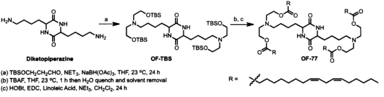
In Chapter 3, we design and synthesize ionizable lipid OF-77. Interestingly, both
OF-02 and OF-77 share the same bis-lysine-diketopiperazine core; however, OF-02 is
AAA based whereas OF-77 incorporates ester linkages. Unlike other materials
(including OF-02) that target the liver, OF-77 LNPs produce more than
85%
of total
functional protein in the spleen. Notably, OF-77 mRNA LNPs also demonstrate the first
example of functional protein production within B lymphocytes.
Finally, in Chapter 4 we modulate the tail length, linker spacing, and total
degrees of unsaturation in OF-77 to afford a new series of materials. In vitro and in vivo
experiments detail interesting trends correlating structural properties to mRNA delivery
potency. We also demonstrate that these materials are also capable of delivering siRNA,
a structurally-distinct nucleic acid cargo that can silence protein production within cells.
C. References
[1]
R. W. Herzog, 0. Cao, A. Srivastava, Discov Med 2010, 45, 105-111.
[2]
F. E. Koehn, G. T. Carter, Nat Rev Drug Discov 2005, 4, 206-220.
[3]
A. Bender, J. Scheiber, M. Glick, J. W. Davies, K. Azzaoui, J. Hamon, L. Urban,
S. Whitebread, J. L. Jenkins, Chemmedchem 2007, 2, 861-873.
[5] V. Escriou, C. Ciolina, A. Helbling-Leclerc, P. Wils, D. Scherman, Cell Biol
Toxicol 1998, 14, 95-104.
[6] S. S. Jiao, P. Williams, R. K. Berg, B. A. Hodgeman, L. J. Liu, G. Repetto, J. A. Wolff, Hum Gene Ther 1992, 3, 21-33.
[7] S. N. Tamkovich, A. V. Cherepanova, E. V. Kolesnikova, E. Y. Rykova, D. V. Pyshnyi, V. V. Vlassov, P. P. Laktionov, Ann Ny Acad Sci 2006, 1075, 191-196. [8] U. Sahin, K. Kariko, 0. Tureci, Nat Rev Drug Discov 2014, 13, 759-780. [9] S. Hacein-Bey-Abina, C. von Kalle, M. Schmidt, F. Le Deist, N. Wulffraat, E.
McIntyre, I. Radford, J. L. Villeval, C. C. Fraser, M. Cavazzana-Calvo, A. Fischer, New Engl J Med 2003, 348, 255-256.
[10] B. Leader,
Q.
J. Baca, D. E. Golan, Nat Rev Drug Discov 2008, 7, 21-39. [11] S. Sorrentino, Cell Mol Life Sci 1998, 54, 785-794.[12] Y. L. Chiu, T. M. Rana, Rna 2003, 9, 1034-1048.
[13] a) R. L. Kanasty, K. A. Whitehead, A. J. Vegas, D. G. Anderson, Mol Ther 2012,
20, 513-524; b) K. A. Whitehead, R. Langer, D. G. Anderson, Nat Rev Drug Discov 2009, 8, 129-138; c) K. A. Whitehead, J. E. Dahlman, R. S. Langer, D. G.
Anderson, Annu Rev Chem Biomol 2011, 2, 77-96.
[14] a) A. Thess, S. Grund, B. L. Mui, M. J. Hope, P. Baumhof, M. Fotin-Mleczek, T. Schlake, Mol Ther 2015,23, 1456-1464; b) K. Kariko, H. Muramatsu, F. A. Welsh, J. Ludwig, H. Kato, S. Akira, D. Weissman, Mol Ther 2008, 16, 1833-1840.
[15] K. J. Kauffman, J. R. Dorkin, J. H. Yang, M. W. Heartlein, F. DeRosa, F. F. Mir, 0. S. Fenton, D. G. Anderson, Nano Lett 2015, 15, 7300-7306.
[16] G. Sahay, W. Querbes, C. Alabi, A. Eltoukhy, S. Sarkar, C. Zurenko, E. Karagiannis, K. Love, D. L. Chen, R. Zoncu, Y. Buganim, A. Schroeder, R. Langer, D. G. Anderson, Nat Biotechnol 2013, 31, 653-U 119.
[17] M. S. D. Kormann, G. Hasenpusch, M. K. Aneja, G. Nica, A. W. Flemmer, S. Herber-Jonat, M. Huppmann, L. E. Mays, M. Illenyi, A. Schams, M. Griese, I. Bittmann, R. Handgretinger, D. Hart, J. Rosenecker, C. Rudolph, Nat Biotechnol 2011, 29, 154-U196.
[18] M. W. Tibbitt, J. E. Dahlman, R. Langer, JAm Chem Soc 2016, 138, 704-717. [19] M. Giacca, S. Zacchigna, J Control Release 2012, 161, 377-388.
[20] I. M. Verma, N. Somia, Nature 1997, 389, 239-242.
[21] B. Scheicher, A. L. Schachner-Nedherer, A. Zimmer, Eur J Pharm Sci 2015, 75,
54-59.
[22] T. Bettinger, R. C. Carlisle, M. L. Read, M. Ogris, L. W. Seymour, Nucleic Acids
Res 2001, 29, 3882-3891.
[23] J. J. Green, R. Langer, D. G. Anderson, Accounts Chem Res 2008, 41, 749-759. [24] a) Y. Z. Dong, K. T. Love, J. R. Dorkin, S. Sirirungruang, Y. L. Zhang, D. L.
Chen, R. L. Bogorad, H. Yin, Y. Chen, A. J. Vegas, C. A. Alabi, G. Sahay, K. T. Olejnik, W. H. Wang, A. Schroeder, A. K. R. Lytton-Jean, D. J. Siegwart, A. Akinc, C. Barnes, S. A. Barros, M. Carioto, K. Fitzgerald, J. Hettinger, V. Kumar, T. I. Novobrantseva, J. N. Qin, W. Querbes, V. Koteliansky, R. Langer, D. G. Anderson, P Natl A cad Sci USA 2014, 111, 5753-5753; b) K. T. Love, K. P. Mahon, C. G. Levins, K. A. Whitehead, W. Querbes, J. R. Dorkin, J. Qin, W. Cantley, L. L. Qin, T. Racie, M. Frank-Kamenetsky, K. N. Yip, R. Alvarez, D. W.
Y. Sah, A. de Fougerolles, K. Fitzgerald, V. Koteliansky, A. Akinc, R. Langer, D. G. Anderson, P Natl Acad Sci USA 2010, 107, 9915-9915; c) K. A. Whitehead, J. R. Dorkin, A. J. Vegas, P. H. Chang, 0. Veiseh, J. Matthews, 0. S. Fenton, Y. L. Zhang, K. T. Olejnik, V. Yesilyurt, D. L. Chen, S. Barros, B. Klebanov, T.
Novobrantseva, R. Langer, D. G. Anderson, Nat Commun 2014, 5.
[25] a) J. J. Lu, R. Langer, J. Z. Chen, Mol Pharmaceut 2009, 6, 763-771; b) T. M. Allen, P. R. Cullis, Adv Drug Deliver Rev 2013, 65, 36-48.
[26] I. S. Zuhom, U. Bakowsky, E. Polushkin, W. H. Visser, M. C. A. Stuart, J. B. F. N. Engberts, D. Hoekstra, Mol Ther 2005, 11, 801-810.
[27] B. L. Mui, Y. K. Tam, M. Jayaraman, S. M. Ansell, X. Y. Du, Y. Y. C. Tam, P. J. C. Lin, S. Chen, J. K. Narayanannair, K. G. Rajeev, M. Manoharan, A. Akinc, M. A. Maier, P. Cullis, T. D. Madden, M. J. Hope, Mol Ther-Nucl Acids 2013, 2.
CHAPTER 2
Bioinspired Alkenyl Amino Alcohol Ionizable Lipid Materials for Highly Potent In Vivo mRNA Delivery
The work presented in this chapter was published in the following manuscript and is reproduced with kind permission from Wiley Periodicals, Inc (Copyright C 2016) Fenton, O.S.; Kauffman, K.J.; McClellan, R.L.; Appel, E.A.; Dorkin, J.R.; Tibbitt, M.W.; Heartlein, M.W.; DeRosa, F.; Langer, R.; Anderson, D.G.* Bioinspired Alkenyl Amino Alcohol Ionizable Lipid Materials for Highly Potent In Vivo mRNA Delivery. Adv. Mat., 2016, in press.
The work presented in this chapter has been filed with the United States Patent and Trademark Office:
Anderson, D.G.; Dorkin, J.R.; Fenton, O.S.; Kauffman, K.J.; McClellan, R.L. "Alkenyl Substituted 2,5-Piperazinediones, Compositions, and Uses Thereof. US Provisional Application Number: 62/182,264. Filing Date: June 19, 2015
A. Discussion
Nucleic acid therapies could be leveraged to treat thousands of genetic disorders, many of which are difficult or impossible to manage with present day
therapeutic approaches. For example, the successful delivery of short interfering RNAs (siRNA) to cells in both rodents and non-human primates has been widely used for the treatment of hereditary diseases and cancer. El By contrast, the delivery of messenger RNA (mRNA) remains largely unexplored. Whereas siRNA sequences are employed to silence gene expression, mRNA therapeutics could be used to treat diseases caused by deficiencies in specific proteins. [2] This is because mRNA
sequences can be translated into proteins once they are successfully transported into the cytoplasm of target cells. The implementation of mRNA therapeutics, therefore, could profoundly impact fields such as protein replacement therapy, vaccine
development, and immune tolerization wherein the selective expression of proteins in vivo could treat disease. 3 1
Before clinical translation can be realized, however, serious limitations with the in vivo delivery of mRNA must still be overcome. The high anionic charge density,
size, and hydrophilicity of nucleic acids prevent meaningful levels of passive diffusion of mRNA across cell membranes. 4 1 To circumvent this barrier, our group and others have developed and implemented an array of lipid nanoparticles (LNPs) for the entrapment and subsequent delivery of nucleic acids in vivo.E'1 Although these LNPs have been largely optimized for siRNA sequences, both Schlake's group 51 and
our research team 61 have recently employed LNPs derived from previously described
components to deliver mRNA in vivo. Successful delivery was confirmed by quantifying serum protein levels, thereby establishing LNPs as viable delivery vehicles for mRNA.
Inspired by these results, we sought to design and synthesize novel LNP components capable of delivering mRNA with unprecedented levels of in vivo efficacy. In practice, LNPs are comprised of cholesterol (aids in stability),71 a phospholipid (modifies bilayer structure),Ia, 8] a polyethylene glycol (PEG) derivative
(decreases aggregation and non-specific uptake),91 and an ionizable lipid (complexes negatively charged RNA and enhances endosomal escape).'O3 Evidence within the siRNA delivery community has implicated the chemical structure and identity of the ionizable lipid as the most pivotal component for efficacy. Accordingly, several rationally designed 111 and combinatorial chemistryIOb, 121 methodologies have been explored to discover novel series of ionizable lipid materials capable of maximizing gene silencing at the lowest possible dose. This strategy both conserves precious therapeutic nucleic acid cargo and also serves to mitigate any possible issues with the toxicity of the LNPs themselves.
Interestingly, however, no reports detailing the creation of a new series of ionizable lipids for the expressed purpose of improving mRNA LNP delivery in vivo have yet been reported. We hypothesized that ionizable lipids based upon alkenyl amino alcohols (AAA), a functional group combination found in sphingosine and other bioactive molecules, could promote high levels of in vivo protein expression when formulated into mRNA LNPs (Figure 2-1).E1 We envisioned that we could
furnish AAA ionizable lipids via a ring opening reaction between alkenyl epoxides (AE) with a polyamine core (Figure 2-2a).E21 It is important to note, however, that no
AEs of suitable tail length are commercially available, nor are they trivial to
synthesize on account of the difficulty of selectively oxidizing singular alkenes in the presence of electronically-similar carbon-carbon double bonds. As such, we report the first detailed procedures for AE synthesis and characterization beginning from
biologically relevant fatty acid starting materials (Experimental Section 4). AE-00 through AE-03 were then each reacted in turn with polyamine 1 to afford AAA
ionizable lipids OF-00 through OF-03. While OF-00 through OF-03 represent the first four members of this series of materials, we hope that the chemical versatility of AE-00 through AE-03 will serve as inspiration for future generations of AAA ionizable lipids for nucleic acid delivery.
Naturally Occurring Lipids
;Flo
OH
Cell
X
,NHAlkenyl Amino Alcohol Lipid Nanoparticle
(AAA) (LNP)
'N&XmRNA (* DOPE 00 Cholesterol
AAA Ionizable lipid PEG-lipid
Figure 2-1. Naturally occurring components of the cell membrane contain alkenyl amino alcohol (AAA) functionality serving as the inspiration for the development of AAA ionizable lipids. These lipids form the basis of lipid nanoparticles exhibiting highly efficient in vivo delivery of mRNA.
a) 0 4 Steps, 3 Pots o 1 R OH 4 R
R OH 1 Chromatography R EtOH, NEt,, hv N NH OH
HO HN
N
FA-XX AE-XX R o OF-XX
HO R
R=
0 -2AcOH AE-00 OF-00 H2N HN NH2 AE-01 OF-01
0 AE-02 OF-02
1 AE-03 OF-03
b) ___________
Figure 2-2. a) Synthesis of OF-00 through OF-03, seminal members of the AAA class of ionizable lipids. b) Representative cryogenic transmission electron
microscopy of
OF-02
LNPs.Lipids OF-00 through OF-03 were then formulated with cholesterol,
1,2-dioleoyl-sn-glycero-3-phosphoethanolamine (DOPE), Cl 4-PEG-2000, and
unmodified mRNA coding for human erythropoietin (EPO) into mRNA LNPs."61 EPO
was selected as a model protein to evaluate the relationship between ionizable lipid identity and mRNA LNP efficacy for two reasons: 1) the associated protein is secreted directly into the bloodstream allowing for robust protein quantification, and 2) EPO has potential therapeutic applications in such areas as anemia. [4,t41 Cryogenic transmission electron microscopy images of OF-02 LNPs detail a spherical
morphology and a multilamellar structure (Figure 2-2b). Additional physical properties include a narrow polydispersity index (0.130) and an average particle
diameter around 100 nm.
The nanoparticle diameters, polydispersity indices, and encapsulation efficiencies for each OF-00 through OF-03 LNP formulation can be found in Table
2-1 (Experimental Section 5). Ionizable lipid cKK-E12 was also formulated alongside these compounds to be used as a positive control in our study. cKK-E12 was chosen because it is capable of silencing Factor VII expression in mice at siRNA doses as low as 0.002 mg/kg, and as such it represents a benchmark ionizable lipid in the field of nucleic acid delivery.E2 1 Each resultant mRNA loaded LNP was then injected intravenously at a 0.75 mg/kg dose in C57BL/6 mice alongside phosphate buffered saline (PBS) as a negative control. At six hours, the serum EPO levels were quantified (Figure 2-3a). The PBS control imparted no significant EPO production in vivo, whereas positive control cKK-E12 LNPs promoted a serum EPO concentration of 7100 700 ng/mL. Excitingly, OF-02 LNPs significantly outperformed benchmark lipid cKK-E12 LNPs, promoting an approximate two-fold increase in EPO
concentration to 14200 1500 ng/mL. Additionally, OF-02 outperformed two other benchmark ionizable lipids from the nucleic acid delivery field, namely 5030131', and C12-200,[10b] whose respective LNP promoted in vivo EPO concentrations were 2800 200 ng/mL and 7100 500 ng/mL at an identical dose. To the best of our knowledge, therefore, OF-02 LNPs represent the most potent mRNA delivery vehicle reported to date in the scientific literature.
a)_ 2000( -Z 150( E ; ;10004 a. un W Z 1- 5004 E (00 C E 50000-3
~
0000 (U 3L 0000 0( 1 00001 b) 20000-E C C Z 15000. o2 0 It E S0 10000- 0.LD5000-0 L)i 0 1 2 3 Batch of OF-02 LNPs d) :3 20000-0 2000 ,I PE 12, 1) 7 r2 * *K-tE 12, 225 r/kjg 1-1F-02, 0 75 mg/Vkg C)OF-02, 2 25 mg/kg 0.0 0.5 1.0 1.5 2.0 2.5 6 hr 24 hr injected Total mRNA Dose (mg/kg) Time after intraveneous injectionFigure 2-3. a) In vivo expression of EPO following administration of AAA LNPs for delivery of mRNA. b) Batch-to-batch variability of OF-02 LNPs for EPO mRNA delivery in vivo. c) In vivo dose response curves for OF-02 and cKK-E12 LNPs. d) EPO expression following administration of OF-02 and cKK-E12 LNPs at 6 and 24 h. Data presented as mean + standard deviation (n = 3).
The OF-00, OF-01, and OF-03 LNPs also allow the deduction of
structure/function relationships within this new series of AAA ionizable lipids. We note two general structure/function trends of interest. First, we note that only alkenes with a cis geometry promote in vivo efficacy - OF-00 and OF-01 exclusively differ in
the cis/trans geometry of their alkenes, and only OF-00 produces meaningful EPO concentrations. Second, the optimal number and placement of two cis alkenes per tail matches those observed in optimized siRNA LNPs.["' 16] While we are still discerning
why these specific trends are observed, these empirical findings could potentially shape subsequent generations of AAA lipids. It is interesting to note, therefore, that only the linoleic acid derivative OF-02 promotes significantly higher levels of EPO expression than the positive control, although oleic acid derivative OF-00 also
40 Ok OR Ok Ok ,- KKE12 P, 99 - CF-02
j
-,F 21-4--
0-demonstrates modest activity promoting a serum EPO concentration of 2100 500
ng/mL using the same dose.
With this information in hand, our attention then shifted from exploring the general properties of the new AAA series of ionizable lipids to further characterizing LNPs made from our lead material OF-02. The clinical translation of nucleic acid delivery vehicles is in part predicated on high reproducibility of the chemical
constituents and formulation of LNPs. To test this, three independent batches of OF-02 were synthesized and then formulated into LNPs. The average serum concentration among all batches was found to be 13700 1700 ng/mL and demonstrated minimal batch-to-batch variability (Figure 2-3b). Next, a dose response curve was collected at
0.75 mg/kg, 1.5 mg/kg, and 2.25 mg/kg total EPO mRNA dose for both OF-02 and
cKK-E12 LNPs (Figure 2-3c). OF-02 LNPs outperformed their cKK-E12 counterparts roughly two-fold across all doses studied, reaching a maximum EPO concentration of 45400 5300 ng/mL at the 2.25 mg/kg dose. It is also interesting to note that both sets of LNPs promote EPO production in a linear fashion with respect to dose. This trend implies that we have not yet reached a saturation point for the intracellular translation machinery, suggesting protein production is currently only limited by the dose of mRNA. Moreover, it is important to note that no animal mortality was observed at all doses studied, and that mice treated with both cKK-E12
and OF-02 LNPs displayed similar weight loss profiles at identical doses (Figure 2-5, Experimental Section 5). OF-02 LNPs therefore represent a tunable handle for in
vivo EPO production readily capable of exceeding normal human EPO levels (40 -250 pg/mL) in our chosen mouse model.E171 Finally, OF-02 LNPs also outperformed their cKK-E12 counterparts at 24 hours, independent of dose (Figure 2-3d). The sharp decrease in EPO concentration as a function of time highlights one of the many
exciting potential therapeutic advantages of mRNA delivery in vivo; in contrast to permanent gene replacement therapies, mRNA delivery offers transient, dose-response dependent protein expression in vivo, a property that could one day prove useful for a variety of genetic disorders.
Finally, we were interested to determine if the efficacy differences observed between ckk-E12 and OF-02 LNPs were due to variations in biodistribution. mRNA
coding for luciferase was independently formulated with both ckk-E12 and OF-02 in the same fashion as for EPO delivery, and mouse organs were harvested 24 hours post injection. The tissues were subsequently imaged ex-vivo to measure the total
luminescence per organ, demonstrating that mRNA from both ckk-E12 and OF-02 LNPs is predominantly translated in the liver with minimal translation in the spleen and negligible translation in other organs (Figure 2-4a,b). Quantification of this data also confirms nearly identical biodistribution profiles for the two formulations, suggesting that the increased efficacy of OF-02 LNPs is not due to a difference in tissue targeting (Figure 2-4c). Since more than 4000 human diseases are caused by liver genetic disorders such as hemophilias A and B, OF-02 LNPs represent a promising delivery vehicle for therapeutic mRNA delivery to the liver. [81
8-F
~
100CD CDs
Figre -4 Rereenttiv lminscncebioisribtin o a cKK-E12 adb F
o 80 d Op d 4
((D
Figreg 2- Representte luinseamces biodistribionizfa) ipids nd s OF
literature, and we hope that their alkene-epoxide precursors AE-00 through AE-03
can
serve as versatile scaffolds for the synthesis of future generations of theseionizable lipids.
OF-02
LNPs yielded a two-fold increase in EPO production in vivoas compared to benchmark LNPs in the literature across a broad
linear
dose-response window. This illustrates thatOF-02
presents a tunable handle overin
v'ivo protein expression, which is important in protein replacement therapies.2 Batch-to-batch variability, dose response curves, and time course studies were coupled withdeliver mRNA to the liver. Future work will study the potential of OF-02 LNPs for therapeutic applications and establish further groundwork necessary for translating this novel mRNA delivery vehicle to the clinic. In total, this study demonstrates efficient mRNA delivery with OF-02 as well as the importance of utilizing synthetic chemistry in tandem with biological inspirations to further improve and understand nucleic acid delivery in vivo.
B. Experimental
1. Instrumentation, Materials, and Animal Protocols
Microwave reactions were performed in a Biotage Initiator. Other reactions were performed in round bottom flasks. Proton nuclear magnetic resonance ('H NMR) spectra were recorded with a Varian inverse probe INOVA-500 spectrometer (with a Magnex Scientific superconducting actively-shielded magnet), are reported in parts per million on the 8 scale, and are referenced from the residual protium in the NMR solvent (CDCl3: 6 7.24; DMSO: 8 2.50). Data are reported as follows: chemical
shift [multiplicity (br = broad, s = singlet, d = doublet, t = triplet, sp = septet, m = multiplet), integration, assignment. All commercial reagents and solvents were used as received.
All animal studies were approved by the M.I.T. Institutional Animal Care and Use Committee and were consistent with local, state and federal regulations as applicable. LNPs were intravenously injected in female C57BL/6 mice (Charles River Labs, 18-22 grams) via the tail vein. After six or 24 hours, blood was collected via the tail vein and serum was isolated by centrifugation in serum separation tubes. Serum EPO levels were quantified with an ELISA assay (Human Erythropoietin Quantikine IVD ELISA Kit, R&D Systems, Minneapolis, MD). 24 hours after injection of Luc-mRNA LNPs, mice were injected intraperitoneally with 130 pL of D-luciferin (30
mg/mL in PBS). After fifteen minutes, mice were sacrificed and the organs were isolated (pancreas, spleen, liver, kidneys, lungs, heart, uterus and ovaries) and imaged with an IVIS imaging system (Perkin Elmer, Waltham, MA). Luminescence was quantified using LivingImage software (Perkin Elmer).
2. General Lipid Nanoparticle Synthesis
The organic phase was prepared by solubilizing with ethanol a mixture of ionizable lipid, 1,2-dioleoyl-sn-glycero-3-phosphoethanolamine (DOPE, Avanti), cholesterol (Sigma), and 1,2-dimyristoyl-sn-glycero-3-phosphoethanolamine-N-[methoxy-(polyethyleneglycol)-2000] (ammonium salt) (C14-PEG 2000, Avanti) at a molar ratio of 35:16:46.5:2.5 and an ionizable lipid:mRNA weight ratio of 10:1. All ethanolic stock solutions were prepared at a concentration of 10 mg/mL. The aqueous phase was prepared in 10 mM citrate buffer (pH 3) with either EPO mRNA (human Erythropoietin mRNA, courtesy of Shire Pharmaceuticals, Cambridge, MA) or Luc mRNA (Firefly luciferase mRNA, Shire). All mRNAs were stored at -80 'C, and were allowed to thaw on ice prior to use. The ethanol and aqueous phases were mixed at a 3:1 ratio in a microfluidic chip device using syringe pumps as previously
described at a final mRNA concentration of 0.1 mg/mL. Resultant LNPs were dialyzed against IX PBS in a 20,000 MWCO cassette at 4*C for 2 hours and were stored at 4*C prior to injection.
3. General Lipid Nanoparticle Characterization
To calculate the mRNA encapsulation efficiency, a modified Quant-iT RiboGreen RNA assay (Invitrogen) was used as previously described. Briefly, RiboGreen fluorescence was compared in the presence and absence of 2% Triton X-100 in TE buffer. The fluorescence was quantified using a Tecan infinite M200 Pro. The diameter and polydispersity (PDI) of the LNPs were measured using dynamic
light scattering (ZetaPALS, Brookhaven Instruments). LNP diameters are reported as the largest intensity mean peak average, which constituted >95% of the nanoparticles present in the sample.
4. Synthetic Procedures for AE-00 through AE-03, all synthetic intermediates, and OF-00 through OF-03
One of the most common and facile synthetic methods to afford epoxides relies on the oxidation of alkenes using meta-chloroperbenzoic acid (mcpba). However, we immediately recognized this as a poor synthetic strategy for
synthesizing alkenyl epoxides AE-00 through AE-03 because the selective oxidation of a terminal alkene in the presence of electronically similar alkenes would be extremely difficult if not impossible. Purification of the reaction medium would also be highly challenging due to the similar polarity of products and the complexity of the mixture ensuing from the reaction. In order to circumvent this problem, we elected to use biologically relevant fatty acids as our general synthetic starting material. We envisioned that fatty acids would serve as excellent synthetic building blocks for our study because they are abundant in large quantities from many commercial vendors and they also offer high levels of regiochemical fidelity in their alkenes. Additionally, fatty acids would allow us to circumvent the forecasted issue with mcpba oxidation; we envisioned that the carboxylic acid termini could be used to directly furnish the epoxide while leaving the alkenes in the substrate fully intact.
Having selected fatty acids as an ideal starting material, we executed our synthesis of alkenyl epoxides. For a general scheme and the fully drawn products, see below. Briefly, fatty acids were subjected to a lithium aluminum hydride reduction
followed by Dess-Martin Periodinane oxidation to afford their corresponding aldehydes. Proline catalyzed alpha-chlorination followed by sodium borohydride
reduction in the same reaction flask afforded the 1,2-chloroalcohols in moderate yields. Finally, gentle heating of these 1,2-chloroalcohols at 35 *C in basic dioxane promoted ring closure to furnish the desired alkenyl epoxides AE-00 through AE-03 in moderate yields in 4 steps with only a single chromatographic purification.
Excitingly, these alkene-containing epoxides represent a virtually unexplored synthetic scaffold for ionizable lipid development. We hope this synthetic route will broadly add to the creation of future generations of ionizable lipids for nucleic acid
therapy. Full synthetic procedures and molecular characterization data for each step of the synthetic procedures for AE-00 through AE-03 are available below, as are the final syntheses of OF-00 through OF-03.
0 1. LAH, THF 0 NCS, L-pro, CH3CN OH NaOH, H20 0
2. DMP, NaHCO3, R H then EtOH, NaBH4 H Dioxane R
CH2CI2 C1
O 0
AE-00 AE-01
o 0
AE-02 AE-03
Versatile Chemical Scaffolds for Future AAA Ionizable Lipid Synthesis
Scheme 2-1: Synthesis of AAA Ionizable Lipid Alkenyl Epoxide Precursors
4.1. AE-00 Synthesis; (Z)-2-(hexadec-7-en-1-y)oxirane 4.1.1 Synthesis of AE-00-aldehyde
0 1. LAH, THF, rt, overnight 0
OH 2. DMP, NaHCO3. DCM, rt, H
3 h 50 min
To a solution of oleic acid (5.01 ml, 15 mmol, 1 eq) in THF (190 ml) at 0C was added lithium aluminum hydride (1 M in THF, 22.5 ml, 22.5 mmol, 1.5 eq) dropwise. The solution was allowed to warm to room temperature and was stirred
overnight. The reaction was quenched with sequential additions of water (0.85 ml), IN NaOH (0.85 ml), and water (2.6 ml) dropwise. The mixture was filtered through celite, and the filtrate was concentrated under reduced pressure. The crude product, AE-00-aldehyde, a yellow oil, was then dissolved in CH2Cl2 (160 ml). NaHCO3
(8.821 g, 105 mmol, 7 eq) was added followed by Dess-Martin Periodinane (7.63 g, 18 mmol, 1.2 eq). The mixture was stirred for 3 hours, 50 minutes. It was then diluted in petroleum ether, washed sequentially with saturated NaHCO3 and brine, dried over anhydrous sodium sulfate, filtered, and concentrated under reduced pressure. The crude product, a yellow oil, was used without further purification.
4.1.2. Synthesis of AE-00-chloroalcohol
0 NCS, L-pro, MeCN, 00C, 1 h 55 min
H OH
then dilute in EtOH, NaBH4, 00C, 3 h 30 min OH
To a solution of AE-00-aldehyde (3.91 g, 14.7 mmol, 1 eq) in MeCN (40 ml) cooled to 0C was added L-proline (0.507 g, 4.41 mmol, 0.3 eq) and
N-chlorosuccinimide (1.8657 g, 14.0 mmol, 0.95 eq). The solution was stirred at 00C for 1 hour, 55 minutes. It was then diluted in ethanol (23 mL) and to it was added NaBH4 (71 mg, 1.875 mmol, 2.5 eq). The mixture was stirred at 0C for 3 hours, 30 minutes. It was then diluted in ethyl acetate, washed with brine, dried over anhydrous sodium sulfate, filtered, and concentrated under reduced pressure. The crude product AE-00-chloroalcohol, a yellow oil, was used without further purification.
4.1.3. Synthesis of AE-00
OH NaOH, H20, 1,4-dioxane,
CI 350C, 4 h
To a solution of AE-00-chloroalcohol (3.495 g, 11.6 mmol, 1 eq) in 1,4-dioxane (35 ml) was added a solution of NaOH (10.44 g, 261 mmol, 22.5 eq) in water (45 ml). The reaction mixture was heated to 35"C and allowed to stir for 4 hours. The resulting mixture was then diluted in hexanes, washed with brine, dried over
anhydrous sodium sulfate, filtered, and concentrated under reduced pressure. The crude product was purified by flash chromatography on silica gel using
acetone/hexanes (0:100 -+ 6:94) to yield AE-00 (0.441 g, 1.65 mmol, 14% yield over 4 steps) as a pale yellow oil.
'H NMR (500 MHz, CDCl3, 20 *C): 5.34 (in, 2H, CHCH), 2.90 (m, 1H, CH2OCH),
2.74 (ddd, 1 H, CH20CH), 2.46 (in, 1H, CH20CH), 2.01 (pd, 4H, CHCHCH2),
1.68-1.18 (m, 22H, CH2), 0.88 (t, 3H, CH3)
4.2. AE-01 Synthesis; (E)-2-(hexadec-7-en-1-yl)oxirane 4.2.1. Synthesis ofAE-01-aldehyde
0
1. LAH, THF, rt, overnight 0OH 2. DMP, NaHCO3, DCM, rt, H
4 h 45 min
To a solution of elaidic acid (4.717 g, 16.7 mmol, 1 eq) in THF (210 ml) at 0*C was added Lithium Aluminum Hydride (1 M in THF, 25 ml, 25 mmol, 1.5 eq) dropwise. The solution was allowed to warm to room temperature and was stirred overnight. The reaction was quenched with sequential additions of water (0.95 ml),
IN NaOH (0.95 ml), and water (2.9 ml) dropwise. The mixture was filtered through celite, and the filtrate was concentrated under reduced pressure. The crude product,
(E)-octadec-9-en-1 -ol, was then dissolved in CH2Cl2 (180 ml). NaHCO3 (9.820 g, 116.9 mmol, 7 eq) was added followed by Dess Martin Periodinane (8.5 g, 20 mmol, 1.2 eq). The mixture was stirred for 4 hours, 45 minutes. It was then diluted in petroleum ether, washed sequentially with saturated NaHCO3 and brine, dried over
anhydrous sodium sulfate, filtered, and concentrated under reduced pressure. The crude product AE-01-aldehyde, a white solid, was used without further purification.
4.2.2. Synthesis ofAE-01-chloroalcohol
0 NCS, L-pro, MeCN, 00C, 2 h 25 min
HI- OH
then dilute in EtOH, NaBH4, 00C, 2 h 15 min C
To a solution AE-01-aldehyde (16.7 mmol, 1 eq) in MeCN (46 ml) cooled to 0*C was added L-proline (0.577 g, 5.01 mmol, 0.3 eq) and N-chlorosuccinimide (2.118 g, 15.8 mmol, 0.95 eq). The solution was stirred at 0C for 2 hours, 25 minutes. It was then diluted in ethanol (26 ml) and to it was added NaBH4 (1.579 g, 41.75 mmol, 2.5 eq). The mixture was stirred at 0C for 2 hours, 15 minutes. It was then diluted in ethyl acetate, washed with brine, dried over anhydrous sodium sulfate, filtered, and concentrated under reduced pressure. The crude product AE-01-chloroalcohol, a white solid, was used without further purification.
4.2.3. Synthesis ofAE-01
NaOH, H20, 1,4-dioxane,
OH _ _ _ _ _ _ _ _ _ 0__
cl 350C, 6 h 20 min
To a solution of AE-01-chloroalcohol in 1,4-dioxane (50 ml) was added a solution of NaOH (15.03 g, 376 mmol, 22.5 eq) in water (65 ml). The reaction mixture was heated to 350C and allowed to stir for 6 hours, 20 minutes. The resulting
sulfate, filtered, and concentrated under reduced pressure. The crude product was purified by flash chromatography on silica gel using acetone/hexanes (0:100
-10:90) to yield AE-01 as an off-white oil (19% yield over 4 steps).
'H NMR (500 MHz, CDCl3, 20 *C): 5.37 (in, 2H, CHCH), 2.87 (tq, 1H, CH2OCH),
2.71 (m, 1H, CH2OCH), 2.43 (dt, 1H, CH20CH), 1.95 (m, 4 H, CHCHCH2),
1.36-1.16 (m, 22 H, CH2), 0.86 (t, 3 H, CH3)
4.3. AE-02 Synthesis; ((7Z,1OZ)-hexadeca-7,10-dien-1-yl)oxirane)
4.3.1. Synthesis of AE-02-aldehyde
0 1. LAH, THF, rt, overnight 0
OH 2. DMP, NaHCO, DOM, rt, H
4 h 30 min
To a solution of linoleic acid (4.66 ml, 15 mmol, 1 eq) in THF (190 ml) at 0C was added Lithium Aluminum Hydride (1 M in THF, 22.5 ml, 22.5 mmol, 1.5 eq) dropwise. The solution was allowed to warm to room temperature and was stirred overnight. The reaction was quenched with sequential additions of water (0.85 ml), IN NaOH (0.85 ml), and water (2.6 ml) dropwise. The mixture was filtered through celite, and the filtrate was concentrated under reduced pressure. The crude product was subsequently dissolved in CH2Cl2 (160 ml). NaHCO3 (8.821 g, 105 mmol, 7 eq)
was added followed by Dess-Martin-Periodinane (7.63 g, 18 mmol, 1.2 eq). The mixture was stirred for 4 hours, 30 minutes. It was then diluted in petroleum ether, washed sequentially with saturated NaHCO3 and brine, dried over anhydrous sodium sulfate, filtered, and concentrated under reduced pressure. The crude product AE-02-aldehyde, a yellow oil, was used without further purification.
4.3.2. Synthesis of AE-02-chloroalcohol
0 NCS, L-pro, MeCN, 00C, 2 h 20 min
H_ OH
H then dilute in EtOH, NaBH4, 00C, 2 h Cl
To a solution AE-02-aldehyde (3.3955 g, 12.7 mmol, 1 eq) in MeCN (35 ml) cooled to 0C was added L-proline (518 mg, 4.5 mmol, 0.3 eq) and
N-chlorosuccinimide (1.903 g, 14.25 mmol, 0.95 eq). The solution was stirred at 0C for 2 hours 20 minutes. It was then diluted in ethanol (20 ml) and to it was added NaBH4 (1.418 g, 37.5 mmol, 2.5 eq). The mixture was stirred at
0
0C for 2 hours. It was then diluted with ethyl acetate, washed with brine, dried over anhydrous sodium sulfate, filtered, and concentrated under reduced pressure. The crude product AE-02-chloroalcohol, a yellow oil was used without further purification.4.3.3. Synthesis of AE-02
OH NaOH, H20, 1,4-dioxane,
-Cl 350C, 5 h 15 min
To a solution of AE-02-chloroalcohol (1.4768 g, 4.91 mmol, 1 eq) in 1,4-dioxane (14.7 ml) was added a solution of NaOH (4.417 g, 110.4 mmol, 22.5 eq) in water (19.4 ml). The reaction mixture was heated to 350C and allowed to stir for 5
hours, 15 minutes. The resulting mixture was then diluted in hexanes, washed with brine, dried over anhydrous sodium sulfate, filtered, and concentrated under reduced pressure. The crude product was purified by flash chromatography on silica gel using ether/petroleum ether (0:100 -+ 20:80) to yield AE-02 (in 41% yield over 4 steps). 'H NMR (500 MHz, CDCl3, 20 "C): 5.33 (m, 4 H, CHCH), 2.88 (tdd, 1H, CH2OCH),
2.73 (m, 3H, CH2OCH and CHCH2CH), 2.44 (m, 1H, CH2OCH), 2.04 (qd, 4H,
4.4. Synthesis of AE-03; 2-((7Z,IOZ,13Z)-hexadeca-7,10,13-trien-1-yl)oxirane
5.4.1. Synthesis of AE-03-aldehyde
S 1. LAH, THF, rt, overnight H
OH 2. DMP, NaHCO3, DCM, rt, H
2 h 15 min
To a solution of linolenic acid (4.57 ml, 15 mmol, 1 eq) in THF (190 ml) at 0 * was added Lithium Aluminum Hydride (1 M in THF, 22.5 ml, 22.5 mmol, 1.5 eq) dropwise. The solution was allowed to warm to room temperature and was stirred overnight. The reaction was quenched with sequential additions of water (0.85 ml),
IN NaOH (0.85 ml), and water (2.6 ml) dropwise. The mixture was filtered through celite, and the filtrate was concentrated under reduced pressure. The crude product was then dissolved in CH2Cl2 (160 ml). NaHCO3 (8.821 g, 105 mmol, 7 eq) was added followed by Dess-Martin-Periodinane (7.63 g, 18 mmol, 1.2 eq). The mixture was stirred for 2 hours, 15 minutes. It was then diluted in petroleum ether, washed
sequentially with saturated NaHCO3 and brine, dried over anhydrous sodium sulfate,
filtered, and concentrated under reduced pressure. The crude product AE-03-aldehyde, a yellow oil, was used without further purification.
4.4.2. Synthesis of AE-03-chloroalcohol
O NCS, L-pro, MeCN, 00C, 2 h 55 min
H then dilute in EtOH, NaBH4, 00C, 2 h 30 min C O
To a solution of AE-03-aldehyde (5.131, 1 eq) in MeCN (14 ml) cooled to 0 C was added L-proline (177 mg, 1.54 mmol, 0.3 eq) and N-chlorosuccinimide (650 mg, 4.87 mmol, 0.95 eq). The solution was stirred at 00C for 2 hours 55 minutes. It
was then diluted in ethanol (8 ml) and to it was added NaBH4 (484 mg, 12.8 mmol,
2.5 eq). The solution was stirred at 00C for 2 hours, 30 minutes. It was then diluted in ethyl acetate, washed with brine, dried over anhydrous sodium sulfate, filtered, and