HAL Id: tel-00756292
https://tel.archives-ouvertes.fr/tel-00756292
Submitted on 22 Nov 2012HAL is a multi-disciplinary open access archive for the deposit and dissemination of sci-entific research documents, whether they are pub-lished or not. The documents may come from teaching and research institutions in France or abroad, or from public or private research centers.
L’archive ouverte pluridisciplinaire HAL, est destinée au dépôt et à la diffusion de documents scientifiques de niveau recherche, publiés ou non, émanant des établissements d’enseignement et de recherche français ou étrangers, des laboratoires publics ou privés.
Nuclear retinoic acid receptor alpha (RARα) : novel
unconventional non-genomic effects and novel partners
Aleksandr Piskunov
To cite this version:
Aleksandr Piskunov. Nuclear retinoic acid receptor alpha (RARα) : novel unconventional non-genomic effects and novel partners. Biochemistry, Molecular Biology. Université de Strasbourg, 2012. English. �NNT : 2012STRAJ023�. �tel-00756292�
THÈSE
Présentée pour obtenir le grade de
Docteur de l’Université de Strasbourg
Discipline: Science du VivantSpécialité: Aspects Moléculaires et Cellulaires de la Biologie
Le Récepteur Nucléaire de l'acide rétinoïque alpha
(RARα): nouveaux effets non-génomiques et nouveaux
partenaires
Aleksandr PISKUNOV
Soutenue publiquement le 25 Juin 2012
Membres du jury :
Directeur de thèse: Dr. Cécile Rochette-Egly
Rapporteur externe: Dr. Muriel Le Romancer, Lyon,France Rapporteur externe: Dr. Antimo Migliaccio, Naples, Italie Examinateur: Dr. Martin Rothkegel, Braunschweig, Allemagne Rapporteur interne: Dr. Bruno Kieffer, Strasbourg, France
THESIS
Submitted in fulfillment of the requirements for the degree of
Doctor of the University of Strasbourg
Discipline: Life SciencesSpecialization: Aspects of Molecular and Cellular Biology
Nuclear Retinoic Acid Receptor alpha (RARα): novel
unconventional non-genomic effects and novel partners
Aleksandr PISKUNOV
Public PhD defense 25 June 2012
Committee members:
Advisor: Dr. Cécile Rochette-Egly
External examiner: Dr. Muriel Le Romancer, Lyon, France External examiner: Dr. Antimo Migliaccio, Naples, Italy Examiner: Dr. Martin Rothkegel, Braunschweig, Germany Internal examiner: Dr. Bruno Kieffer, Strasbourg, France
Nuclear Retinoic Acid Receptor alpha (RARα): novel unconventional non-genomic
effects and novel partners
By Aleksandr Piskunov
Institute of Genetics and Molecular and Cellular Biology (IGBMC) Department of Functional Genomics and Cancer
BP 10142, 1 rue Laurent Fries 67404 Illkirch Cedex. FRANCE Tél +33 (0) 3 88 65 3466 E-mail: piskunov@igbmc.fr
Advisor Dr. Cécile Rochette-Egly
Institute of Genetics and Molecular and Cellular Biology (IGBMC) Department of Functional Genomics and Cancer
BP 10142, 1 rue Laurent Fries 67404 Illkirch Cedex. FRANCE Tél +33 (0) 3 88 65 3459 E-mail: cegly@igbmc.fr
Committee members:
Dr. Muriel Le Romancer (University of Lyon, Cancer Research Center of Lyon, France) Dr. Antimo Migliaccio (Department of General Pathology, II University of Naples, Italy)
Dr. Bruno Kieffer (University of Strasbourg, Institute of Genetics and Molecular and Cellular Biology, France) Dr. Martin Rothkegel (Braunschweig University of Technology, Institute of Zoology, subdivision of Cellular neurobiology, Germany)
Aleksandr Piskunov was supported by the Fondation pour la Recherche Médicale and by the Lady TATA Memorial Trust.
© 2012
Aleksandr Piskunov All rights reserved
RESUME
L’acide rétinoïque (AR), dérivé actif de la vitamine A, régule de nombreux processus biologiques comme la prolifération et la différenciation des cellules, l’embryogénèse et l’homéostasie des tissus. L’AR agit en se fixant à des récepteurs nucléaires appelés RAR pour lesquels 3 sous-types α, β et γ ont été caractérisés et qui se comportent comme des facteurs de transcription inductibles par le ligand. Selon le modèle classique, la transcription des gènes cibles induite par l’AR, nécessite la fixation des RAR au niveau de séquences spécifiques des promoteurs et met en jeu des changements conformationnels des récepteurs qui, en initiant l’association/dissociation de toute une panoplie de corégulateurs, vont permettre le recrutement de la machinerie transcriptionnelle. Cependant, en plus de ce modèle génomique et nucléaire bien établi, l’équipe du Dr Cécile Rochette-Egly a montré récemment que l’AR a aussi des effets non-génomiques et induit rapidement la voie de signalization p38MAPK/MSK1 qui ensuite cible les RAR pour des cascades de phosphorylations.
Mon travail de thèse porte sur deux nouveaux aspects de la voie de signalization de l’AR: (i) la mise en évidence d’une sous-population de RARα dans les membranes et impliquée dans les effets non-génomiques de l’AR (Piskunov and Rochette-Egly, 2011a), (ii) l’interaction de RARα dans le noyau avec un nouveau partenaire, la profiline IIA, (Piskunov et al. manuscrit en preparation) et (iii) la mise en évidence d’un nouveau rôle de RARα dans l’adhésion cellulaire.
- J’ai mis en exergue un nouveau concept, la présence de RARα dans des microdomaines membranaires, les radeaux lipiques ou “lipid rafts”. J’ai montré que l’activation de la voie de la p38MAPK par l’AR résulte de l’interaction de RARα présent dans ces microdomaines avec les proteines Gαq. Pour mettre en évidence cette interaction in vivo des proteines endogènes j’ai mis au point une technique nouvelle et sensible, appelée « Proximity Ligation Assay » (PLA). Le principe de cette technique est très semblable a celui du FRET, mais exploite la capacité de dimérization d’oligonucléotides couplés à des anticorps lorsque ceux-ci sont assez proches, comme dans le cas de deux anticorps dirigés contre deux proteines d’un même complexe. Cette technique est tres specifique et permet une amplification importante du signal avec une meilleure sensibilité.
L’interaction RARα/Gαq, comme l’activation de la p38MAPK, ont été corrélés à l’activation des gènes cibles des RAR. De tels résultats confirment l’hypothèse du laboratoire selon laquelle les effets non-génomiques interfèrent avec les effets génomiques et sont de ce fait indispensables. De manière interessante, j’ai aussi montré que dans des cellules de cancer mammaires surexprimant le recepteur à activité tyrosine kinase erbB-2, RARα n’interagit pas avec Gαq dans les radeaux lipidiques et, par conséquent, la voie de la p38MAPK n’est pas activée. Ces résultats mettent encore en éxerque l’importance des effets non-génomiques dans le mécanisme d’action des RAR. Finalement ce travail allonge encore la liste des récepteurs nucléaires présents dans les membranes.
- J’ai aussi identifié un nouveau partenaire de RARα, la profiline IIA, en utilisant la technique du double hybride dans la levure. La profiline IIA, comme toutes les profilines, est une proteine de petite taille (14-17 kDa) aux fonctions multiples et exprimée essentiellement dans les cellules nerveuses. J’ai montré que la profiline IIA interagit spécifiquement avec RARα (et non avec les autres RAR), et que l’interaction met en jeu le motif N-terminal riche en prolines de RARα et le domaine SH3-like de la profiline IIA. J’ai aussi déterminé l’affinité de l’interaction en utilisant la technique Biacore. In vivo, cette interaction a été analysée en utilisant la technique PLA et les complexes RARα/profiline IIA ont été détectés dans les noyaux. Finalement la profiline IIA s’est révélée être un régulateur transcriptionnel de RARα et est recrutée avec RARα au niveau des promoteurs des gènes cibles.
- Finalement, étant donné que RARα n’est pas impliqué dans la différenciation neuronale des cellules souches embryonnaires de souris induite par l’AR (Al Tanoury et al, manuscript in preparation), j’ai mis en évidence une nouvelle fonction de RARα dans le contrôle de l’adhésion et de l’étalement des cellules. Des expériences sont en cours pour déterminer si RARα contrôle avec la profiline IIA l’expression des proteines d’adhésion via des effets génomiques. Cependant, de manière inattendue, j ‘ai identifié une nouvelle population de RARα dans le cytoplasme de ces cellules. D’ou l’hypothèse de nouveaux effets non-génomiques via l’interaction de RARα avec des proteines d’adhésion.
En conclusion, j’ai montré que RARα peut être exprimé dans trois compartiments subcellulaires différents, avec trois fonctions différentes.
- une population membranaire avec des effets non-génomiques (activation de kinases)
- une population nucléaire majoritaire impliquée dans des effets génomiques. Dans ce contexte j’ai identifié un nouveau partenaire transcriptionnel de RARα, la profiline IIA
SUMMARY
Retinoic Acid (RA) is the active metabolite of Vitamin A, which modulates a wide variety of biological processes such as cell proliferation and differentiation, embryogenesis and homeostasis. These effects are mediated by nuclear receptors (RARα, β and γ), which act as ligand-dependent regulators of transcription. According to the classical model, RAR-mediated transcription of cognate target genes involves the binding of the receptors to specific DNA sequences located in promoters and RA-induced conformational changes that initiate cascades of protein-protein associations/dissociations leading to communication with the transcriptional machinery. However, in addition to this well-established nuclear genomic function, recent studies from the laboratory of Dr. Cécile Rochette-Egly demonstrated that RA also has non-genomic effects and rapidly induces the p38MAPK/MSK1 pathway, which then targets RARs for phosphorylation cascades.
The work of my thesis focused on three novel and original aspects of RA and RARα signaling: (i) the characterization of a RARα pool located in membrane lipid rafts and involved in non-genomic effects (Piskunov and Rochette-Egly, 2011a), (ii) the interaction of nuclear RARα with a new partner profilin IIA (Piskunov et al, manuscript in preparation), and finally (iii) a novel role of RARα in cell adhesion.
- I highlighted a novel paradigm, in which a fraction of the cellular RARα pool is present in membrane lipid rafts, where it interacts with G protein alpha Q in response to RA. To explore the endogenous RARα/Gαq complexes in vivo and in situ, I set up a new technique called proximity ligation assay (PLA). The assay is similar to Fluorescence Resonance Energy Transfer (FRET), but is based on the use of two primary antibodies raised in different species that recognize the antigens of interest and on species-specific secondary antibodies attached to unique DNA strand that can hybridize when in close proximity and then be amplified. The technique is very sensitive and specific.
This interaction is the signal for the activation of p38MAPK and of the downstream kinase MSK1. Both the RARα-Gαq interaction and the p38MAPK pathway have been correlated to the activation of RA-target genes, highlighting its physiological relevance. It also corroborates the hypothesis raised by the team according to which the non-genomic effects crosstalk with the genomic ones. Remarkably, in RA-resistant breast cancer cells characterized by the overexpression of the receptor tyrosine kinase erbB-2, RARα associated to the membrane lipid rafts does not interact with Gαq and p38MAPK is not activated, outlining again the essential contribution of this non-genomic mechanism in RA signaling. Finally this work extends the long list of nuclear receptors already shown to be present in diverse membrane structures.
- I also identified a new binding partner of RARα, profilin IIA. Profilin IIA, a small (14-17 kDa) protein with multiple functions essentially in neuronal cells. However, I found that profilin IIA is also present in mouse embryonic fibroblasts (MEF cell line) and in human breast cancer cells (MCF7 cell
line). I demonstrated that profilin IIA interacts specifically with the RARα subtype (and not with the other RARs) and that the interaction involves the N-terminal proline-rich motif of RARα and the SH3-like domain of profilin IIA. I also analyzed the affinity of the interaction by using the Biacore technology. The interaction of the endogenous proteins has been analyzed by using the PLA technique and found to occur in nuclei. Remarkably, I found that profilin IIA modulates positively the expression of RA-target genes and is recruited with RARα to target genes promoters.
- Finally, in an attempt to decipher the relevance of the RARα interaction with profilin IIA, the laboratory found that RARα is not involved in RA-induced differentiation of ES cells into neurons (Al Tanoury et al, manuscript in preparation). However, I found that RARα controls adhesion and spreading of these cells. This might suggest a novel genomic effect of RARα, i.e the control of the expression of genes involved in adhesion. However, preliminary experiments indicate that, in these cells, as well as in fibroblasts that are well adherent cells, a pool of RARα is present in the cytosol, suggesting non-genomic effects. Whether RARα controls adhesion via its interaction with profilin IIA or other proteins will require further investigations.
In conclusion, I have highlighted that RARα can depict three different subcellular localizations with three different functions:
- A membrane pool for non-genomic effects (activation of kinase cascades)
- A nuclear pool for genomic effects. In this context I identified a new partner, profilin IIA, which acts as coregulator of RARα-mediated transcription
ACKNOWLEDGEMENTS
I am grateful to my supervisor Dr. Cécile Rochette-Egly who helped and encouraged me during my PhD studies and who also gave me all the opportunities to progress as much as possible in the scientific field, for her support and guidance, for sharing her vast knowledge of my dissertation topic over the past four years and for her dedicated mentorship, strong commitment to my development as a scientist.
I am thankful to Drs. Antimo Migliaccio, Bruno Kieffer, Muriel Le Romancer and Martin Rothkegel for their valuable expertise and serving on my thesis committee.
I thank Prof. Jean-Marc Egly for the invitation to work at the institute.
I thank my current and former labmates and colleagues Ziad Al Tanoury, Dina Andriamoratsiresy, Samia Gaouar, Régis Lutzing, Eric Samarut, Gabriella Pankotai-Bodo, Marilyn Carrier, Nathalie Bruck, Vanessa Duong, Christine Ferry, Annie Bauer, Sébastien Lalevée and Alexander Imhoff for their guidance, technical assistance, friendship, help and support that they gave me during my PhD study and work. I feel very fortunate to have worked with and learned from each of you.
I would like to thank the staff of Doctoral School of University of Strasbourg particularly Catherine Florentz, Michel Labouesse, Céline Montibeller and Fanny Hummel.
Thanks to all people from the Institute of Genetics and Molecular and Cellular Biology especially to common services (Cell culture, Monoclonal antibodies, Peptides, Imaging) and all members from the Functional Genomics & Cancer Department.
I also thank the Russian Higher Education system and Russian Academy of Science. All professors, lecturers and administration from Novosibirsk State University, especially Faculties of Natural Sciences and Medicine.
I am thankful to my wife Dr. Madina Iskakova for her advices, infinite patience, support and love that continuously accompanies and helps me all the way.
I am especially grateful to all my relatives, particularly to my parents who instilled in me from a very early age the value of education and work, and also to my sister for fully supporting me in my academic endeavors. Мама, Папа и Вика спасибо за вашу любовь, терпение и поддержку.
LIST OF ABBREVIATIONS
ABP actin-binding proteins ACTR acetyltransferase AD activation domains ADA2 adaptor protein 2
ADF actin depolymerasing factor ADH alcohol dehydrogenase AF activation function
Akt serine/threonine protein kinase AlB1 amplified in breast cancer I ANCO1 ankyrin repeats cofactor-1 AP-1 activator protein-1
AR Androgen receptor
ARAT acyl-CoA retinol acyltransferase
ARNT aryl hydrocarbon receptor nuclear translocator ArpM1 actin-related protein M1
BAF57 barrier-to-autointegration factor 57 BCMO β-carotene-monooxygenase
bHLH/PAS helix-loop-helix/Per/ARNT/Sim CAFs carcinoma-associated fibroblasts CAK CDK-activating kinase
CAMKII calmodulin-dependent protein kinase II CAR constitutive androstane receptor
CARM1 coactivator-associated arginine methyltransferase1 Cdk cyclin-dependent kinase
ChIP chromatin immunoprecipitation CoCoA coiled-coil coactivator CoRNR corepressor nuclear receptor
CRABPII cellular retinoic acid binding protein II CRBP cellular retinol binding protein
CREB cAMP response element-binding c-Src a tyrosine kinase
CTE COOH-terminal extension cycH cyclin H
CYP26 cytochrome P450
DAD deacetylase activation domain DBD DNA binding domain
DGAT diacylglycerol O-acyltransferase
DHRS dehydrogenase/reductase SDR family member DR direct repeats
DRIP205 vitamin D-interacting protein 205 EB embryoid body
EGF epidermal growth factor
Ena/VASP vasodilator-stimulated phosphoprotein ER estrogen receptor
ERK extracellular signal-regulated kinase ES embryonic stem
FliI flightless-1 homolog FXR farnesoid x receptor
GAC63 GRIP1-associated coactivator 63 GPS2 G-protein pathway suppressor 2 GR glucocorticoid receptor
GRIP-1 glucocorticoid receptor interacting protein GST glutathione S transferase
GTFs general transcription factors HDAC3 histone deacetylase 3 HID histone interaction domain HNF hepatocyte nuclear factor Hox homeobox
IDs interactions domains
IUPAC-IUB international union of pure and applied chemistry – international union of biochemistry JNKs c-Jun N-terminal kinases
LBD ligand binding domain LBP ligand binding pocket
LCoR ligand-dependent corepressor LRAT lecithin retinol acyl transferase LXR oxysterols liver X receptor
MAPK mitogen-activated protein kinase
MAT1 cyclin-dependent kinase-activating kinase (ménage á trois 1)
MEF-2C myocyte-specific enhancer factor 2C MSK1 mitogen- and stress-activated protein kinase
NCoA nuclear receptor coactivator complex N-CoR nuclear receptor corepressor complex NFmB nuclear factor-mB
NMDA N-methyl-D-aspartate NTD N-terminal domain
p300/CBP CREB-binding protein PCAF P300/CBP-associated factor pCIP p300/CBP-interacting protein Per period
PI3K phosphoinositide-3-kinase
PIP2 phosphatidylinositol (4,5)-bisphosphate PKA protein kinase A
PKC protein kinase C PLA proximity ligation assay
PNPLA4 patatin-like phospholipase domain-containing protein 4
PPAR peroxisomal proliferator activated receptor PR progesterone receptor
PRAME preferentially expressed antigen in melanoma PRM proline-rich motif
PRMT1 protein arginine N-methyltransferase 1 PXR xenobiotics pregnane X receptor
RA retinoic acid
RAC3 receptor-associated coactivator RALDH retinaldehyde dehydrogenase RAR retinoic acid receptor
RAREs retinoic acid response elements RBP retinol binding protein
RDH retinol dehydrogenase
RETSAT all-trans-retinol 13,14-reductase
RPE65 retinal pigment epithelium-specific 65 kDa protein RXR retinoid x receptor
SDR short-chain dehydrogenase/reductase SH3 Src-homology-3
sim single-minded protein SMN survival of motor neuron
SMRT silencing mediator for retinoic acid and thyroid hormone receptors
snRNP small nuclear ribonucleoprotein SPR surface plasmon resonance SRC steroid receptor coactivators SRF serum response factor SUG-1 suppressor of Gal 1
SWI/SNF SWItch/Sucrose NonFermentable TACC1 transforming acidic coiled coil TBL1 transducin β-like 1
TBLR1 TBL1-related protein 1 TEF-4 transcriptional enhancer factor 4 TFIIB transcription factor IIB
THIIH transcription factor IIH
TIF1α/Trim24 transcription intermediary factor-1 α TIF-2 transcriptional intermediary factor 2
TRAM1 thyroid hormone receptor-activator molecule I TRAP220 thyroid hormone receptor-associated protein 220
TTR transthyretin
UGT glucuronosyltransferase VAD vitamin A deficiency VDR vitamin D receptor
WASP/WAVE Wiskott-Aldrich syndrome family protein/WASP family Verprolin-homologous protein WD40 protein containing WD40 (tryptophan-aspartic acid) repeat
WW (tryptophan-tryptophan)
XPB (Xeroderma Pigmentosum B) ATP dependent DNA helicase
THIS THESIS IS BUILT ON THE FOLLOWING PUBLICATIONS:
1. Piskunov, A. and C. Rochette-Egly. 2011. A retinoic acid receptor RARalpha pool present in membrane lipid rafts forms complexes with G protein alphaQ to activate p38MAPK.
Oncogene. In press.
2. Piskunov A, Andrimoratsiresy D, Al Tanoury Z, Rochette-Egly C. Profilin IIA: a novel coregulator of the N-terminal domain of the Retinoic acid receptor alpha (RARα). Manuscript in preparation.
3. Piskunov, A., and C. Rochette-Egly. 2011b. MSK1 and Nuclear Receptors Signaling. Landes Bioscience Books, Austin, TX, USA. 2012.
OTHER PUBLICATIONS:
Ferry, C., S. Gaouar, B. Fischer, M. Boeglin, N. Paul, E. Samarut, A. Piskunov, G. Pankotai-Bodo, L. Brino, and C. Rochette-Egly. 2011. Cullin 3 mediates SRC-3 ubiquitination and degradation to control the retinoic acid response. Proc Natl Acad Sci U S A. 108:20603-8.
ORAL COMMUNICATIONS:
Première journée des Récepteurs Nucléaires, Muséum National d’Histoire Naturelle Amphithéâtre Rouelle, 47 Rue Cuvier 75005, Paris, France, 22 April 2011. “Novel concepts of nuclear retinoic acid receptors regulation through phosphorylation”.
EMBO Workshop “Retinoids“, IGBMC, Strasbourg, September 2011. “Non-genomic effects of retinoic acid receptors: crosstalk with genomic effects”
POSTER PRESENTATIONS:
EMBO Workshop “Retinoids“, IGBMC, Strasbourg, France, September 2011. “The Retinoic acid receptor RARα is present in membrane lipid rafts and activates p38MAPK through interaction with Gαq proteins”
The 12th Hunter Meeting Australia's Premier Cellular Biology Meeting, the Sebel-Kirkton Park, Pokolbin, Hunter Valley, NSW Australia March 2012. “The Retinoic acid receptor RARα is present in membrane lipid rafts and activates p38MAPK through interaction with Gαq proteins”.
...
...
...
...
... CHAPTER 1: VITAMIN A AND RETINOIDS ...
- HEALTH BENEFITS OF VITAMIN A ... - ORIGIN OF VITAMIN A ... - NOMENCLATURE, STRUCTURE, AND CHEMICAL PROPERTIES OF RETINOIDS... - RETINOID METABOLISM ...
1. Enzymatic Conversion of Proretinoids (Carotenoids) to Retinoids ...
2. Enterocyte Esterification of Retinol ...
3. Hepatic retinoid metabolism ...
4. Retinol processing ...
5. Retinoic acid degradation ...
CHAPTER 2: NUCLEAR RETINOIC ACID RECEPTORS ...
- RETINOIC ACID RESPONSE ELEMENTS (RARES) ... - STRUCTURE OF RARS ...
2. Ligand- binding domain (LBD)... • Ligand-binding pocket (LBP) ... • Heterodimerization surface ... • C-terminal helix 12, named AF-2 ...
3. N-terminal AF-1 domain (NTD)...
4. D-region ...
5. The F-region ...
CHAPTER 3: THE CLASSICAL MODEL OF RAR-MEDIATED REGULATION OF
TRANSCRIPTION ...
- REPRESSION OF TRANSCRIPTION IN THE ABSENCE OF LIGAND ...
- ACTIVATION OF TRANSCRIPTION UPON LIGAND BINDING ... - UNCONVENTIONAL COREGULATORS OF RARS ...
1. Corepressors with LXXLL motifs ...
2. Coregulators devoid of LXXLL motifs ...
CHAPTER 4: NEW PICTURES OF RARS: RARS HAVE NON-GENOMIC EFFECTS AND ARE PHOSPHOPROTEINS ...
- THE NON-GENOMIC EFFECTS OF RARS: ACTIVATION OF KINASE PATHWAYS ... - RARS ARE PHOSPHOPROTEINS ... - RARS PHOSPHORYLATION INVOLVES A CASCADE OF KINASES ... - CONSEQUENCES OF PHOSPHORYLATION ON RARS ACTIVITY ...
1. RAR phosphorylation and the transcription of RA target genes ... 2. RARs phosphorylation and their subcellular localization ...
3. RARs phosphorylation and binding proteins with WW or SH3 domains ...
CHAPTER 5: IMPORTANCE OF ACTIN-BINDING PROTEINS WITH SH3 DOMAINS ...
- PROFILIN ...
1. Profilin Isoforms ...
2. Profilin structure ...
3. Profilin interaction domains ... • Actin-binding site ... • Polyphosphoinositide lipid binding site and association with plasmamembrane ... • Poly-L-Proline binding site ...
4. Cellular localization of profilin ...
5. Profilin functions ... • Role in actin dynamics and cell signaling ... • Profilin in the nucleus ...
...
... 1. NOVEL NON-GENOMIC EFFECTS OF RA: ACTIVATION OF THE P38MAPK PATHWAY VIA A MEMBRANE-ASSOCIATED POOL OF RARα ...
- RA ACTIVATES P38MAPK VIA GαQ PROTEINS AND A POOL OF RARα PRESENT IN
MEMBRANE LIPID RAFTS ... - RARα INTERACTS WITH GαQ IN VITRO AND IN VIVO ... - RARα INTERACTS WITH GαQ ONLY IN CELLS THAT RESPOND TO RA VIA THE
2. THE NON-GENOMIC EFFECTS OF RA CONTROL THE GENOMIC ONES ...
3.PROFILIN IIA, A NOVEL COREGULATOR OF RARα ...
- PROFILIN IIA INTERACTS WITH THE N-TERMINAL PRM OF RARα BUT INDEPENDENTLY OF
ITS PHOSPHORYLATION ... - PROFILIN IIA INTERACTS WITH RARα IN NUCLEI ... - PROFILIN IIA AND RARα: ROLE IN THE TRANSCRIPTION OF RARα TARGET GENES ...
4. NEW UNCONVENTIONAL ROLE OF RARα IN CELL ADHESION ...
- RARα KNOCKOUT CELLS ARE DEFICIENT IN ADHESION ... - A NOVEL UNCONVENTIONAL LOCALIZATION OF RARα IN THE CYTOSOL OF NEURONAL
AND FIBROBLASTIC CELLS ... - THE POOL OF CYTOSOLIC RARα IS INCREASED IN CARCINOMA-ASSOCIATED
FIBROBLASTS (CAFS) ... - MATERIALS AND METHODS ...
... - A POOL OF RARα IS PRESENT IN LIPID RAFTS FOR NON-GENOMIC EFFECTS ... - RELEVANCE OF THE NON-GENOMIC EFFECTS OF RARα: CROSS TALK WITH THE GENOMIC
EFFECTS ... - NUCLEAR RARα INTERACTS WITH AN ACTIN-BINDING PROTEIN, PROFILIN IIA ... - PROFILIN IIA MODULATES THE TRANSCRIPTIONAL ACTIVITY OF RARα ...
- NOVEL UNCONVENTIONAL ROLE OF RARα IN CELL ADHESION ...
...
CHAPTER 1: VITAMIN A AND RETINOIDS
- HEALTH BENEFITS OF VITAMIN A
Vitamin A is very important for the life of all chordates. This vitamin has a large number of functions in vision, maintenance of epithelial surfaces, immune competence, reproduction, embryonic growth and development. Therefore, in humans, insufficient consumption of vitamin A, called Vitamin A Deficiency (VAD) is characterized by ocular features (xerophthalmia) and a generalized impaired resistance to infection (Blomhoff and Blomhoff, 2006).
For children, VAD is one of the major causes of preventable blindness and increases the risk of illness and even death from serious childhood infections such as diarrheal disease and measles.
For pregnant women, VAD causes night time blindness and builds up the risk of maternal mortality. In high-risk areas, VAD occurs particularly during the last trimester of pregnancy, when demand by both the unborn child and the mother is highest. The impact of VAD on mother-to-child HIV transmission needs additional investigation.
Thus VAD is a public health problem in more than half of all countries, especially in low-income countries (Africa and South-East Asia) (Figure 1), hard-hitting small children and pregnant women.
An estimated 250 million preschool children are vitamin A deficient and in vitamin A deficient areas a considerable percentage of pregnant women are vitamin A deficient.
An estimated 250 000 to 500 000 vitamin A-deficient children lose their sight every year, half of them dying within one year (http://www.who.int/en/).
Figure 1. Map of the global prevalence of vitamin A deficiency (World Health Organization, 2009).
- ORIGIN OF VITAMIN A
No animal species have the potential for de novo synthesis of vitamin A (Retinol). Thus in most animals the only source is diet-derived.
Retinol is derived from carotenoids, which are present in plants and some microorganisms (bacteria and fungi) and are responsible for the yellow, orange and red colors of many vegetables, fruits, and plants (Fraser and Bramley, 2004). About 600 carotenoids are known in nature, but only 10% can be metabolized to vitamin A. Among carorenoids, β-carotene has the best biological activity (Yeum and Russell 2002).
Animals obtain vitamin A by consuming plants containing carotenoids or animal tissues that store carotenoids such as eggs, poultry and fish (Fraser and Bramley, 2004). Then they converte carotenoids to vitamin A. Alternatively, animals can also obtain vitamin A by ingesting animal tissues that have already transformed carotenoids into retinol. In general, retinol is stored as retinyl esters in fish, avian, and mammalian livers.
- NOMENCLATURE, STRUCTURE, AND CHEMICAL PROPERTIES OF RETINOIDS
The term “retinoids” was introduced by Sporn et al. in 1976 and later was designated by the International Union of Pure and Applied Chemistry – International Union of Biochemistry (IUPAC-IUB) as a class of compounds consisting of four isoprenoid units joined in a head-to-tail manner. All retinoids may be formally derived from a monocyclic parent compound containing five carbon-carbon double bonds and a functional terminal group at the terminus of the acyclic portion.
Retinoids include retinol as well as its analogs and derivatives (Figure 2A). There are six biologically active isoforms of vitamin A that include all-trans, 11-cis, 13-cis, 9, 13-di-cis, 9-cis, and 11, 13-di-cis retinol, all-trans being the main physiological form. The active metabolites of retinol include all-trans Retinoic Acid (RA), 9-cis RA, 11-cis retinaldehyde, 3,4-didehydro RA, and perhaps 14-hydroxy-4, 14-retro retinol, 4-oxo RA, and 4-oxo retinol (Achkar et al., 1996; Buck et al., 1991; Napoli, 1996).
However, some synthetic compounds (Figure 2B) that do not fit with this chemical definition are much more active than retinol or retinoic acid in several assays for vitamin A or retinoid activity. Therefore it has been proposed that retinoids should be outlined as substances eliciting a particular biologic response via binding to and activating a specific receptor or set of receptors (Sporn and Roberts 1985). Today the definition of the retinoid term includes not only retinol analogues and derivatives (with or without biologic activity), but also a number of compounds that are not structurally related to retinol but elicit biological vitamin A or retinoid activity (Blomhoff and Blomhoff 2006).
Chemically, retinoids are composed of a β-ionone ring, a polyunsaturated side chain and a polar end group (Figure 3). Such an amphipathic chemical structure makes them poorly soluble in water and provides easy transfer through membrane lipid bilayers. The polar end group can exist at several oxidation states, varying from the low oxidation state of retinol to a higher oxidation state in RA (Figure 2A).
Figure 3: Structure of retinoids. Adapted from (Hong and Lotan, 1993).
- RETINOID METABOLISM
1. Enzymatic Conversion of Proretinoids (Carotenoids) to Retinoids
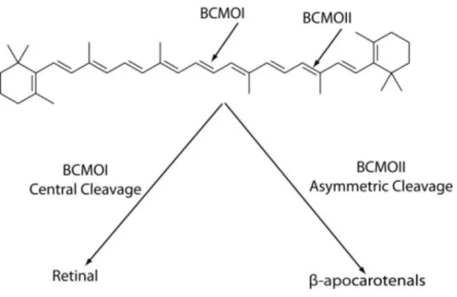
As early as 1930, it has been reported that β-carotene can be transformed to retinoids within the small intestine (Moore, 1930). Later on, two independent groups (Goodman and Huang, 1965; Olson and Hayaishi, 1965) showed that one molecule of β-carotene yields two molecules of retinal via a central cleavage catalyzed by an enzyme termed ββ-carotene-15, 15’-monooxygenase(BCMO-I) (Figure 4).
Another enzyme, β,β-carotene-9’10’-dioxygenase (BCMO-II) is also able to cleave β-carotene (Kiefer et al., 2001). However, BCMO-II catalyzes an asymmetrical cleavage of β-carotene at non-central double bonds of the polyene chain, yielding apocarotenals like 8′-, 10′- and β-apo-12′-carotenals (Figure 4). This alternative pathway is very important as it implies that, in tissues expressing BCOM-II, retinoic acid can be produced in the absence of ‘‘classical RA synthesis pathway’’ enzymes, such as alcohol dehydrogenase (ADH), short-chain dehydrogenase/reductase (SDR), retinaldehyde dehydrogenase (RALDH) (Simoes-Costa et al., 2008) or certain cytochrome P450s (Chen et al., 2000) (see below).
Figure 4: Carotenoids undergo cleavage either symmetrically by BCMOI or asymmetrically by BCMOII. Adapted from (D'Ambrosio et al., 2011).
2. Enterocyte Esterification of Retinol
Retinal obtained by carotenoid cleavage is then reduced to retinol and taken up by enterocytes. Enterocytes also take dietary consumed retinyl esters after hydrolysis into retinol within the intestinal lumen (Blomhoff and Blomhoff, 2006).
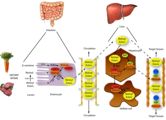
In enterocytes, retinol binds to the cellular retinol binding protein II (CRBPII), which is specifically highly expressed in the intestinal mucosa. CRBPII belongs to the greater family of fatty acid binding proteins and its role is to solubilize fat-soluble retinol (Sporn et al., 1994) (Figure 5).
Then, the majority of CRBPII-bound retinol is re-esterified via the enzyme lecithin retinol acyl transferase (LRAT). The remaining esterification activity would be assumed by two other enzymes diacylglycerol acyltransferase 1 (DGAT1) and acyl-CoA:retinol acyltransferase ARAT (D'Ambrosio et al., 2011) (Figure 5).
Afterwards retinyl esters are included into chylomicrons and secreted into general circulation (Blomhoff et al., 1982), where they are taken up by hepatocytes (Blomhoff et al., 1982), bone marrow, peripheral blood cells, spleen, adipose tissue, skeletal muscle and kidney (Paik et al., 2004). In mammals, 50-80% of the body’s overall retinoids are collected in hepatic stellate cells as retinyl esters (Blomhoff and Blomhoff, 2006; Fontana and Rishi, 2002) (Figure 5).
Figure 5: Metabolism of vitamin A. In the intestine, carotenoids are converted to retinol, which binds
CRBP. Then retinol is transformed into retinyl esters, exported into the circulation and taken up by the liver. In hepatocytes, retinyl esters are reconverted to retinol and bind RBP for transport to target cells. In stellate cells of the liver, retinol is retransformed to retinyl esters for storage. In the bloodstream, the retinol:RBP complex is bound to TTR to avoid removing by the kidney and for guaranteed delivery to target tissues. Figure adapted from (Theodosiou et al., 2010).
3. Hepatic retinoid metabolism
In hepatocytes, retinyl esters are re-converted to retinol, which then binds retinol binding-protein (RBP) and is secreted into circulation. The majority of the retinol:RBP complexes are associated with transthyretin (TTR), which prevents elimination by the kidney and ensures delivery to target tissues. However, a big part of hepatic retinol is reesterified into retinyl esters and packed within cytoplasmic lipid droplets in stellate cells (Blomhoff and Blomhoff, 2006) (Figure 5).
4. Retinol processing
In target tissues, retinol either associates with CRBP or is transformed into active metabolites. Indeed retinol undergoes oxidation to retinaldehyde by enzymes termed retinol dehydrogenases (Figure 6) (Gottesman et al., 2001; Pares et al., 2008). These enzymes are members of the ADH (alcohol dehydrogenase) or SDR (short-chain dehydrogenase/reductase) families, which depict cytosolic and microsomal localizations respectively. Given that the next step, the irreversible oxidation of retinaldehyde to retinoic acid, takes place in cytosol, it has been suggested that ADHs would be more important than microsomal SDRs for RA synthesis (Duester et al., 2003).
Then the oxidation of retinaldehyde to RA is carried out by retinaldehyde dehydrogenases (RALDHs), such as RALDH 1, 2, 3, and 4 depending on tissue types (Niederreither et al., 2002). Finally, newly synthesized RA associates to cellular RA binding proteins types I and II (CRABP-I and CRABP-II), and then either enters nuclei for activation of transcription (autocrine) or is transported to adjacent target cells (paracrine) (Napoli, 1996).
5. Retinoic acid degradation
Catabolism is necessary to control RA levels in cells and tissues. It occurs mainly through enzymes of the Cytochrome P450 enzyme family (CYP26). There are several CYP26 enzymes and the first one to be cloned was CYP26A1, which generates several hydroxylated forms of RA, such as 4-hydroxy retinoic acid, 4-oxo retinoic acid, 18-4-hydroxy retinoic acid, 5,6-epoxy retinoic acid, and 5,8-epoxy retinoic acid (Swindell and Eichele, 1999). Other similar enzymes (CYP26B1 and 26C1) have been identified later and are also able to metabolize RA (Taimi et al., 2004; White et al., 2000). The expression patterns of CYP26A1, CYP26B1, and CYP26C1 normally do not overlap, suggesting specific and distinct roles for each enzyme in the catabolism of RA (Reijntjes et al., 2004).
Figure 6: Biochemical pathway of retinoids. The enzymes responsible for conversion of the retinoids: ADH
alcohol dehydrogenase, BCMO-I β,β-carotene-15’,15’-monooxygenase, BCMO-II
β,β-carotene-9’,10’-dioxygenase, DGAT diacylglycerol O-acyltransferase, CYP26 cytochrome P450, DHRS
dehydrogenase/reductase SDR family member, LRAT phosphatidylcholine-retinol O-acyltransferase, PNPLA4 patatin-like phospholipase domain-containing protein 4, RDH retinol dehydrogenase, RETSAT all-trans-retinol 13,14-reductase , RPE65 retinal pigment epithelium-specific 65 kDa protein, SDR short-chain dehydrogenase/reductase, UGT glucuronosyltransferase. Adapted from (Theodosiou et al., 2010).
CHAPTER 2: NUCLEAR RETINOIC ACID RECEPTORS
The biological effects of RA are mediated through two main families of nuclear receptors, which belong to the nuclear receptor superfamily: the retinoic acid receptors (RAR) and the retinoid x receptors (RXR). For both RARs and RXRs, there are three subtypes - α, β, and γ. They act as ligand-inducible transcription factors and usually form RAR/RXR heterodimers.
Note however that RXRs can also form homodimers and heterodimerize with other nuclear receptors. Indeed, RXRs are promiscuous receptors, which have the capacity to form heterodimers with several different nuclear receptors such as the receptors for fatty acids [peroxisomal proliferator activated receptors (PPAR)], bile acids [farnesoid x receptor (FXR)], oxysterols [liver×receptor (LXR)], xenobiotics [pregnane×receptor (PXR), androstanes [constitutive androstane receptor (CAR)], and vitamin D [vitamin D receptor (VDR)] (Germain et al., 2006b).
- RETINOIC ACID RESPONSE ELEMENTS (RARES)
RARs together with RXRs form asymmetrically oriented heterodimers, which bind to specific DNA sequences, called RA response elements (RAREs), located in the regulatory sequences of target genes.
The classical RAREs are composed of two direct repeats of a core hexameric motif, PuG(G/T)TCA, separated by 1 base pair, 2 base pairs or 5 base pairs and named DR1, DR2 and DR5 respectively (Figure 7) (Germain et al., 2003; Leid et al., 1992; Mangelsdorf and Evans, 1995). Such RAREs have been identified in the promoters of a large number of RA target genes involved in a wide variety of functions. For example, the classical DR5 elements are found in the promoters of the RARβ2 gene itself (de The et al., 1990), of the CYP26A1 gene (cytochrome 450, family 26, subfamily a, polypeptide 1) (Loudig et al., 2000), and of several Homeobox (Hox) and hepatocyte nuclear factor
(HNF) genes (Dupe et al., 1997; Qian et al., 2000). Recently, in silico studies revealed new DR5
RARE-associated genes (Meis2 and Bhlhbe40) (Lalevee et al. 2011). DR2 elements were identified in the CRBPI (Cellular retinol binding protein I) and CRABPII (Cellular retinoic acid binding protein II) gene promoters (Durand et al., 1992; Smith et al., 1991). The only natural DR1 element has been found in the rat CRBPII gene promoter (Mangelsdorf et al., 1991).
Remarkably, recent ChIP-seq analysis revealed novel natural DR0 and DR8 RAREs, the latter being composed of a DR2 juxtaposed to a DR0 (I. Davidson et al, unpublished results).
Figure 7: The classical retinoid response elements are composed of two direct repeats of the hexameric,
motif 5′-PuG(G/T)TCA spaced by 1 (DR1), 2 (DR2) or 5 (DR5) base pairs. Several examples of natural retinoid response elements from the promoters of RA-target genes are shown. Adapted from (Bastien and Rochette-Egly, 2004).
- STRUCTURE OF RARS
As most nuclear receptors, RARs have a modular structure consisting of 6 regions named A to F, from the N-terminal to the C-terminal end (Figure 8) (Chambon, 1996; Laudet and Gronemeyer, 2002). Some of these regions overlap with functional domains. Indeed, the C and E modules correspond to the DNA binding domain (DBD) and the Ligand Binding Domain (LBD), respectively. These domains are highly conserved between RARs and nuclear receptors and play critical roles in the classical model of RAR transcriptional activity. Oppositely, the A/B, D and F modules are less conserved.
DR5
RARβ2 AG CGTTCA CCGAA AGTTCA CT
CYP26 TT AGTTCA CCCAA AGTTCA TC
Hoxa1 CA GGTTCA CCGAA AGTTCA AG
DR2
mCRBP-I GT AGGTCA AA AGGTCA GA
mCRABP-II CC AGTTCA CC AGGTCA GG
DR1 mCRBP-II AC AGGTCA C AGGTCA CA
Figure 8: Modular structure of RARs. Adapted from (Rochette-Egly and Germain, 2009).
1. DNA binding domain (DBD)
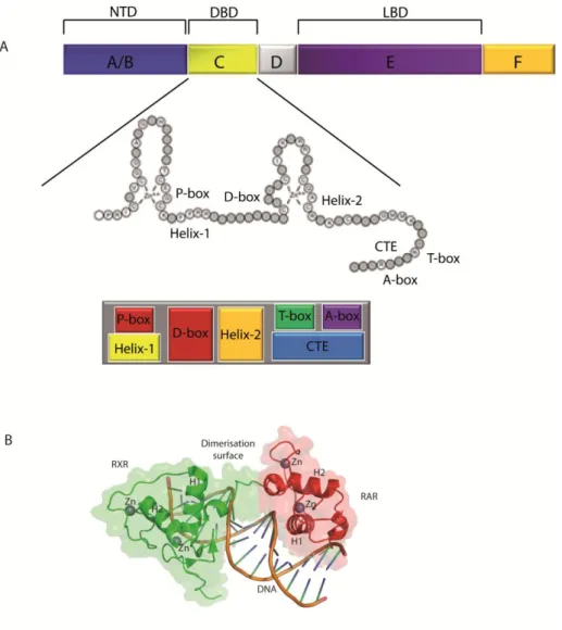
The DBD, which is responsible for sequence-specific DNA recognition, consists of two zinc-nucleated modules, two α-helices and a COOH-terminal extension (CTE) (Zechel et al., 1994). Functionally, the DBD is divided into 4 boxes: the P box within the first helix and the A box in the CTE, are responsible for discrimination between the half sites, while the D and T boxes are involved in the heterodimerization interface (Figure 9A).
According to nuclear magnetic resonance and crystallographic studies, when the DBD is complexed with DNA, helix 1 and helix 2 cross at right angles and fold into a globular conformation to form the core of the DBD (Lee et al., 1993) (Figure 9B). Indeed, helix 1 fits specifically into the major groove of the DNA through the P box, while helix 2 and the CTE cooperate to create the interface between the heterodimerization partners.
Figure 9: Structure of the DNA binding domain
A. Schematic view of the DBD, showing the relative locations of the P-box, D-box, T-box, A-box, Helix 1, Helix 2, and the C-terminal extension (CTE). Adapted from (Aranda and Pascual, 2001; Bain et al., 2007). B. Structure of a RAR/RXR DBD heterodimer in complex with a DR1 DNA response element. Zn = atome of zinc, H1 and H2 = helices 1 and 2. Adapted from (Rastinejad et al., 2000) pdb1dsz.
Depending on the RARE, the heterodimers are differently oriented. For example on DR2 and DR5 elements, the RXR partner occupies the 5’ hexameric motif and the RAR partner the 3’ motif (5’-RXR-RAR-3’) (Chambon, 1996; Laudet and Gronemeyer, 2002). However, for DR1 elements, the polarity is opposite, with the RAR in 5’ side and the RXR in 3’ (5’-RAR-RXR-3’) (Figure 10). This variation of orientation depending on the DR type, would explain why the activity of the heterodimer switches from an activator (DR5) to a repressor (DR1).
Moreover, also depending on the DR type, different regions of the DBD of each partner participate in the dimerization interface, in order to achieve the required binding to the response
elements. Indeed, the binding of RXR-RAR heterodimers to DR5 elements requires the D box of the RXR second zinc-finger, and the tip of the RAR first zinc finger. However, binding with reverse polarity to the DR1 elements involves the second zinc finger of RAR and the T box of the RXR CTE (Renaud and Moras, 2000).
Figure10: Different orientations of RXR/RAR hetodimers depending on the DR type. On DR5 elements,
the 5’ hexameric motif is occupied by RXR. On DR1 elements, the 5’ hexameric motif is occupied by RAR. Adapted from (Bastien and Rochette-Egly, 2004).
2. Ligand- binding domain (LBD)
The structures of the RAR LBDs were demonstrated by crystallographic studies (Moras and Gronemeyer, 1998; Renaud and Moras, 2000; Wurtz et al., 1996). The LBD is composed of 12 conserved α helices and a β-turn (located between H5 and H6) (Figure 11). Helices 1-11 are folded into a three-layered, anti parallel helical sandwich where H4, H5, H8, H9 and H11 are sandwiched between H1, H2 and H3 on one side and H6, H7 and H10 on the other side. In this structure, H12 that encompasses the AF-2 activation domain, points away from the LBD core. Functionally, the LBD is divided into three main functional domains: the ligand-binding pocket (LBP), the major dimerization interface and the ligand-dependent activation function-2 (AF-2).
Ligand-binding pocket (LBP)
The ligand-binding pocket (LBP) contains hydrophobic residues mainly from helices H3, H5, H11 and the β-sheet, which establish van der Waals interactions with the ligand (Klaholz et al., 2000). Several crystallographic studies revealed the structural basis of ligand recognition (Bourguet et al., 2000a; Li et al., 2003; Renaud et al., 1995). The carboxylic group of the retinoid molecules is buried deep inside the LBP, engaging hydrogen bonds with specific amino acids of H3 and the β-sheet. The rest of the molecule has to adapt to the overall structure of the LBP, which causes the ligand molecule to bend to accommodate the RAR LBP.
Figure 11: The 3D structure of the LBD. Helices are shown as ribbons and labelled from H1 to H12. The
LBD is consisted of 12 α helices which form a three-layered, antiparallel helical sandwich (on the left side of the figure). H4, H5, H8, H9 and H11 are sandwiched between H1, H2 and H3 on one side and H6, H7 and H10 on another side. Adapted from (Bourguet et al., 1995) pdb1lbd.
The precise contacts with ligands involve three divergent residues, which are unique for each subtype receptor-cognate ligand pair and are located in H3, H5 and H11 (Table 1). Therefore, it has been possible to generate subtype-selective ligands (Germain et al., 2004). For example, the unique polar residues S232 and M272 located within LBP of RARα and RARγ respectively have been used to develop specific ligands for RARα (Am580) or RARγ (BMS270394 or CD666). Via their amino group, these ligands form hydrogen bonds with S232 of RARα or M27 of RARγ leading to increased affinity and selectivity for RARα and RARγ respectively (Table1).
Table 1. Residues within the α-helices H3, H5 and H11 of the different RAR isotypes involved in ligand
binding
Heterodimerization surface
The main heterodimerization surface between the RAR and RXR partners is located in the LBD. It involves residues from helices H7, H9, H10 and H11, as well as loops L8-9 and L9-10 (Bourguet et al., 2000b; Gampe et al., 2000; Pogenberg et al., 2005). The core of the dimer is formed mostly by helices H9 and H10, which contribute to more than 75% of the total dimerization surface. In contrast to the almost perfect symmetric organization of RXR homodimers (so called butterfly shape), the heterodimer interfaces are asymmetric. Indeed, helix H7 of RXR contacts loop L8-9 of RAR, but loop L8-9 of RXR and helix H7 of RAR are not involved in the interaction (Figure 12).
Helices
Receptor H3 H5 H11
RARα Ser232 Ile270 Val395
RARβ Ala225 Ile263 Val388
Figure 12: The three-Dimensional structure of RXR–RAR heterodimer with 9‑cis-retinoic acid and DNA. Pointed lines indicate domains with unresolved structures. Helices are shown as ribbons and labelled
from H1 to H12 (LBD) or α1 and α2 (DBD). Helix H12 (AF-2) is represented in red in each subunit. The short LBD β-strands are labelled S1 and S2. 9-cis-retinoic acid in RAR and RXR LBDs is shown by green sticks lines. The orange spheres in the DBD indicate atoms of zinc. Image modified from (de Lera et al., 2007).
Recently Rochel et al revealed new structural features of the RAR-RXR heterodimers architecture on different RAREs. They demonstrated that the RXR–RAR–DR5 complex is elongated and asymmetric, with two separate DBDs and LDBs connected by a narrow segment (Figure 13 B) (Rochel et al., 2011). The RAR–RXR–DR1 complex is similarly elongated but with a larger connecting volume between the DBDs and LBDs (Figure 13 A). Interestingly, in both cases (DR5 or DR1), the LBD dimers are always positioned at the 5′ end of the target DNA. The observed asymmetry of the overall architecture and the relative position of the domains point to the essential role played by the hinge domains in establishing and maintaining the integrity of the functional structures.
Figure 13: A. RAR-RXR-DR1 complex B. RAR-RXR-DR5 complex. Adapted from (Rochel et al.,
2011).
C-terminal helix 12, named AF-2
The C-terminal helix 12, known as AF-2, regulates the interaction of RARs with coregulators. The analysis of the crystal structures of the unliganded and ligand-bound LBDs of RXRα and RARα respectively (Bourguet et al., 1995; Renaud et al., 1995), highlighted the conformational flexibility of H12 and how AF-2 becomes transcriptionally active (Figure 14 A) (Egea et al., 2001; Steinmetz et al., 2001).
In the unliganded receptor (so called apo-conformation), H11 is almost perpendicular to H10 and points towards the ligand-binding pocket and some of the hydrophobic residues of H11 partially fill and stabilize the LBP. H12 extends away from the core LBD, pointing away from the dimer axis at an angle of about 45° (Bourguet et al., 1995).
Upon ligand binding, H11 is repositioned in the continuity of H10, causing the concomitant swinging of H12, which moves in a ‘mouse trap’ model and packs tightly against H3 and H4 (Figure 14B). Consequently the lid of the LBD is sealed and ligand binding is stabilized (Moras and Gronemeyer, 1998). Moreover, a new hydrophobic cleft between H3, H4 and H12 is formed, creating a defined surface for the interaction with transcriptional coactivators. This liganded conformation is referred as “holo” or “active” conformation (Figure 14B). Note that in the case of the RARβ and RARγ subtypes, some biochemical studies proposed that even in the absence of ligand, H12 interacts with H3 and adopts a constitutively closed conformation that approximately corresponds to the conformation of liganded RARα (Farboud et al., 2003; Farboud and Privalsky, 2004; Hauksdottir et al., 2003).
Figure 14: Three-dimensional structure of LBD and structural changements induced by ligand binding. Helices are represented as ribbons and labelled from H1 to H12. A. Structure of the LBD of RXRα
in apo conformation. Adapted from (Bourguet et al., 1995) pdb1lbd. B. Structure of the LBD of RARγ with ligand (holo conformation). Adapted from (Renaud et al., 1995) pdb2lbd.
3. N-terminal AF-1 domain (NTD)
The N-terminal domain (NTD) of RARs (activation function AF-1) corresponds to the A and B regions (Nagpal et al., 1993; Nagpal et al., 1992). Within the NTD, the A regions are comparatively variable between the different RAR subtypes and isoforms (Chambon, 1996). In contrast, the B region is well conserved and holds a proline-rich motif containing phosphorylation sites (see below, Figure 26 page 42).
In contrast to the DBD and the LBD, there are still no high-resolution structures available for the NTD of RARs and most nuclear receptors. Even the relatively very short NTD of peroxisome proliferator-activated receptor-γ (PPAR-γ) failed to show any signature of structure (Chandra et al., 2008). According to several studies, the NTDs of the Estrogen Receptor (Warnmark et al., 2001), the Glucocorticoid Receptor (Warnmark et al., 2001), the Progesterone Receptor (Bain et al., 2000) and the Androgen Receptor (Reid et al., 2002) possess an intrinsically disordered (ID) conformation.
Importantly, intrinsically unstructured proteins are functional and the NTD of RARs has been shown to play an important role in the regulation of transcription (Nagpal et al., 1993). Though the mechanism of this functionality has not been elucidated yet, it has been shown that disordered domains provide the flexibility that is necessary for modifications by kinases or ubiquitin ligases (Dyson and Wright, 2005). Such modifications may change structural properties of the domain and
subsequently affect the dynamics of neighboring structural domains (Pufall et al., 2005) and therefore interactions with co-regulators and/or DNA (Dyson and Wright, 2005; Liu et al., 2006). In line with this, it is interesting to note that the NTD of RARs contains phosphorylation sites located in a proline-rich motif (PRM). As PRMs have the capacity to bind proteins with SH3 (Src-homology-3) or WW (tryptophan-tryptophan) domains (Kay et al., 2000b), one can speculate that the NTD of RARs might regulate transcription via the phospho-dependent association or dissociation of coregulators (See below chapter 5).
4. D-region
The D domain or hinge region is poorly conserved and serves as a hinge between the DBD and the LBD. It has been proposed that its flexibility would permit the adaptation of the RAR/RXR heterodimers to different types of RAREs (Glass, 1994; Rochel et al., 2011). In RARs, this domain is very small (12 amino acids) and shares 50% of identity between RARα and RARβ and 33% between RARα and RARγ. The hinge region would also harbor a nuclear localization signal (Hamy et al., 1991).
5. The F-region
The F region is the most carboxy-terminal region of RARs and is absent in RXRs. It is not conserved between RARs and its three-dimensional structure is not known. So far the precise functions of region F are not well understood. It has been proposed that in the absence of ligand this region might stabilize the H12 of RARα in open conformation, thus enhancing binding of corepressors (Farboud and Privalsky, 2004). According to recent studies, this region would be able to bind specific mRNA motifs (Poon and Chen, 2008). Interestingly, the F region is phosphorylated at multiple sites and such modificatins might change the properties of RARs (Bastien et al., 2000; Rochette-Egly et al., 1997).
CHAPTER 3: THE CLASSICAL MODEL OF RAR-MEDIATED
REGULATION OF TRANSCRIPTION
The canonical mechanism of action of RARs involves the activation or repression of target-gene transcription (Figure 15).
Figure 15: Classical model of activation of RA-target genes.
A. In the absence of ligand, RARα/RXR heterodimers bind DNA in association with corepressor complexes. B. Ligand binding induces the release of corepressors and the recruitment of coactivator complexes. C. Upon decompaction of chromatin, the transcriptional machinery, which consists of the Mediator, RNA polymerase II and the general transcription factors (GTFs), is recuited to the promoter, resulting in the initiation of transcription. Adapted from (Bour et al., 2007b).
This mechanism is at the basis of the regulation by RARs of gene networks involved in a wide number of functions such as homeostasis, development and reproduction. According to such a model, genes are silenced through the recruitment of corepressor-containing complexes to unliganded (apo) DNA-bound receptors. Conversely, genes are activated subsequent to corepressors release from liganded receptors (holo) and recruitment of coactivator complexes (Figure 15). At the molecular level, the discrimination between coactivators and corepressors relies in the positioning of H12 within the LBD.
- REPRESSION OF TRANSCRIPTION IN THE ABSENCE OF LIGAND
In the absence of ligand, RAR/RXR heterodimers occupy RAREs in association with large multiprotein complexes with several enzymatic activities (histone deacetylases, methylases, ubiquitin ligases etc), which maintain histones and chromatin in a compacted repressed state.
Basically, the corepressor core is composed of NCoR (nuclear receptor co-repressor) or SMRT (silencing mediator for retinoic acid and thyroid hormone receptors). NCoR and SMRT were the first identified corepressors (Chen and Evans, 1995; Lee et al., 1995; Sande and Privalsky, 1996). They bind NRs and serve as platforms for the binding of other proteins such as GPS2 (G-protein pathway suppressor 2), TBL1 (Transducin β-like 1), TBLR1 (TBL1-related protein 1), HDAC3 (Histone deacetylase 3) and Sin3 (Figure 16) (Li et al., 2000; Zhang et al., 2002).
Figure 16: Corepressors and associated complexes. Core of the complex (pointed) and associated
Of note is that SMRT is the preferential corepressor of RARs. It is an ubiquitous 270 kDa protein, which belongs to a variety of multiprotein complexes containing histone deacetylases. These complexes repress transcription by deacetylation of lysine residues located in the N-terminal tails of histones.
NCoR and SMRT are structurally and functionally similar and share about 40% amino-acid identity. Both have been shown to repress the transcriptional activity of several nuclear receptors and of a variety of unrelated transcription factors involved in several cellular processes. For example SMRT represses serum response factor (SRF), activator protein-1 (AP-1), and nuclear factor-mB (NFmB), which are all transcription factors involved in stimulation of cell proliferation (Lavinsky et al., 1998; Shibata et al., 1997; Zamir et al., 1996). SMRT as well as NCoR display specific domain structures (Figure 17).
Figure 17: Schematic N-CoR/SMRT domains with the associated proteins. Adapted from (O'Malley and
Kumar, 2008).
The C-terminus contains corepressor nuclear receptor (CoRNR) boxes, also called NR interactions domains (IDs), which interact with the LBD of NRs (Hu and Lazar, 1999; Nagy et al., 1999; Perissi et al., 1999). The ID contains the sequence (L/I)XX(I/V)I or LXXX(I/L)XXX(I/L) (where X is any amino acid), and forms an extended α helix that interacts with the hydrophobic groove generated by H3, L3-4 and H4 of RARs, the N-terminal extension of the motif masking the H12 interaction interface (Figure 18). As this surface is topologically related to that involved in coactivator interaction, but without H12; this may explain why the binding of corepressors and coactivators is mutually exclusive (Hu and Lazar, 1999).
In contrast, the amino terminus of N-CoR and SMRT contains the domains responsible for transcriptional repression (Figure 17). Three repressive regions (RD1, RD2, and RD3) were originally described due to their ability to act as autonomous repression domains when assosiated to DNA binding proteins (Horlein et al., 1995). RD1 interacts with GPS2 but little is known about the role of GPS2 in NR repression (Zhang et al., 2002). RD1 also interacts with TBL1 and TBLR1, which are members of the WD40 family.
Figure 18: Interaction of NR LBDs with corepressors.
A. Structure of the LBD of PPARα complexed with an antagonist and a CoRNR box. Adapted from (Xu et al., 2002) pdb1kkq. B. Structure of apo RXR LBD. Adapted from (Bourguet et al., 1995) pdb1lbd. C. Structure of holo RARγ LBD (with an agonist). Adapted from (Renaud et al., 1995) pdb2lbd
TBL1 and TBLR1 interact simultaneously with deacetylated histone H4 (Yoon et al., 2003), thereby stabilizing the corepressor complex on chromatin and facilitating repression. The other repressive domains interact with HDAC3 and with SIN3, a component of the Sin3A corepressor complex (Alland et al., 1997; Heinzel et al., 1997; Nagy et al., 1997). Remarkably, SIN3 can recruit additional enzymes with repressive activity such as the histone H3K9 methyltransferase ESET/SETDB1 and the ATP-dependent chromatin remodeling complex SWI/SNF (Underhill et al., 2000; Yang et al., 2003).
Between RD1 and RD2 there are two SANT (SWI3, ADA2, N-CoR, and TFIIB) motifs, which are also important for corepressor function. The first SANT motif forms a deacetylase activation domain (DAD), which stably associates with and activates histone deacetylase 3 (HDAC3) (Danielian et al., 1992; Umesono et al., 1991). The second one named histone interaction domain (HID), interacts directly with unacetylated histone H4 N-terminus tails (Yu et al., 2003).
- ACTIVATION OF TRANSCRIPTION UPON LIGAND BINDING
Ligand binding induces conformational changes of RARα, with reorientation of H12 (Figure 19), resulting in the formation of a charge clamp between a conserved glutamate residue in H12 and a lysine residue in H3. Such a charge clamp can form hydrogen bonds with the LxxLL motif of coactivators. However it does not fit with the extended LxxI/HIxxxI/L motif of corepressors. Thus, it has been proposed that the length difference of the interacting motifs might be at the origin of the alternative interactions of the hydrophobic cleft in the apo or holo conformations, with corepressors and coactivators (Germain et al., 2006b; Perissi et al., 1999).
Figure 19: Conformational changes in LBD and interaction with coactivators.
A. Structure of the LBD of RXR bound to an agonist and to the NR box of coactivators. Adapted from (Lippert et al., 2009) pdb2zxz.B. Structure of apo RXR LBD. Adapted from (Bourguet et al., 1995) pdb1lbd. C. Structure of holo RARγ LBD complexed with a ligand. Adapted from (Renaud et al., 1995) pdb2lbd.
According to the classical model of transcriptional activation, ligand binding releases bound corepressors and promotes the recruitment of coactivators that serve as a platform for larger complexes with chromatin modifying and remodeling activities (Figure 20).
The coactivators of the p160/SRC (Steroid Receptor Coactivators) family have been the most extensively studied among the large spectrum of identified coactivators. This family comprises three members: SRC-1 (also referred to as NCoA-1), SRC-2 [NCoA2, TIF-2 (transcriptional intermediary factor 2), GRIP-1 (glucocorticoid receptor interacting protein 1)] and SRC-3 [pCIP (p300/CBP-interacting protein), ACTR (Acetyltransferase), AlB1 (amplified in breast cancer I), TRAM1 (thyroid
hormone receptor-activator molecule I), RAC3 (Receptor-Associated Coactivator 3)] (Chatterjee and Kashfi, 2011).
Figure 20: Coactivators and associated complexes. The RAR/RXR heterodimers in holo conformation
recruit the p160 coactivators, which serve as a platform for chromatin modifying and remodeling complexes. Adapted from (Perissi and Rosenfeld, 2005).
The p160 SRCs are approximately 160 kDa in size and share an overall of 50-55% sequence similarity and 43-48% of sequence identity. Structurally p160/SRCs are composed of several domains (Figure 21). The central Receptor-Interacting Domain (RID) contains three LXXLL motifs or NR boxes and is responsible for interaction with the hydrophobic cleft of ligand-bound RARs. The C-terminal transcriptional activation domains (AD1 and AD2) recruit proteins that contribute to chromatin remodelling. AD1 is responsible for the recruitment of histone acetyltransferases such as p300/CBP (CREB-binding protein) and PCAF (P300/CBP-associated factor) (Chen et al., 1997; Stallcup et al., 2003; Torchia et al., 1997). AD2 usually recruits histone methyltransferases such as CARM1 (coactivator-associated arginine methyltransferase 1) and PRMT1 (Protein arginine N-methyltransferase 1) (Chen et al., 1999; Lee et al., 2005; Stallcup et al., 2003). Moreover, the C-terminal domain of SRCs itself shows a weak HAT activites (Chen et al., 1997; Spencer et al., 1997). The N-terminal domain called bHLH/PAS (helix-loop-helix/Per/ARNT/Sim) is highly conserved and functions as a third AD (AD3) domain. It serves as a binding site for DNA-binding transcription factors such as TEF-4, MEF-2C, p53 and myogenin. Additionally, bHLH/PAS can recruit other coactivators including GAC63, CoCoA, FliI, G9a,BAF57 and ANCO1 (Kim et al., 2003; Lee et al., 2006).
Figure 21: Schematic representation of functional domains and interacting proteins of p160/SRC family. Adapted from (O'Malley and Kumar, 2008).
The complexes with enzymatic activities associated to coactivators alter the chromatin structure around the promoter of target genes and create modifications of histone tails according to a “histone code”. Indeed these modifications create new sites for the recruitment of other complexes such as SWI/SNF (SWItch/Sucrose NonFermentable) that also contribute to chromatin remodeling using the energy of ATP hydrolysis (Huang et al., 2003; Sims and Reinberg, 2008).
Finally, chromatin remodeling and modifications pave the way for the recruitment of the transcriptional machinery that includes the multisubunit Mediator complex DRIP205/TRAP220, RNA polymerase II and the general transcription factors (Bastien and Rochette-Egly, 2004; Dilworth and Chambon, 2001; Rochette-Egly, 2005; Rosenfeld et al., 2006). Note that the recruitment of the transcription machinery involves interaction of RARs with a specific subunit of the Mediator complex, DRIP205/TRAP220, which contains two LxxLL motifs (Lefebvre et al., 2005).
It must be noted that depending on the target gene’s promoter context, RARs can carry out different programs for gene activation. Indeed, recent chromatin immunoprecipitation experiments showed that even in the absence of RA, RARα can occupy the promoters of some genes in association with the Mediator complex and RNA PolII (Flajollet et al., 2006; Mendoza-Parra et al., 2011; Pavri et al., 2005; Perissi et al., 2004). In this case, initiation of transcription relies on the dissociation of cdk8 (cyclin-dependent kinase 8 inhibitory subunit) from the Mediator complex (Andrau et al., 2006; Elmlund et al., 2006) and on the subsequent recruitment of the general transcription factors such as TFIIH.