HAL Id: hal-01191121
https://hal.archives-ouvertes.fr/hal-01191121
Submitted on 14 Nov 2015
HAL is a multi-disciplinary open access
archive for the deposit and dissemination of
sci-entific research documents, whether they are
pub-lished or not. The documents may come from
teaching and research institutions in France or
abroad, or from public or private research centers.
L’archive ouverte pluridisciplinaire HAL, est
destinée au dépôt et à la diffusion de documents
scientifiques de niveau recherche, publiés ou non,
émanant des établissements d’enseignement et de
recherche français ou étrangers, des laboratoires
publics ou privés.
Classical swine fever virus npro limits type I interferon
induction in plasmacytoid dendritic cells by interacting
with interferon regulatory factor 7
Ana R Fiebach, Laurence Guzylack-Piriou, Sylvie Python, Artur
Summerfield, Nicolas Ruggli
To cite this version:
Ana R Fiebach, Laurence Guzylack-Piriou, Sylvie Python, Artur Summerfield, Nicolas Ruggli.
Clas-sical swine fever virus npro limits type I interferon induction in plasmacytoid dendritic cells by
inter-acting with interferon regulatory factor 7. Journal of Virology, American Society for Microbiology,
2011, 85 (16), pp.8002-8011. �10.1128/JVI.00330-11�. �hal-01191121�
JOURNAL OFVIROLOGY, Aug. 2011, p. 8002–8011 Vol. 85, No. 16 0022-538X/11/$12.00 doi:10.1128/JVI.00330-11
Copyright © 2011, American Society for Microbiology. All Rights Reserved.
Classical Swine Fever Virus N
pro
Limits Type I Interferon Induction in
Plasmacytoid Dendritic Cells by Interacting with Interferon
Regulatory Factor 7
䌤
Ana R. Fiebach,† Laurence Guzylack-Piriou,†‡ Sylvie Python,
Artur Summerfield,* and Nicolas Ruggli*
Institute of Virology and Immunoprophylaxis, Mittelha¨usern, Switzerland
Received 17 February 2011/Accepted 23 May 2011
Viruses are detected by different classes of pattern recognition receptors that lead to the activation of interferon regulatory factors (IRF) and consequently to the induction of alpha/beta interferon (IFN-␣/). In turn, efficient viral strategies to escape the type I IFN-induced antiviral mechanisms have evolved. Previous studies established that pestivirus Npro
antagonizes the early innate immune response by targeting the transcription factor IRF3 for proteasomal degradation. Here, we report that Npro
of classical swine fever virus (CSFV) interacts also with IRF7, another mediator of type I IFN induction. We demonstrate that the Zn-binding domain of Npro
is essential for the interaction of Npro
with IRF7. For IRF3 and IRF7, the DNA-binding domain, the central region, and most of the regulatory domain are required for the interaction with Npro
. Importantly, the induction of IRF7-dependent type I IFN responses in plasmacytoid dendritic cells (pDC) is reduced after wild-type CSFV infection compared with infection with virus mutants unable to interact with IRF7. This is associated with lower levels of IRF7 in pDC. Consequently, wild-type but not Npro
mutant CSFV-infected pDC show reduced responses to other stimuli. Taken together, the results of this study show that CSFV Npro
is capable of manipulating the function of IRF7 in pDC and provides the virus with an additional strategy to circumvent the innate defense.
Innate immune activation following viral infection is depen-dent on recognition by pattern recognition receptors (PRRs) and on the production of alpha/beta interferon (IFN-␣/) (32, 38). Toll-like receptors (TLR) and the cytosolic RIG-I and MDA5 recognize viral RNA (11, 31, 58) and respond by acti-vating a signaling network that culminates in the induction of type I IFN and establishes an antiviral state (22). The central players of this signaling cascade are interferon regulatory fac-tor 3 (IRF3) and IRF7. They are essential to modulate the activity of the innate immune response (45, 55) but have dis-tinct roles in the induction of IFN-stimulated genes. As a result of the signaling initiated by the cytoplasmic PRRs, IRF3 is phosphorylated by the kinases IKKε and TANK-binding kinase 1, dimerizes, and translocates to the nucleus, where it eventu-ally upregulates transcription of IFN-␣/ mRNA (13, 50). TLR7, TLR8, and TLR9 trigger the induction of IFN-␣ through the adaptor molecule MyD88, which forms a complex with IRF7 in pDC (33). IRF7 is in turn phosphorylated by interleukin-1 (IL-1) receptor-associated kinase (IRAK)-1 and translocates to the nucleus (56). IRF3 is expressed mainly constitutively and appears to be an important element in the early phase of the IFN-␣/ induction. In contrast, IRF7 is a protein with a short half-life that is usually expressed at low
levels, but it is highly inducible by type I IFN (20) and hence is expressed in larger amounts in the later phase of the type I IFN induction (19, 36, 45). However, in mice, constitutive expres-sion of IRF7 in plasmacytoid dendritic cells (pDC) enables them to render fast and efficient IFN-␣/ responses (24). Fur-thermore, pDC are the only cell type described to produce type I IFN in IRF3 knockout mice (24).
During evolution, viruses have acquired numerous mecha-nisms to evade or subvert key elements of the host viral re-sponse (15, 19, 21, 57). For pestiviruses, it is known that they counteract the IFN-␣/ induction by mediating the protea-somal degradation of IRF3 in their primary target cells, such as myeloid DC and endothelial and epithelial cells, allowing the virus to establish a productive infection (6, 9, 23, 48). The viral protein responsible for this function is Npro, encoded at the N
terminus of the single open reading frame of the positive-sense RNA genome. Npro harbors an autoprotease domain in the
N-terminal half of the protein, resulting in cotranslational re-lease of Nprofrom the nascent polyprotein. In the C-terminal
half of Npro, a metal-binding TRASH motif consisting of C 112
-X21-C134-X3-C138mediates the coordination of one zinc (Zn) atom per molecule (53). This domain is essential for Nproto
interact with IRF3 and to mediate the degradation of IRF3 by the proteasome.
In the present study, we identified IRF7 as a novel interac-tion partner of Npro. We show that Npro modulates the IRF7
turnover, limiting the IFN-␣ induction in pDC, which is of functional relevance for pathogenesis.
MATERIALS AND METHODS
Cells.The porcine kidney cell lines SK-6 (30) and PK-15 (ATCC) were prop-agated in Earle’s minimal essential medium (EMEM) with 7% horse serum and * Corresponding author. Mailing address: Institute of Virology and
Immunoprophylaxis (IVI), Sensemattstrasse 293, CH-3147 Mittel-ha¨usern, Switzerland. Phone: 41 31 848 9211. Fax: 41 31 848 9222. E-mail for Nicolas Ruggli: nicolas.ruggli@ivi.admin.ch. E-mail for Artur Summerfield: artur.summerfield@ivi.admin.ch.
† These authors contributed equally.
‡ Present address: Institut National de la Recherche Agronomique, 180 Chemin de Tournefeuille, F-31027 Toulouse, France.
䌤Published ahead of print on 15 June 2011.
in Dulbecco’s minimal essential medium (DMEM) supplemented with 5% horse serum, nonessential amino acids, and 1 mM Na pyruvate. HEK293T cells (ATCC) were cultured in EMEM supplemented with 10% fetal bovine serum (Biochrom AG). Peripheral blood mononuclear cells (PBMC) were obtained from blood of specific-pathogen-free pigs using Ficoll-Paque density centrifuga-tion (1.077 g/liter; Amersham Pharmacia Biotech). pDC were enriched as de-scribed previously (18) by cell sorting of CD172a⫹PBMC using the magnetic cell sorting system (MACS) with LD columns (Miltenyi Biotec GmbH). This permits a 10-fold enrichment of functional pDC to 2 to 5% (52). To obtain a purified pDC preparation, PBMC were centrifuged over a 1.065-density iodixanol gradi-ent (Optiprep; Nycomed Pharma) (47). Subsequgradi-ently, the cells were depleted of T cells using anti-CD3, followed by CD4 enrichment on LS columns (MACS). This protocol yielded 60 to 80% pure pDC, identified as CD4high
CD172alow
(17).
Viruses.The classical swine fever virus (CSFV) strains 1 and vA187-⌬Npro
(44) were derived from the respective full-length cDNA clones. The vA187-1-derived viruses vA187-C112R and vA187-D136N (referred to as the
C112R and D136N mutants, respectively) with single-amino-acid mutations in
Nprowere described previously (43). All virus titers were determined on SK-6
and PK-15 cells by standard endpoint dilution and were expressed as 50% tissue culture infectious doses (TCID50) per ml. The avian influenza virus H5N1
NIBRG14, used for stimulation of pDC, was obtained from Jim Robertson (National Institute for Biological Standards and Control, Hertfordshire, United Kingdom).
Plasmid constructs.The plasmids pCI-Ub-Core, IRF3, and pFLAG-IRF7 were described elsewhere (6, 42). For the mammalian two-hybrid assays (Promega), plasmid pFN10A(ACT)-IRF3 expressing the porcine IRF3 fused to the VP16 transactivator domain and pFN11A(BIND)-derived plasmids express-ing GAL4-Npro
and the mutants GAL4-Npro
(C69A), Npro(C112R), Npro(C134A),
Npro(D
136N), Npro(C138A), and Npro(C161A) were used; these are described
elsewhere (43, 53). Mutants with deletions in the IRF3 and IRF7 genes were obtained by PCR-based site-directed mutagenesis using standard techniques. Briefly, the mutants were constructed using the plasmids pIRF3 and pFLAG-IRF7 (6). Mutagenesis was performed by PCR with appropriate oligonucleotides (Microsynth) and AccuPrime Pfx DNA polymerase (Invitrogen). The integrity of the mutagenized DNA fragments was verified by nucleotide sequencing using the Thermo Sequenase DYEnamic direct cycle sequencing kit (GE Healthcare) and the Global IR2 System with e-Seq software (Li-Cor Biosciences).
Antibodies.The rabbit polyclonal antisera against recombinant affinity-puri-fied CSFV Npro
(42), CSFV C, and porcine IRF3 were described previously (6). The viral nonstructural protein NS3 was detected using the monoclonal antibody (MAb) C16 (16), kindly provided by Irene Greiser-Wilke, Hannover Veterinary School, Hannover, Germany. The anti-FLAG M2 MAb and the anti-␣-tubulin MAb were purchased from Sigma. The antiserum against the porcine IRF7 was produced in rabbits with a recombinant protein encompassing amino acids 122 to 487 of the porcine IRF7 expressed in Escherichia coli M15 with the pQE31 vector (Qiagen) and purified by nickel chelate affinity chromatography as described previously for Npro
(42). For pDC phenotyping and isolation, MAbs against the following cell surface molecules were used: CD172a (MAb 74-22-15A), CD4 (MAb 74-12-4 and PT90A), and CD3 (MAb PPT3). Hybridomas for MAbs 74-22-15A and 74-12-4 were kindly donated by Armin Saalmu¨ller (Institute of Immunology, Department of Pathobiology, University of Veterinary Medicine, Vienna, Austria), and the hybridoma for MAb PTT3 was a gift from Karin Haverson (University of Bristol, United Kingdom). MAb PT90A was purchased from VMRD (Pullman, WA).
Immunoprecipitation.Coimmunoprecipitation was performed essentially as described by Rottenberg et al (41). Briefly, cells were lysed for 20 min on ice with a buffer containing 20 mM Tris (pH 7.4), 75 mM NaCl, 0.1% Triton X-100, and 2.5 mM MgCl2, supplemented with 5l/ml protease inhibitor cocktail (BD) and
10l/ml phosphatase inhibitor cocktail II (Sigma). After clarification by centrif-ugation at 15⬘000 ⫻ g for 10 min at 4°C, the lysates were incubated overnight at 4°C with 50l anti-FLAG M2 agarose (Sigma) in Tris-buffered saline (TBS) (20 mM Tris-HCl, 75 mM NaCl, pH 7.6). After incubation, the agarose was washed three times with TBS and the protein complexes were eluted from the beads with 20l of 2⫻ sample buffer (125 mM Tris-HCl, pH 6.8, 20% glycerol, 1% bro-mphenol blue, 4% sodium dodecyl sulfate [SDS]). The eluted proteins were analyzed by sodium dodecyl sulfate-polyacrylamide gel electrophoresis (SDS-PAGE) and Western blotting.
SDS-PAGE and Western blotting.Cells were lysed in a hypotonic buffer containing 20 mM morpholinepropanesulfonic acid, 10 mM NaCl, 1.5 mM MgCl2, 1% Triton X-100 (pH 6.5), and 1l/106cells protease inhibitor cocktail
(Sigma). Proteins were separated by SDS-PAGE under reducing conditions and
analyzed by Western blotting as previously described (43). The signal was ac-quired using the Odyssey infrared imaging system (Li-Cor Biosciences).
FCM analysis.The detection of the viral NS3 protein and the porcine IRF3 and IRF7 by flow cytometry (FCM) was performed as described previously (6). For staining of cell surface molecules, unfixed cells were incubated for 20 min on ice with the respective primary and secondary antibodies diluted in Cellwash (Becton Dickinson). For the detection of the intracellular antigens NS3, IRF3, and IRF7, the cells were fixed and permeabilized with FIX&PERM solution (An der Grub Bio Research GmbH) and stained with rabbit anti-NS3, anti-IRF3, and anti-IRF7 polyclonal antibody. For detection by FCM, isotype-specific fluores-cein isothiocyanate (FITC), R-phycoerythrin (RPE) (Southern Biotechnology Associates), and RPE-Cy5 (Dako) conjugates were used as described previously (17).
Mammalian two-hybrid assay.Protein interactions were analyzed with the CheckMate/Flexi Vector mammalian two-hybrid system (Promega) as described before (43). Briefly, HEK293T cells were seeded at a density of 105cells/well in
a 24-well plate. One day later, the cells were transfected with a mixture of the pFN10A(ACT)-derived and pFN11A(BIND)-derived plasmids at a concentra-tion of 100 ng/well each and the pGL4.31(luc2P/GAL4UAS/Hygro) reporter plasmid (Promega) at 50 ng/well. The plasmids pACT-Myo and pBind-Id (Pro-mega) were used as controls. The transfections were performed with FuGENE 6 transfection reagent (Roche) according to the manufacturer’s protocol. After 48 h of incubation at 37°C, the cells were lysed with passive lysis buffer (Pro-mega). Firefly and Renilla luciferases were quantified using the dual luciferase reporter assay system (Promega). The relative luminescence measured with firefly luciferase induced from the reporter plasmid was normalized with the corresponding relative luminescence obtained with Renilla luciferase constitu-tively expressed from the pFN11A(BIND)-derived plasmid. The fold firefly lu-ciferase induction was calculated relative to the normalized firefly lulu-ciferase activity obtained with the empty vectors set to 1. The expression of the proteins fused to VP16 and GAL4 was analyzed by SDS-PAGE and Western blotting as described above.
Type I IFN detection.The bioactivity of porcine IFN-␣/ in SK-6 cells was
assayed as described previously (42, 44) using the Mx/CAT reporter gene assay developed for the quantification of bovine IFN-␣/ (14) and kindly provided by Martin D. Fray (Institute of Animal Health, Compton, Newbury, Berkshire, United Kingdom). To this end, the culture supernatants were added to MDBK-t2 cells at appropriate dilutions allowing measurement in the linear range of the assay. Recombinant porcine IFN-␣ produced in HEK293-EBNA cells (4) was used as standard. Chloramphenicol acetyltransferase (CAT) was quantified using a CAT enzyme-linked immunosorbent assay (ELISA) (Roche). For the detection of IFN-␣ in purified pDC, the cells were stimulated with CpG 2216 at 10g/ml or with avian influenza virus (AIV) strain H5N1 NIBRG-23 for 24 h. Secreted IFN-␣ was detected by ELISA using anti-pig IFN-␣ MAbs K9 and F17 (kindly provided by Bernard Charley, INRA, Jouy-en-Josas, France) as described previously (18).
RNA extraction and quantitative TaqMan RT-PCR for porcine IRF3, IRF7, and IFN-. To determine mRNA levels in pDC, total RNA was prepared using
the RNeasy minikit (Qiagen, Hombrechtikon, Switzerland), followed by treat-ment with RNase-free DNase I (Ambion, Huntingdon, United Kingdom). Re-verse transcription-PCR (RT-PCR) was performed using the ABI PRISM 7700 sequence detector system (Applied Biosystems, Foster City, CA). The oligonu-cleotides for the quantification of IRF7 and 18S mRNAs were designed based on the coding sequence of the respective genes using Primer Express version 1.5 software (Applied Biosystems). The sense IRF7 primer 5⬘-CTGCGATGGCTG GATGAA-3⬘ and the antisense IRF7 primer 5⬘-TAAAGATGCGCGAGTCGG A-3⬘ were used in combination with the IRF7 probe 5⬘-CCGCGTGCCCTGGA AGCACTT-3⬘. The sense 18S primer 5⬘-CGCCGCTAGAGGTGAAATTC-3⬘ and the antisense 18S primer 5⬘-GGCAAATGCTTTCGCTCTG-3⬘ were used together with the 18S probe 5⬘-TGGACCGGCGCAAGACGGA-3⬘. The rela-tive expression of each mRNA was determined with the⌬CTmethod by
calcu-lating the amount of target mRNA in relation to 18S mRNA. The data are presented as the ratio of target mRNA to 18S mRNA.
RESULTS Npro
interacts with IRF7. Pestivirus Npro inhibits
dsRNA-mediated IFN-␣/ induction by mediating the proteasomal degradation of IRF3 (6, 9, 23). Although Nprodoes not
accel-erate the degradation of IRF7 expressed exogenously (6), we investigated whether Nprointerferes with the functions of IRF7
by other means. We used a mammalian two-hybrid assay to analyze whether Nprocan interact with IRF7, using IRF3 as a
control. As expected from previous experiments, coexpression of Nprofused to the GAL4 DNA-binding domain and of IRF3
fused to the VP16 activation domain resulted in strong induc-tion of GAL4 promoter-driven luciferase expression compared with that with the empty vectors (Fig. 1A). The C112R muta-tion in Npro completely abolished this interaction, as shown
before (43). In the same assay, coexpression of Nprowith
por-cine IRF7 also produced a strong induction of luciferase ex-pression, indicating an interaction of Nprowith IRF7.
Impor-tantly, coexpression of increasing amounts of FLAG-IRF3 diminished the interaction of Npro with IRF7 gradually,
con-firming the specificity of the interaction between Npro and
IRF7. While an excess of IRF3 displaced IRF7, the cotitration of the C protein of CSFV as a control had no influence on the interaction of Npro with IRF7 (Fig. 1A). To further confirm
these findings, HEK293T cells were cotransfected with plas-mids expressing FLAG-IRF7 and untagged Nproor with each
plasmid individually, and proteins were coimmunoprecipitated with anti-FLAG agarose. Npro was immunoprecipitated with
anti-FLAG from cell lysates containing both FLAG-IRF7 and untagged Npro but not from cells expressing Npro or
FLAG-IRF7 alone (Fig. 1B). Conversely, FLAG-IRF7 was immunoprecipi-tated with anti-FLAG from cell lysates containing both FLAG-Npro and untagged IRF7 but not when these proteins were
expressed alone (Fig. 1B). These experiments show that Npro
interacts with IRF7.
The Zn-binding domain of Npro
is required for the interac-tion with IRF7.A recent report described a novel Zn-binding TRASH motif in Npro, involving the residues C
112, C134, D136,
and C138. Zn binding by Nprois a prerequisite for the
interac-tion of Nprowith IRF3, leading to the proteasomal degradation
of IRF3 and to the inhibition of type I IFN induction (53). Considering the structural similarity of IRF3 and IRF7, we asked whether Nproalso relays on the same Zn-binding domain
to interact with IRF7. To this end we analyzed the interaction of TRASH domain mutants of Npro with porcine IRF3 and
IRF7, using the mammalian two-hybrid assay. Coexpression of GAL4-Nproand VP16-IRF3 or of GAL4-Nproand VP16-IRF7
caused a strong induction of the GAL4 promoter-driven lucif-erase compared to the negative control, as expected (Fig. 2). The mutations C112R, C112A, C134A, D136N, and C138A, which disrupt the TRASH domain, resulting in proteins incapable of coordinating Zn, completely abolished the interaction of Npro
FIG. 1. Nprointeracts with IRF7. The interaction of Nprowith IRF7 was analyzed using a mammalian two-hybrid assay (A) and by immu-noprecipitation (B). (A) For the mammalian two-hybrid assay, HEK293T cells were cotransfected with pFN10A(ACT)-derived plas-mids expressing IRF7 or IRF3 fused to the VP16 transactivator (VP16-IRF7 and VP16-IRF3, respectively) and with pFN11A(BIND)-derived plasmids expressing the viral Nproor the Npro(C
112R) mutant fused to the GAL4 DNA-binding domain [GAL4-Nproand GAL4-Npro(C
112R), respectively]. These constructs were transfected alone or in combina-tion with increasing amounts of 1 ng, 25 ng, 100 ng, 250 ng, and 500 ng (indicated with open triangles) of plasmids for CMV promoter-driven expression of FLAG-IRF3 or of the C protein (core) of CSFV. The presence and the absence of the plasmids are indicated with⫹ and ⫺, respectively. The empty vectors pACT and pBIND served as negative controls, whereas the VP16-Myo and GAL4-Id control proteins ex-pressed from pFN10A(ACT) and pFN11A(BIND), respectively, were used as positive controls. After 48 h of incubation, the cells were lysed and the firefly luciferase activity was measured and normalized with Renilla luciferase activity constitutively expressed by the pFN11A(BIND)-plasmid. The results are shown as fold induction compared to the negative control and represent the mean of triplicates from a representative experiment, with error bars showing the stan-dard deviation. In parallel, the VP16-IRF7, FLAG-IRF3, GAL4-Npro,
core, and␣-tubulin proteins from the cells used for luciferase quanti-fication were analyzed by Western blotting and detection with rabbit antisera against the porcine IRF7, the viral Npro, and core and with anti-FLAG and anti-␣-tubulin MAbs. (B) For immunoprecipitation, HEK293T cells were transfected with plasmids expressing FLAG-tagged IRF7 and unFLAG-tagged Nprotogether or individually (left panels) or FLAG-tagged Npro and untagged IRF7 together or individually (right panels). At 24 h after transfection, the cells were lysed and the FLAG-tagged proteins were immunoprecipitated (FLAG-IP) with anti-FLAG M2 agarose. The precipitated proteins (IP) and the lysates subjected to the immunoprecipitation (input) were analyzed by West-ern blotting (WB) and detection using the anti-Nprorabbit antiserum, the anti-FLAG MAb, and the anti-␣-tubulin MAb as indicated.
with IRF7. Consistent with the results obtained earlier with IRF3 (53), mutation of C161 to alanine did not disrupt the interaction with IRF7. In addition, the C69A mutation within
the protease domain, which had no effect on the Npro-mediated
IRF3 degradation (43), also did not abrogate the interaction of Nprowith IRF7. The expression of the IRF3 and
VP16-IRF7 chimeric proteins and of all the Npromutants was verified
by Western blotting (Fig. 2). These data indicate that the Zn-binding domain but not the protease domain is required for the interaction of Nprowith IRF7, similar to the case for IRF3.
For IRF3 and IRF7, the DNA-binding domain, the central region, and most of the regulatory domain are required for the interaction with Npro
. There are several examples of viruses
that inhibit type I IFN induction by antagonizing the function of more than one IRF simultaneously (5, 7, 10, 27, 37). The IRF family members share conserved features in their struc-tural architecture and have inter- and intraspecies sequence similarities (1, 5, 26, 49). In line with this, we asked whether Nprocould recognize a common motif of the porcine IRF3 and
IRF7. For this purpose we used the domain prediction pro-grams SMART (46) and InterProScan (39) to identify and map conserved motifs in porcine IRF3 and IRF7 that may be in-volved in the interaction with Npro. We found that IRF3 and
IRF7 contain a highly conserved N-terminal DNA-binding do-main and a variable C-terminal regulatory dodo-main (Fig. 3A). In order to investigate which of these domains are crucial for the interaction with Npro, we constructed deletion mutant
IRF3 and IRF7 lacking the DNA-binding domain or the reg-ulatory domain, with or without the central region (Fig. 3B). The expression of the chimeric proteins IRF3 and VP16-IRF7 and of the different deletion mutants was confirmed by Western blot analysis (Fig. 3C and D). The rabbit antisera detected multiple bands. For each construct, the presence of the VP16-IRF fusion protein with the expected molecular weight (asterisks in Fig. 3C and D) was confirmed with the anti-VP16 MAb (not shown). Lower levels of the truncated proteins IRF3(195-419), IRF7(1-132), and IRF7(133-487) than of the other proteins were detected. Transfection of larger amounts of these three plasmids did not result in the expression of more protein (data not shown), suggesting that the truncated proteins were unstable. As expected, transfection of HEK293T cells with the GAL4-Npro and VP16-IRF3
chi-meric proteins resulted in strong induction of the luciferase gene, which was completely abolished by the C112R mutant (Fig. 3E). Interestingly, the carboxy-terminal IRF3(1-114) de-letion mutant and the amino-terminal IRF3(115-419) and IRF3(195-419) deletion mutants did not interact with Npro,
suggesting that the DNA-binding domain and the regulatory domain alone are not sufficient for interaction with Npro. The
mutant IRF3(1-194), encompassing the DNA-binding domain and the central region and lacking the regulatory domain, showed a weak interaction that was slightly increased when half of the regulatory domain was present in the IRF3(1-272) construct. Similar results were obtained for the interaction of Npro with the different IRF7 deletion mutants. The
IRF7(1-268), mutant in which the DNA-binding domain and the cen-tral region were maintained, resulted in weak interaction with Npro (Fig. 3F). When part of the regulatory domain in the
IRF7(1-336) construct was included, binding to Npro was
nearly completely restored. The interactions of Nprowith the
truncated IRF3 and IRF7 were specific, since the C112R
mu-tant of Nprocompletely abrogated the interaction. These data
indicate that nearly the complete structure of IRF3 and IRF7 rather than the DNA-binding and the regulatory domain alones is required for the interaction with Npro.
Npro
can inhibit IRF7-driven IFN-␣/ responses in SK-6
cells.Having demonstrated that Nprointeracts with IRF7, we
asked whether this interaction enhances the degradation of IRF7. Previous studies showed that Nprodoes not
downregu-late IRF7 expression (2, 6), in contrast to IRF3 (6, 9, 23, 48), in permanent cell lines. To have an applicable cell system to study the proteasomal degradation, we transfected SK-6 cells with plasmids expressing FLAG-IRF7 under the control of the
FIG. 2. The zinc-binding TRASH domain of Nprois essential for the interaction of Nprowith IRF7. HEK293T cells were cotransfected with pFN10A(ACT)-derived plasmids expressing VP16-IRF3 or VP16-IRF7 and with pFN11A(BIND)-derived plasmids for the expression of GAL4-Nproor the indicated mutants thereof. The empty vectors pACT and pBIND were used as negative controls, and the respective plasmids ex-pressing VP16-Myo and GAL4-Id served as positive controls. The pres-ence and the abspres-ence of the plasmids are indicated with⫹ and ⫺, re-spectively. The fold induction of firefly luciferase activity normalized to Renilla luciferase activity was calculated relative to the negative control. The error bars represent the standard deviation. The expression of VP16-IRF7, VP16-IRF3, and GAL4-Nprowas analyzed in parallel from the cells used for luciferase quantification by Western blotting and detection with rabbit antisera against the porcine IRF3 and IRF7 and the viral Npro, respectively. The␣-tubulin content was determined as loading control using the anti-␣-tubulin MAb.
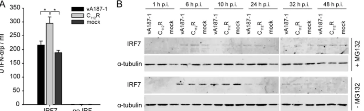
cytomegalovirus (CMV) promoter. Subsequently, the cells were mock treated or infected with vA187-1 or the C112R
mutant, and IFN-␣/ production was measured. Stimulation of the cells by transfection with the IRF7-expressing plasmid re-sulted in the production of high levels of type I IFN. Impor-tantly, infection with the C112R mutant virus significantly
en-hanced IFN-␣ expression in IRF7-transfected cells compared with the vA187-1 virus-infected cells and mock-treated cells, indicating an inhibitory effect of Nproon IRF7-mediated type I
IFN responses to viral stimuli.
In order to analyze the effect of Nproon IRF7 turnover, we
tested how the proteasome inhibitor MG132 influences the content of IRF7 in the presence of Npro. SK-6 cells were
transfected with a plasmid expressing FLAG-tagged IRF7 in the presence or absence of MG132, followed by infection with vA187-1 or C112R mutant CSFV. Comparable levels of IRF7
were detected in mock-, CSFV-, and C112R mutant-infected cells at each time point in the absence of MG132 (Fig. 4B, bottom panel). The decrease of IRF7 in all samples at 24 h after infection is consistent with the known high turnover of IRF7. Note that in the presence of MG132, the overall IRF7 expression levels were low (Fig. 4B, top panel), probably due to the toxic effect of MG132. From previous data we know that the level of IRF3 is clearly reduced at 24 h after infection with vA187-1 and can be rescued by inhibition of the proteasomal activity (6). Here we show that in contrast to the case for IRF3, the amount of IRF7 was not influenced by infection with either
vA187-1 or the C112R mutant virus. In conclusion, in SK-6 cells there is no evidence of accelerated proteasomal degradation of IRF7 resulting from the interaction of Nprowith IRF7.
The interaction of Npro
with IRF7 results in reduced
CSFV-induced IFN-␣ responses in pDC. pDC are considered to be
the main source of the high serum levels of IFN-␣ found during the first 2 to 5 days after CSFV infection in vivo (29, 51), and they are known to respond to viruses in an IRF3-indepen-dent, IRF7-dependent pathway (24). Therefore, we analyzed the induction of IFN-␣ in enriched porcine pDC at different times after infection with wild-type CSFV or with CSFV car-rying the D136N or the C112R mutation in Npro, abolishing the
interaction of Npro with IRF7. No IFN-␣ was observed in
mock-treated cells, whereas infection with wild-type CSFV re-sulted in the production of low levels of IFN-␣ found at 6 h, 24 h, and 48 h postinfection (p.i.) (Fig. 5A). Related to their inability to interact with IRF7, the D136N or C112R mutant CSFV led to a 5-fold-higher production of IFN-␣. Indepen-dently of the virus dose, the relative difference in the induction of IFN-␣ between the wild-type and the D136N mutant viruses
remained visible (Fig. 5B). The differences between wild-type and Npro mutant CSFV in the induction of IFN-␣ were also
observed in the context of the highly virulent Eystrup strain (Fig. 5C). Interestingly, the latter virus consistently induced higher levels of IFN-␣ than the moderately virulent vA187-1 strain. At the IFN-␣ and IFN- mRNA levels, the D136N and
C112R mutants also induced higher responses than the
wild-FIG. 3. Truncations at the amino terminus or the carboxy terminus of IRF3 and IRF7 affect the interaction with Npro. (A) Schematic representation of porcine IRF3 and IRF7 based on the domain prediction tool SMART; the DNA-binding domain (DNA-BD) and the regulatory domain (RD) are indicated. (B) Schematic representation of the N-terminal and C-terminal deletion mutants of IRF3 and IRF7; all the truncated fragments were expressed fused to the C terminus of the transactivator VP16 of the pFN10A(ACT) plasmid. (C and D) The expression of IRF3 (C) and IRF7 (D) and of mutants thereof was analyzed by Western blotting and detection with rabbit antisera against porcine IRF3 and IRF7, respectively. The␣-tubulin content was determined as a loading control using the anti-␣-tubulin MAb. (E and F) The interaction of Nprowith the truncated forms of IRF3 (E) and IRF7 (F) was analyzed using the mammalian two-hybrid assay. HEK293T cells were transfected with plasmids for the expression of the GAL4-Nprochimeric protein and of the VP16-IRF3 (E) or VP16-IRF7 (F) proteins and the indicated deletion mutants. The fold induction of firefly luciferase activity normalized to Renilla luciferase activity was calculated relative to the negative-control pACT and pBIND. The error bars represent the standard deviation.
FIG. 4. Nprodoes not induce proteasomal degradation of IRF7 in SK-6 cells. (A) SK-6 cells were seeded at a density of 105cells/well and transfected with the plasmid pFLAG-IRF7 at a concentration of 100 ng/well or mock transfected (no IRF). Twenty-four hours later, the cells were mock treated or infected with CSFV vA187-1 or the C112R mutant of vA187-1 at a multiplicity of infection (MOI) of 2 TCID50/cell. After 48 h of incubation, the supernatants were collected and IFN-␣/ bioactivity was measured with the MxCAT assay. Statistically significant differences (P⬍ 0.05, calculated with Student’s t test) are shown with an asterisk. (B) SK-6 cells were transfected with the plasmid pFLAG-IRF7 for CMV promoter-driven expression of FLAG-tagged porcine IRF7 in the presence (⫹MG132) or absence (⫺MG132) of 0.2 M proteasomal inhibitor MG132. At 24 h after transfection, the cells were mock treated or infected with vA187-1 or the C112R mutant at an MOI of 2 TCID50/cell. The cells were lysed at 1, 6, 10, 24, 32, and 48 h p.i., and the extracts were analyzed for IRF7 expression using mouse anti-FLAG MAb and for␣-tubulin using anti-␣-tubulin MAb.
type CSFV at 6 and 10 h p.i. (Fig. 5D and E). The ability of the Npro mutants to induce higher IFN-␣/ responses than the
wild-type CSFV was not due to different levels of infection by the viruses, since the percentages of NS3⫹pDC were compa-rable at various virus doses (Fig. 5F). Taken together, the results show that CSFV has the ability to significantly limit IFN-␣/ induction in pDC and that this function is mediated by the viral protein Npro, probably through its ability to
inter-fere with IRF3 and/or IRF7 signaling.
Npro
negatively regulates IRF7 levels in pDC. In order to determine the mechanisms by which Nprolimits the production
of IFN-␣ in pDC, we examined the fate of IRF7 following infection of purified pDC with wild-type and with D136N and C112R mutant CSFV. With no IRF7 detectable in
mock-in-fected cells, the D136N mutant induced a strong upregulation of IRF7 that was visible with the first IFN-␣ found at 6 h p.i. In wild-type-infected pDC, IRF7 was detectable as a weak band in the Western blot as early as 6 h p.i. Interestingly, despite the high levels of IFN-␣ (several thousand units) also found in the wild-type CSFV-infected purified pDC cultures at 10 h and 24 h p.i., the IRF7 levels remained at a constant low level and never reached the high levels found in pDC infected with the D136N mutant virus (Fig. 6A). These observations were con-firmed by flow cytometry. While infection with the parent
CSFV resulted in IRF7 expression similar to that with mock infection (Fig. 6B, right panel), the D136N and C112R mutant
CSFV induced higher levels of IRF7. In addition, we observed a clear reduction of IRF3 expression in wild-type CSFV-in-fected pDC compared with mock treatment or infection with D136N and C112R mutant CSFV (Fig. 6B, left panel),
confirm-ing previous data showconfirm-ing the degradation of IRF3 in other cell types (6). In order to determine whether the differences in the IRF7 protein expression levels seen in Fig. 6A and B resulted from different transcriptional activities, we quantified IRF3 and IRF7 mRNAs by real-time RT-PCR in wild-type CSFV-infected pDC versus pDC infected with D136N mutant
CSFV. IRF7 was strongly induced by both wild-type and mu-tant CSFV. Although at 10 h p.i., the D136N mutant induced
higher levels of IRF7, with both viruses similar maximum levels of 170- to 190-fold over that with the mock controls were found (Fig. 6C). Compared to that of IRF7, the induction of IRF3 was lower, and no difference was found between the wild type and the Npromutant at most time points analyzed (Fig. 6D).
The higher levels of IRF3 induced by vA187-1 found at 16 h p.i. were not reproducible. From this we conclude that the lower IRF7 protein levels found in wild-type CSFV infection did not result from lower transcription. Taken together, these findings indicate that Npro negatively regulates IRF7 at the
FIG. 5. Nprolimits the IFN-␣ responses in pDC. (A) Enriched pDC were mock treated or infected at a MOI of 2 TCID50/cell with CSFV strain vA187-1 or with CSFV vA187-1 carrying the C112R or the D136N mutation in Npro. The supernatants were collected at 6, 24, and 48 h p.i., and IFN-␣ was measured by ELISA. The bars show the means of triplicates from a representative experiment, with error bars showing the standard deviation. (B) Enriched pDC were mock treated or infected with CSFV vA187-1 or with the D136N mutant at an MOI of 5, 2, 1, or 0.04 TCID50/cell. Supernatants were collected at 20 h p.i., and IFN-␣ was quantified by ELISA. (C) Enriched pDC were mock treated or infected at an MOI of 1 TCID50/cell with the wild-type (WT) CSFV strains vA187-1 or Eystrup as indicated, and with the respective D136N mutant viruses. The supernatants were collected at 20 h p.i., and IFN-␣ was measured by ELISA. (D and E) The contents of IFN-␣ (D) and IFN- (E) mRNAs of purified pDC at different times after mock treatment or infection with CSFV strain vA187-1 or the D136N and C112R mutants were measured by real time RT-PCR. The amount of mRNA is shown relative to the levels of the 18S mRNA. (F) Enriched pDC were mock treated or infected with CSFV vA187-1 or the D136N mutant at an MOI of 5, 2, 1, or 0.04 TCID50/cell. At 20 h p.i., triple staining for surface CD172a, CD4, and intracellular NS3 was performed. The percentage of NS3-positive cells was calculated on CD172alowCD4⫹pDC. The results of one representative experiment out of three are shown. Statistically significant differences (P⬍ 0.05, calculated by Student’s t test) are shown with an asterisk.
protein level in pDC, probably through interference with IRF7 turnover.
Infection with CSFV inhibits the IFN-␣ response in pDC. In
order to investigate whether CSFV can also inhibit the IFN-␣ response of pDC to other TLR ligands or viruses, we infected enriched pDC with wild-type and D136N mutant CSFV for 20 h and subsequently treated the cells with CpG as a TLR9 agonist or with H5N1 AIV as a TLR7 agonist (Fig. 7). Interestingly, infection with parental CSFV impaired the ability of pDC to secrete high levels of IFN-␣ in response to the other stimuli. In pDC infected with D136N mutant CSFV, this inhibition was not
observed with CpG stimulation and was significantly less po-tent for AIV activation. These data demonstrate that CSFV is able to negatively regulate the IFN-␣ response in pDC, pre-sumably through its ability to reduce the levels of IRF7.
DISCUSSION
Nproof pestiviruses antagonizes the induction of the early
innate immune response in non-pDC such as epithelial cells, macrophages, and monocyte-derived DC. This function is re-lated to a Zn-binding motif in the C-terminal half of Nprothat
is essential for Npro to interact with IRF3, resulting in the
proteasomal degradation of IRF3 (53). Considering that in mouse pDC type I IFN induction was found to be essentially IRF7 dependent (24), it was not surprising to observe that pDC represent the only cell type able to respond to CSFV infection by production of IFN-␣ (4, 6, 8, 35). However, here we demonstrate a novel function of Nproin innate immunity,
namely, its interaction with IRF7, which reduces IRF7-depen-dent IFN-␣ induction in SK-6 cells and pDC.
In contrast to other cells, human pDC express IRF7 consti-tutively, although this basal expression is rapidly upregulated after stimulation (28, 34, 54) and is maintained in vitro only in
FIG. 6. Npronegatively regulates the IRF7 protein but not the IRF7 mRNA levels in pDC. (A) Purified pDC were mock treated or infected with CSFV vA187-1 or the D136N mutant at an MOI of 2 TCID50/cell. The supernatants were collected at 1, 6, 10, and 24 h p.i., and IFN-␣ was measured by ELISA. The bars show the means of triplicates from one representative experiment out of three, with error bars displaying the standard deviation. In parallel, the expres-sion of IRF7 from the same wells was analyzed by Western blotting and detection with rabbit antiserum against the porcine IRF7 (the asterisk indicates an unspecific band shown as loading control). (B) Enriched pDC were mock treated or infected with CSFV vA187-1 or with the C112R or the D136N mutant at an MOI of 1 TCID50/cell. At 20 h p.i., triple staining was performed for surface CD172a and CD4 and for intracellular IRF3 (left panel) or IRF7 (right panel). The CD172lowCD4⫹ pDC subset was analyzed for IRF3 (left panel) and IRF7 (right panel) expression. The results of one representative experiment out of two are shown. (C and D) Purified pDC were mock treated or infected with CSFV strain vA187-1 or with the D136N mutant at an MOI of 1 TCID50/cell. The levels of IRF7 (C) and IRF3 (D) mRNAs relative to the 18S mRNA were measured by real-time RT-PCR at 1, 6, 10, 16, and 24 h p.i. The bars show the means for triplicate cultures from one represen-tative experiment out of three. In panels A and C, statistically significant differences (P⬍ 0.05, calculated by Student’s t test) are shown with an asterisk. ns, not significant.
FIG. 7. Infection with CSFV but not with virus carrying the D136N mutation in Nproinhibits the IFN-␣ response in pDC. Enriched pDC were mock treated or infected with CSFV vA187-1 or vA187-D136N at an MOI of 2 TCID50/cell. At 18 h p.i., the cells were stimulated by adding CpG 2216 (10g/ml) to the cell culture medium or by infection with H5N1 AIV at an MOI of 10 TCID50/cell. The supernatants were collected after 24 h and tested for IFN-␣ by ELISA. Statistically sig-nificant differences (P⬍ 0.05, calculated by Student’s t test) are shown with an asterisk. ns, not significant.
the presence of a stimulant (12). Related to this, studies with human pDC describe that nuclear translocation of IRF7 occurs earlier than IRF7 induction, implying that the first wave of IFN-␣ is induced by the constitutively expressed IRF7 in pDC (12, 54). This would explain our finding that CSFV induces considerable amounts of IFN-␣ despite the interference of Nprowith IRF3 and IRF7 functions. However, the IFN-␣ levels
found with the Zn-binding-deficient Npro are never reached
during infection with the parent CSFV due to a limited IRF7 expression observed with CSFV infection. Our data with pDC suggest that IRF7 is regulated at the protein level, since no or only minor differences were found in the IRF7 mRNA quan-tities. In the porcine cell lines SK-6 and PK-15, endogenous IRF7 protein was not detectable. Our results obtained with SK-6 cells expressing plasmid-driven IRF7 did not show any evidence of accelerated proteasomal degradation of IRF7, sup-porting earlier studies in which a downregulation of IRF7 expression in PK-15 cells was not detected (6). Nevertheless, it is clear from this study that under “natural” conditions in pDC, functional Nprolimits the level of IRF7 protein, which in turn
limits IFN-␣ induction. However, it is not clear whether Npro
accelerates proteasomal degradation of IRF7 in pDC or whether alternative mechanisms are involved. Experiments with proteasome inhibitors such as MG132 and epoxomycin did not answer this question due to the toxicity of these drugs in pDC. It is possible that Nproutilizes a different mechanism
to interfere with IRF7 than to interfere with IRF3. An alter-native could be that Nproinfluences the localization of IRF7
and retains the transcription factor in the cytoplasm, as has been reported, for example, in the context of hepatitis C virus (40). A reduced translocation of IRF7 following CSFV infec-tion not only may result in a reducinfec-tion of IFN-␣ induction but also may modulate the overall natural proteasome-mediated IRF7 turnover in the infected pDC.
Several examples in the literature demonstrate that viral proteins interfere with the induction of type I IFN by antago-nizing the function of multiple IRFs (5, 7). One explanation for this might be that the members of the IRF family contain related features, including an N-terminal DNA-binding do-main (55). As shown here, porcine IRF3 and IRF7 share at least structural elements, namely, the N-terminal DNA-bind-ing domain and a regulatory domain in the C-terminal half of the protein. This led us to hypothesize that Npromay interact
with a common domain in certain IRF family members. How-ever, mutational analyses indicate that Nprodoes not interact
with IRF3 and IRF7 upon recognition of a single domain but that nearly the complete IRF protein is necessary for full interaction. Importantly, Npro relies on an intact Zn-binding
domain for the interaction with IRF7, as with IRF3. These data provide a further indication that Nprouses partially
over-lapping but distinct mechanisms to interact with IRF3 and IRF7.
Our results add a new piece of information to understand the mechanisms involved in the pathogenesis of CSF. At the initial sites of replication, involving macrophages, conventional DCs, and endothelial and epithelial cells, CSFV Npro would
inhibit viral RNA-induced IFN-␣/ production, allowing the virus to rapidly replicate and resulting in viremia and spread to secondary sites of replication. By targeting IRF3 for degrada-tion, Nproremoves an upstream transcription factor needed for
inducing the IRF7-dependent IFN-␣/ feedback loop, which is essential in non-pDC (25). As the speed and level of replica-tion are critical for the outcome of the disease, this inhibireplica-tion of the innate immune response would allow CSFV to establish a productive infection. Once the virus has spread to secondary sites of replication representing lymphoid tissue, it would in-fect pDC that can produce IFN-␣ independently of IRF3 (24). This activation of pDC would result in the production of pro-inflammatory cytokines and IFN-␣ related to the typical im-munopathological syndrome of CSF (3, 43, 51). In this context it is important to note that the IFN-␣ responses during CSF are correlated with the severity of the disease (29, 43, 51). We speculate that in this phase of acute disease the interaction of Nprowith IRF7 could be beneficial to limit IFN-␣ and cytokine
responses. This would explain earlier results showing disease exacerbation with increased serum IFN-␣ levels in pigs in-fected with the highly virulent vEy-37 strain in which the Zn-binding function of Nprowas knocked out (43). It could thus be
of evolutionary benefit for the virus to establish more chronic persisting infections rather than peracute disease with rapid death of the animals, as a consequence of “cytokine storm” and shock typical of severe hemorrhagic fevers. However, it is im-portant to note that the influence of Nproon the outcome of
the infection is not absolute but acts in concert with other virulence factors, as the impact of Npro in the pathogenesis
differs between moderate and highly virulent strains (43). Taken together, our results present a novel function of Npro
in subverting the innate immune system. Considering that IRF3 is the initial trigger of IFN- expression and IRF7, the “master regulator” of type I IFN (25), the dual actions of Npro
would help the virus to interfere with autocrine and paracrine pathways of type I IFN induction in a cell type-dependent manner. In the future it will be of interest to study whether Nprois also capable of interacting with other members of the
IRF family and to define the molecular mechanisms in more detail. Understanding of the diverse molecular interactions of CSFV with targets of the innate immune system will help us to gain a closer insight into pathogenesis and to understand virus-host coevolution of CSF and other hemorrhagic fevers.
ACKNOWLEDGMENTS
This work was supported by Swiss National Science Foundation grants 31003A-116608 and 310030-116800.
We thank Irene Greiser-Wilke (Hannover Veterinary School, Han-nover, Germany) for MAb C16, Martin D. Fray (Institute of Animal Health, Compton, Newbury, Berkshire, United Kingdom) for the Mx/ CAT reporter gene assay, Armin Saalmu¨ller (Institute of Immunology, Department of Pathobiology, University of Veterinary Medicine Vi-enna, ViVi-enna, Austria) for the hybridomas for MAbs 74-22-15A and 74-12-4, Karin Haverson (University of Bristol, United Kingdom) for the hybridoma for MAb PTT3, Bernard Charley (INRA, Jouy-en-Josas, France) for the anti-pig IFN-␣ MAbs K9 and F17, and Jim Robertson (National Institute for Biological Standards and Control, Hertfordshire, United Kingdom) for the AIV H5N1 NIBRG14. We also thank Jon Duri Tratschin for critical reading of the manuscript and Christian Griot, Director of IVI, for continuous support.
REFERENCES
1. Au, W. C., W. S. Yeow, and P. M. Pitha. 2001. Analysis of functional domains of interferon regulatory factor 7 and its association with IRF-3. Virology
280:273–282.
2. Baigent, S. J., S. Goodbourn, and J. W. McCauley. 2004. Differential acti-vation of interferon regulatory factors-3 and -7 by non-cytopathogenic and cytopathogenic bovine viral diarrhoea virus. Vet. Immunol. Immunopathol.
100:135–144.
3. Balmelli, C., N. Ruggli, K. McCullough, and A. Summerfield. 2005. Fibro-cytes are potent stimulators of anti-virus cytotoxic T cells. J. Leukoc. Biol.
77:923–933.
4. Balmelli, C., et al. 2005. FcgammaRII-dependent sensitisation of natural interferon-producing cells for viral infection and interferon-alpha responses. Eur. J. Immunol. 35:2406–2415.
5. Barro, M., and J. T. Patton. 2007. Rotavirus NSP1 inhibits expression of type I interferon by antagonizing the function of interferon regulatory factors IRF3, IRF5, and IRF7. J. Virol. 81:4473–4481.
6. Bauhofer, O., et al. 2007. Nproof classical swine fever virus interacts with
interferon regulatory factor 3 and induces its proteasomal degradation. J. Vi-rol. 81:3087–3096.
7. Bentz, G. L., R. Liu, A. M. Hahn, J. Shackelford, and J. S. Pagano. 2010. Epstein-Barr virus BRLF1 inhibits transcription of IRF3 and IRF7 and suppresses induction of interferon-beta. Virology 402:121–128.
8. Carrasco, C. P., et al. 2001. Porcine dendritic cells generated in vitro: mor-phological, phenotypic and functional properties. Immunology 104:175–184. 9. Chen, Z., et al. 2007. Ubiquitination and proteasomal degradation of
inter-feron regulatory factor-3 induced by Npro
from a cytopathic bovine viral diarrhea virus. Virology 366:277–292.
10. Cheng, T. F., et al. 2006. Differential activation of IFN regulatory factor (IRF)-3 and IRF-5 transcription factors during viral infection. J. Immunol.
176:7462–7470.
11. Childs, K., et al. 2007. mda-5, but not RIG-I, is a common target for paramyxovirus V proteins. Virology 359:190–200.
12. Dai, J. H., N. J. Megjugorac, S. B. Amrute, and P. FitzgeraldBocarsly. 2004. Regulation of IFN regulatory factor-7 and IFN-alpha production by envel-oped virus and lipopolysaccharide in human plasmacytoid dendritic cells. J. Immunol. 173:1535–1548.
13. Fitzgerald, K. A., et al. 2003. IKKepsilon and TBK1 are essential compo-nents of the IRF3 signaling pathway. Nat. Immunol. 4:491–496.
14. Fray, M. D., G. E. Mann, and B. Charleston. 2001. Validation of an Mx/CAT reporter gene assay for the quantification of bovine type-I interferon. J. Im-munol. Methods 249:235–244.
15. Goodbourn, S., L. Didcock, and R. E. Randall. 2000. Interferons: cell sig-nalling, immune modulation, antiviral response and virus countermeasures. J. Gen. Virol. 81:2341–2364.
16. Greiser-Wilke, I., K. E. Dittmar, B. Liess, and V. Moennig. 1992. Hetero-geneous expression of the non-structural protein p80/p125 in cells infected with different pestiviruses. J. Gen. Virol. 73:47–52.
17. Guzylack-Piriou, L., F. Bergamin, M. Gerber, K. C. McCullough, and A.
Summerfield.2006. Plasmacytoid dendritic cell activation by foot-and-mouth disease virus requires immune complexes. Eur. J. Immunol. 36:1674–1683. 18. Guzylack-Piriou, L., C. Balmelli, K. C. McCullough, and A. Summerfield.
2004. Type-A CpG oligonucleotides activate exclusively porcine natural in-terferon-producing cells to secrete interferon-alpha, tumour necrosis factor-alpha and interleukin-12. Immunology 112:28–37.
19. Haller, O., G. Kochs, and F. Weber. 2006. The interferon response circuit: induction and suppression by pathogenic viruses. Virology 344:119–130. 20. Hata, N., et al. 2001. Constitutive alpha/beta signal for efficient
IFN-alpha/beta gene induction by virus. Biochem. Biophys. Res. Commun. 285: 518–525.
21. Hengel, H., U. H. Koszinowski, and K. K. Conzelmann. 2005. Viruses know it all: new insights into IFN networks. Trends Immunol. 26:396–401. 22. Hertzog, P. J., L. A. O’Neill, and J. A. Hamilton. 2003. The interferon in
TLR signaling: more than just antiviral. Trends Immunol. 24:534–539. 23. Hilton, L., et al. 2006. The Npro
product of bovine viral diarrhea virus inhibits DNA binding by interferon regulatory factor 3 and targets it for proteasomal degradation. J. Virol. 80:11723–11732.
24. Honda, K., et al. 2005. IRF-7 is the master regulator of type-I interferon-dependent immune responses. Nature 434:772–777.
25. Honda, K., H. Yanai, A. Takaoka, and T. Taniguchi. 2005. Regulation of the type I IFN induction: a current view. Int. Immunol. 17:1367–1378. 26. Huang, B., Z. T. Qi, Z. Xu, and P. Nie. 2010. Global characterization of
interferon regulatory factor (IRF) genes in vertebrates: glimpse of the di-versification in evolution. BMC Immunol. 11:22.
27. Island, M. L., et al. 2002. Repression by homeoprotein pitx1 of virus-induced interferon a promoters is mediated by physical interaction and trans repres-sion of IRF3 and IRF7. Mol. Cell. Biol. 22:7120–7133.
28. Izaguirre, A., et al. 2003. Comparative analysis of IRF and IFN-alpha ex-pression in human plasmacytoid and monocyte-derived dendritic cells. J. Leukoc. Biol. 74:1125–1138.
29. Jamin, A., S. Gorin, R. Cariolet, M. F. Le Potier, and G. Kuntz-Simon. 2008. Classical swine fever virus induces activation of plasmacytoid and conven-tional dendritic cells in tonsil, blood, and spleen of infected pigs. Vet. Res.
39:7.
30. Kasza, L., J. A. Shadduck, and G. J. Christofinis. 1972. Establishment, viral susceptibility and biological characteristics of a swine kidney cell line SK-6. Res. Vet. Sci. 13:46–51.
31. Kato, H., et al. 2005. Cell type-specific involvement of RIG-I in antiviral response. Immunity 23:19–28.
32. Kawai, T., and S. Akira. 2006. Innate immune recognition of viral infection. Nat. Immunol. 7:131–137.
33. Kawai, T., et al. 2004. Interferon-alpha induction through Toll-like receptors involves a direct interaction of IRF7 with MyD88 and TRAF6. Nat. Immu-nol. 5:1061–1068.
34. Kerkmann, M., et al. 2003. Activation with CpG-A and CpG-B oligonucle-otides reveals two distinct regulatory pathways of type I IFN synthesis in human plasmacytoid dendritic cells. J. Immunol. 170:4465–4474. 35. Knoetig, S. M., A. Summerfield, M. Spagnuolo-Weaver, and K. C.
Mc-Cullough.1999. Immunopathogenesis of classical swine fever: role of mono-cytic cells. Immunology 97:359–366.
36. Lin, R., P. Genin, Y. Mamane, and J. Hiscott. 2000. Selective DNA binding and association with the CREB binding protein coactivator contribute to differential activation of alpha/beta interferon genes by interferon regulatory factors 3 and 7. Mol. Cell. Biol. 20:6342–6353.
37. Lubyova, B., M. J. Kellum, A. J. Frisancho, and P. M. Pitha. 2004. Kaposi’s sarcoma-associated herpesvirus-encoded vIRF-3 stimulates the transcrip-tional activity of cellular IRF-3 and IRF-7. J. Biol. Chem. 279:7643–7654. 38. Malmgaard, L. 2004. Induction and regulation of IFNs during viral
infec-tions. J. Interferon Cytokine Res. 24:439–454.
39. Quevillon, E., et al. 2005. InterProScan: protein domains identifier. Nucleic Acids Res. 33:W116–W120.
40. Raychoudhuri, A., et al. 2010. Hepatitis C virus infection impairs IRF-7 translocation and alpha interferon synthesis in immortalized human hepa-tocytes. J. Virol. 84:10991–10998.
41. Rottenberg, S., J. Schmuckli-Maurer, S. Grimm, V. T. Heussler, and D. A.
Dobbelaere.2002. Characterization of the bovine IkappaB kinases (IKK)al-pha and IKKbeta, the regulatory subunit NEMO and their substrate Ikappa-Balpha. Gene 299:293–300.
42. Ruggli, N., et al. 2005. Npro
of classical swine fever virus is an antagonist of double-stranded RNA-mediated apoptosis and IFN-alpha/beta induction. Virology 340:265–276.
43. Ruggli, N., et al. 2009. Classical swine fever virus can remain virulent after specific elimination of the interferon regulatory factor 3-degrading function of Npro. J. Virol. 83:817–829.
44. Ruggli, N., et al. 2003. Classical swine fever virus interferes with cellular antiviral defense: evidence for a novel function of Npro
. J. Virol. 77:7645– 7654.
45. Sato, M., et al. 2000. Distinct and essential roles of transcription factors IRF-3 and IRF-7 in response to viruses for IFN-alpha/beta gene induction. Immunity 13:539–548.
46. Schultz, J., F. Milpetz, P. Bork, and C. P. Ponting. 1998. SMART, a simple modular architecture research tool: identification of signaling domains. Proc. Natl. Acad. Sci. U. S. A. 95:5857–5864.
47. Schwartz-Cornil, I., M. Epardaud, and M. Bonneau. 2006. Cervical duct cannulation in sheep for collection of afferent lymph dendritic cells from head tissues. Nat. Protoc. 1:874–879.
48. Seago, J., et al. 2007. The Npro
product of classical swine fever virus and bovine viral diarrhea virus uses a conserved mechanism to target interferon regulatory factor-3. J. Gen. Virol. 88:3002–3006.
49. Servant, M. J., B. Tenoever, and R. Lin. 2002. Overlapping and distinct mechanisms regulating IRF-3 and IRF-7 function. J. Interferon Cytokine Res. 22:49–58.
50. Sharma, S., et al. 2003. Triggering the interferon antiviral response through an IKK-related pathway. Science 300:1148–1151.
51. Summerfield, A., M. Alves, N. Ruggli, M. G. de Bruin, and K. C.
Mc-Cullough. 2006. High IFN-alpha responses associated with depletion of lymphocytes and natural IFN-producing cells during classical swine fever. J. Interferon Cytokine Res. 26:248–255.
52. Summerfield, A., et al. 2003. Porcine peripheral blood dendritic cells and natural interferon-producing cells. Immunology 110:440–449.
53. Szymanski, M. R., et al. 2009. Zinc binding in pestivirus Npro
is required for interferon regulatory factor 3 interaction and degradation. J. Mol. Biol.
391:438–449.
54. Takauji, R., et al. 2002. CpG-DNA-induced IFN-alpha production involves p38 MAPK-dependent STAT1 phosphorylation in human plasmacytoid den-dritic cell precursors. J. Leukoc. Biol. 72:1011–1019.
55. Taniguchi, T., K. Ogasawara, A. Takaoka, and N. Tanaka. 2001. IRF family of transcription factors as regulators of host defense. Annu. Rev. Immunol.
19:623–655.
56. Uematsu, S., et al. 2005. Interleukin-1 receptor-associated kinase-1 plays an essential role for Toll-like receptor (TLR)7- and TLR9-mediated interferon-alpha induction. J. Exp. Med. 201:915–923.
57. Weber, F., G. Kochs, and O. Haller. 2004. Inverse interference: how viruses fight the interferon system. Viral Immunol. 17:498–515.
58. Yoneyama, M., et al. 1998. Direct triggering of the type I interferon system by virus infection: activation of a transcription factor complex containing IRF-3 and CBP/p300. EMBO J. 17:1087–1095.