HAL Id: hal-02944115
https://hal.archives-ouvertes.fr/hal-02944115
Submitted on 23 Sep 2020HAL is a multi-disciplinary open access archive for the deposit and dissemination of sci-entific research documents, whether they are pub-lished or not. The documents may come from teaching and research institutions in France or
L’archive ouverte pluridisciplinaire HAL, est destinée au dépôt et à la diffusion de documents scientifiques de niveau recherche, publiés ou non, émanant des établissements d’enseignement et de recherche français ou étrangers, des laboratoires
Advances in bacterial pathways for the biosynthesis of
ubiquinone
Sophie Saphia Abby, Katayoun Kazemzadeh, Charles Vragniau, Ludovic
Pelosi, Fabien Pierrel
To cite this version:
Sophie Saphia Abby, Katayoun Kazemzadeh, Charles Vragniau, Ludovic Pelosi, Fabien Pierrel. Advances in bacterial pathways for the biosynthesis of ubiquinone. Biochimica biophysica acta (BBA) -Bioenergetics, Elsevier, 2020, 1861 (11), pp.148259. �10.1016/j.bbabio.2020.148259�. �hal-02944115�
Advances in bacterial pathways for the biosynthesis of ubiquinone 1
2 3
Sophie Saphia Abby1, Katayoun Kazemzadeh1$, Charles Vragniau1$, Ludovic Pelosi1*, Fabien Pierrel1* 4 5 6 7 1 Univ. Grenoble Alpes, CNRS, CHU Grenoble Alpes, Grenoble INP, TIMC‐IMAG, F‐38000 Grenoble, France 8 $ authors contributed equally to the work 9 10 11 12 * Correspondence: Fabien Pierrel and Ludovic Pelosi 13 TIMC Laboratory, UMR5525, Jean Roget building, Domaine de la Merci, 38700 La Tronche, France 14 Tél +33 4 76 63 74 79 15 fabien.pierrel@univ‐grenoble‐alpes.fr, ludovic.pelosi@univ‐grenoble‐alpes.fr 16 17 18 Keywords 19 Ubiquinone, biosynthesis, evolution, isoprenoid quinone, bacteria, respiration, anaerobiosis, pathway 20 21 22 Highlights 23 Ubiquinone is a crucial component of bacterial bioenergetics 24 The bacterial pathways to produce ubiquinone are highly diverse 25 Ubiquinone is produced in anoxic conditions by a dioxygen‐independent pathway 26 Ubi‐, mena‐ and plasto‐quinone biosynthetic pathways are evolutionary related 27 28
Abstract 29
Ubiquinone is an important component of the electron transfer chains in proteobacteria and 30 eukaryotes. The biosynthesis of ubiquinone requires multiple steps, most of which are common to bacteria 31 and eukaryotes. Whereas the enzymes of the mitochondrial pathway that produces ubiquinone are highly 32 similar across eukaryotes, recent results point to a rather high diversity of pathways in bacteria. This review 33 focuses on ubiquinone in bacteria, highlighting newly discovered functions and detailing the proteins that 34
are known to participate to its biosynthetic pathways. Novel results showing that ubiquinone can be 35 produced by a pathway independent of dioxygen suggest that ubiquinone may participate to anaerobiosis, 36 in addition to its well‐established role for aerobiosis. We also discuss the supramolecular organization of 37 ubiquinone biosynthesis proteins and we summarize the current understanding of the evolution of the 38 ubiquinone pathways relative to those of other isoprenoid quinones like menaquinone and plastoquinone. 39 40
1) Introduction 41 Isoprenoid quinones are central to bioenergetics as they constitute obligate electrons and protons 42 shuttles in the respiratory chains of most organisms. These molecules are composed of a quinone head 43 group (in most cases a naphtho‐ or a benzo‐ ring) to which is attached a polyprenyl tail with a length that 44 varies depending on the organisms (from 4 to 14 isoprene units, indicated as subscript, Q4‐14) (Figure 1) 1. 45 A specific number of isoprene units characterizes the quinone pool (Qn) of a given species, but several 46
lower abundance isoprenologs, typically Qn‐1 and Qn+1, are also usually produced 2,3. The polyprenyl tail is 47
hydrophobic and localizes isoprenoid quinones inside membranes. The polar head group is the functional 48
part of the molecule and undergoes a two‐steps redox chemistry between quinone (oxidized) and quinol 49
(reduced) forms 1. Historically, isoprenoid quinones have been used as chemotaxonomic markers 2,3 and 50
more recently, quinone profiles served as markers of bacterial communities in complex ecosystems 4,5. 51
Isoprenoid quinones are classified based on the nature of the head group and also according to 52
their midpoint redox potential. Menaquinone (MK) belongs to naphthoquinones, whereas ubiquinone 53
(UQ), plastoquinone (PQ) and rhodoquinone (RQ) are benzoquinones (Figure 1). Classically, MK and RQ 54
are considered low potential quinones (E’ ‐70 mV), whereas UQ and PQ are high potential quinones (E’ 55
+100 mV) 6. The redox potential of quinones determines the protein partners with which they 56
functionally interact in respiratory chains. For example, complex II reduces UQ in the oxidation of succinate 57
into fumarate, and it oxidizes RQ in the reduction of fumarate into succinate 7 ; the related 58
quinol:fumarate reductase oxidizes MK 8. Excellent reviews have covered the taxonomic distribution, the 59
functions and the biosynthesis of isoprenoid quinones 1,9 and the case of RQ is discussed in detail by 60 Shepherd and colleagues in this issue of BBA‐Bioenergetics 10. 61 Figure 1: Chemical structures of common isoprenoid quinones. 62 The polyprenyl chain of various lengths are in blue and the polar 63 head groups are in black. The molecules are represented in their 64 oxidized form. However, in this review, the abbreviations used 65
for quinones (UQ, RQ, MK, PQ) refer to the molecules 66 irrespective of their redox state, which will be specified in the 67 text, when necessary. 68 69
MK, which is present in most bacterial phyla and in archaea, was proposed to have been a 70
component of the bioenergetic toolbox of the last universal common ancestor (LUCA) 6,11. In contrast, UQ 71
evolved later and is restricted to specific classes of proteobacteria (, , ) and to eukaryotes, in which UQ 72
participates to oxidative phosphorylation in mitochondria. Much attention has been paid to the 73
biosynthesis and functions of UQ (also called coenzyme Q) in eukaryotes, and these topics ‐ including the 74
pathologies resulting from coenzyme Q deficiencies in humans ‐ have been covered recently in 75 authoritative reviews 9,12–15. 76 In contrast, an update is needed for UQ in bacteria. The historical model to study bacterial UQ 77 biosynthesis has been Escherichia coli and several recent discoveries advanced our understanding of the 78
UQ pathway in this species. At the same time, studies on other bacterial models revealed substantial 79
differences with the E. coli pathway and highlighted an unsuspected diversity of solutions evolved by 80
bacteria to synthesize UQ. The numerous sequences of bacterial genomes now available in public 81 databases also appear to be a very relevant source of information with this respect. In this review, we 82 summarize the recent results obtained on bacterial UQ biosynthesis and functions, and we emphasize how 83 they advanced our current understanding of the field. 84 85 2) New functions for UQ in bacteria 86
The functions of UQ related to respiration, gene regulation and oxidative stress have been 87 reviewed elsewhere 16,17 and will not be covered here. In 2014, Sevin and Sauer reported that UQ promotes 88 tolerance to osmotic stress in E. coli 18. The authors showed that the growth of a UQ‐deficient strain was 89 impaired when the medium contained high concentrations of salt. Furthermore, they observed a 100 90 fold increase of the UQ content in response to osmotic stress 18. In such conditions, UQ represented 1% 91 of the lipids constituting the plasma membrane of E. coli. UQ and structural analogs had a stabilizing effect 92
on liposomes, which led the authors to propose that the polyprenyl tail of UQ mediates a mechanical 93
stabilization of the plasma membrane that likely explains the osmoprotective effect observed in vivo 18. 94
However, these results have been challenged recently 19. Indeed, a new study suggests that the impaired 95
growth of UQ‐deficient E. coli cells at high osmotic pressure was simply caused by the compromised 96
function of the respiratory chain, which affected the proton‐solute symporter ProP 19. ProP mediates the 97
uptake of zwitterionic osmolytes such as proline and glycine betaine, and requires high proton‐motive 98 force for function. As the proton gradient is compromised in the absence of UQ, the function of ProP is 99 impaired, impacting osmotic regulation 19. The authors also found that UQ amounted to 1% of the total 100 lipids, but the UQ content was not significantly modulated by the osmotic pressure of the growth medium 101
19, consistent with unpublished results from several laboratories (personal communications of David 102
Pagliarini and Gilles Basset) and ours. Overall, it appears that UQ levels do not respond to osmotic stress 103
and that the decreased tolerance to osmotic stress observed in UQ‐deficient E. coli cells results from an 104
indirect effect of the inactivation of the respiratory chain 19. Even though multiple in vitro studies reported 105
that UQ modifies the mechanical and physical properties of liposomes (18,20 and references therein), 106 sometimes at UQ concentrations hardly compatible with biological levels, the direct impact of UQ on the 107 properties of membranes does not seem relevant for protection against osmotic stress in vivo. 108 A new contribution of UQ to cell metabolism was described by Chaba and colleagues who showed 109 that UQ is required by E. coli to grow on medium containing long‐chain fatty acids (LCFAs) as a carbon 110
source 21. Interestingly, mutants with intermediate UQ levels (15‐20% compared to wild‐type) grew 111 normally on various non‐fermentable carbon sources but not on the LCFA oleate. Thus, in addition to its 112 role as an electron shuttle in the respiratory chain, UQ has another function in oleate metabolism. The 113 authors suggested that the antioxidant function of the reduced form of UQ was important based on the 114 observations that the level of reactive oxygen species (ROS) increased in cells metabolizing oleate, and 115
that supplementation with antioxidants improved growth and decreased ROS levels of UQ‐deficient 116
mutants in oleate‐containing medium 21. Remarkably, UQ levels increased 1.8 fold in cells metabolizing 117
oleate. Overall, the authors proposed that UQ is the preponderant antioxidant system during LCFA 118 degradation and acts to mitigate ROS production by the acyl‐CoA dehydrogenase FadE. In this regard, UQ 119 might be particularly important for pathogenic bacteria that use LCFAs derived from host tissues as their 120 main nutrient 21. 121 122 3) UQ biosynthesis in E. coli 123 3.1 Biochemical steps of the classical pathway and enzymes involved 124 Over a period of several decades, the UQ biosynthetic pathway has been extensively studied in E. 125 coli, a bacterium that synthesizes UQ8 as its main isoprenolog. Nowadays, the pathway is known to require 126
twelve proteins (UbiA to UbiK and UbiX), most of them being involved in reactions that modify the 127
aromatic ring originating from 4‐hydroxybenzoic acid (4‐HB) (Figure 2). UbiC is the first committed enzyme 128
in the biosynthesis of UQ8, catalyzing the removal of pyruvate from chorismate to produce 4‐HB 22. Then, 129
the membrane‐bound UbiA prenylates 4‐HB using octaprenyl diphosphate (a molecule with 40 carbon 130
atoms) as a side chain precursor 23. The octaprenyl diphosphate synthase IspB synthesizes the C
40 chain by 131
successive condensation of five isopentenyl diphosphate units onto a C15 precursor formed by the 132
diphosphate synthase IspA 24. The length of the octaprenyl diphosphate moiety is controlled by bulky 133
residues at the bottom of the active site tunnel, Met 123 and Met 135 in E. coli IspB 25. Recent three 134
dimensional (3D) structures of two members in the UbiA superfamily 26,27 revealed an all α‐helical structure 135
that is completely different from the α/β barrel structure of soluble aromatic prenyltransferases, in 136
agreement with a catalysis that occurs in lipid bilayers. Both UbiA homologs contain nine transmembrane 137
helices arranged counterclockwise in a U‐shape presenting a large central cavity with an opening 138 assimilated to a lateral portal that is largely buried in the membrane. It was proposed that this lateral 139 portal may facilitate the binding of long‐chain isoprenyl diphosphate substrates, the prenylated products 140 being directly released into membranes through this portal 26,27. We note that the two crystallized UbiA 141 homologs belong to archaeal species and as such do not participate to UQ biosynthesis. However, given 142 the conservation of important catalytic residues with E. coli UbiA 27, we believe that the structural insights 143 provided by these structures are largely applicable to UbiA family members involved in UQ biosynthesis. 144 145 Figure 2: Model of UQ8 biosynthesis in E. coli and supramolecular organization of the pathway. The names 146 of precursors and intermediates are indicated in blue and the molecules are represented in their reduced 147 forms. The red dotted rectangle delimits the Ubi‐complex, which is composed of UbiE to UbiK proteins and 148 encompasses the last six reactions of the pathway. The numbering of the aromatic carbon atoms is shown 149
on OPP. Abbreviations used are: 4‐HB, 4‐hydroxybenzoate; OHB, octaprenyl‐4‐hydroxybenzoate; OPP, 150
octaprenyl phenol; DDMQ8, C2‐demethyl‐C6‐demethoxy‐ubiquinone 8; DMQ8, C6‐demethoxy‐ubiquinone 151
8; DMeQ8, 6‐demethyl‐ubiquinone 8; UQ8, ubiquinone 8. The 3D‐structures of UbiC (PDB ID: 1G81), UbiI 152
(PDB ID: 4K22), UbiD (PDB ID: 5M1B) and UbiG (PDB ID: 4KDR) correspond to the proteins from E. coli and 153
the 3D‐structures of UbiX (PDB ID: 4RHE) and UbiA (PDB ID: 4TQ5) correspond to the proteins from 154
Pseudomonas aeruginosa and Archaeoglobus fulgidus, respectively. The UbiJ monomer is colored grey and
155
the UbiK dimer is colored green in the model of the E. coli UbiK–UbiJ 2:1 heterotrimer complex 28. The 156
oligomerization state of the 3D‐models is indicated in brackets when it is greater than one. The ribbon 157 diagrams were drawn using PyMOL (DeLano Scientific LLC). 158 159 Following its prenylation by UbiA, 4‐HB is decarboxylated by the UbiD‐UbiX system, which consists 160 of the decarboxylase UbiD and its associated flavin prenyltransferase UbiX that produces the prenylated 161
FMN (pFMN) used as a cofactor by UbiD 29. Recent studies have provided structural insights into the 162
mechanism of both enzymes, detailing unusual chemistry in each case 30–32. Crystal structures of UbiD from 163
E. coli in complex or not with pFMN have been solved, showing the quarternary structure as
164
homohexamers 31. The 3D‐structure of UbiX from Pseudomonas aeruginosa is organized as a 165
homododecamer 29. Interestingly, Blue Native‐PAGE of E. coli’s soluble extracts showed a co‐migration of 166
UbiD and UbiX at ~700 kDa, compatible with a UbiD6‐UbiX12 complex (theoretical mass of 582 kDa) 167
suggested by the individual 3D‐multimeric structures 33. The apparent difference in mass may reflect 168
aberrant migration in native gels or the presence of additional proteins in the complex. 169
Both O‐methylation reactions in the biosynthesis of UQ8 are catalyzed by the S‐adenosyl‐L‐ 170
methionine (SAM)‐dependent UbiG protein (Figure 2). Crystal structures of UbiG in complex with S‐ 171
adenosyl‐L‐homocysteine have been determined, with the proteins organized as monomers 34,35. 172
Interestingly, the conserved region from amino acid 165 to 187 was identified in UbiG as essential for in 173
vivo UQ production and for in vitro interaction with liposomes. The authors hypothesized that, upon
174 interaction with membrane lipids, this region may promote the entrance of SAM into the protein 34,35. 175 However, whether or not the membrane association of UbiG contributes to its catalytic activity has not 176 yet been investigated. Moreover, UbiG purified from E. coli extracts exhibits in vitro methyltransferase 177
activity 36 and a large part of UbiG is detected in the soluble fraction 33. Thus, the relevance of the lipid 178
binding region of UbiG remains unclear, especially when considering that UbiG is part of the soluble Ubi 179
complex 33 (see 3.3). The C‐methylation reaction of the pathway is catalyzed by UbiE, a SAM‐dependent 180
methyltransferase that is involved in the biosynthesis of UQ and MK 37. UbiE, for which no structural 181
information is yet available, converts DDMQ8 to DMQ8 (2‐octaprenyl‐6‐methoxy‐1,4‐benzoquinone to 2‐ 182 octaprenyl‐3‐methyl‐6‐methoxy‐1,4‐benzoquinone, Figure 2) and demethyl‐menaquinone to MK 37. 183 Finally, three related class A flavoprotein monooxygenases (FMOs) – UbiH, UbiI and UbiF ‐ catalyze 184 hydroxylation reactions on the aromatic ring at carbon atoms C‐1, C‐5, and C‐6, respectively 38,39 (Figure 185 2). These FMOs use dioxygen as a source of hydroxyl 40 and use the flavin adenine dinucleotide (FAD) to 186
activate O2. UbiI and UbiH seem specific of the position that they modify, whereas UbiF has a broader 187 regio‐selectivity since it has a limited ability to hydroxylate C‐5 in addition to C‐6 39. The 3D‐structure of a 188 truncated form of UbiI revealed an association as a tetramer, with each monomer containing a typical FAD‐ 189 binding domain with a Rossman‐like β/α/β‐fold 39. It is important to note that in vitro assays have still not 190 been developed for most Ubi enzymes, owing in part to the difficulty to obtain isolated purified proteins 191 and to manipulate highly hydrophobic substrates. 192 3.2 Accessory proteins in UQ biosynthesis 193 Besides the enzymes discussed above, accessory proteins are also involved in UQ biosynthesis. 194 UbiB is an important accessory factor given the nearly complete absence of UQ in E. coli mutants lacking 195
a functional ubiB gene 41. UbiB was originally assigned to the C5‐hydroxylation step 41, which is now known 196
to depend on UbiI 39. The UbiB family, composed of bacterial UbiB proteins and of the eukaryotic homologs 197
Coq8‐ACDK3/4, belongs to the atypical protein kinase‐like family 42. Biochemical studies of Coq8 and 198
ADCK3 showed that these proteins interact with UQ intermediates and possess ATPase activity but lack 199
kinase activity in trans 42,43. Furthermore, the ATPase activity is stimulated by the interaction with 200 membranes containing cardiolipin and by compounds that resemble UQ intermediates 44. Overall, UbiB 201 family members were hypothesized to couple the hydrolysis of ATP to the extraction of UQ precursors out 202 of the membrane in order to make them available for UQ biosynthetic enzymes 44, but this hypothetical 203 role remains to be confirmed. 204 Two other accessory factors, UbiJ and UbiK (formerly YigP and YqiC), were identified recently 28,45. 205 Cells lacking ubiJ show a complete absence of UQ, while ubiK mutants retain ~ 20% UQ compared to wild‐ 206 type. The UQ deficiency is apparent only when the cells are grown in oxic conditions, suggesting that UbiJ 207 and UbiK do not play important functions for UQ biosynthesis under anoxic conditions 28,45,46. Purified UbiJ 208
and UbiK interact and form an elongated UbiJ1:UbiK2 complex 28 (Figure 2). UbiJ is able to bind UQ 209
biosynthetic intermediates via its Sterol Carrier Protein 2 (SCP2) domain , which crystal structure was 210 solved recently 33. The current hypothesis is that UbiJ and UbiK assist several steps of UQ biosynthesis by 211 presenting UQ intermediates to Ubi enzymes inside the Ubi complex (see 3.3) 33. In addition to producing 212 a protein, the ubiJ locus was proposed to encode a small non‐coding RNA (sRNA) termed EsrE 47,48. EsrE is 213 composed of 252 nucleotides and resides in the 3’ half of the ubiJ gene 48. Our group showed that the C‐ 214
terminal part of the UbiJ protein is sufficient to maintain a minimal level of UQ biosynthesis and we 215
provided evidence to rule out the implication of a sRNA 45. In contrast, another group reported that both 216
the UbiJ protein and the sRNA EsrE are involved in UQ biosynthesis 48,49. While some controversy remains, 217
data from both groups agree that the main contribution of the ubiJ locus to UQ biosynthesis is carried out 218
by the UbiJ protein rather than by the sRNA EsrE 45,49. 219
Finally, a new E. coli gene (named pasT in the uropathogenic strain CFT073, and ratA in the 220
MG1655 laboratory strain) was very recently connected to aerobic respiration and UQ functioning in ETC 221
50. CFT073 cells lacking PasT exhibited a mild defect for de novo UQ biosynthesis in early exponential 222
growth phase but had normal steady state levels of UQ. The pasT/ratA mutant cells displayed several 223
phenotypes consistent with impaired aerobic respiration, among which decreased membrane potential, 224
sensitivity to H2O2 and small colony size 50. These phenotypes were complemented with Coq10, the 225 eukaryotic homolog of PasT/RatA, which was proposed to function as a lipid chaperone that facilitates the 226 implementation of UQ in the electron transport chain 51,52. Overall, PasT/RatA do not seem important for 227 UQ biosynthesis, but rather act to promote the function of UQ in aerobic respiration 50. 228 3.3 Supramolecular organization of the E. coli UQ biosynthetic pathway 229 Since UbiA adds the polyprenyl tail onto 4‐HB early on (Figure 2), most biosynthetic intermediates 230
of the UQ pathway contain the octaprenyl tail and are therefore highly hydrophobic. Surprisingly, Ubi 231
proteins acting downstream of UbiA are mostly soluble and the last six reactions of the pathway take place 232
in soluble extracts, and not in the membrane fraction as the hydrophobicity of the biosynthetic 233 intermediates would predict 33. In fact, we showed that a ~1 MDa Ubi complex composed of seven proteins 234 (UbiE‐K) exists in the soluble fraction of E. coli extracts 33. This complex contains the five enzymes (UbiE‐I) 235 that catalyze the reactions downstream of OPP (Figure 2) and the accessory factors UbiJ and UbiK, the 236 former being essential to the stability of the Ubi complex 33. We also demonstrated that the biosynthetic 237 intermediates OPP and DMQ8 are bound in the Ubi complex and we proposed that the N‐terminal SCP2 238 domain of UbiJ mediates the interaction 33. Altogether, the current model is that the Ubi complex forms a 239
soluble metabolon that synthesizes UQ from OPP (Figure 2). The trafficking of these two hydrophobic 240 molecules between the membrane and the Ubi complex might involve the UbiB protein with its ATPase 241 activity and its predicted C‐terminal transmembrane domain 33,44. 242 Interestingly, a similar organization of the UQ pathway has also been described in eukaryotes with 243 complex Q (also termed the ‘CoQ‐synthome’). Complex Q groups the enzymes of the late steps 12,53, but it 244
is associated to the membrane, contrary to the Ubi complex which is soluble. Outstanding questions 245
remain regarding the supramolecular assembly of the UQ pathway, notably the conservation of the Ubi 246
complex in other bacterial species, the exact composition and stoichiometry of the complexes, their 3D 247
structures, their potential dynamic nature and their cellular localization. A recent study began to address 248 the two latter points in yeast 54. 249 3.4 Discovery of a conserved O2‐independent pathway 250
Based on the observation that E. coli was able to synthesize UQ under anoxic conditions, the 251
existence of a UQ biosynthesis pathway independent from O2 had long been hypothesized 55. This pathway 252
remained uncharacterized until 2019, when we identified three genes, ubiT, ubiU and ubiV which are 253
required for UQ biosynthesis under anoxic conditions but are dispensable under oxic conditions 46. The 254
only reactions that differ between the O2‐dependent and O2‐independent pathways are the three 255
hydroxylation steps catalyzed by the O2‐consuming flavin hydroxylases UbiI, UbiH and UbiF 46. UbiU and 256 UbiV, which belong to the U32 peptidase family, form a heterodimer that is required for the hydroxylation 257 of DMQ8 in vivo. This result is in line with the demonstration that UbiU from P. aeruginosa co‐purifies with 258 UQ8 and DMQ8 56. Besides obtaining evidence for a role of UbiU and UbiV in the hydroxylation of DMQ8 46, 259
we also hypothesized that UbiU and UbiV may participate in the two other hydroxylation steps of the 260 anaerobic UQ pathway, thus substituting for UbiI and UbiH of the O2‐dependent pathway (Figure 3A) 46,56. 261 A role for UbiU and UbiV in O2‐independent hydroxylation reactions is supported by recent studies showing 262 that two other U32 peptidase family members ‐ RlhA and TrhP – are required for the hydroxylation of 263
C2501 on 23S rRNA 57 and of U34 on some tRNAs 58,59, respectively. The source of oxygen used in the 264 hydroxylation reactions involving U32 peptidase family members is unknown at this stage but prephenate, 265 a metabolite of the shikimate pathway, is a candidate since it is required for the function of RlhA and TrhP 266 57,59. The presence of an iron‐sulfur cluster might be another feature common to U32 proteins. Indeed, we 267 showed that UbiU and UbiV each carry a 4Fe‐4S cluster ligated by a motif of conserved cysteine residues, 268
which is found in most U32 peptidase family members 46. Interestingly, the function of RlhA and TrhP 269 depends on these Cys residues and on the genes of the isc operon that catalyze the biogenesis of Fe‐S 270 clusters 57,59. Some additional players may also be involved in the function of UbiU and UbiV, like the low 271 potential ferredoxin YhfL, which is required for the hydroxylation of tRNAs by TrhP 58. Overall, the U32 272 peptidase family emerges as a new class of O2‐independent hydroxylases and additional work is required 273
to elucidate the mechanism of these enzymes and the precise function of UbiU and UbiV in UQ 274 biosynthesis. 275 The role of UbiT in the O2‐independent UQ biosynthetic pathway is still unclear. Yet, the presence 276 of a SCP2 domain in the sequence of UbiT and the demonstration that UbiT binds the lipid phosphatidic 277
acid 60 suggests that UbiT’s function is linked to lipids. Moreover, we recently showed that UbiT from P. 278
aeruginosa binds UQ8 by recognizing its isoprenoid tail 56, suggesting that UbiT may perform a role similar 279
to UbiJ in presenting the hydrophobic intermediates of the UQ pathway to Ubi enzymes. Interestingly, UbiJ 280
is important for UQ biosynthesis only in oxic conditions, whereas the role of UbiT is limited to anoxic 281
conditions 45,46. The possibility that UbiJ and UbiT may functionally replace each other depending on 282 environmental conditions is an appealing hypothesis, given that both SCP2 proteins need to assist different 283 sets of UQ biosynthetic enzymes, the O2‐dependent hydroxylases (UbiI, UbiH, UbiF) in one case, and the 284 O2‐independent hydroxylases (likely UbiU and UbiV) in the other case. 285 The ubiT, ubiU and ubiV genes are widespread in proteobacterial genomes that possess the O2‐ 286 dependent UQ pathway, suggesting that numerous bacteria have the previously unrecognized capacity to 287
synthesize UQ over the entire O2 range 46. The low potential quinones MK and RQ are typically involved in 288
transferring electrons in anaerobic respiratory chains, thus the physiological function(s) of UQ synthesized 289
in anoxic conditions remains to be clarified in proteobacteria possessing both UQ and MK pathways. 290
Whether bacteria synthesizing RQ possess or not the O2‐independent UQ biosynthesis pathway is an 291
interesting question, which has not been investigated yet. Several gram‐negative bacteria, such as P. 292 aeruginosa, contain UQ as sole quinone 2. We found that the ubiT, ubiU and ubiV genes are essential for 293 UQ production by P. aeruginosa in anoxic conditions and that these genes are required for denitrification 294 56, a metabolism on which P. aeruginosa heavily relies to develop in the lungs of cystic fibrosis patients. 295 Overall, the discovery of a widespread UQ pathway independent of O2 certainly changes our perspective 296
of the relative contribution of various quinones to bacterial metabolism under hypoxic and anoxic 297 conditions. Interestingly, substantial amounts of UQ were reported lately in the anoxic zone of the water 298 column of the Black Sea 5, suggesting that an O 2‐independent pathway could have been at work in this 299 ecosystem. It remains to be investigated whether or not bacteria containing ubiT, ubiU and ubiV genes are 300 found in this ecological niche. By extension, assessing the contribution of the O2‐independent UQ pathway 301 to anaerobiosis constitutes an exciting new research avenue. 302 303 4) Variations in UQ biosynthesis pathways across bacteria 304 Our current view of the biosynthesis of UQ in bacteria is mostly based on the E. coli pathway 17. 305 Even though numerous discoveries on E. coli are applicable to other bacterial species, recent studies using 306
other bacterial models revealed an unsuspected diversity in the composition of the UQ biosynthesis 307 pathway across bacteria. 308 4.1 Synthesis of the aromatic ring precursor 309 So far, only 4‐HB has been described as an aromatic ring precursor for UQ in bacteria. In contrast, 310 the eukaryote Saccharomyces cerevisiae is able to use additional molecules like para‐aminobenzoic acid 311 (pABA) 61,62. Note that pABA was shown to be processed through several steps of the UQ pathway in E. coli 312
62. However, the amino‐substituted intermediates were not converted into UQ 62, thus pABA is not 313
considered a precursor for UQ in E. coli. The first gene identified to synthesize 4‐HB for bacterial UQ 314
biosynthesis was ubiC, which encodes a chorismate pyruvate‐lyase 22. The xanB2 gene of Xanthomonas 315
campestris was later shown to encode a chorismatase that produces 4‐HB for UQ biosynthesis and 3‐
316 hydroxybenzoic acid for the biosynthesis of pigments from the xanthomonadin family 63. Even though UbiC 317 and XanB2 use the same substrate – chorismate, the end product of the shikimate pathway – they do not 318 share sequence or structural identities and belong to different protein families, chorismate pyruvate‐lyase 319 and chorismatase, respectively 63. xanB2 is present in several proteobacterial genera that do not contain 320 ubiC 63, supporting a strong anti‐occurrence of the two genes, although this has not been analyzed in detail. 321 It is currently unclear if all UQ producing bacteria contain UbiC or XanB2 or if additional unidentified 4‐HB 322 generating systems might also be involved in some species. Interestingly, a new subfamily of chorismatase 323 (type IV) was shown to produce only 4‐HB (and not a mixture of 3‐HB and 4‐HB as the type III chorismatase 324 XanB2) 64 and may therefore represent a new candidate to produce 4‐HB for UQ biosynthesis. 325 4.2 Hydroxylases 326 Three hydroxylation reactions on contiguous positions of the aromatic ring are required during 327 the biosynthesis of UQ. The enzymes (UbiI, UbiH and UbiF) involved in the O2‐dependent E. coli pathway 328 each hydroxylate one position and belong to the same family of flavin monooxygenases. An unrelated di‐ 329 iron monooxygenase Coq7 is implicated in the C‐6 hydroxylation instead of UbiF in some bacterial species 330 65–67. In 2016, a search for these four monooxygenases over representative proteobacterial genomes led 331
to the identification of two new flavin monooxygenases, UbiL and UbiM 68. This study revealed an 332
astonishing diversity of combinations of monooxygenases used by bacteria to synthesize UQ (19 333
combinations in 67 species) 68. Interestingly, some genomes contained less than three UQ 334
monooxygenases 68. We demonstrated that the UbiL protein from Rhodospirillum rubrum hydroxylates 335
two positions (C‐1 and C‐5) and that the UbiM protein from Neisseria meningitidis hydroxylates three 336
positions, rationalizing the presence of respectively two and one UQ monooxygenase genes in these 337
species. Some genes are restricted to specific classes (ubiL to ‐ and ubiF to ‐proteobacteria), while the 338
distribution of ubiM across , , ‐proteobacteria is likely the result of horizontal gene transfer 68. 339
Intriguingly, some species such as Xanthomonas campestris or Alteromonas macleodii contain four UQ 340
monooxygenases 68. The reason as to why bacteria evolved such a diversity of O
2‐dependent UQ 341
monooxygenases is still unknown. Of note, the putative hydroxylases of the O2‐independent pathway 342 show probably less diversity since a very high co‐occurrence of UbiU and UbiV was observed 46. 343 4.3 Incomplete UQ biosynthesis pathways 344 The decarboxylation step of the pathway seems also variable. Indeed, the only enzyme implicated 345
so far is the UbiD decarboxylase assisted by the prenyl‐transferase UbiX 69. However, several authors 346
recently noticed the absence of ubiX‐ubiD genes from genomes containing most of the other ubi genes, 347
suggesting that another enzymatic system could be involved in the decarboxylation reaction 67,70,71. A 348
candidate gene ubiZ was proposed based on its co‐localization with ubiE and ubiB in the genomes of 349 Acinetobacter spp. and Psychrobacter sp. PRwf‐1 71. However, the sequence of UbiZ is quite short ( 160 350 aa) and does not resemble any known decarboxylases, so a careful investigation of its potential role as a 351 decarboxylase is needed. In any case, the fact that the ubiZ gene is not conserved in all the genomes lacking 352 ubiD and ubiX suggests the existence of yet another decarboxylation system in UQ biosynthesis (Table 1). 353
Another intriguing possibility is that incomplete quinone biosynthesis pathways might 354
nevertheless be functional. Indeed, organisms with incomplete pathways might be able to scavenge 355
particular metabolites from their environment rather than to synthesize them intracellularly. As such, 356 genetic gaps in Wolbachia for the biosynthesis of 4‐HB and of isopentenyl diphosphate (one of the building 357 blocks of the polyprenyl tail of UQ), led the authors to propose that these compounds might be acquired 358 exogenously in order to support UQ biosynthesis 72. Remarkably, Streptococcus agalactiae synthesizes its 359
demethylmenaquinone thanks to a partial MK biosynthesis pathway and the uptake of the late 360
intermediate 1,4‐dihydroxy‐2‐naphthoic acid (DHNA) from the extracellular environment 73. Several 361
Lactobacillus species also contain a partial MK pathway 71, suggesting that these bacteria might also rely 362
on the import of exogenous intermediates to synthesize MK. Exchanges of metabolites between species 363
are common in bacterial communities as in the gut of vertebrates, and small soluble components like 364
DHNA are likely exchanged more easily than the large hydrophobic intermediates of the UQ pathway. 365
Therefore, this strategy of complementing a partial pathway by importing extracellular intermediates is 366
certainly more applicable to quinone pathways with a prenylation reaction occurring at a late stage (like 367
the MK pathway in which most intermediates are small and hydrophilic 9) rather than to UQ and PQ 368
pathways with early prenylation steps, and consequently large and hydrophobic intermediates. 369
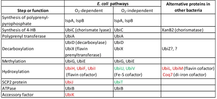
Overall, the large diversity of combination of enzymes used to synthesize UQ in various 370 environmental conditions (Table 1) leads us to refer to UQ biosynthesis pathways and not anymore to a 371 single pathway, as already proposed by Degli Esposti 70. We envision that even more UQ pathways will be 372 revealed by systemic bioinformatic approaches aimed at studying the variations of UQ biosynthesis in the 373 ever expanding diversity of bacterial genomes available. Let’s mention here that the task faces several 374 difficulties, one of which is that only some ubi genes tend to group into operonic structures whereas others 375 are dispersed around the chromosome 17. 376 Table 1: Protein composition of the bacterial UQ biosynthetic pathways. green: proteins involved only in 377
the O2‐independent pathway; red: proteins involved only in the O2‐dependent pathway, ?: suspected 378 existence of unidentified alternative proteins 379 380 381 5) An evolutionary perspective on (ubi)quinone biosynthetic pathways 382 The rise of O2 concentrations on Earth caused a shift from globally reducing to oxidizing conditions 383
around 2.4 billion years ago 74. This transition had far‐reaching consequences, notably for quinones. 384
Indeed, the low potential MK, which was present at the time of the great oxidation event, is readily 385
oxidized by O2 75. Thus, it was proposed that microorganisms had to evolve higher potential quinones, like 386
UQ and PQ, to sustain electron transport in bioenergetic chains operating under oxidizing conditions 75. 387
Step or function O2‐dependent O2‐independent
Synthesis of polyprenyl‐
pyrophosphate IspA, IspB IspA, IspB
Synthesis of 4‐HB UbiC (chorismate lyase) UbiC XanB2 (chorismatase) Polyprenyl transferase UbiA UbiA
Decarboxylation UbiD (decarboxylase) UbiX (flavin prenyltransferase) UbiD UbiX UbiZ?, ?
Methylation UbiG, UbiE UbiG, UbiE Hydroxylation UbiH, UbiF, UbiI (flavin cofactor) UbiU, UbiV (Fe‐S cofactor) UbiL, UbiM (flavin cofactor) Coq7 (di‐iron cofactor) SCP2 protein UbiJ UbiT
ATPase UbiB UbiB
Accessory factor UbiK
E. coli pathways Alternative proteins in other bacteria
This scenario is in line with the presence of O2‐requiring steps, respectively three and one, in the 388
biosynthetic pathways for UQ and PQ (Figure 3). However, our recent discovery of an O2‐independent 389
pathway for UQ production, widespread across proteobacterial lineages, suggests that UQ biosynthesis 390
might have emerged in a less favorable O2 context than previously thought 46. One way to tackle the 391 question of the relative origins of the quinone pathways is to study the evolution of the involved enzymes 392 provided homologs are shared between pathways. 393 394 395 396 397 398 399 Figure 3: Homology between bacterial UQ‐ and cyanobacterial PQ‐ pathways. A) Biosynthetic pathway of 400
UQ in Escherichia coli with enzymes specific of the O2‐dependent and O2‐independent pathways in red and 401
green respectively. B) Biosynthetic pathway of PQ in the cyanobacterium Synechocystis sp. PCC 6803. 402
Reactions 1‐3 in the UQ pathway and 1’‐3’ in the PQ pathway are catalyzed by homologous enzymes. 403 Proposed candidates for the PQ pathway (Slr1300? and Sll0418?) are homologous to UbiH and UbiG (see 404 text). Enzymes with homologs in MK pathways are designated with (*). 405 406 5.1 Evolution of the UQ and PQ pathways 407 It should be possible to address the relative appearance of the UQ and the PQ pathways since they 408
share several homologs. Here, we consider only the cyanobacterial PQ pathway which consist of six 409
reactions (Figure 3), as opposed to the pathway found in plants which is entirely different 76. In the 410 cyanobacterium Synechocystis sp., the first three steps of PQ biosynthesis involve homologs to UbiC, UbiA, 411 and UbiD – UbiX of the UQ pathway: respectively, the chorismate lyase Sll1797, the 4‐HB prenyltransferase 412 Slr0926, and the decarboxylase ‐ flavin prenyltransferase Sll0936 ‐ Slr1099 77,78 (Figure 3). The following 413 hydroxylation and methylation steps are still to be experimentally validated, but candidates have been 414 proposed (Slr1300 and Sll0418) based on their homology to UbiH and UbiG enzymes of the UQ pathway 415 79. Degli Esposti conducted a phylogenetic analysis of the UbiA, ‐C, ‐D, ‐H homologs and proposed that the 416 UQ pathway derived from the PQ pathway and appeared twice independently in Alphaproteobacteria and 417
in Zetaproteobacteria 70. Yet the trees built in this study are missing outgroups to root the phylogenies and 418 as such do not definitively address the question of the relative origins of the UQ and PQ pathways 70. 419 420 5.2 Relationships between the UQ and the MK pathways 421 Two pathways are known for the biosynthesis of MK 9: a fully characterized, long‐known “classical MK 422
pathway” and a still incomplete, more recently identified “futalosine pathway” 80. The classical MK 423
pathway has only two steps related to the UQ pathway: the prenylation step catalyzed by the 424
prenyltransferase MenA (homologous to UbiA) and the methylation of the aromatic ring catalyzed by the 425
literally shared enzyme MenG/UbiE. The characterized mqnA‐E genes are specific to the futalosine 426 pathway 81, but the still putative MqnP, MqnL, MqnM, and UbiE/MenG have homologs in the UQ pathway 427 (UbiA, UbiD and UbiX, respectively) 71,80. These later mqnP, ‐L, ‐M genes were found to strictly co‐occur 428 with mqnA‐E in many bacterial genomes, which reinforces their potential to participate in the futalosine 429 pathway 71. In 2014, Zhi and colleagues observed that the futalosine pathway was found in more phyla of 430
Bacteria and Archaea than the classical MK pathway 11. Furthermore, phylogenies of MenB, ‐C, ‐F 431
suggested that the classical MK pathway was acquired in Archaea, specifically in Halobacteriaceae, as a 432
result of lateral gene transfers from bacteria. In contrast, phylogenies for the MqnA, ‐D, and ‐C enzymes 433
(specific to the futalosine pathway) globally retrieved the delineation of major bacterial and archaeal 434 lineages, suggesting a vertical inheritance of the futalosine pathway and an early emergence predating 435 that of the classical pathway 11. 436 In 2016, Ravcheev and Thiele built phylogenies for genes of the two MK, and the UQ pathways 71. Their 437 trees showed that homologs from the different pathways separated well (including those of the candidate 438 MqnP, ‐L and –M, homologs of UQ enzymes). Interestingly, the only enzyme supposedly shared by the 439 three pathways, the prenyltransferase UbiA/MenA/MqnP family, had a phylogeny displaying a dichotomy 440 between the classical MK pathway on one side, and the futalosine and UQ pathways on another side 71. 441 However, in the tree of the methyltransferase family (UbiE/MenG), candidate enzymes of the futalosine 442 pathway positioned within those of the classical MK pathway, and apart from those of the UQ pathway. 443 The authors therefore suggested that the likely younger pathway of UQ evolved from parts of the two pre‐ 444
existing MK pathways, with some enzymes being more closely related to the futalosine pathway, and 445
others to the classical pathway. 446
Future studies in the context of recent discoveries, including that of new pathways (e.g. the O2‐ 447 independent UQ pathway) or new taxonomic groups of Archaea and Bacteria 82, are very likely to further 448 enlighten the origins of quinones. Elucidating the evolutionary relationships of the quinone pathways is 449
indeed important as it bears strong implications for understanding the evolution of bioenergetics and 450 adaptation to extant oxidizing environments. 451 452 6) Conclusion and Perspectives 453
Recent results have significantly expanded our view of the biosynthesis of UQ in bacteria. Several 454
functional homologs have now been identified at various steps (Table 1) and a pathway independent from 455
O2 has been characterized 46. The first proof of a supramolecular structuration of the E. coli O2‐dependent 456
UQ pathway was recently provided with the characterization of the Ubi complex 33. Whether such 457
multiprotein complexes exist or not in other bacterial species and how they accommodate the variability 458
of the constituting proteins (notably the hydroxylases) remains to be investigated. Understanding the 459 regulation of the various UQ pathways and establishing their cellular localization will also be of interest. 460 Indeed, we may expect the UQ biosynthesis apparatus to localize close to active bioenergetic enzymes, 461 and some of them adopt a specific localization, as recently observed for the fumarate dehydrogenase and 462 nitrate reductase in respiring E. coli 83. Whether the UQ pathways indeed originated from the MK pathways 463 71 and how they evolved in the past 2 billion years is also a challenging and interesting question. 464
To obtain a satisfactory understanding of the composition, regulation and evolution of the UQ 465
pathways across bacteria, it will certainly be fruitful to combine biochemical and bioinformatic approaches 466
in order to extract information from the multiple genomes now available in public databases. Besides 467
increasing our basic knowledge of UQ pathways, such studies will also benefit bioengineering projects 468
aimed at increasing the production of UQ 84 or that of related natural products like antroquinonol, a 469
molecule currently in clinical trials for non‐small‐cell lung cancer 85. In addition, a better understanding of 470
the UQ pathways may refine possible strategies to target them in order to develop novel antibiotics, and 471
may also provide valuable information to help pinpoint the nature of the bacterial ancestor of 472 mitochondria 86. 473 474 Acknowledgments: This work was supported by the Agence Nationale de la Recherche (ANR), projects 475 (An)aeroUbi ANR‐15‐CE11‐0001‐02, O2‐taboo ANR‐19‐CE44‐0014, by the Grenoble Alpes Data Institute 476
funded under the “Investissements d’avenir” program (ANR‐15‐IDEX‐02), by the Centre National de la 477 Recherche Scientifique (CNRS) and by the University Grenoble Alpes (UGA). 478 References 479
(1) Nowicka, B.; Kruk, J. Occurrence, Biosynthesis and Function of Isoprenoid Quinones.
480
Biochim Biophys Acta 2010, 1797 (9), 1587–1605. 481
https://doi.org/10.1016/j.bbabio.2010.06.007.
482
(2) Collins, M. D.; Jones, D. Distribution of Isoprenoid Quinone Structural Types in Bacteria
483
and Their Taxonomic Implication. Microbiol Rev 1981, 45 (2), 316–354.
484
(3) Hiraishi, A. Isoprenoid Quinones as Biomarkers of Microbial Populations in the
485
Environment. J Biosci Bioeng 1999, 88 (5), 449–460.
486
(4) Kunihiro, T.; Veuger, B.; Vasquez-Cardenas, D.; Pozzato, L.; Le Guitton, M.; Moriya, K.;
487
Kuwae, M.; Omori, K.; Boschker, H. T. S.; van Oevelen, D. Phospholipid-Derived Fatty
488
Acids and Quinones as Markers for Bacterial Biomass and Community Structure in Marine
489
Sediments. PLoS One 2014, 9 (4), 14. https://doi.org/10.1371/journal.pone.0096219.
490
(5) Becker, K. W.; Elling, F. J.; Schroder, J. M.; Lipp, J. S.; Goldhammer, T.; Zabel, M.; Elvert,
491
M.; Overmann, J.; Hinrichs, K. U. Isoprenoid Quinones Resolve the Stratification of Redox
492
Processes in a Biogeochemical Continuum from the Photic Zone to Deep Anoxic Sediments
493
of the Black Sea. Appl. Environ. Microbiol. 2018, 84 (10), 20.
494
https://doi.org/10.1128/aem.02736-17.
495
(6) Schoepp-Cothenet, B.; van Lis, R.; Atteia, A.; Baymann, F.; Capowiez, L.; Ducluzeau, A.
496
L.; Duval, S.; ten Brink, F.; Russell, M. J.; Nitschke, W. On the Universal Core of
497
Bioenergetics. Biochimica et biophysica acta 2013, 1827 (2), 79–93.
498
https://doi.org/10.1016/j.bbabio.2012.09.005.
499
(7) VanHellemond, J. J.; Klockiewicz, M.; Gaasenbeek, C. P. H.; Roos, M. H.; Tielens, A. G.
500
M. Rhodoquinone and Complex II of the Electron Transport Chain in Anaerobically
501
Functioning Eukaryotes. Journal of Biological Chemistry 1995, 270 (52), 31065–31070.
502
https://doi.org/10.1074/jbc.270.52.31065.
503
(8) Singh, P. K.; Sarwar, M.; Maklashina, E.; Kotlyar, V.; Rajagukguk, S.; Tomasiak, T. M.;
504
Cecchini, G.; Iverson, T. M. Plasticity of the Quinone-Binding Site of the Complex II
505
Homolog Quinol:Fumarate Reductase. J. Biol. Chem. 2013, 288 (34), 24293–24301.
506
https://doi.org/10.1074/jbc.M113.487082.
507
(9) Kawamukai, M. Biosynthesis and Applications of Prenylquinones. Biosci Biotechnol
508
Biochem 2018, 82 (6), 963–977. https://doi.org/10.1080/09168451.2018.1433020. 509
(10) Salinas, G.; Langelaan, D. N.; Shepherd, J. N. Rhodoquinone in Bacteria and Animals: Two
510
Distinct Pathways for Biosynthesis of This Key Electron Transporter Used in Anaerobic
511
Bioenergetics. BBA Bioenergetics 2020.
512
(11) Zhi, X. Y.; Yao, J. C.; Tang, S. K.; Huang, Y.; Li, W. J. The Futalosine Pathway Played an
513
Important Role in Menaquinone Biosynthesis during Early Prokaryote Evolution. Genome
514
Biol Evol 2014, 6 (1), 149–160. https://doi.org/10.1093/gbe/evu007. 515
(12) Stefely, J. A.; Pagliarini, D. J. Biochemistry of Mitochondrial Coenzyme Q Biosynthesis.
516
Trends in biochemical sciences 2017, 42 (10), 824–843. 517
https://doi.org/10.1016/j.tibs.2017.06.008.
518
(13) Wang, Y.; Hekimi, S. The Complexity of Making Ubiquinone. Trends in endocrinology and
519
metabolism: TEM 2019. https://doi.org/10.1016/j.tem.2019.08.009. 520
(14) Awad, A. M.; Bradley, M. C.; Fernandez-Del-Rio, L.; Nag, A.; Tsui, H. S.; Clarke, C. F.
521
Coenzyme Q10 Deficiencies: Pathways in Yeast and Humans. Essays in biochemistry 2018.
522
https://doi.org/10.1042/EBC20170106.
523
(15) Salviati, L.; Trevisson, E.; Doimo, M.; Navas, P. Primary Coenzyme Q10 Deficiency. In
524
GeneReviews®; Adam, M. P., Ardinger, H. H., Pagon, R. A., Wallace, S. E., Bean, L. J., 525
Stephens, K., Amemiya, A., Eds.; University of Washington, Seattle: Seattle (WA), 2017.
526
(16) Soballe, B.; Poole, R. K. Microbial Ubiquinones: Multiple Roles in Respiration, Gene
527
Regulation and Oxidative Stress Management. Microbiology-(UK) 1999, 145, 1817–1830.
528
(17) Aussel, L.; Pierrel, F.; Loiseau, L.; Lombard, M.; Fontecave, M.; Barras, F. Biosynthesis
529
and Physiology of Coenzyme Q in Bacteria. Biochimica et biophysica acta 2014, 1837 (7),
530
1004–1011. https://doi.org/10.1016/j.bbabio.2014.01.015.
531
(18) Sevin, D. C.; Sauer, U. Ubiquinone Accumulation Improves Osmotic-Stress Tolerance in
532
Escherichia Coli. Nat Chem Biol 2014, 10 (4), 266–272.
533
https://doi.org/10.1038/nchembio.1437.
534
(19) Tempelhagen, L.; Ayer, A.; Culham, D. E.; Stocker, R.; Wood, J. M. Cultivation at High
535
Osmotic Pressure Confers Ubiquinone 8-Independent Protection of Respiration on
536
Escherichia Coli. J. Biol. Chem. 2020, 295 (4), 981–993.
537
https://doi.org/10.1074/jbc.RA119.011549.
538
(20) Eriksson, E. K.; Edwards, K.; Grad, P.; Gedda, L.; Agmo Hernández, V. Osmoprotective
539
Effect of Ubiquinone in Lipid Vesicles Modelling the E. Coli Plasma Membrane.
540
Biochimica et Biophysica Acta (BBA) - Biomembranes 2019, 1861 (7), 1388–1396. 541
https://doi.org/10.1016/j.bbamem.2019.04.008.
542
(21) Agrawal, S.; Jaswal, K.; Shiver, A. L.; Balecha, H.; Patra, T.; Chaba, R. A Genome-Wide
543
Screen in Escherichia Coli Reveals That Ubiquinone Is a Key Antioxidant for Metabolism
544
of Long Chain Fatty Acids. The Journal of biological chemistry 2017, 292 (49), 20086–
545
20099. https://doi.org/10.1074/jbc.M117.806240.
546
(22) Siebert, M.; Severin, K.; Heide, L. Formation of 4-Hydroxybenzoate in Escherichia Coli:
547
Characterization of the UbiC Gene and Its Encoded Enzyme Chorismate Pyruvate-Lyase.
548
Microbiology 1994, 140 ( Pt 4), 897–904. 549
(23) Melzer, M.; Heide, L. Characterization of Polyprenyldiphosphate-4-Hydroxybenzoate
550
Polyprenyltransferase from Escherichia-Coli. Biochimica Et Biophysica Acta-Lipids and
551
Lipid Metabolism 1994, 1212 (1), 93–102. https://doi.org/10.1016/0005-2760(94)90193-7. 552
(24) Kainou, T.; Okada, K.; Suzuki, K.; Nakagawa, T.; Matsuda, H.; Kawamukai, M. Dimer
553
Formation of Octaprenyl-Diphosphate Synthase (IspB) Is Essential for Chain Length
554
Determination of Ubiquinone. J Biol Chem 2001, 276 (11), 7876–7883.
555
https://doi.org/10.1074/jbc.M007472200.
556
(25) Han, X.; Chen, C.-C.; Kuo, C.-J.; Huang, C.-H.; Zheng, Y.; Ko, T.-P.; Zhu, Z.; Feng, X.;
557
Wang, K.; Oldfield, E.; Wang, A. H.-J.; Liang, P.-H.; Guo, R.-T.; Ma, Y. Crystal Structures
558
of Ligand-Bound Octaprenyl Pyrophosphate Synthase from Escherichia Coli Reveal the
559
Catalytic and Chain-Length Determining Mechanisms. Proteins 2015, 83 (1), 37–45.
560
https://doi.org/10.1002/prot.24618.
561
(26) Huang, H.; Levin, E. J.; Liu, S.; Bai, Y.; Lockless, S. W.; Zhou, M. Structure of a
562
Membrane-Embedded Prenyltransferase Homologous to UBIAD1. PLoS biology 2014, 12
563
(7), e1001911. https://doi.org/10.1371/journal.pbio.1001911.
564
(27) Cheng, W.; Li, W. Structural Insights into Ubiquinone Biosynthesis in Membranes. Science
565
2014, 343 (6173), 878–881. https://doi.org/10.1126/science.1246774. 566
(28) Loiseau, L.; Fyfe, C.; Aussel, L.; Hajj Chehade, M.; Hernandez, S. B.; Faivre, B.; Hamdane,
567
D.; Mellot-Draznieks, C.; Rascalou, B.; Pelosi, L.; Velours, C.; Cornu, D.; Lombard, M.;
568
Casadesus, J.; Pierrel, F.; Fontecave, M.; Barras, F. The UbiK Protein Is an Accessory
569
Factor Necessary for Bacterial Ubiquinone (UQ) Biosynthesis and Forms a Complex with
570
the UQ Biogenesis Factor UbiJ. The Journal of biological chemistry 2017, 292 (28), 11937–
571
11950. https://doi.org/10.1074/jbc.M117.789164.
572
(29) White, M. D.; Payne, K. A. P.; Fisher, K.; Marshall, S. A.; Parker, D.; Rattray, N. J. W.;
573
Trivedi, D. K.; Goodacre, R.; Rigby, S. E. J.; Scrutton, N. S.; Hay, S.; Leys, D. UbiX Is a
574
Flavin Prenyltransferase Required for Bacterial Ubiquinone Biosynthesis. Nature 2015, 522
575
(7557), 502-+. https://doi.org/10.1038/nature14559.
576
(30) Payne, K. A. P.; White, M. D.; Fisher, K.; Khara, B.; Bailey, S. S.; Parker, D.; Rattray, N. J.
577
W.; Trivedi, D. K.; Goodacre, R.; Beveridge, R.; Barran, P.; Rigby, S. E. J.; Scrutton, N. S.;
578
Hay, S.; Leys, D. New Cofactor Supports Alpha,Beta-Unsaturated Acid Decarboxylation
579
via 1,3-Dipolar Cycloaddition. Nature 2015, 522 (7557), 497-+.
580
https://doi.org/10.1038/nature14560.
581
(31) Marshall, S. A.; Fisher, K.; Cheallaigh, A. N.; White, M. D.; Payne, K. A. P.; Parker, D. A.;
582
Rigby, S. E. J.; Leys, D. Oxidative Maturation and Structural Characterization of Prenylated
583
FMN Binding by UbiD, a Decarboxylase Involved in Bacterial Ubiquinone Biosynthesis.
584
Journal of Biological Chemistry 2017, 292 (11), 4623–4637. 585
https://doi.org/10.1074/jbc.M116.762732.
586
(32) Marshall, S. A.; Payne, K. A. P.; Fisher, K.; White, M. D.; Cheallaigh, A. N.; Balaikaite, A.;
587
Rigby, S. E. J.; Leys, D. The UbiX Flavin Prenyltransferase Reaction Mechanism
588
Resembles Class I Terpene Cyclase Chemistry. Nature Communications 2019, 10, 2357.
589
https://doi.org/10.1038/s41467-019-10220-1.
590
(33) Hajj Chehade, M.; Pelosi, L.; Fyfe, C. D.; Loiseau, L.; Rascalou, B.; Brugiere, S.;
591
Kazemzadeh, K.; Vo, C. D.; Ciccone, L.; Aussel, L.; Coute, Y.; Fontecave, M.; Barras, F.;
592
Lombard, M.; Pierrel, F. A Soluble Metabolon Synthesizes the Isoprenoid Lipid
593
Ubiquinone. Cell chemical biology 2019, 26 (4), 482-492 e7.
594
https://doi.org/10.1016/j.chembiol.2018.12.001.
595
(34) Zhu, Y.; Jiang, X.; Wang, C.; Liu, Y.; Fan, X.; Zhang, L.; Niu, L.; Teng, M.; Li, X.
596
Structural Insights into the Methyl Donor Recognition Model of a Novel
Membrane-597
Binding Protein UbiG. Scientific reports 2016, 6, 23147. https://doi.org/10.1038/srep23147.
598
(35) Zhu, Y.; Wu, B.; Zhang, X.; Fan, X.; Niu, L.; Li, X.; Wang, J.; Teng, M. Structural and
599
Biochemical Studies Reveal UbiG/Coq3 as a Class of Novel Membrane-Binding Proteins.
600
The Biochemical journal 2015, 470 (1), 105–114. https://doi.org/10.1042/BJ20150329. 601
(36) Poon, W. W.; Barkovich, R. J.; Hsu, A. Y.; Frankel, A.; Lee, P. T.; Shepherd, J. N.; Myles,
602
D. C.; Clarke, C. F. Yeast and Rat Coq3 and Escherichia Coli UbiG Polypeptides Catalyze
603
Both O-Methyltransferase Steps in Coenzyme Q Biosynthesis. J. Biol. Chem. 1999, 274
604
(31), 21665–21672.
605
(37) Lee, P. T.; Hsu, A. Y.; Ha, H. T.; Clarke, C. F. A C-Methyltransferase Involved in Both
606
Ubiquinone and Menaquinone Biosynthesis: Isolation and Identification of the Escherichia
607
Coli UbiE Gene. J Bacteriol 1997, 179 (5), 1748–1754.
608
(38) Kwon, O.; Kotsakis, A.; Meganathan, R. Ubiquinone (Coenzyme Q) Biosynthesis in
609
Escherichia Coli: Identification of the UbiF Gene. FEMS Microbiol Lett 2000, 186 (2), 157–
610
161.
611
(39) Hajj Chehade, M.; Loiseau, L.; Lombard, M.; Pecqueur, L.; Ismail, A.; Smadja, M.;
612
Golinelli-Pimpaneau, B.; Mellot-Draznieks, C.; Hamelin, O.; Aussel, L.; Kieffer-Jaquinod,