THÈSE
En vue de l'obtention du
DOCTORAT DE L’UNIVERSITÉ DE TOULOUSE Délivré par Institut National Polytechnique De Toulouse Discipline ou spécialité : Ingénieries microbiennes et enzymatique
Présentée et soutenue par Muhammad Hussnain SIDDIQUE Le 05/11/2012
Study of the biosynthesis pathway of the geosmin in Penicillium expansum
JURY
M. AZIZ Aziz Maître de Conférences,
Université de Reims Champagne-Ardenne M. HAFIDI Mohamed Professeur, Université de Bordeaux Mme. MATHIEU Florence Professeur, Université de Toulouse M. LEBRIHI Ahmed Professeur, Université de Toulouse
Ecole doctorale : École doctorale: Sciences Ecologiques, Vétérinaires, Agronomiques et Bioingénieries
Unité de recherche : LGC UMR 5503 (CNRS/UPS/INPT) Directeur de Thèse : Pr. LEBRIHI Ahmed (INP-ENSAT)
Co-Directeur de Thèse : Dr. LIBOZ Thierry (INP-ENSAT)
Acknowledgements
First of all I am thankful to the almighty ALLAH, whose blessings are always with me.
I offer my humble thanks from the deepest core of my heart to Holy Prophet Muhammad (Peace be upon him) who is forever a torch of guidance and knowledge for humanity as a whole.
I have the deepest sense of gratitude to my Saain Gee Soofi Nisar Ahmad Dogar Naqshbandi Khaliqi who has always been a source of elevation in my whole life.
My sincere appreciation goes to my supervisor Professor Ahmed LEBRIHI and co- supervisor Doctor Thierry LIBOZ, whose scientific approach, careful reading and constructive comments were valuable. Their timely and efficient contributions helped me to shape my research work into its final form and I express my sincerest appreciation for their assistance in any way that I may have asked.
I deem it utmost pleasure to avail this opportunity to express the heartiest gratitude and deep sense of obligation to Ahmed LEBRIHI for their dexterous guidance, critical judgment, sympathetic attitude and inspiring efforts to inculcate in me the spirit of motivation during the course of my research work. They were always available whenever I need their assistance and guidance, especially, during thesis writing.
Sincere thanks are due to Professor Florence MATHIEU for her kindness and helping in thesis work.
I also wish to thank the ―Higher Education Commission (H EC)‖ of Pakistan, its leadership and the staff who in their limited resources supported me financially for my studies in France. I must also mention the services provided by SFERE (Société Française d' Exportation des Resources Educatives) to facilitate my living in France.
I fervently extend my zealous thanks to the members of my thesis jury Dr. AZIZ Aziz, Pr. HAFIDI Mohamed, Pr. MATHIEU Florence and Pr. Ahmed LEBRIHI .
I would like to reflect my gratitude to Professor Nasserdine SABAOU, Professor
Atika MEKLAT, Nafees BACHA, Saima MUZAMMIL, Philippe ANSON, Rafik, Hafsa, Elida, Carol, Marion, Safwan, Patricia and Rayenne who always helped me and gave me strong support.
I am also indebted to my friends who were always with me in every situation and helped me morally and I would like to reflect my gratitude particularly Ali, Saqlain, Ramiz, Tusawar, Tausif and Imran.
Finally, I am forever indebted to my family: my father Noor MUHAMMAD and my mother Hameeda BIBI. A special thanks to my wife Dr. Saima MUZAMMIL who always shares my problems with an ultimate solution, and cooperated at each and every crucial step of my life. My humble thanks to my brothers: Shaukat Ali (late), Abdul Razzaq and Muhammad Riaz and my sisters: Shagufta Parveen, Tasleem Kousar and Afshan Sahar, and my niece Tehmina. Last but not the least I feel pleasure to acknowledge of those who love me and whom I love.
I dedicate this thesis in honor of my family especially my dear brother Abdul Razzaq
whose love, affection and confidence enabled me to achieve my goals.
Summary
Résumé ... 7
Abstract ... 8
List of Abbreviations ... 9
List of Tables ... 11
List of Figures ... 12
Chapter I: Literature Review ... 14
1.1. Fungal secondary metabolites ... 15
1.2. Penicillium expansum ... 18
1.2.1. Classification and morphological description ... 18
1.2.2. Hosts ... 21
1.2.3. Secondary metabolites produced by P. expansum ... 21
1.3. Cytochrome P450 monooxygenase ... 23
1.3.1. Characteristics of cytochrome P450s ... 23
1.3.2. Structure of P450 ... 23
1.3.3. Reactions catalyzed by P450s ... 26
1.3.4. Involvement of P450s in biosynthesis of secondary metabolites and their different functions ... 26
1.4. Geosmin ... 27
1.4.1. General characteristics ... 27
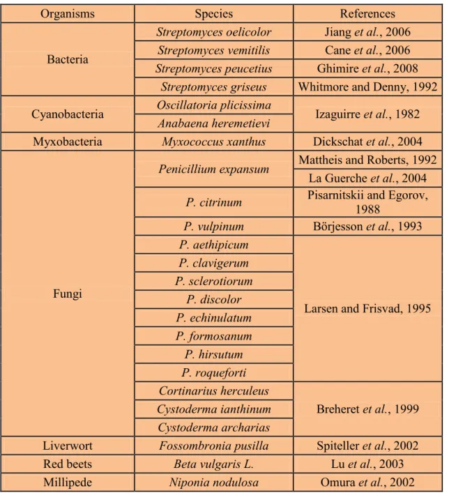
1.4.2. Production of geosmin by microorganisms ... 28
1.4.3. Different methods to analyze geosmins ... 31
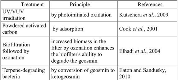
1.4.4. Treatments to control geosmin ... 33
1.4.5. Biosynthesis pathways of gesmin ... 36
1.4.5.1. Biocchemical pathway of geosmin synthesis in bacteria ... 36
1.4.5.2. Genes involved in geosmin biosynthetic pathway in bacteria ... 38
1.4.5.3. Mechanism and stereochemistry of the conversion of farnesyldiphosphate to germacradienol and germacrene D ... 40
Objectives ………42
Chapter III: Results and Discussion ... 63
3.1. Whether cytocrome P450 monooxygenase genes can be involved in geosmin production? ... 64
3.1.1. Bioinformatics analysis to identify germacradienol/geosmin synthase in Penicillium ... 64
3.1.2.Role of P450 in terpens biosynthesis ... 65
3.1.3. Amplification of P. expansum P450 (gpe1)gene sequence by PCR ... 66
3.1.4. Alignment of gpe1 with other cytochrom P450 monooxygenases ... 69
3.1.5. gpe1 gene presence in geosmin producing Penicillium species ... 71
3.2. How to explore different aspects of the gpe1 gene using bioinformatics tools? ... 72
3.2.1. Implication of P450 enzymes in the biosynthesis of different secondary metabolites… ... 72
3.2.2. Conserved domains of cytochrome P450s ... 73
3.2.3.Which functional domains of cytochrome P450 monooxygenase enzymes are present in gpe1. ... 74
3.3. Does the gpe1 gene require for geosmin biosynthesis in P. expansum?. ... 76
3.3.1. Production of mutants by the gene disruption method and their screening by PCRs. ... 76
3.2.3. Production of reverse compliments and their screening by PCRs. ... 77
3.3.3. Quantifictaion of geosmin by gas chromatography-mass spectrometry(GC/MS)..78
3.4. Where does the gpe1 gene intervene in the biosynthesis pathway of geosmin?. ... 79
3.4.1. Dependence of secondary metabolites pathways upon the relative abundance of their precursors. ... 79
3.4.2: Is the biosynthesis pathway of geosmin same in P. expansum as that suggested in bacteria?.. ... 81
3.4.3. Use of Penicillium marneffei genome to know about the neighbor genes of the gpe1. ... 81
3.5. Chapter IV: General Conclusions and Future Prospects. ... 84
References. ... 89
Annexes ... 109
Résumé
La géosmine est un terpénoïde, provoquant un goût moisi-terreux associée à des flaveurs atypiques dans l'eau et le vin. Chez les bactéries, la voie de biosynthèse de la géosmine est bien caractérisée, mais peu de connaissance sont disponibles au sujet de sa biosynthèse chez les eucaryotes, en particulier dans les champignons filamenteux. L'origine de la géosmine dans la vigne est en grande partie attribuable à la présence de Penicillium expansum sur les raisins. Dans cette thèse, afin de mieux comprendre la voie de biosynthèse de la géosmine chez Penicillium expansum, nous avons décrit la caractérisation et l'analyse de "gpe1", un gène codant pour une cytochrome P450 monooxygénase impliquée dans la biosynthèse de la géosmine.
Nous avons démontré que les deux fragments d'ADN: p450-1 et p450-2 appartiennent à un seul gène du cytochrome p450 (gpe1). La séquence d'acides aminés déduite de gpe1 a une identité moyenne de 40 % avec les enzymes PbP450-2 et P450-4 qui ont été trouvées impliquées respectivement dans la synthèse d'indole diterpène et dans la synthèse des gibbérellines. Les amplifications par PCR effectuée sur quatorze espèces de Penicillium ont montré que seules les espèces producteurices de la géosmine ont donné le même fragment de ~1,2 kb que gpe1. L'analyse du gène gpe1 nous a permis d'identifier la présence de certains domaines conservés de cytochromes P450 monooxygénases. Ensuite, la caractérisation fonctionnelle du gène gpe1 chez P. expansum M2230 a été décrite. Nous avons montré que les mutants de gpe1 ont perdus leur pouvoir de produire la géosmine alors que les révertants de gpe1 ont rétablis leur pouvoir de production. Enfin, nous avons démontré qu'une polykétide synthase putative et une putative NRPS sont présentes sur le côté droit du gène gpe1 proposant que le gène gpe1 pourrait être une partie d'un "Cluster"
codant pour la biosynthèse de métabolites secondaires.
Mots clés: Cytochrome P450 monooxygénase, géosmine, gpe1, Penicillium
expansum
.Abstract
Geosmin is a terpenoid, an earthy-musty compound associated with off-flavors in water and wine. In bacteria, the biosynthesis pathway of geosmin is well characterized, but little is known about its biosynthesis in eukaryotes, especially in filamentous fungi. The origin of geosmin in grapevine is largely attributable to the presence of Penicillium expansum on grapes. In this thesis, we have described the characterization and analysis of
― gpe1 ‖ , a gene encoding a cytochrome P450 monooxygenase probably involved in the biosynthesis of geosmin in P. expansum M2230, in order to better understand of the biosynthesis pathway of geosmin in this species.
. We demonstrated that the two DNA fragments i.e. p450-1 and p450-2 belong to a single cytochrome p450 gene (gpe1). We showed that the deduced amino acid sequence of gpe1 has an average identity of 40 % with PbP450-2 and P450-4 enzymes which have been found involved in indole diterpene synthesis and in gibberellin synthesis respectively. Then, the results of PCRs performed on the fourteen Penicillium species showed that only Penicillium species which were producers of geosmin gave the same fragment of ~1.2 kb like gpe1. Analysis of the gpe1 gene enabled us to identify the presence of some conserved domains of cytochromes P450 monoxygenases in the amino acid sequence of gpe1. Then, the functional characterization of the gpe1 gene in P. expansum M2230 was described. We illustrated that the mutants of gpe1 lost their potential to produce geosmin whereas the reverse complements of gpe1 restored their potential to produce geosmin. Finally, we demonstrated that a putative polyketide synthase and a putative NRPS-like enzyme are present on the right side of the gpe1 gene suggesting that gpe1 gene might be the part of a gene cluster encoding the biosynthesis of secondary metabolites.
Key words: Cytochrome P450 monooxygenase, geosmin, gpe1, Penicillium
expansum.
List of Abbreviations
2-MIB: 2-methylisoborneol aa: Amino acid
BLAST: Basic local alignment search tool bp: Base pair
CoA: Coenzyme-A
CPR: Cytochrome P450 reductase CTAB: Cetyltrimethylammonium bromide CYA Czapek yeast agar
CYP Cytochrome P450 DNA: Deoxyribonucleic acid
EDTA: Ethylenediaminetetraacetic acid ER: Endoplasmatic reticulum
FAD: Flavine adenine dinucleotide FCPD: Fungal cytochrome P450 database FMN: Flavin mononucleotide
FPP: Farnesyl diphosphate GAC: Granular activated carbon
GC–MS: Gas chromatography-mass spectrometry GGPP: Geranylgeranyl diphosphate
hph: Hygromycin B phosphotransferase LQ: Limit of quantification
MEA: Malt Agar Extract
NADPH: Nicotinamide adenine dinucleotide phosphate (reduced form) NRPS: Non-ribosomal peptide synthase
OD: Optical Density
PAC: Powdered activated carbon
PCR: Polymerase chain reaction
PKS: Polyketide synthase
RNA: Ribonucleic acid
SDS: Sodium dodecyl sulfate TOC: Total organic carbon UV: Ultraviolet
YES: Yeast extract sucrose
List of Tables
Table 1: Production of geosmin by different organisms ... 30 Table 2: Extraction / enrichment techniques used to preconcentrate geosmin prior
to quantification by gas chromatography-mass spectrometry ... 32
Table 3: Different treatments to control geosmin in water ... 35
Table 4: Dosage of geosmin ... 79
List of Figures
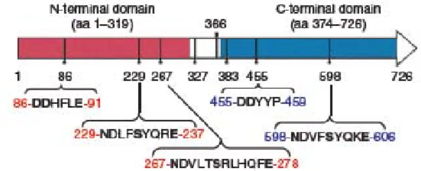
Figure 1: Penicillium expansum A-C, 7-days-old colonies on A. CYA, B. MEA, C. YES at 25 °C. ... 20 Figure 2: Penicillium expansum, D-H. Conidiophores. I. Conidia. Scale bar = 10 µm. 20 Figure 3: Schematic representation of the eukaryotic endoplasmatic reticulum type cytochrome P450 enzyme system ... 25 Figure 4: Chemical structure of geosmin ... 28 Figure 5: Simplified biosynthetic scheme for the formation of geosmin in streptomycetes and myxobacteria. ... 37 Figure 6: Mechanism of cyclization of FPP (2) to Germacradienol (3), Germacrene D (4), Hydrocarbon 5, and Geosmin (1) ... 39 Figure 7: Organization of protein domain and conserved Mg
2+-binding motifs in S.
coelicolor germcradienol-geosmin synthase ... 40
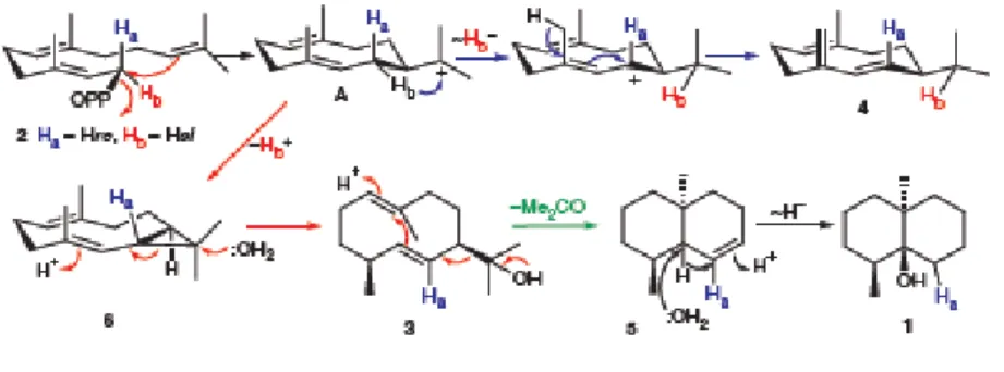
Figure 8: Mechanism and stereochemistry of the cyclization of FPP (2) to germacradienol
(3), germacrene D (4), octalin (5) and geosmin (1)... 41
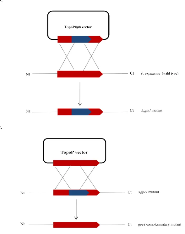
Figure 9: Schematic representation of transformation vector formation and gpe1 gene
disruption. ... 58
Figure 10: A BLAST search with the protein sequence of the S. peucetius strept13
(ATCC 27952) as a query did not show any gene having homology with the genes
encoding germacradienol/geosmin synthase, in the genus Penicillium ... 65
Figure 11: Alignment of the deduced amino acid sequences of p450-1 and p450-2 of
Penicillium expansum with CYP619C2 and CYP619C3 of Aspergillus clavatus involved
in patulin biosynthesis ... 67
Figure 12: Alignment of the deduced amino acid sequence of p450-1 and p450-2 of Penicillium expansum with P450-4 of Gibberella fujikuroi involved in the biosynthesis of gibberellins ... 68 Figure 13: Amplification of the gpe1 using the primers mhsF and mhsR in P. expansum.
... 69 Figure 14: Alignment of the deduced amino acid sequence of gpe1 with other cytochrome P450 monooxygenases ... 70 Figure 15: gpe1 PCR amplification on geosmin productive and non-productive Penicillium species ... 71 Figure 16: Typical features of an ER-bound P450 protein ... 74 Figure17: Conserved domains of cytochromes P450 monooxygenases present in gpe .. 75 Figure 18: InterPro Scan visual output showing different domains of gep1………….76 Figure 19: PCR transformants screening ... 78 Figure 20: Metabolic pathway diagram, the mevalonate (MVA) and non-mevalonate (MEP) pathways that link geosmin synthesis ... 80 Figure21: A simple diagram showing neighbor genes of the particular gene of
Penicillium marneffei showing highest resemblance with gpe1of P. expansum
as a result of BLAST search ... 83
.
Chapter I
Literature Review
1. Literature Review
1.1. Fungal Secondary Metabolite
The primary metabolism of an organism is the summation of an interrelated series of enzyme-catalyzed chemical reactions (both degradative and synthetic) which provide the organism with its energy, its synthetic intermediates and its key macromolecules such as protein and DNA. On the other hand, secondary metabolism involves mainly synthetic processes whose end-products, the secondary metabolites, play no obvious role in the internal economy of the organism.
Plants and microorganisms produce a vast number of natural compounds known as secondary metabolites. Kossel (1891) introduced the concept of ―secondary metaboli tes‖ to distinguish these compounds from primary metabolites that these products are not necessary for the growth, survival or reproduction of their producers. Secondary metabolites are substances of limited molecular weight (normally < 3000 Daltons) which display an enormous structural diversity. However each of them is synthesized only by a limited taxonomic group of organisms whereas primary metabolites are found in all living organisms since they perform essential functions in growth and development.
Many species in the fungal kingdom have unique and unusual biochemical
pathways. Important pharmaceuticals such as penicillin, cyclosporin and statins; potent
poisons, including aflatoxins and trichothecenes; and some Janus-faced metabolites that are
both toxic and pharmaceutically useful, such as the ergot alkaloids are the products of these
pathways. All of these natural products, along with many other low-molecular-weight fungal
metabolites, are classified together as secondary metabolites. Secondary metabolites are
produced as families of related compounds at restricted parts of the life cycle, with
production often correlated with a specific stage of morphological differentiation. Secondary
produces each metabolite and the producer organisms can grow without synthesizing these metabolites. Secondary metabolites are, often synthesized after active growth has ceased, which do not have an obvious function in producer species (Keller et al., 2005).
In fungi, in the case of the Basidiomycetes and the larger Ascomycetes, secondary metabolites may be obtained simply by extraction of the organism collected in the field. But the great advantage of the fungi as sources of secondary metabolites is their ability to produce the compounds on aqueous media. As a result, secondary metabolites of diverse type are conveniently available in the laboratory for chemical, biochemical and biological studies, and a few are manufactured on a commercial scale. In some cases the same secondary metabolites have been obtained from fruiting-bodies and from aqueous culture of Basidomycetes, though in most cases the compounds have so far only been obtained from one of the sources. The laboratory cultures of Basidiomycetes are, of course, mycelial;
Basidiomycetes do not normally form fruiting-bodies under laboratory conditions and in some cases have resisted all attempts to induce them to do so. In aqueous cultures, secondary metabolites accumulate both in the medium and in the mycelium. For related compounds, the distribution between medium and mycelium can often be correlated with water-solubility, though this apparent correlation may be a result of some other factor such as ease of transport across cell membranes (Turner, 1971).
Fungal secondary metabolites encompass over 30,000 known compounds with an
extremely diverse array of chemical structures. It is intriguing that all these secondary
metabolites originate from a few common biosynthetic pathways utilizing precursors (small
biosynthetic units or building blocks) formed during primary metabolism. The intermediates
resulting from condensation of these small biosynthetic units are further elaborated
(―tailored ‖ or ―decorate d‖ ) by numerous enzyme-catalyzed reactions, leading to products
with a diversity of structures. Thus, fungal secondary metabolites are conveniently classified
based on their biosynthetic origin as polyketides (e.g. aflatoxin and fumonisins), nonribosomal peptides (e.g. sirodesmin, peramine and siderophores such as ferricrocin), terpenes (e.g. T-2 toxin, deoxynivalenol (DON)), and indole alkaloids (e.g. paxilline, fumigaclavines and fumitremorgens) (Keller et al., 2005; Gunatilaka, 2006).
Biosynthesis of fungal secondary metabolites often involves elaborate biochemical pathways and is regulated by a group of genes known as biosynthetic genes. The insights that have been gained from recent advances in genetics, genomics, molecular biology, and bioinformatics have contributed to the understanding and manipulation of these genes for improved production, or inhibition of production, of fungal secondary metabolites.
Fungal secondary metabolites are well known for their biological activity and represent some of today's important and useful pharmaceuticals and agrochemicals. Among the pharmaceuticals, most noteworthy are penicillins, cephalosporins, and fusidic acid with antibacterial activity; echinocandin B, pneumocandins, griseofulvin, and strobilurins with antifungal activity; integric acid and integresone with antiviral activity; cyclosporin A and mycophenolic acid with immunosuppressive activity; fumagillin and rhizoxin with antitumor activity; lovastatin and pravastatin with cholesterol-lowering activity; and ergot alkaloids (for example, ergotamine) with antimigraine activity. Gibberellins and zearalenones are fungal secondary metabolites used in agriculture as plant growth hormones and in animal husbandry as growth promoters, respectively. Some fungal secondary metabolites such as mycotoxic aflatoxins and mutagenic fusarin C possess potent toxic and carcinogenic activities and are therefore important in human, animal, and plant health (Vining, 1990; Fox and Howlett, 2008) whereas some volatile non-toxic secondary metabolites such as geosmin and 2-methylisoborneol (2-MIB) haves also concerns for humans as they are responsible for off-flavors in drinking water and wines (Gerber, 1979;
Darriet et al., 2000).
In some fungi, secondary metabolism (the process that results in the production of secondary metabolites) has been found to commence during the stationary or resting phase of their development and is often associated with sporulation and colony formation. Some well-documented functions of fungal secondary metabolites include enhancement of spore survival by acting as virulence factors and protecting against ultraviolet (UV) light, and augmentation of their fitness and competitive ability against other fast-growing organisms.
Fungal metabolites associated with sporulation may activate sporulation (for example, linoleic acid analogs produced by Aspergillus nidulans), provide pigmentation required for sporulation structures (for example, melanins produced by Alternaria alternata), or have toxic properties to ward off competing organisms (for example, mycotoxins produced by some Aspergillus species). The relationship between production of secondary metabolites and regulation of asexual sporulation by a G-protein–mediated growth pathway in Aspergillus species was established over a decade ago. Also, it has been speculated that secondary metabolites in fungi function as metal chelators (combining with metal ions and removing them from their sphere of action), which is important in mineral nutrition, and that pathways leading to their formation act as safety-valve shunts that prevent the accumulation of toxic intermediates of primary metabolism under conditions of unbalanced growth (Calvo et al., 2002; Fox and Howlett, 2008).
1.2. Penicillium expansum
1.2.1. Classification and morphological description
Penicillium expansum is the typical fungus of the genus Penicillium and is therefore
also one of the most studied species in the genus (Pitt, 1979). This fungus belongs to phylum
Ascomycota, class Eurotiomycetes, subclass Eurotiomycetidae, order Eurotiales and family
Trichocomaceae.
After 7 days of incubation on Petri dish containing CYA (Czapek Yeast Agar) medium at 25 °C, a colony of P. expansum attains a diameter of 26-50 mm, on MEA (Malt Agar Extract) medium, its colony could be 16-34 mm in diameter, while on YES (Yeast Extract Sucrose) agar medium, the diameter of a colony could be 38-65 mm (Figure 1) but there is no growth at 37 °C (Frisvad and Samson, 2004). Cultural characteristics of this fungus include: the colonies grow rapidly on the culture media, with radial wrinkles up to 2 mm deep, spore heavily, very variable, from velvety with conidiophores occurring singly to granular with conidiophores grouped together in fascicles or producing quite distinct coremia, often showing radial zonation; white, rapidly becoming dull yellow green to greyish green with the production of conidia (Figure 2); reverse variable, colorless to yellow brown to deep brown. The conidial heads are asymmetric, once or twice branched, elongate, bearing long tangled chains of conidia. The conidiophores are smooth or in some strains slightly roughened, moderately long, up to 400 µm long but occasionally up to 600-700 µm long and 3-3.5 µm wide; branches 15-25 x 2.5-3.5 µm, occasionally longer. But the metulae arising from branches at about the same level, 3 to 6 in number, and about 10-15 x 2-3 µm.
The phialides are in groups of 5-9, often about 8-12 x 2-2.5 µm, occasionally longer. The
conidia are smooth, elliptical to cylindrical when first formed and usually remain elliptical,
generally 4-5 x 2.5-3.5 µm (Link, 1809; Onions, 1966)
(http://www.mycobank.org/MycoTaxo.aspx?Link=T&Rec=159382).
Figure1: Penicillium expansum, A-C. 7-days-old colonies on A. CYA, B. MEA, C.
YES at 25 °C.
Figure 2: Penicillium expansum, D-H. Conidiophores. I. Conidia. Scale bar = 10 µm.
1.2.2. Host
Penicillium expansum is commonly present in soil and in a wide variety of organic material including grains and cereal products, and though generally isolated from mouldy fruit, particularly apples, it also occurs on other pomaceous fruits such as cherries, peaches, pears, grapes, olives, pineapple and sometimes on citrus and avocado. It is also common on walnuts, pecans, hazelnuts and acorns. P. expansum is responsible for the postharvest decay of these fruits leading to important economic losses in the fruit industry (Filtenborg et al., 1996; Karabulat et al., 2002; Karabulat and Bakyal, 2002; Venturini et al., 2002).
1.2.3. Secondary metabolites produced by P. expansum
P. expansum has been reported to produce many secondary metabolites such as:
chaetoglobosins A and C, communesin B which are cytotoxic metabolites (Bridge et al., 1989; Frisvad and Filtenborg, 1989; Frisvad, 1992; Andersen et al., 2004), the bioactive compounds expansolides A and B (Massias et al., 1990; Andersen et al., 2004), an antibiotic penicillic acid (Leistner and Pitt, 1977), roquefortine C which is neurotoxic (Frisvad and Filtenborg, 1983; Bridge et al., 1989), patulin which is carcenogenic, citrinin which is nephrotoxic (Leistner and Pitt, 1977; Frisvad and Filtenborg, 1983; Paterson et al., 1987;
Andersen et al., 2004) and geosmin which is an aromatic volatile secondary metabolite (Mattheis and Roberts, 1992). Among the above mentioned extrolites of P. expansum, chaetoglobosins A and C, penicillic acid, patulin and citrinin belong to polyketides.
Geosmin and expansolides A and B are terpenes whereas roquefortine C and communesin B belong to indole alkaloid family.
In literature, it has been reported that citrinin biosynthesis was originated from a
pentaketide in Penicillium and Aspegillus species (Barber and Staunton, 1980; Sankawa et
al., 1983). It was demonstrated in literature that in the genus Aspergillus, the condensation
of one acetyl coenzyme A (acetyl-CoA) molecule with four malonyl-CoA molecules, followed by the addition of three methyl units has synthesized the citrinin (Colombo et al., 1981; Hill et al., 1981). In the contrary, Hajjaj et al. (1999) revealed that citrinin is formed from a tetraketide precursor arising from the condensation of one acetyl-CoA molecule with three malonyl-CoA molecules in the filamentous fungus Monascus ruber instead of a pentaketide as reported in Penicillium and Aspergillus. The patulin production pathway from the polyketide, 6-methylsalicylic acid (6-MSA) has been established and is thought to involve at least 10 different enzymatic steps (Moake et al., 2005). However, two of the genes namely the 6-methylsalicylic acid synthase (6-msas) gene (Beck et al., 1990) and the isoepoxydon dehydrogenase gene (idh) (Gaucher & Fedeshko, 2000) encoding these enzymes have been cloned and sequenced, both from Penicillium urticae. Precursor feeding experiments revealed that tryptophan, histidine, and mevalonate are involved in the biosynthesis of roquefortine C (Barrow et al., 1979; Gorst-Allman et al., 1982). Garcia- Estrada et al. (2011) cloned 5 genes from a single gene cluster of Penicillium chrysogenum involved in the biosynthesis and secretion of the mycotoxin roquefortine C and proved that the roquefortine C derive from a single pathway. Communesins are of mixed biosynthetic origin, predictably derived from tryptophan, mevalonate, acetate and a methyl group from methionine (Wigley et al., 2006). In bacteria, MEP or/and MVA pathway may lead to the synthesis of the geosmin (Dickschat et al., 2005; Jüttner and Watson, 2007). Biosynthesis of the geosmin has been discussed in detail in the next part.
The cytochtochromes P450 monooxygenases could be involved in the biosynthesis
of geosmin in P. expansum. Therefore, an inclusive introduction of these enzymes has been
given in the following section.
1.3. Cytochrome P450 monooxygenase 1.3.1. Charecteristics of cytochrome P450s
Cytochrome P450 (CYP) genes encode a superfamily of heme-thiolate-containing enzymes. These enzymes are found in all life forms from prokaryotes (archea, bacteria) and lower eukaryotes (fungi and insects) to higher eukaryotes (plants and animals including humans) (Cresnar and Petric, 2011) and reported to be involved in an array of diverse endogenous and exogenous oxidative processes (Guengerich, 1991). Cytochromes P450 are external monooxygenases. Monooxygenases (mixed function oxidases) catalyse the incorporation of a single atom of molecular oxygen into a substrate with the concomitant reduction of the other atom to water. There are two classes of monooxygenases: the internal and the external monooxygenases. Their character as hemoproteins and their unusual spectral properties displaying a typical absorption maximum of the reduced CO-bound complex at 450 nm gave them a name as cytochromes P450: cytochrome stands for a hemoprotein, P for pigment and 450 reflects the absorption peak of the CO complex at 450 nm. The following reaction is catalysed by cytochrome P450 systems:
RH + O
2+ NAD(P)H + H+ → ROH + H
2O + NAD(P)+
A separate electron donating system donates the electrons needed for the oxygen insertion in the substrate molecule (R). The electron donating system is either a two-protein system (adrenodoxin and adrenodoxin reductase) for mitochondrial and prokaryotic P450s or a single protein (cytochrome P450 reductase, CPR) for cytochrome P450 enzymes that are located in the endoplasmatic reticulum (ER). Most fungal cytochrome P450s identified thus far are expected to be located in the ER.
1.3.2. Structure of P450
Three dimensional structures of cytochrome P450s have shown somewhat similarity
although cytochrome P450 amino acid sequences are not well conserved between different
families. The conserved structures of cytochrome P450s include the heme binding region at the C-terminus of the protein and the putative substrate binding region (Figure 3) (van den Brink et al., 1998). An additional N-terminal peptide is present in eukaryotic endoplasmic reticulum (ER) localized cytochrome P450s. This noncleavable signal peptide is responsible for the localization in the ER membrane.
The other component of the cytochrome P450 enzyme system is cytochrome P450
reductase (CPR). This protein is able to reduce cytochrome P450 enzymes. CPR is a
flavoprotein of about 78 kDa, containing 1 mol each of the prosthetic factors FAD (flavine
adenine dinucleotide) and FMN (flavin mononucleotide) per mole protein (Figure 3). CPR
consists of a small membrane spanning region of 6 kDa (TR) and a hydrophilic,
cytoplasmatic part of approximately 72 kDa (Black et al., 1979). The hydrophilic part can be
divided into four structural domains interacting with the cytochrome P450, NADPH, and the
cofactors FAD and FMN (Porter and Kasper, 1986; Shen et al., 1989). The cofactors are
important for the electron flow from NADPH to FAD to FMN and finally to the electron
acceptor cytochrome P450 (Vermillion et al., 1981; Kurzban and Strobel, 1986; Porter,
1991).
Figure 3. Schematic representation of the eukaryotic endoplasmatic reticulum type cytochrome P450 enzyme system (van den Brink et al., 1998). (A) Cytochrome P450.
Indicated are the membrane-spanning domain (TR), the two regions involved in heme
binding (HR1 and HR2), and the completely conserved cysteine residue (C). SB indicates
the putative substrate binding region. (B) Cytochrome P450 reductase (CPR). The
transmembrane region is indicated by TR. FMN and FAD indicate regions involved in the
interaction with these prosthetic factors. P450 indicates charged regions putatively involved
in interaction with cytochrome P450 enzymes and NADPH indicates the region involved in
NADPH binding and recognition.
1.3.3. Reactions catalyzed by P450s
They are found involved in reactions as diverse as e.g. hydroxylation, N-, O- and S- dealkylation, sulphoxidation, epoxidation, deamination, desulphuration, dehalogenation, peroxidation, and N-oxide reduction. More than 20 different reactions, which can be catalysed by cytochromes P450s have been listed: hydrocarbon hydroxylation, alkene epoxidation, alkyne oxygenation, arene epoxidation, aromatic hydroxylation, N- dealkylation, S-dealkylation, O-dealkylation, N-hydroxylation, N-oxidation, S-oxidation, oxidative deamination, oxidative dehalogenation, alcohol and aldehyde oxidations, dehydrogenation, dehydrations, reductive dehalogenation, N-oxide reduction, epoxide reduction, reductive β-scission of alkyl peroxide, NO reduction, isomerizations, oxidative C- C bond cleavage (Sono et al., 1996). They have different substrates as: fatty acids, steroids, prostaglandins, as well as a multitude of foreign compounds such as drugs, anaesthetics, organic solvents, ethanol, alkylaryl hydrocarbon products, pesticides, and carcinogens.
1.3.4. Involvement of P450s in biosynthesis of secondary metabolites and different functions
33 cytochromes P450 (CYPs) and 18 CYP genes have been identified in Streptomyces avermitilis and Streptomyces coelicolor A3, respectively. At least one-third of them were proposed to be involved in the biosynthesis of secondary metabolites, in both organisms. The probable contribution of many of the remaining CYP genes to secondary metabolite production was also reported but they were not linked to a specified gene cluster (Lamb et al., 2003). In literature, cytochrome P450 enzymes have been reported to involve in many metabolic pathways, including terpenes and their derivatives (Nelson et al., 1993;
Werck-Reichhart and Feyereisen, 2000; Bernhardt, 2006). White et al. (2006) have cloned
and characterized part of two putative cytochrome P450 monooxygenase genes P-450 1 and
P-450 2 in Penicillium expansum. They reported the involvement of these genes in patulin
biosynthesis as their increased expression was observed under patulin-permissive conditions. Saikia et al. (2007) demonstrated the involvement of two cytochrome P450 monooxygenases, PaxP and PaxQ in paxilline biosynthesis in Penicillium paxilli.
Cytochrome P450 enzymes have been reported involved in many complex fungal bioconversion processes (van den Brink et al., 1998). The conversion of hydrophobic intermediates of primary and secondary metabolic pathways of fungi is catalyzd by cytochrome P450 monooxygenases. They also detoxify natural and environmental pollutants and allow fungi to grow under different conditions. 4,538 putative P450 genes have been identified in the genomes of 66 fungal and 4 oomycete species. The systematic identification and multifaceted analyses of P450s at multiple taxon levels via the web are facilitated by the Fungal Cytochrome P450 Database (FCPD). All data and functions are available at the web site http://p450.riceblast.snu.ac.kr/ (Park et al., 2008).
1.4. Geosmin
1.4.1. General characteristics
Geosmin (trans-1,10-dimethyl-trans-9-decalol) (Figure 4) is a small aromatic
volatile secondary metabolite responsible for the characteristic odor of freshly plowed earth
and belongs to the class of sesquiterpenes. The name geosmin is derived from two Greek
words: ―ge ‖ meaning earth and ― osme‖ meaning odor. This compound was first isolated by
Gerber and Lechevalier in 1965 (Gerber and Lechevalier, 1965). Geosmin exists as (+) and
(–) enantiomers and odor outbreaks are caused by biological production of the naturally
occurring (–) enantiomers which are some 10 times more potent than the (+) molecules
(Watson et al., 2007). The molecular formula of geosmin is C
12H
22O having a molecular
mass of 182.3 g / mol. Geosmin is responsible for undesirable musty or off-flavors in
drinking water, wine, grape juices, fish and other food stuffs (Gerber, 1979; Heil and
Lindsay, 1988; Darriet et al., 2000; La Guerche et al., 2005). Geosmin has been identified,
often associated with another volatile secondary metabolite i.e 2-methylisoborneol (2-MIB) which is also found responsible for the earthy/musty smell (Buttery and Garibaldi, 1976).
Figure 4. Chemical structure of geosmin