HAL Id: hal-02636162
https://hal.inrae.fr/hal-02636162
Submitted on 27 May 2020
HAL is a multi-disciplinary open access
archive for the deposit and dissemination of sci-entific research documents, whether they are pub-lished or not. The documents may come from teaching and research institutions in France or abroad, or from public or private research centers.
L’archive ouverte pluridisciplinaire HAL, est destinée au dépôt et à la diffusion de documents scientifiques de niveau recherche, publiés ou non, émanant des établissements d’enseignement et de recherche français ou étrangers, des laboratoires publics ou privés.
Characterization of Cowpea-Infecting Viruses in
Burkina Faso
E. Palanga, Denis Filloux, D. P. Martin, Emmanuel Fernandez, Daniel
Gargani, Romain Ferdinand, J. Zabre, Z. Bouda, J. B. Neya, M. Sawadogo, et
al.
To cite this version:
E. Palanga, Denis Filloux, D. P. Martin, Emmanuel Fernandez, Daniel Gargani, et al.. Metagenomic-Based Screening and Molecular Characterization of Cowpea-Infecting Viruses in Burkina Faso. PLoS ONE, Public Library of Science, 2016, 11 (10), pp.21. �10.1371/journal.pone.0165188�. �hal-02636162�
Metagenomic-Based Screening and
Molecular Characterization of
Cowpea-Infecting Viruses in Burkina Faso
EssowèPalanga1,2,4, Denis Filloux3, Darren P. Martin5, Emmanuel Fernandez3,
Daniel Gargani3, Romain Ferdinand3, Jean Zabre´2,4, Zakaria Bouda2,4, James Bouma Neya2,4, Mahamadou Sawadogo2, Oumar Traore2,4, Michel Peterschmitt3,
Philippe Roumagnac3*
1 Laboratoire de Ge´ne´tique et Biotechnologies Ve´ge´tales, Universite´ de Ouagadougou, 03 BP 7021, Ouagadougou, Burkina Faso, 2 Laboratoire de Virologie et de Biotechnologies Ve´ge´tales, INERA, 01 BP 476, Ouagadougou, Burkina Faso, 3 CIRAD-INRA-SupAgro, UMR BGPI, F-34398, Montpellier, France, 4 LMI Patho-Bios, 01 BP 476, Ouagadougou, Burkina Faso, 5 Computational Biology Group, Institute of Infectious Disease and Molecular Medicine, Faculty of Health Sciences, University of Cape Town, Observatory, South Africa
*philippe.roumagnac@cirad.fr
Abstract
Cowpea, (Vigna unguiculata L. (Walp)) is an annual tropical grain legume. Often referred to as “poor man’s meat”, cowpea is one of the most important subsistence legumes cultivated in West Africa due to the high protein content of its seeds. However, African cowpea pro-duction can be seriously constrained by viral diseases that reduce yields. While twelve cow-pea-infecting viruses have been reported from Africa, only three of these have so-far been reported from Burkina Faso. Here we use a virion-associated nucleic acids (VANA)-based metagenomics method to screen for the presence of cowpea viruses from plants collected from the three agro-climatic zones of Burkina Faso. Besides the three cowpea-infecting virus species which have previously been reported from Burkina Faso (Cowpea aphid borne mosaic virus [Family Potyviridae], the Blackeye cowpea mosaic virus—a strain of Bean common mosaic virus—[Family Potyviridae] and Cowpea mottle virus [Family
Tom-busviridae]) five additional viruses were identified: Southern cowpea mosaic virus
(Sobe-movirus genus), two previously uncharacterised polerovirus-like species (Family
Luteoviridae), a previously uncharacterised tombusvirus-like species (Family Tombusviri-dae) and a previously uncharacterised mycotymovirus-like species (Family TymoviriTombusviri-dae).
Overall, potyviruses were the most prevalent cowpea viruses (detected in 65.5% of sam-ples) and the Southern Sudan zone of Burkina Faso was found to harbour the greatest degrees of viral diversity and viral prevalence. Partial genome sequences of the two novel polerovirus-like and tombusvirus-like species were determined and RT-PCR primers were designed for use in Burkina Faso to routinely detect all of these cowpea-associated viruses.
a11111
OPEN ACCESS
Citation: Palanga E, Filloux D, Martin DP,
Fernandez E, Gargani D, Ferdinand R, et al. (2016) Metagenomic-Based Screening and Molecular Characterization of Cowpea-Infecting Viruses in Burkina Faso. PLoS ONE 11(10): e0165188. doi:10.1371/journal.pone.0165188
Editor: Mikhail M. Pooggin, University of Basel,
SWITZERLAND
Received: July 28, 2016 Accepted: October 8, 2016 Published: October 20, 2016
Copyright:© 2016 Palanga et al. This is an open access article distributed under the terms of the
Creative Commons Attribution License, which permits unrestricted use, distribution, and reproduction in any medium, provided the original author and source are credited.
Data Availability Statement: All relevant data are
within the paper and its Supporting Information files.
Funding: This work was supported by NN:
808087J, http://www.ambafrance-tg.org/Resultats-de-la-campagne-de. French Embassy of Togo (PhD fellowship grant), EP. Seventh Framework Programme PIOF-GA-2013-622571, PR. The funders had no role in study design, data collection and analysis, decision to publish, or preparation of the manuscript.
Introduction
Cowpea, (Vigna unguiculata L. (Walp)), which is one of the most important subsistence legumes cultivated in West Africa [1] is an annual tropical grain legume that has seeds and leaves with a 25–30% protein content [2–4]. Cowpea is therefore one of the most important subsistance crops that are cultivated in West Africa.
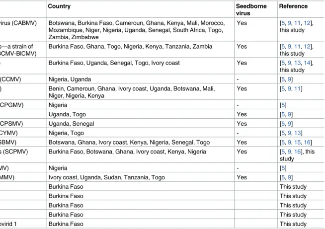
Viral diseases, which can often occur as multiple infections, are a major constraint on cow-pea production [5], and can cause plant stunting, reduced foliage, decreased seed protein con-tent, and, in individual plants, yield losses of up to 93% [3,6,7]. While members of 140 virus species can naturally or artificially infect cowpea (reviewed in [8]), only twelve of these species have so far been found in Africa (Table 1), from which, only three have been reported from Burkina Faso. Viruses in eight of these twelve species are seedborne (Table 1): a factor that seri-ously hampers their effective control [9,10]. For example, seed transmission can reach 2% for CMV, 6.9% for Blackeye cowpea mosaic virus—a strain of Bean common mosaic virus (BCMV-BlCM), and 13.3% for Cowpea aphid-borne mosaic virus (CABMV; [11]. Given that a far broader diversity of cowpea-infecting viruses has been discovered elsewhere in Africa, it is likely that additional cowpea-infecting viruses remain to be discovered in Burkina Faso. The limited available knowledge on cowpea infecting viruses in this country hinders the control of diseases, particularly with respect to the production of disease-free seeds and the creation of virus-resistant cowpea varieties. Hence, the main objective of this study was to further investi-gate the diversity of cowpea viruses in Burkina Faso.
The rapid advances in both nucleic acid sequencing technologies (next generation sequenc-ing, NGS) and metagenomics-based approaches to study viromes at scales ranging from
Table 1. List of cowpea-infecting viruses present in Africa.
Virus Country Seedborne
virus
Reference
Cowpea aphid-borne mosaic virus (CABMV) Botswana, Burkina Faso, Cameroun, Ghana, Kenya, Mali, Morocco, Mozambique, Niger, Nigeria, Uganda, Senegal, South Africa, Togo, Zambia, Zimbabwe
Yes [5,9,11,12], this study
Blackeye cowpea mosaic virus—a strain of Bean common mosaic virus (BCMV-BlCMV)
Burkina Faso, Ghana, Togo, Nigeria, Kenya, Tanzania, Zambia Yes [5,9,11,12], this study Cowpea mottle virus (CPMoV) Burkina Faso, Uganda, Senegal, Togo, Ivory coast Yes [5,9,13,14],
this study
Cowpea chlorotic mottle virus (CCMV) Nigeria, Uganda - [5,9]
Cucumber mosaic virus (CMV) Benin, Cameroun, Ghana, Ivory coast, Uganda, Botswana, Mali, Niger, Nigeria, Kenya
Yes [5,9,11]
Cowpea golden mosaic virus (CPGMV) Nigeria - [5]
Cowpea mosaic virus (CPMV) Uganda, Togo Yes [5,9]
Cowpea severe mosaic virus (CPSMV) Uganda, Senegal Yes [5,9]
Cowpea yellow mosaic virus (CYMV) Nigeria, Togo - [5,9,13]
Southern bean mosaic virus (SBMV) Botswana, Ghana, Ivory coast, Kenya, Nigeria, Senegal, Togo Yes [5,9,15,16] Southern cowpea mosaic virus (SCPMV) Burkina Faso, Botswana, Ghana, Ivory coast, Kenya, Nigeria Yes [5,9,16], this
study
Sunn-hemp mosaic virus (SHMV) Nigeria - [5]
Cowpea mild mottle virus (CPMMV) Ivory coast, Uganda, Sudan, Tanzania, Togo Yes [5,9]
Cowpea polerovirus 1 Burkina Faso This study
Cowpea polerovirus 2 Burkina Faso This study
Cowpea tombusvirid 1 Burkina Faso This study
Cowpea tombusvirid 2 Burkina Faso This study
Cowpea associated mycotymovirid 1 Burkina Faso This study
doi:10.1371/journal.pone.0165188.t001
Competing Interests: The authors have declared
individual organisms to entire communities, have enabled the discovery of increasing numbers of viruses in both wild ecosystems and agro-ecosystems [17–21]. Metagenomics-based approaches have also provided estimation of the plant community prevalence of plant viruses at the agro-ecosystem scale [22,23].
Here, we used a virion-associated nucleic acids (VANA) based metagenomics approach [24–27] to screen for the presence of cowpea viruses within cowpea plants collected from the Sudan (humid), Sudan-Sahel (sub-humid), and Sahel (dry) agro-climatic zones of Burkina Faso. Besides detecting four viruses that have so far been found infecting cowpea in Africa, we report the discovery of three novel plant virus species that have never before been found infect-ing cowpea plants, and one novel mycotymovirus, which probably infects a fungus species that is associated with cowpea plants.
Materials and Methods
Plant sampling
Three hundred and twelve leaf samples were randomly collected (i.e. irrespective of the pres-ence of potential symptoms) in 2013 (S1 Table). 104 plants were sampled in the humid Sudan zone, 142 in the sub-humid Sudan-Sahel zone and 66 from the dry Sahel zone. The sampled plants were collected from 110 farmer’s fields or experimental plots. We confirm that owners of the cowpea fields gave permission to conduct the study on their sites. We confirm that the field studies did not involve endangered or protected species. Leaf samples were dried in the presence of CaCl2 and stored at 4°C until virion-associated nucleic acid extraction. Addition-ally, in 2014, 103 samples were collected in Burkina Faso, including 25 samples from the Sudan-Sahel zone and 78 from the Sudan zone (S1 Table).
Detection of seed-borne viruses from cowpea seedlings
Eight cowpea cultivars (Komcallé, Nafi, Tiligré, Gorgou, Niizwé, Yiis-yandé, Kvx61-1, and Moussa local) obtained from Burkina Agricultural institute (INERA, Institut de l’Environne-ment et de Recherches Agricoles) and one unknown cultivar from Togo were grown at Mont-pellier, France within an insect-proof plant growth-chamber. Eighty-one seeds of each Burkina accession and twenty seeds of the Togo cultivar were sown in single use plastic pots containing sterilized peat and compost. Germinated seeds were examined daily during two weeks for the presence of symptoms on the primary and trifoliate leaves (S1 Table).
Virion-associated nucleic acids extraction, cDNA amplification,
sequencing and sequence analysis
The VANA-based 454 pyrosequencing approach [24] was used to analyse 384 cowpea plants, including 312 field plants sampled in Burkina Faso in 2013 and 72 plants grown in a growth-chamber at CIRAD (S1 Table). 150–250 mg of dried leaf material from the 384 plants were ground in Hanks’ buffered salt solution (HBSS) (1:10) with four ceramic beads (MP Biomedi-cals, USA) using a tissue homogeniser (MP biomediBiomedi-cals, USA). The homogenised plant extracts were centrifuged at 3,200 X g for 5 min and 6 ml of the supernatants were further cen-trifuged at 8,228 X g for 3 min. The resulting supernatants were then filtered through a 0.45 μm sterile syringe filter. The filtrate was then centrifuged at 148,000 X g for 2.5 hrs at 4°C to concentrate viral particles. The resulting pellet was resuspended overnight at 4°C in 200 μl of HBSS. Unencapsidated nucleic acids were eliminated by adding 15 U of bovine pancreas DNase I (Euromedex) and 1.9 U of bovine pancreas RNase A (Euromedex, France) followed by incubation at 37°C for 90 min. Total nucleic acids were finally extracted from 100μl of
resuspended virions using a NucleoSpin 96 Virus Core Kit (Macherey-Nagel, Germany) fol-lowing the manufacturer’s protocol. Viral cDNA synthesis was performed by incubation of 10 μl of extracted viral nucleic acids with 100 pmol of primer DoDec (5’-CCT TCG GAT CCT CCN NNN NNN NNN NN-3’) at 85°C for 2 min. The mixture was immediately placed on ice. Subsequently, 10 mM dithiothreitol, 1 mM of each deoxynucloside triphosphate (dNTP), 4 μl of 5X Superscript buffer, and 5 U of SuperScript III (Invitrogen, USA) were added to the mix-ture (final volume of 20 μl), which was then incubated at 25°C for 10 min, followed by 42°C incubation for 60 min and 70°C incubation for 5 min before being placed on ice for 2 min. cDNAs were purified using the QiaQuick PCR cleanup kit (Qiagen). Priming and extension was then performed using Large (Klenow) Fragment DNA polymerase (Promega). First, 20 μl of cDNA in the presence of 2 μM of primer DoDec were heated to 95°C for 2 min and then cooled to 4°C. 2.5 U of Klenow Fragment, 10X Klenow reaction buffer and 0.4 mM of each dNTP (final volume of 25 μl) were added. The mixture was incubated at 37°C for 60 min fol-lowed by 75°C for 10 min. PCR amplification was carried out using 5 μl of the reaction described above in a 20 μl reaction containing 2 μM of one of the 96 primers listed inS2 Table, and 10 μl of HotStarTaq Plus Master Mix Kit (Qiagen). The following cycling conditions were used: one cycle of 95°C for 5 min, five cycles of 95°C for 1 min, 50°C for 1 min, 72°C for 1.5 min, 35 cycles of 95°C for 30 sec, 50°C for 30 sec, 72°C for 1.5 min +2 sec at each cycle. An additional final extension for 10 min at 72°C was then performed. DNA products obtained from 96 cowpea samples were pooled, cleaned using the Wizard SV Gel and PCR Clean-Up System (Promega) and sequenced on 1/8th of a 454 pyrosequencing plate using GS FLX Tita-nium reagents (Beckman Coulter Cogenics, USA). The resulting reads were processed using a custom-built computational pipeline dedicated to the processing of multiplex identifier (MID) tagged DNA samples. Briefly, MID-tags and primers were identified in each raw read using agrep [28] in order to assign them to the particular samples from which they originated. Sepa-rated raw reads were then cleaned to eliminate MID-tags, primers and low quality regions (cut-off Phred quality score of 25) using cutadapt [29]. De novo assemblies of cleaned reads were performed using CAP3 [30]. Contigs and non-assembled reads with a minimum length of 45 bp were compared to sequences in the GenBank database using BlastN and BlastX meth-ods [31]. Open reading frames (ORFs) were identified using the ORF Finder NCBI analysis tool (http://www.ncbi.nlm.nih.gov/gorf/gorf.html). Primary sequence outputs have been deposited in the sequence read archive of GenBank (accession number: SRP083221).
Virus prevalence
The prevalence of a particular group of viruses was defined as the proportion of the 307 field sampled cowpea plants containing at least one VANA-read with a high degree of similarity (either BlastN or BlastX e-values <0.001) to that group of viruses. Five samples were consid-ered to have failed because no VANA-reads were produced.
RT-PCR, nested PCR and semi-nested PCR detection of viruses
A subset of fifty-two cowpea plants (S1 Table) that were initially processed by the VANA-based metagenomics approach was tested by RT-PCR to verify the presence of viruses identi-fied during the metagenomic screen (S1 Table). This subset of plants included 20 plants within which one or more of these eight viruses were detected together with (i) twenty-seven plants that were collected within close proximity to these 20 plants and (ii) five seedlings grown at Montpellier in which potyvirus sequences were identified. In addition to these 52 plants, a fur-ther 103 cowpea plants collected in 2014 were tested by RT-PCR for the presence of the eight viruses.
Total RNA was extracted from 35–40 mg of CaCl2dried cowpea leaves with the Qiagen1 RNeasy Plant Mini Kit (Qiagen, Valencia, CA) as described by the manufacturer. The detection of potyviruses was carried out using the primer pair Oligo1N/Oligo2N [32]. For the other viruses, contigs and reads produced in this study were aligned with related sequences obtained from GenBank (S3 Table) using ClustalW with default settings [33] and primers were designed (Table 2) using Oligo Explorer version 1.1.0 (www.uku.fi/~kuulasma/OligoSoftware) with cus-tomized settings (Tm, ~60°C; 40%<%GC<60%).
RT-PCR reactions were performed using the Qiagen1OneStep RT-PCR Kit. The 25 μL RT-PCR reaction mix consisted of 1 μL of eluted RNA (concentration range of 12–350 ng/μL), 14 μL of RNAse-free water, 5 μL of RT-PCR buffer (5X), 1 μL of dNTP mix (10 mM), 1.5 μL of each primer (10 μM) and 1 μL of RT-PCR enzyme mix. The RT-PCR program was as follows with the annealing temperature (Ta) and extension time (Ext) for each targeted virus listed in
Table 2: 50°C for 30 min, 95°C for 15 min, 35 cycles at 94°C for 1 min, Ta for 1 min and 72°C for Ext with a final 72°C extension for 10 min. PCR products were analyzed by electrophoresis on a 1.2% agarose gel in TAE buffer stained with ethidium bromide and visualized under UV light.
Specific nested or semi-nested-PCR assays were also designed to improve the detection of Cowpea mottle virus (CPMoV), Southern cowpea mosaic virus (SCPMV), tombusvirus-like
Table 2. List of detection primers designed in this study.
Cowpea viruses Primers Sequences Gene Annealing
temperature (˚C)
Extension duration (Sec)
Amplicon length (pb)
Cowpea polerovirus 1 and PoleroNB3897F GAGTTCATCTCCGAGGCC cp 55 30 263
Cowpea polerovirus 2 PoleroNB4160R CDTCTACCTATTTSGGRTTHTG
SCPMV SCPMVNB2698F CTGGGARTTRTGGGCTGATG RdRp 63 60 721
SCPMVNB3419R CTGAGCAATAGGGGCCATG
SCPMVNB2783F TCRTGYTTCATGAACTCAGTC 53 30 133
SCPMVNB2916R AGYTCAGCCATRAGGCAWCG
CPMoV CPMoV1138F TGAGYACTTTCATCAAAGCWGA RdRp 53 60 548
CPMoV1686R ACACARTCRTCWCCGTTGTT
CPMoV1138F TGAGYACTTTCATCAAAGCWGA 51 30 455
CPMoV1593R GTGTTCATRTCMCCACTCAT
Cowpea tombusvirid 1 Tombus3NB31F CAAGGTTCGACCAACATGTG RdRp 57 30 412
Tombus4NB79R CCAGTTTACAACCTTGAGGAG Tombus2NB237F TGTCTCTCGTGCCGATGCT 55 30 308 Tombus3NB52R GGTTCGACCAACATGTGGG Tombus2NB237F TGTCTCTCGTGCCGATGCT RdRp/ cp 55 120 1772 Tombus1NB44R CCTGGTGTCGATGTGGCC Tombus3NB31F CAAGGTTCGACCAACATGTG 55 90 1485 Tombus1NB44R CCTGGTGTCGATGTGGCC
Cowpea tombusvirid 2 Tomb2NB50F CTGTGTGCTGTTCGTGGAG RdRp 55 30 122
Tomb2NB172R TCAATCTTCTCTATATCGTAAAC
Cowpea tombusvirid 3 Tomb1NB18F TATCGGGGAGCGTTTGTACA RdRp 55 30 175
Tomb1NB193R TGCATGTCGGGTGTAATACC Cowpea associated mycotymovirid 1 TymoNB120F CTTTGGGTAGCACTATCCAC RP 55 30 295 TymoNB415R GAGTTTTGCTCCTTGAGACG TymoNB42F GCTGCCATAGAAAAGCGCC RP 55 30 154 TymoNB196R TAAAGAAGCTCGTCGAAGGG
cp: coat protein; RdRp: RNA dependant RNA polymerase; RP: replication-associated polyprotein doi:10.1371/journal.pone.0165188.t002
viruses and mycotymovirus. RT-PCRs were performed as described above using the follow-ing primers: CPMoV1138F/CPMoV1686R for CPMoV; SCPMVNB2698F /SCPMVNB3419R for SCPMV, Tombus2NB237F/Tombus4NB79R for Cowpea tombusvirid 1 and
TymoNB120F/TymoNB415R for Cowpea associated mycotymovirid 1 (Table 2). PCR ampli-fications were carried out using 1 μL of the reaction volume described above in a 25 μL reac-tion mix containing 0.5 μl at 10 μM of each primer, 10.5 μL of RNAse-free water and 12.5 μL of the HotStarTaq Plus Master Mix Kit (Qiagen). The following cycling conditions were used: one cycle at 95°C for 5 min, 35 cycles at 94°C for 1 min, Ta (Table 2) for 1 min, Ext (Table 2) at 72°C. An additional final extension for 10 min at 72°C was then performed. Amplification products were sequenced using the Sanger method (Beckman Coulter Cogenics, USA).
Recovery of partial genomes of Cowpea polerovirus 1 and Cowpea
polerovirus 2
Twenty specific primers (S4 Table) were designed from the VANA-contigs assigned to Cow-pea polerovirus 1. These primers were scattered along the VANA-contigs and were expected to amplify 1 Kb amplicons with 500 bp of sequence overlap between adjacent amplicons. In addition, two small products of 161 bp and 201 bp were amplified to confirm the 5’ end of the genome using primers PoleroNB1F/PoleroNB162R and PoleroNB1F/PoleroNB202R (S4 Table). Twelve specific primers were also designed, as described to amplify fragments of the Cowpea polerovirus 2 genome (S4 Table). RT-PCRs were performed as described above and amplicons were sequenced using the Sanger method (Beckman Coulter Cogenics, USA). Nucleotidic sequences were further assembled using DNAMAN v 7.0.2 (Lynnon
Corporation).
Cloning and sequencing of partial genome of Cowpea tombusvirid 1
VANA-contigs potentially coding RdRp and coat proteins of a novel virus hereafter referred to as Cowpea tombusvirid 1 were used to design primers for amplifying the genomic region encompassing these two positive sense single stranded RNA virus genes (Tombus2NB237F/ Tombus1NB44R and Tombus3NB31F/Tombus1 NB44R primer pairs;Table 2). RT-PCR was performed as described above using an annealing temperature of 55°C for the two primer com-binations and an extension time of 2 min for Tombus2NB237F/Tombus1NB44 R (1772 bp) and 1 min 30 sec for Tombus3NB31F/Tombus1 NB44R (1485 bp). Amplified products were gel purified with the QIAquick Gel Extraction Kit (Promega), inserted into the pGEM1-T vector as recommended by the manufacturer (Promega) and sequenced by the Sanger method (Beck-man Coulter Cogenics, USA) using the universal primers, T7 and SP6.
GenBank accession numbers
Partial genome of Cowpea polerovirus 1 (KX599154), partial RdRp gene of Cowpea polerovirus 1 (KX599155-KX599163), partial genome of Cowpea polerovirus 2 (KX599164), partial RdRp gene of Cowpea mottle virus (KX599165-KX599169), partial genome of Southern cowpea mosaic virus (KX599170), partial RdRp gene of Southern cowpea mosaic virus
(KX599171-KX599173), partial genome of Cowpea tombusvirid 1 (KX599174), partial RdRp gene of Cowpea tombusvirid 1 (KX599175-KX599177), partial RdRp gene of Cowpea tombus-virid 2 (KX599183), partial RdRp gene of Cowpea tombustombus-virid 3 (KX599184), partial RP gene of Cowpea associated mycotymovirid 1 (KX599178-KX599182).
Phylogenetic analysis
Sanger sequences were assembled using DNAMAN and were used as queries to perform BlastN and BlastX searches [31]. Sequences were subsequently aligned using MUSCLE 3.7 with default settings [34]. Maximum likelihood phylogenetic trees were produced from this alignment using PhyML 3.1 [35] implemented in MEGA version 6.06 [36] with a K2+G+I (Polerovirus) and K2 +G (Potyvirus, Carmovirus, Sobemovirus and Tombusviridae) nucleotidic substitution models (selected as best fit by MEGA) and 1000 bootstrap replicates as a test for the support of branches.
Results and Discussion
Exploration of cowpea virus diversity using the VANA-based
metagenomics-approach
A total of 669,589 reads were obtained from the 384 cowpea samples that were processed using the VANA approach (S1 Table). No reads were obtained in five of the 312 field plants. The average read count for each plant sample was 2848 reads/plant (standard deviation: 3037 reads/plant). A total of 45,901 reads (6.85%) were discarded after the quality control process. BlastN and BlastX comparisons between the VANA-reads and GenBank sequences indicated that 20.89% of the processed reads were potentially related to plant RNA viruses and that among the 307 field plants in which reads were obtained, 203 were positive for the presence of virus-related reads (66.1%;S1 Table). Unexpectedly, no reads corresponding to plant DNA viruses were obtained. Five family-level plant viral lineages were identified, including the
Poty-viridae, LuteoPoty-viridae, Tombusviridae and Tymoviridae families and the unassigned
Sobemo-virus genus (Table 3).
Detection of known cowpea viruses
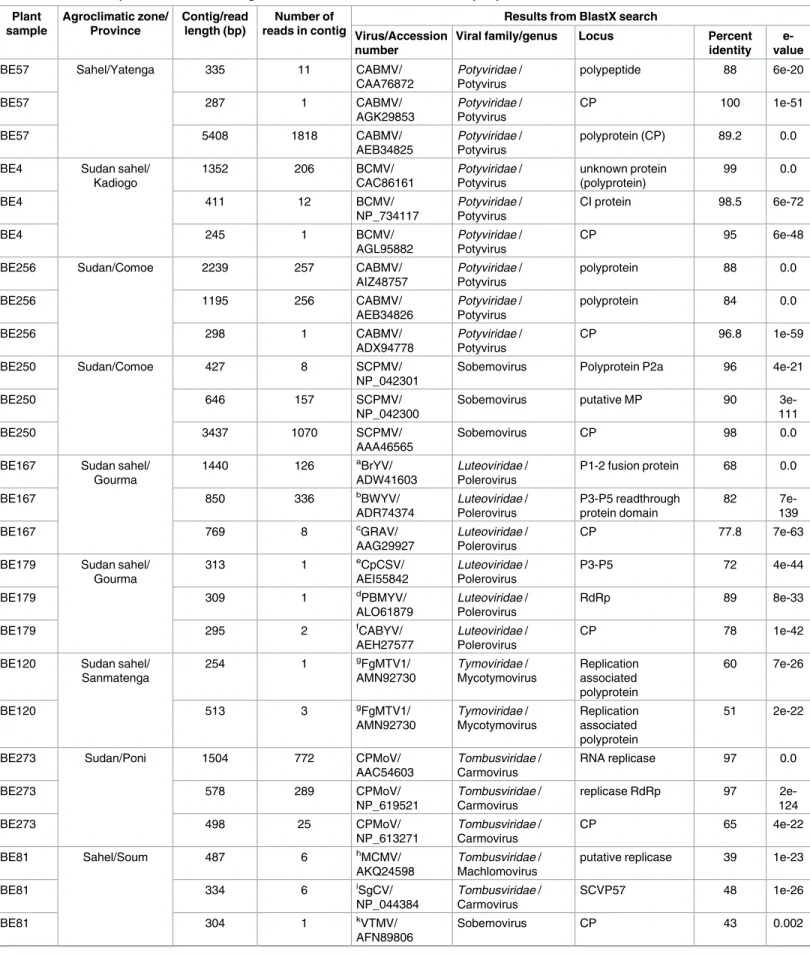
BlastX comparisons between the 3510 VANA-contigs that were produced by de novo assembly of potyvirus-, sobemovirus- and carmovirus-related reads and GenBank sequences yielded identity scores of 78–93% with CABMV, 98–100% with BCMV-BlCM, 90–96% with SCPMV and 65–97% with CPMoV (Table 3). These contigs apparently correspond with potyvirus genes (coat protein [cp], cytoplasmic inclusion protein [ci]), sobemovirus genes (polyprotein P2a, movement protein [mp] and cp), and carmovirus genes (RNA replicase, RNA dependent RNA polymerase [RdRp] and cp;Table 3). The degrees of similarity between these contigs and the amino acid (aa) or nucleotidic (nt) sequences of classified viruses in GenBank are above the species demarcation thresholds recommended for potyviruses (80% aa identity in the coat pro-tein; [37]), carmoviruses (52% aa identity of the polymerase, 41% aa identity of the coat pro-tein; [38]) and sobemoviruses (72% genome-wide pairwise nt sequence identity; [39])
indicating that the viral isolates from which these genomic sequences were obtained could rea-sonably, albeit tentatively, belong to the CABMV, BCMV-BlCM, SCPMV and CPMoV species. Of the 203 virus positive plants, 197 contained CABMV (97.04%), six contained BCMV-BlCM (2.96%), three contained SCPMV (1.48%) and three contained CPMoV (1.48%).
It is noteworthy that SCPMV is, to our knowledge, identified here for the first time in Bur-kina Faso. One of the three contigs is 3437 nt long (Table 3), which corresponds to slightly more than 80% of a typical SCPMV genome. Three large ORFs were identified within this con-tig: two overlapping ORFs corresponding to the P2a polyprotein encoding region (SCPMV, accession number NP_042301, highest percent identity = 96%, e-value = 0.0) and the P2ab polyprotein encoding region (SCPMV, accession number NP_042302, highest percent iden-tity = 97%, e-value = 0.0) and an ORF3 corresponding to the CP protein encoding region (SCPMV, accession number ABW34399, highest percent identity = 98%, e-value = 0.0).
Table 3. Selection of plant virus VANA-contigs and VANA-reads recovered from cowpea plants collected in Burkina Faso. Plant sample Agroclimatic zone/ Province Contig/read length (bp) Number of reads in contig
Results from BlastX search Virus/Accession
number
Viral family/genus Locus Percent identity
e-value
BE57 Sahel/Yatenga 335 11 CABMV/
CAA76872 Potyviridae / Potyvirus polypeptide 88 6e-20 BE57 287 1 CABMV/ AGK29853 Potyviridae / Potyvirus CP 100 1e-51 BE57 5408 1818 CABMV/ AEB34825 Potyviridae / Potyvirus polyprotein (CP) 89.2 0.0
BE4 Sudan sahel/ Kadiogo 1352 206 BCMV/ CAC86161 Potyviridae / Potyvirus unknown protein (polyprotein) 99 0.0 BE4 411 12 BCMV/ NP_734117 Potyviridae / Potyvirus CI protein 98.5 6e-72 BE4 245 1 BCMV/ AGL95882 Potyviridae / Potyvirus CP 95 6e-48
BE256 Sudan/Comoe 2239 257 CABMV/
AIZ48757 Potyviridae / Potyvirus polyprotein 88 0.0 BE256 1195 256 CABMV/ AEB34826 Potyviridae / Potyvirus polyprotein 84 0.0 BE256 298 1 CABMV/ ADX94778 Potyviridae / Potyvirus CP 96.8 1e-59 BE250 Sudan/Comoe 427 8 SCPMV/ NP_042301
Sobemovirus Polyprotein P2a 96 4e-21
BE250 646 157 SCPMV/
NP_042300
Sobemovirus putative MP 90
3e-111
BE250 3437 1070 SCPMV/
AAA46565
Sobemovirus CP 98 0.0
BE167 Sudan sahel/ Gourma 1440 126 aBrYV/ ADW41603 Luteoviridae / Polerovirus P1-2 fusion protein 68 0.0 BE167 850 336 bBWYV/ ADR74374 Luteoviridae / Polerovirus P3-P5 readthrough protein domain 82 7e-139 BE167 769 8 cGRAV/ AAG29927 Luteoviridae / Polerovirus CP 77.8 7e-63
BE179 Sudan sahel/ Gourma 313 1 eCpCSV/ AEI55842 Luteoviridae / Polerovirus P3-P5 72 4e-44 BE179 309 1 dPBMYV/ ALO61879 Luteoviridae / Polerovirus RdRp 89 8e-33 BE179 295 2 fCABYV/ AEH27577 Luteoviridae / Polerovirus CP 78 1e-42
BE120 Sudan sahel/ Sanmatenga 254 1 gFgMTV1/ AMN92730 Tymoviridae / Mycotymovirus Replication associated polyprotein 60 7e-26 BE120 513 3 gFgMTV1/ AMN92730 Tymoviridae / Mycotymovirus Replication associated polyprotein 51 2e-22
BE273 Sudan/Poni 1504 772 CPMoV/
AAC54603 Tombusviridae / Carmovirus RNA replicase 97 0.0 BE273 578 289 CPMoV/ NP_619521 Tombusviridae / Carmovirus replicase RdRp 97 2e-124 BE273 498 25 CPMoV/ NP_613271 Tombusviridae / Carmovirus CP 65 4e-22 BE81 Sahel/Soum 487 6 hMCMV/ AKQ24598 Tombusviridae / Machlomovirus
putative replicase 39 1e-23
BE81 334 6 iSgCV/ NP_044384 Tombusviridae / Carmovirus SCVP57 48 1e-26 BE81 304 1 kVTMV/ AFN89806 Sobemovirus CP 43 0.002 (Continued )
Discovery of novel cowpea viruses
Reads and contigs showing high degrees of similarity with viruses in the families Luteoviridae and Tymoviridae—families with no previously known cowpea-infecting viruses—were identi-fied from several cowpea plants collected during the 2013 sampling survey. In addition, reads and contigs showing low degrees of similarity with CPMoV, a member of the Tombusviridae family, were also identified.
Reads related to sequences of viruses in the family Luteoviridae were found in 10/203 (4.92%) of the evaluated plants (S1 Table). Eleven contigs were produced by de novo assembly of reads from two plants (BE167 and BE179;Table 3). These contigs apparently encoded partial CPs (two contigs), partial RdRps (two contigs) and partial P3-P5 readthrough proteins (two contigs,Table 3). Contigs obtained from both plants were further compared to one another. The pairwise identity scores that we obtained ranged from 57.9% (for the partial CP aa sequences) to 54.08% (for the partial P3-P5 aa sequences), suggesting that the reads may origi-nate from two or more different luteovirus-like species. Further, a single 231 nt long read obtained from plant BE179 displayed a relatively high degree of similarity (highest percent identity = 79%, e-value = 8e-09) with a polerovirus mp gene (ORF4 of Pepo aphid-borne yel-lows virus, accession number CRL92752).
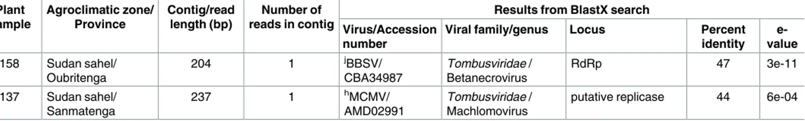
Reads and contigs showing low degrees of similarity with CPMoV, a virus in the family
Tombusviridae, were also identified from 3/203 plants (Table 3). Two contigs, both sharing similarities with tombusvirus sequences were assembled from plant BE81 (Table 3). One of these contigs potentially encodes a sobemovirus-like coat protein that is most similar to that of Velvet tobacco mosaic virus (accession number AFN89806, identity = 35%, e-value = 4e-09). In addition, single reads that were most similar to tombusvirus-like RdRp genes (47–54%,
Table 3), were recovered from two other plants (Table 3).
One 513 nt long contig and one 254 nt long single read showing detectable degrees of simi-larity with viruses in the family Tymoviridae were obtained from one plant (Table 3). Both of these sequence fragments may encode partial replication-associated polyproteins that are most similar to that of Fusarium graminearum mycotymovirus 1 (accession number AMN92730,
Table 3. (Continued) Plant sample Agroclimatic zone/ Province Contig/read length (bp) Number of reads in contig
Results from BlastX search Virus/Accession
number
Viral family/genus Locus Percent identity
e-value BE158 Sudan sahel/
Oubritenga 204 1 jBBSV/ CBA34987 Tombusviridae / Betanecrovirus RdRp 47 3e-11
BE137 Sudan sahel/ Sanmatenga
237 1 hMCMV/
AMD02991
Tombusviridae / Machlomovirus
putative replicase 44 6e-04
a: Brassica yellows virus, b
: Beet western yellows virus,
c: Groundnut rosette assistor virus, d: Phasey bean mild yellows virus, e
: Chickpea chlorotic stunt virus,
f: Cucurbit aphid borne yellow virus, g: Fusarium graminearum mycotymovirus 1, h
: Maize chlorotic mottle virus,
i: Saguaro cactus virus, j: Beet black scorch virus, k
: Velvet tobacco mottle virus
BlastX highest percent identity = 51% and 60%, e-value = 2e-22 and = 7e-26, respectively). These results suggest that these fragments are likely derived from a cowpea-associated fungus, that potentially belongs to the recently proposed lineage mycotymovirus in the family
Tymovir-idae [40].
The seven putative plant viruses identified here using the VANA-based approach (two poty-viruses, one sobemovirus, one carmovirus, two poleropoty-viruses, and one tombusvirus-like virus) sometimes occurred in mixed infections (14/307 plants, 4.6%;S1 Table). While the co-infected cowpea plants mostly contained two detectable viruses (13/14), a single case of triple infection was also observed (S1 Table). There was no correlation between average read count and the occurrence of multiple virus infection.
Molecular detection and characterisation of known and novel cowpea
viruses
To validate the results of the metagenomic screen, RT-PCR detection assays using virus-spe-cific primers (Table 2) were carried out on a subset of 52 samples collected in 2013 survey and the 103 cowpea plants collected in a further 2014 survey.
Potyviruses (CABMV and BCMV-BlCM). Fourty out of the 52 plants collected in 2013
tested positive for potyviruses (S1 Table). Among these 40 samples, potyvirus-related VANA reads went undetected in only a single plant (BE121), suggesting that the potyvirus detection results obtained with both these molecular virus detection approaches were consistent. In addi-tion, 94.1% of a subset of 17 plants collected in 2014 tested positive for potyviruses (S1 Table). Phylogenetic analysis based on the 182 nt partial nuclear inclusion gene indicated that these Burkinabe isolates all belong to either the CABMV or BCMV-BlCM species (S1 Fig).
Cowpea mottle virus (CPMoV). RT-PCR detection was most successful using the
CPMoV1138F/CPMoV1686R and CPMoV1138F/CPMoV1593R primer pairs (Table 2). When the sensitivity of the detection test was critical, the second pair could be used for a semi-nested RT-PCR. Whereas three of the 52 tested plant samples (BE273, BE276 and BE287) from the 2103 survey were found to contain detectable CPMoV-like sequence fragments using the VANA-based metagenomics approach, four of these 52 plants were found to potentially con-tain CPMoV RNA using the RT-PCR test (BE273, BE274, BE275 and BE276;S2 Fig). Unex-pectedly, CPMoV-like sequences were detected by both approaches in only two of these plants (BE273, BE276). As the detection of CPMoV by RT-PCR required the semi-nested PCR approach, it is plausible that the concentration of viral nucleic acids in plants that tested posi-tive by RT-PCR but negaposi-tive by the VANA-based metagenomics approach may have simply been too low to detect using the metagenomics approach. However it is not understood why the detection of CPMoV by RT-PCR was negative for sample BE287 in which four reads and one contig were detected with the VANA-based metagenomics approach. Only 1/103 plants collected in Burkina Faso in 2014 tested positive for CPMoV by RT-PCR (S1 Table). Phyloge-netic analysis based on a 415 nt partial RdRp gene unambiguously reveals that the Burkinabe CPMoV isolates are nested within the CPMoV species (Fig 1).
Southern cowpea mosaic virus (SCPMV). While the four primers pairs (Table 2) enabled the amplification of the three SCPMV isolates identified using the VANA-based metagenomics approach, the primer pair SCPMVNB2698F/SCPMVNB3419R was further chosen for the detection of SCPMV (S2 Fig). Noteworthy, the primer pairs SCPMVNB2783F/
SCPMVNB2916R and SCPMVNB2698F/SCPMVNB2916R could be further used for setting up either a nested PCRs or a semi-nested PCR in order to improve the sensitivity of SCPMV detection. Overall, six plant samples tested positive, including 3/307 samples collected in 2013 (that also tested positive using the VANA approach,S1 Table) and 3/103 samples collected in
Fig 1. Maximum-likelihood phylogenetic trees depicting the relatedness of cowpea viruses from Burkina Faso. A) Maximum-likelihood phylogenetic trees of partial cp genes from nine isolates of Cowpea polerovirus 1 and representative species from the family Luteoviridae. SCYLV, Sugarcane yellow leaf virus; PLRV, Potato leafroll virus; PeVYV, Pepper vein yellows virus; CpCSV, Chickpea chlorotic stunt virus; BrYV, Brassica yellows virus; BWYV, Beet western yellows virus; BYDV, Barley yellow dwarf virus; BLRV, Bean leafroll virus; SbDV, Soybean dwarf virus; PEMV-1, Pea enation mosaic virus-1; GRAV, Groundnut rosette assistor virus; PBMYV, Phasey bean mild yellows virus. B) Maximum-likelihood phylogenetic trees of partial RdRp genes from four isolates of Cowpea tombusvirid-1 and representative species from the family
2014 (S1 Table). Phylogenetic analysis based on a 495 nt partial RdRp gene unambiguously revealed that the SCPMV Burkinabe isolates are nested within the SCPMV species (Fig 1).
Cowpea-associatedpoleroviruses. A consensus sequence of the partial genome (5012 nt
in length) of the luteovirus-like isolate infecting plant BE167 was obtained using specific prim-ers designed from the luteovirus-like related VANA-contigs recovered from this plant. This consensus sequence corresponds to >83% of a typical polerovirus genome length. A BlastN search of GenBank returned Chickpea chlorotic stunt virus (CpCSV; accession number AY956384) as the closest match (highest percent identity = 68%, e-value = 0.0). Six ORFs were identified from this contig, including ORF0 (Beet mild yellowing virus (BMYV), accession number ACA61672, highest percent identity = 27%, e-value = 0.049), ORF1 (Phasey bean mild yellows virus (PBMYV), accession number ALR87184, highest percent identity = 33%, e-value = 9e-86), ORF2 (Brassica yellows virus (BrYV), accession number ADW41603, highest percent identity = 69%, e-value = 0.0), ORF3 (Groundnut rosette assistor virus (GRAV), acces-sion number AAG29926, highest percent identity = 85%, e-value = 1e-94), ORF4 (CpCSV, accession number YP_667842, highest percent identity = 67%, e-value = 4e-55) and
ORF3-ORF5 (Beet western yellows virus (BWYV), accession number ADR74374, highest per-cent identity = 81%, e-value = 2e-117). The partial genome that was obtained had an organiza-tion typical of poleroviruses in that it was comprised of six ORFs, including ORF0, which is absent in viruses of the genus Luteovirus, and ORF4, which is absent in Pea enation mosaic virus-1; which is presently the only member of the genus Enamovirus [41,42]. Based on the current species demarcation criteria used by the ICTV Lutoviridae study group (less than 90% aa identity to any previously described species in any of the genes), it is likely that this virus represents a new Polerovirus species (it is hereafter referred to as Cowpea polerovirus 1;
Table 1).
A second partial luteovirus-like genome fragment (3164 nt in length) was obtained by RT-PCR from plant BE179. A BlastN search revealed that this partial genome shares ~81% nucleotidic identity with PBMYV (accession number: KT963000, e-value = 0.0). Three com-plete ORFs identified in this sequence are most similar to the ORF2 of PBMYV (accession number: ALR87185, identity = 93%, e-value = 0.0), the ORF3 of PBMYV (accession number: ALR87186, identity = 76%, e-value = 7e-75) and the ORF4 of Suakwa aphid-borne yellows virus (SABYV; accession number: AHJ59956, identity = 56% and e-value = 7e-50). In addition, two partial ORFs were also found which were most similar to the ORF1 of PBMYV (accession
Tombusviridae. TurCV, Turnip crinkle virus; MNSV, Melon necrotic spot virus; MCMV, Maize chlorotic mottle virus; JCSMV, Johnsongrass chlorotic stripe mosaic virus; OCSV, Oat chlorotic stunt virus; TNV A, Tobacco necrosis virus A; OLV1, Olive latent virus 1; PMV, Panicum mosaic virus; CMMV, Cocksfoot mild mosaic virus; CarMV, Carnation mottle virus; MWLMV, Maize white line mosaic virus; PNSV, Pelargonium necrotic spot virus; CIRV, Carnation Italian ringspot virus; GaMV, Galinsoga mosaic virus; FNSV, Furcraea necrotic streak virus; LWSV, Leek white stripe virus; BBSV, Beet black scorch virus; SCNMV, Sweet clover necrotic mosaic virus; CRSV, Carnation ringspot virus; CkMV, Cocksfoot mottle virus. C) Maximum-likelihood phylogenetic trees of partial RdRp gene from 5 isolates of CPMoV from Burkina Faso and representative species from Carmovirus genus and from the family Tombusviridae. CCFV, Cardamine chlorotic fleck virus; SYMMV, Soybean yellow mottle mosaic virus; HCRV, Hibiscus chlorotic ringspot virus; PSNV, Pea stem necrosis virus; MNSCG, Melon necrotic spot virus; CymRSV, Cymbidium ringspot tombusvirus; MPV-PM75, Moroccan pepper virus. D: Maximum-likelihood phylogenetic trees of partial RdRp genes from five isolates of SCPMV from Burkina Faso and representative species of the Sobemovirus genus. SCPMV, Southern cowpea mosaic virus; RYMV, Rice yellow Mottle virus; CfMV, Cocksfoot mottle virus_sobemovirus; SCMoV, Subterranean clover mottle virus; SYCMV, Soybean yellow common mosaic virus; SBMV, Southern bean mosaic virus; SeMV, Sesbania mosaic virus; CarMV, Carnation mottle virus. For all four trees, branches associated with a filled dot have bootstrap support above 90 per cent whereas those with an unfilled dot have bootstrap support above 70 per cent. All branches with less than 50 percent bootstrap support were collapsed.
number: ALR87184, identity = 71% and e-value = 8e-42) and the ORF5 of CpCSV (accession number: YP_667840, identity = 79% and e-value = 7e-85). The only canonical polerovirus ORF that was completely missing from this 3184 nt long fragment was ORF0. It is nevertheless likely that this luteovirus-like sequence is from a virus that should be classified as belonging to the Polerovirus genus of the Luteoviridae. It is also noteworthy that ORF2 shares >80% nucleo-tide sequence identity with that of PBMYV, a novel polerovirus also isolated from phasey bean (Macroptilium lathyroides), a legume of the Fabaceae family [43]. However, ORF3 and ORF4 share <80% nucleotidic identity with the corresponding ORFs of PBMYV, suggests that the new virus could potentially be considered as either a new variant of PBMYV or a new Polero-virus species. Although sequencing of the full genome of this second cowpea poleroPolero-virus will likely be required to resolve its taxonomic placement, we hereafter refer to this virus as Cowpea polerovirus 2 (Table 1).
Ten out of 52 plants collected in 2013 tested positive for the presence of poleroviruses using the primer pair designed in this study (Table 2), including plant BE168, from which no polero-virus-related reads were found using the VANA-based metagenomics approach (S1 Tableand
S2 Fig). Conversely, plant BE186 tested negative using the RT-PCR approach despite the recov-ery of polerovirus-related VANA-reads from this plant during the metagenomic screen (S1 Table). None of the samples from the 2014 sampling survey tested positive for poleroviruses using the RT-PCR assays. Based on the 233 nt partial cp gene sequences of these ten isolates, possible evolutionary relationships with other poleroviruses were investigated using phyloge-netic analyses. While nine isolates tightly cluster around the isolate from plant BE167, from which we recovered the 5012 nt long partial genome of Cowpea polerovirus 1, the isolate BE179 branches from a different part of the tree, suggesting that this virus (Cowpea polerovirus 2) is probably a new species of cowpea-infecting polerovirus (Fig 1). However, due to the possi-bility of recombination (which is common in poleroviruses; [44]), further studies involving the characterization of the full genomes of these viruses are needed before it can be definitively confirmed whether or not these poleroviruses are new species.
Cowpea associated tombusvirids. The consensus 2142 nt long tombusvirus-like sequence
obtained from plant BE81 was most similar to Panicum mosaic virus (PMV, accession number: U55002, identity = 73%, e-value = 0.006). One ORF was identified within this consensus sequence, encoding a tombusvirus-like RdRp protein that is most similar to that of Saguaro cactus virus (SCV, accession number: NP_044384, identity = 42%, e-value = 4e-79). While this result suggests that this virus, hereafter referred to as Cowpea tombusvirid 1 (Table 1), should belong to the family Tombusviridae, the VANA study also revealed an ORF potentially encod-ing a sobemovirus-like coat protein from plant BE81. However, since sobemovirus coat pro-teins are most similar to those found in the genus Necrovirus within the family Tombusviridae [45], Cowpea tombusvirid 1 can tentatively be classified in the family Tombusviridae.
RT-PCR detection was most successful using the Tombus2NB237F/Tombus 4NB79R primer pair (Table 2), yielding a 700 bp fragment from plant BE81 as well as from three other plants (BE137, BE190 and BE197). Primer pairs Tombus3NB31F/Tombus4NB7 9R (Table 2
andS2 Fig) and Tombus2NB237F/Tombus3NB52 R (Table 2) could be further used for setting up either a nested PCRs or a semi-nested PCR in order to improve the sensitivity of Cowpea tombusvirid 1 detection. Phylogenetic analysis based on a 660 nt RdRp gene fragment revealed that the four Cowpea tombusvirid 1 isolates from Burkina Faso cluster together on a branch that is not closely associated with any sequences classified within any of the established
Tom-busviridae species, suggesting that Cowpea tombusvirid 1 genome fragment is likely derived
from a previously unknown tombusvirus species (Fig 1).
In addition, two other potentially novel tombusvirus-like sequences were detected in plants BE137 and BE158 using the primer pairs Tomb1NB18F/Tomb1NB193R and Tomb2NB50F/
Tomb2NB172R, respectively. However, no additional plants collected in either 2013 or 2014 tested positive for these viruses. Based on the sequence of a 127 nt RdRp gene fragment phylo-genetic analyses indicated that while the four isolates of Cowpea tombusvirid 1 cluster together tightly, the tombusvirus-like sequence from plant BE158, which we have named Cowpea tom-busvirid 2 (Table 1), fall on an isolated branch in another part of the tree: suggesting that it is possibly derived from a novel tombusvirus species (S1 Fig). However, further studies will be needed to fully characterize these two tombusviruses before it can be decided whether they actually constitute new species in the family Tombusviridae.
Cowpea-associatedtymovirus-like viruses. RT-PCR detection was most successful using
the TymoNB120F/TymoNB415R primer pair (Table 2), yielding a 255 bp partial replication-associated polyprotein gene fragment from 5 cowpea samples collected in 2013 and 1 in 2014 (S1 TableandS2 Fig). Because of the extremely distant relationships that existed between these 255 nt amplicons and homologous sequences found in known tymovirus species, it was not possible to accurately align the sequences. However, tymovirus-like amplicons shared high degrees of similarity with sequences of a novel mycotymovirus species that has recently been characterized from the plant pathogenic fungus Fusarium graminearum, suggesting that the probable tymovirus-like virus species detected here (which will hereafter referred to as Cowpea associated mycotymovirid 1) is potentially a second member of the new mycotymovirus lineage of the family Tymoviridae [40].
Symptomatology of cowpea plants collected in Burkina Faso
Field-sampled plants displayed a large range of symptom types (S1 TableandS2 Fig), including mild mosaic, severe mosaic, yellowing, mottling, leaf distortion, vein chlorosis and necrosis. However, since the majority of the cowpea plants infected by the novel viruses were also co-infected by potyviruses, it was not possible to clearly assign specific types of symptom to partic-ular viruses. It is, however, noteworthy that plant BE81, which is apparently only infected by Cowpea tombusvirid 1 (S1 Table), displayed symptoms of leaf distortion (S3 Fig). Altogether, these results indicate that the virus pressure on cowpea plants is relatively high in Burkina Faso and suggests that the virus-related sequences identified in this study are probably part of func-tional viruses that could potentially have a detrimental impact on cowpea production.
Detection sensitivities of VANA-based metagenomics and RT-PCR
methods
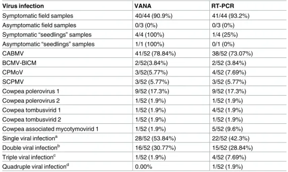
Overall, RT-PCR assay sensitivities were found to be slightly higher than that of the VANA-based metagenomic assay (Table 4). While neither approach detected any viruses in the field plants scored as asymptomatic, RT-PCR assay sensitivities were slightly better for detecting plant viruses from field cowpea samples scored as being symptomatic (Table 4). Several plants infected with CPMoV (3/52 detected by VANA and 4/52 detected by RT-PCR), Cowpea tom-busvirid 1 (1/52 by VANA, 4/47 by RT-PCR) and the Cowpea associated mycotymovirid 1 (1/ 47 by VANA, 5/47 by RT-PCR) were missed by the VANA-based approach (Table 4). Conse-quently, RT-PCR revealed a few more cases of viral co-infection than were revealed by the VANA-based metagenomics screen, including cases of triple and quadruple infections (Table 4). We hypothesize that the reduced efficiency of the random priming VANA-based approach compared to the specific priming RT-PCR approach can be accounted for by the rel-atively high numbers of mixed infections occurring in the subset of 52 cowpea samples (20/52; 38.46%), that may have hampered the detection of all co-infecting viruses using the VANA-based approach.
Geographic distribution and prevalence of cowpea-infecting viruses in
Burkina Faso
Among the various groups of viruses identified using the VANA-based metagenomic approach, the potyvirus CABMV is the most prevalent within cowpea grown in Burkina Faso. Whereas CABMV was found in 195/201 (97.0%) of the plants testing positive for potyviruses, BCMV-BlCM was found in only 6/201 (2.99%) of these plants.
The prevalence of viruses from other families were low: 10/307 (3.26%) for the polero-viruses, 3/307 (0.98%) for the carmopolero-viruses, and 3/307 (0.98%) for the sobemoviruses. Never-theless, the prevalence of all cowpea viruses (other than perhaps the potyviruses) was probably slightly under-estimated because the detection rate of the VANA-based approach may have been reduced due to the high frequency of viral co-infections as evidenced by the comparison of RT-PCR/VANA cowpea virus detection approaches.
While the five taxonomic viral groups occur in the Sudan zone (Potyviridae, sobemovirus,
Luteoviridae, Tombusviridae and Tymoviridae) and four in the Sudan-Sahel zone (Potyviridae, Luteoviridae, Tombusviridae and Tymoviridae), only two taxonomic groups are present in the
Sahel zone (Potyviridae and Tombusviridae,Fig 2). The percentage of plants infected with potyviruses decreased between the Sudan zone (87/101, 86.1%) and Sahel zone (15/65, 23.07%). This gradient, which was already reported in a previous study [46] can be accounted
Table 4. Virus prevalence and mixed infection prevalence of 52 cowpea plants based on VANA-based and RT-PCR-VANA-based detection results.
Virus infection VANA RT-PCR
Symptomatic field samples 40/44 (90.9%) 41/44 (93.2%)
Asymptomatic field samples 0/3 (0%) 0/3 (0%)
Symptomatic “seedlings” samples 4/4 (100%) 1/4 (25%) Asymptomatic “seedlings” samples 1/1 (100%) 0/1 (0%)
CABMV 41/52 (78.84%) 38/52 (73.07%) BCMV-BlCM 2/52(3.84%) 2/52 (3.84%) CPMoV 3/52(5.77%) 4/52 (7.69%) SCPMV 3/52 (5.77%) 3/52 (5.77%) Cowpea polerovirus 1 9/52 (17.3%) 9/52 (17.3%) Cowpea polerovirus 2 1/52 (1.9%) 1/52 (1.9%) Cowpea tombusvirid 1 1/52 (1.9%) 4/52 (1.9%) Cowpea tombusvirid 2 1/52 (1.9%) 1/52 (1.9%)
Cowpea associated mycotymovirid 1 1/52 (1.9%) 5/52 (9.6%) Single viral infectiona 28/52 (53.84%) 22/52 (42.3%)
Double viral infectionb 16/52 (30.77%) 15/52 (28.84%)
Triple viral infectionc 1/52 (1.9%) 4/52 (7.69%)
Quadruple viral infectiond 0.00% 1/52 (1.9%)
a: single infections consist of infection of: CABMV; BCMV-BlCM; Cowpea tombusvirid 1 or SCPMV b: double infections consist of mixed infection of: CABMV / SCPMV; CABMV / CPMoV; CABMV / Cowpea
associated mycotymovirid 1; CABMV / Cowpea tombusvirid 2; CABMV / Cowpea polerovirus 1; BCMV-BlCM / Cowpea polerovirus 1 or CABMV / Cowpea polerovirus 2
c: triple infections consist of mixed infection of: CABMV / SCPMV / CPMoV; CABMV / Cowpea tombusvirid 2
/ Cowpea associated mycotymovirid 1 or CABMV / Cowpea polerovirus 1 / Cowpea tombusvirid 1
d: quadruple infections consist of mixed infection of: CABMV / Cowpea polerovirus 1 / Cowpea tombusvirid 1
/ Cowpea associated mycotymovirid 1
for by climatic conditions in Burkina Faso, which are more favourable for the growth and maintenance of insect populations in the Sudan zone which, in turn, favors the transmission of plant viruses in the Sudan and Sudan-Sahel zones [47].
By contrast, both CPMoV and SCPMV are only present in the Sudan zone (Fig 2). While CPMoV was already reported in Burkina Faso in 1989 [48], this is the first report of the occur-rence of SCPMV in this country. The epidemiological dynamics of SCPMV will need to be monitored because, as has been reported for other African countries, this virus could become an important constraint on cowpea production in Burkina [16,49,50].
Poleroviruses were mainly detected from the Sudan-Sahel zone (9/141, 6.38%) although one isolate was found in the Sudan zone (1/101, 0.99%). Tombusvirus-like viruses were identified from the Sahel zone (Cowpea tombusvirid 1, 1/65) and the Sudan-Sahel zone (Cowpea tom-busvirid 1, 4/65, and Cowpea tomtom-busvirid 2, 1/141). Finally, the Cowpea associated mycotymo-virid 1 was identified in the Sudan-Sahel zone firstly with VANA-based approach in one sample (1/141, 0.7%), whereas in RT-PCR method other positive samples were detected in both the Sudan and Sudan-Sahel zones.
The occurrence of potyviruses and poleroviruses in mixed infection can be related to the fact that these viruses are both aphid transmitted [51,52], while the occurrence of SCPMV and CPMoV may be linked to the fact that both these viruses are beetle transmitted [53,54].
Detection of seed-borne cowpea-infecting viruses
Plant virus-associated VANA-reads were found from 16.66% (12/72) of the cowpea seedlings grown in an insect-proof growth chamber in France (UMR BGPI, CIRAD, Montpellier). While
Fig 2. Geographical distribution and prevalence of Cowpea viruses in Burkina Faso.
eight cowpea-infecting viruses (BCMV-BlCM, CABMV, SCPMV, CPMoV, CPMV, CPSMV, CPMMV and CMV) are reported to be potentially seed-transmissible (reviewed in [55]), only potyvirus-related reads were obtained from the cultivars Nafi, Tiligré, Yiis-yandé, Kvx61-1 and the unknown Togo cultivar (S1 Table). The rate of seedlings infected by potyviruses was highly variable and ranged from 0% to 100% for specific cultivars. Seedlings from the unknown Togo cultivar were all infected (100%), while seedlings from the Burkina Faso cultivars were hetero-geneously infected, ranging from 0% (for 4 cultivars) to 12.5% (cultivars Tiligré, Yiis yandé and Kvx61-1) and 25% (cultivar Nafi). BCMV-BlCM was the only potyvirus species that was detected from the Togo cultivar while both BCMV-BlCM and CABMV were detected from the Burkina cultivars.
Overall, these results highlight the fact that potyvirus seed-transmission rates are likely high in Togo and Burkina Faso: a fact that could certainly have a major impact on the recurrence of diseases associated with potyviruses in this African region and can partly account for the very high prevalence of potyvirus infections in cowpeas grown throughout Burkina Faso. Minimiz-ing or removMinimiz-ing this primary source of viral inoculum would probably be a first step towards better control of potyvirus diseases of cowpea within this country.
Conclusion
Overall, a combination of VANA-based metagenomics and classical RT-PCR- based molecular detection approaches have strengthened our knowledge about the diversity of viruses infecting cowpea in Burkina Faso; which is a first step towards minimizing the economic burden of these viral diseases on the smallholder farmers whose are the principal producers of legumes both in this country, and the rest of west Africa. The cowpea viruses identified in this study should be further studied and taken into account in future efforts to control diseases in this important crop.
Supporting Information
S1 Fig. Maximum-likelihood phylogenetic trees depicting the relatedness of potyvirus and tombusvirus-like viruses from Burkina Faso. A) Maximum-likelihood phylogenetic trees of
partial nuclear inclusion genes from 21 isolates of Cowpea polerovirus and representative spe-cies from the family Potyviridae. CABMV, Cowpea aphid-borne mosaic virus; BCMV, Black-eye cowpea mosaic strain of Bean common mosaic virus; PPST, Passiflora virus; ABMV, Azuki bean mosaic virus; HMV, Hardenbergia mosaic virus. B) Maximum-likelihood phylogenetic trees of partial RdRp genes from five isolates of Cowpea tombusvirus-like virus and representa-tive species from the family Tombusviridae. OCSV, Oat chlorotic stunt virus; TBTV, Tobacco bushy top virus; SCNMV, Sweet clover necrotic mosaic virus; CRSV, Carnation ringspot virus; FNSV, Furcraea necrotic streak virus; GaMV, Galinsoga mosaic virus; OLV1, Olive latent virus 1; TNV A, Tobacco necrosis virus A; JCSMV, Johnsongrass chlorotic stripe mosaic virus; MWLMV, Maize white line mosaic virus; MNSV, Melon necrotic spot virus; PNSV, Pelargo-nium necrotic spot virus; CIRV, Carnation Italian ringspot virus; LWSV, Leek white stripe virus; BBSV, Beet black scorch virus; TurCV, Turnip crinkle virus; CarMV, Carnation mottle virus; MCMV, Maize chlorotic mottle virus; PMV, Panicum mosaic virus; CkMMV, Cocksfoot mild mosaic virus; PMV, Panicum mosaic panicovirus.
(EPS)
S2 Fig. Agarose gel illustrating RT-PCR detection of cowpea-infecting viruses. (A) RT-PCR
for detection of potyviruses (B), RT-PCR for detection of Cowpea mottle virus (C), RT-PCR for detection of Southern cowpea mosaic virus (D), RT-PCR for detection of Cowpea
polerovirus1 and Cowpea polerovirus2 (E) Nested RT-PCR for detection of Tombusvirid1 and (F) RT-PCR for detection of Cowpea associated mycotymovirid 1.
(DOC)
S3 Fig. Symptoms observed on plants naturally infected by Cowpea-infectingviruses.
(DOCX)
S1 Table. List of cowpea viruses detected using VANA-based metagenomic and RT-PCR approaches.
(DOCX)
S2 Table. 10-nucleotide multiplex identifier (MID) tagged DNA primers used for PCR from cDNA.
(DOC)
S3 Table. List of NCBI GenBank sequences used for designing primers.
(DOCX)
S4 Table. List of primers used for Cowpea polerovirus 1 and Cowpea polerovirus 2 partial genome amplification.
(DOCX)
Acknowledgments
We acknowledge INERA for kindly providing with us cowpea cultivars. EP’s PhD fellowship was funded by the French Embassy of Togo (N°: 808087J). This study was partially funded by EU grant FP7-PEOPLE-2013-IOF (N° PIOF-GA-2013-622571).
Author Contributions
Conceptualization: EP DF MP PR. Data curation: EP DF PR. Formal analysis: EP DPM MP PR. Funding acquisition: MS OT MP PR. Investigation: EP EF DG RF JZ ZB JBN. Methodology: EP DF EF DG RF MP PR. Project administration: MS OT MP PR. Resources: EP DF EF DG RF. Software: DF DPM. Supervision: MS OT MP PR. Validation: DF DPM MP PR. Visualization: EP DF DPM MP PR. Writing – original draft: EP PR.References
1. Timko MP, Ehlers JD, Roberts PA. Cowpea. K C, editor. Berlin: Springer-Verlag; 2007. 49–68 p. 2. Nielsen SS, Brandt WE, Singh BB. Genetic-Variability for Nutritional Composition and Cooking Time of
Improved Cowpea Lines. Crop Science. 1993; 33(3):469–72. WOS:A1993LM11100010.
3. Taiwo MA, Akinjogunla OJ. Cowpea viruses: Quantitative and qualitative effects of single and mixed viral infections. Afr J Biotechnol. 2006; 5(19):1749–56. WOS:000248655800012.
4. Hall AE, Cisse N, Thiaw S, Elawad HOA, Ehlers JD, Ismail AM, et al. Development of cowpea cultivars and germplasm by the Bean/Cowpea CRSP. Field Crop Res. 2003; 82(2–3):103–34. doi:10.1016/ S0378-4290(03)00033-9WOS:000183397900004.
5. Taiwo MA, Shoyinka SA. Viruses infecting cowpeas in Africa with special emphasis on the potyviruses. In: Williams AO, Mbiele AL, Nkouka N, editors. Virus Diseases of Plants in Africa. Lagos, Nigeria: OAU/STRC Scientific Publication; 1988. p. 93–115
6. Kareem KT, Taiwo MA. Interactions of viruses in Cowpea: effects on growth and yield parameters. Virol J. 2007; 4:15. PMID:17286870; PubMed Central PMCID: PMC1805424. doi: 10.1186/1743-422X-4-15
7. Booker HM, Umaharan P, McDavid CR. Effect of Cowpea severe mosaic virus on crop growth charac-teristics and yield of cowpea. Plant Dis. 2005; 89(5):515–20. doi:10.1094/PD-89-0515
WOS:000228593600014.
8. Hampton RO, Thottappilly G. Cowpea. Loebenstein G, Thottappilly G, editors. Dordrecht: Kluwer Academic Publishers.; 2003.
9. Salem NM, Ehlers JD, Roberts PA, Ng JCK. Biological and molecular diagnosis of seedborne viruses in cowpea germplasm of geographically diverse sub-Saharan origins. Plant Pathol. 2010; 59(4):773– 84.
10. Gillaspie AG, Hopkins MS, Pinnow DL, Hampton RO. Seed-borne viruses in preintroduction cowpea seed lots and establishment of virus-free accessions. Plant Dis. 1995; 79(4):388–91. WOS: A1995QV23800014.
11. Bashir M, Hampton RO. Detection and identification of seed-borne viruses from cowpea (Vigna ungui-culata (L.) Walp.) germplasm. Plant Pathol. 1996; 45(1):54–8. doi:10.1046/j.1365-3059.1996.d01-97. xWOS:A1996TW41500006.
12. Huguenot C, Furneaux MT, Clare JA, Hamilton RI. Improved diagnosis of cowpea aphid-borne mosaic virus in Africa: significance for cowpea seed-indexing, breeding programs and potyvirus taxonomy. Arch Virol. 1996; 141(1):137–45. doi:10.1007/bf01718594WOS:A1996TR99700011. PMID:8629941
13. Gumedzoe MY, Sunu DY, Thottappilly G, Asselin A. Importance of the Cowpea Mottle Virus and Cow-pea Yellow Mosaic-Virus in Togo. Phytoprotection. 1990; 71(2):85–91. WOS:A1990DV50100005. 14. Thouvenel JC, Tia E, Fishpool LDC. Characterization of Cowpea Mottle Virus on Cowpea
(Vigna-Unguiculata) in the Ivory-Coast and the Identification of a New Vector. Trop Agr. 1990; 67(3):280–2. WOS:A1990DK61300020.
15. Amayo R, Arinaitwe AB, Mukasa SB, Tusiime G, Kyamanywa S, Rubaihayo PR, et al. Prevalence of viruses infecting cowpea in Uganda and their molecular detection. Afr J Biotechnol. 2012; 11 (77):14132–9. doi:10.5897/AJB11.398
16. Gumedzoe MYD, T G, A A. Occurrence of southern bean mosaic virus (SBMV) in Togo and its interac-tion with some cowpea cultivars. African Crop Science Journal. 1996; 4(2):215–22.
17. Boonham N, Kreuze J, Winter S, van der Vlugt R, Bergervoet J, Tomlinson J, et al. Methods in virus diagnostics: from ELISA to next generation sequencing. Virus Res. 2014; 186:20–31. PMID:
24361981. doi:10.1016/j.virusres.2013.12.007
18. Massart S, Olmos A, Jijakli H, Candresse T. Current impact and future directions of high throughput sequencing in plant virus diagnostics. Virus Res. 2014; 188:90–6. PMID:24717426. doi:10.1016/j. virusres.2014.03.029
19. Roossinck MJ, Martin DP, Roumagnac P. Plant Virus Metagenomics: Advances in Virus Discovery. Phytopathology. 2015; 105(6):716–27. PMID:26056847. doi:10.1094/PHYTO-12-14-0356-RVW
20. Bernardo P, Golden M, Akram M, Naimuddin, Nadarajan N, Fernandez E, et al. Identification and char-acterisation of a highly divergent geminivirus: Evolutionary and taxonomic implications. Virus Res. 2013; 177(1):35–45. PMID:23886668. doi:10.1016/j.virusres.2013.07.006
21. Roumagnac P, Granier M, Bernardo P, Deshoux M, Ferdinand R, Galzi S, et al. Alfalfa leaf curl virus: An aphid-transmitted geminivirus. J Virol. 2015; 89(18):9683–8. PMID:26109720. doi:10.1128/JVI. 00453-15
22. Scheets K, Blinkova O, Melcher U, Palmer MW, Wiley GB, Ding T, et al. Detection of members of the Tombusviridae in the Tallgrass Prairie Preserve, Osage County, Oklahoma, USA. Virus Res. 2011; 160(1–2):256–63. PMID:21762736. doi:10.1016/j.virusres.2011.06.023
23. Thapa V, Melcher U, Wiley GB, Doust A, Palmer MW, Roewe K, et al. Detection of members of the Secoviridae in the Tallgrass Prairie Preserve, Osage County, Oklahoma, USA. Virus Res. 2012; 167 (1):34–42. PMID:22487310. doi:10.1016/j.virusres.2012.03.016
24. Candresse T, Filloux D, Muhire B, Julian C, Galzi S, Fort G, et al. Appearances can be deceptive: revealing a hidden viral infection with deep sequencing in a plant quarantine context. PLoS One. 2014; 9(7):e102945. PMID:25061967; PubMed Central PMCID: PMC4111361. doi:10.1371/journal.pone. 0102945
25. Filloux D, Dallot S, Delaunay A, Galzi S, Jacquot E, Roumagnac P. Metagenomics approaches based on virion-associated nucleic acids (VANA): An innovative tool for assessing without a priori viral diver-sity of plants. Methods in molecular biology. 2015; 1302:249–57. PMID:25981259. doi: 10.1007/978-1-4939-2620-6_18
26. Francois S, Bernardo P, Filloux D, Roumagnac P, Yaverkovski N, Froissart R, et al. A Novel Itera-Like Densovirus Isolated by Viral Metagenomics from the Sea Barley Hordeum marinum. Genome announcements. 2014; 2(6). PMID:25477401; PubMed Central PMCID: PMC4256182. doi:10.1128/ genomeA.01196-14
27. Kraberger S, Farkas K, Bernardo P, Booker C, Arguello-Astorga GR, Mesleard F, et al. Identification of novel Bromus- and Trifolium-associated circular DNA viruses. Arch Virol. 2015; 160(5):1303–11. PMID:25701210. doi:10.1007/s00705-015-2358-6
28. Wu S, Manber U, editors. A fast approximate pattern-matching tool. Usenix Winter 1992 Technical Conference; 1992; San Francisco.
29. Martin M. Cutadapt removes adapter sequences from high-throughput sequencing reads. EMBnet. 2011; 17(1):10.
30. Huang XQ, Madan A. CAP3: A DNA sequence assembly program. Genome Research. 1999; 9 (9):868–77. PMID:10508846
31. Altschul SF, Gish W, Miller W, Myers EW, Lipman DJ. Basic Local Alignment Search Tool. Journal of Molecular Biology. 1990; 215(3):403–10. doi:10.1016/S0022-2836(05)80360-2PMID:2231712
32. Marie-Jeanne V, Ioos R, Peyre J, Alliot B, Signoret P. Differentiation of Poaceae potyviruses by reverse transcription-polymerase chain reaction and restriction analysis. J Phytopathol. 2000; 148 (3):141–51. doi:10.1046/j.1439-0434.2000.00473.xWOS:000086416500002.
33. Larkin MA, Blackshields G, Brown NP, Chenna R, McGettigan PA, McWilliam H, et al. Clustal W and Clustal X version 2.0. Bioinformatics. 2007; 23(21):2947–8. PMID:17846036. doi:10.1093/ bioinformatics/btm404
34. Edgar RC. MUSCLE: multiple sequence alignment with high accuracy and high throughput. Nucleic Acids Res 2004; 32:1792–7. doi:10.1093/nar/gkh340PMID:15034147
35. Guindon S, Dufayard JF, Lefort V, Anisimova M, Hordijk W, Gascuel O. New algorithms and methods to estimate maximum-likelihood phylogenies: assessing the performance of PhyML 3.0. Syst Biol. 2010; 59(3):307–21. PMID:20525638. doi:10.1093/sysbio/syq010
36. Tamura K, Stecher G, Peterson D, Filipski A, Kumar S. MEGA6: Molecular Evolutionary Genetics Analysis version 6.0. Molecular Biology and Evolution. 2013; 30: 2725–9. doi:10.1093/molbev/mst197
PMID:24132122
37. Adams MJ, Antoniw JF, Fauquet CM. Molecular criteria for genus and species discrimination within the family Potyviridae. Arch Virol. 2005; 150(3):459–79. PMID:15592889. doi: 10.1007/s00705-004-0440-6
38. Robertson NL, Cote F, Pare C, Leblanc E, Bergeron MG, Leclerc D. Complete nucleotide sequence of Nootka lupine vein-clearing virus. Virus Genes. 2007; 35(3):807–14. PMID:17657600. doi:10.1007/ s11262-007-0139-3
39. Truve E, Fargette D. Genus Sobemovirus. In: King AMQ, Carstens E, Adams M, Lefkowitz E, editors. Ninth Report of the International Committee on Taxonomy of Viruses: Elsevier; 2012. p. 1185–9. 40. Li P, Lin Y, Zhang H, Wang S, Qiu D, Guo L. Molecular characterization of a novel mycovirus of the
family Tymoviridae isolated from the plant pathogenic fungus Fusarium graminearum. Virol J. 2016; 489:86–94.
41. Mayo MA. Developments in plant virus taxonomy since the publication of the 6th ICTV Report. Arch Virol 1999; 144 (8):1659–66. PMID:10486120
42. Domier LL, D’Arcy CJ. Luteoviruses. In: Mahy BWJ, Van Regenmortel MHV, editors. Desk Encyclope-dia of Plant and Fungal Virology: Oxford: Academic; 2010. p. 197–204.
43. Sharman M, Kehoe M, Coutts B, van Leur J, Filardo F, Thomas J. Two Complete Genome Sequences of Phasey Bean Mild Yellows Virus, a Novel Member of the Luteoviridae from Australia. Genome announcements. 2016; 4 (1): e01569–15. doi:10.1128/genomeA.01569-15PMID:26847905
44. Moonan F, Molina J, Mirkov TE. Sugarcane yellow leaf virus: an emerging virus that has evolved by recombination between luteoviral and poleroviral ancestors. Virology. 2000; 269(1):156–71. PMID:
10725208. doi:10.1006/viro.1999.0162
45. Tamm T, Truve E. Sobemoviruses. Journal of Virology. 2000; 74(14):6231–41. doi:10.1128/Jvi.74.14. 6231-6241.2000WOS:000087817900001. PMID:10864632
46. Neya BJ. Se´rologie, pathoge´nie, e´pide´miologie et controˆle de la mosaique Cowpea aphid-borne mosaic virus (CABMV) du nie´be´ (Vigna unguiculata (L.) WALP.) transmise par des pucerons (Aphis craccivora, A. gossypii) au Burkina Faso: University of Ouagadougou. PhD thesis; 2011.
47. Dabire´ C, Suh JB, editors. Insectes nuisibles du nie´be´ et lutte contre leurs de´gaˆts au Burkina Faso. Etat de la recherche sur la culture du nie´be´ en Afrique centrale et Occidentale semi-aride; 1988 14–25 Nov; Ibada, Nigeria.
48. Some KKJ. Contributionàl’e´pide´miologie du virus de la mosaïque du nie´be´ transmis par les pucerons au Burkina Faso: Universite´ de Ouagadougou; 1989.
49. Bashir M, Hampton RO. Natural occurrence of five seedborne cowpea viruses in Pakistan. Plant Dis. 1993; 77:948–51.
50. Ndiaye M, Bashir M, Keller KE, Hampton RO. Cowpea Viruses in Senegal, West-Africa—Identification, Distribution, Seed Transmission, and Sources of Genetic-Resistance. Plant Dis. 1993; 77(10):999– 1003. WOS:A1993MD93700009.
51. Blanc S, Lopez-Moya JJ, Wang R, Garcia-Lampasona S, Thornbury DW, Pirone TP. A specific interac-tion between coat protein and helper component correlates with aphid transmission of a potyvirus. Virology. 1997; 231(1):141–7. PMID:9143313.
52. Brault V, Perigon S, Reinbold C, Erdinger M, Scheidecker D, Herrbach E, et al. The polerovirus minor capsid protein determines vector specificity and intestinal tropism in the aphid. J Virol. 2005; 79 (15):9685–93. PMID:16014930; PubMed Central PMCID: PMC1181584. doi:10.1128/JVI.79.15. 9685-9693.2005
53. Walters HJ, Henry DG. Bean leaf beetle as a vector of the cowpea strain of Southern bean mosaic virus. Phytopathology. 1970; 60:177–8.
54. Shoyinka SA, Bozarth RF, Reese J, Rossel HW. Cowpea mottle virus: a seed-borne virus with distinc-tive properties infecting cowpeas in Nigeria. Phytopathology. 1978; 68: 693–9.
55. Hema M, Sreenivasulu P, Patil BL, Kumar PL, Reddy DV. Tropical food legumes: virus diseases of economic importance and their control. Advances in virus research. 2014; 90:431–505. doi:10.1016/ B978-0-12-801246-8.00009-3PMID:25410108