HAL Id: hal-02152278
https://hal.archives-ouvertes.fr/hal-02152278
Submitted on 11 Jun 2019
HAL is a multi-disciplinary open access
archive for the deposit and dissemination of
sci-entific research documents, whether they are
pub-lished or not. The documents may come from
teaching and research institutions in France or
abroad, or from public or private research centers.
L’archive ouverte pluridisciplinaire HAL, est
destinée au dépôt et à la diffusion de documents
scientifiques de niveau recherche, publiés ou non,
émanant des établissements d’enseignement et de
recherche français ou étrangers, des laboratoires
publics ou privés.
Distributed under a Creative Commons Attribution| 4.0 International License
between dengue viruses and mosquito vectors
Louis Lambrechts, Christine Chevillon, Rebecca Albright, Butsaya
Thaisomboonsuk, Jason Richardson, Richard Jarman, Thomas Scott
To cite this version:
Louis Lambrechts, Christine Chevillon, Rebecca Albright, Butsaya Thaisomboonsuk, Jason
Richard-son, et al.. Genetic specificity and potential for local adaptation between dengue viruses and mosquito
vectors. BMC Evolutionary Biology, BioMed Central, 2009, 9, pp.160. �10.1186/1471-2148-9-160�.
�hal-02152278�
Open Access
Research article
Genetic specificity and potential for local adaptation between
dengue viruses and mosquito vectors
Louis Lambrechts*
1,2, Christine Chevillon
2, Rebecca G Albright
1,
Butsaya Thaisomboonsuk
3, Jason H Richardson
4, Richard G Jarman
3and
Thomas W Scott
1Address: 1Department of Entomology, University of California, One Shields Avenue, Davis, CA 95616, USA, 2Génétique et Evolution des Maladies
Infectieuses, UMR CNRS-IRD 2724, Centre de Recherche IRD, 911 Avenue Agropolis, B.P. 64501, 34394 Montpellier Cedex 5, France,
3Department of Virology, Armed Forces Research Institute of Medical Sciences, 315/6 Rajvithi Road, Bangkok, 10400, Thailand and 4Department
of Entomology, Armed Forces Research Institute of Medical Sciences, 315/6 Rajvithi Road, Bangkok, 10400, Thailand Email: Louis Lambrechts* - Louis.Lambrechts@mpl.ird.fr; Christine Chevillon - Christine.Chevillon@mpl.ird.fr; Rebecca G Albright - rgalbright@ucdavis.edu; Butsaya Thaisomboonsuk - ButsayaT@afrims.org;
Jason H Richardson - Jason.Richardson@afrims.org; Richard G Jarman - Richard.Jarman@afrims.org; Thomas W Scott - twscott@ucdavis.edu * Corresponding author
Abstract
Background: Several observations support the hypothesis that vector-driven selection plays an important role in shaping dengue virus (DENV) genetic diversity. Clustering of DENV genetic diversity at a particular location may reflect underlying genetic structure of vector populations, which combined with specific vector genotype × virus genotype (G × G) interactions may promote adaptation of viral lineages to local mosquito vector genotypes. Although spatial structure of vector polymorphism at neutral genetic loci is well-documented, existence of G × G interactions between mosquito and virus genotypes has not been formally demonstrated in natural populations. Here we measure G × G interactions in a system representative of a natural situation in Thailand by challenging three isofemale families from field-derived Aedes aegypti with three contemporaneous low-passage isolates of DENV-1.
Results: Among indices of vector competence examined, the proportion of mosquitoes with a midgut infection, viral RNA concentration in the body, and quantity of virus disseminated to the head/legs (but not the proportion of infected mosquitoes with a disseminated infection) strongly depended on the specific combinations of isofemale families and viral isolates, demonstrating significant G × G interactions.
Conclusion: Evidence for genetic specificity of interactions in our simple experimental design indicates that vector competence of Ae. aegypti for DENV is likely governed to a large extent by G × G interactions in genetically diverse, natural populations. This result challenges the general relevance of conclusions from laboratory systems that consist of a single combination of mosquito and DENV genotypes. Combined with earlier evidence for fine-scale genetic structure of natural
Ae. aegypti populations, our finding indicates that the necessary conditions for local DENV
adaptation to mosquito vectors are met. Published: 9 July 2009
BMC Evolutionary Biology 2009, 9:160 doi:10.1186/1471-2148-9-160
Received: 17 March 2009 Accepted: 9 July 2009 This article is available from: http://www.biomedcentral.com/1471-2148/9/160
© 2009 Lambrechts et al; licensee BioMed Central Ltd.
This is an Open Access article distributed under the terms of the Creative Commons Attribution License (http://creativecommons.org/licenses/by/2.0), which permits unrestricted use, distribution, and reproduction in any medium, provided the original work is properly cited.
Background
Dengue viruses (DENV) are mosquito-borne viruses which, like many RNA viruses, exhibit substantial genetic diversity [1]. This diversity can be hierarchically organized in large clusters of lineages sometimes termed 'genotypes' (the terms lineage and genotype will be used interchange-ably hereafter) within each one of the four distinct sero-types (DENV-1, -2, -3 and -4) [2]. In the last 200 years, the number of DENV lineages worldwide has been increasing exponentially [3], along with increasing epidemic fre-quency and occurrence of severe forms of the disease [4,5]. Dengue is now the most common human arthro-pod-borne viral (arboviral) disease and a major public health threat [6]. Although DENV genetic variation alone is not sufficient to completely explain the incidence of severe disease or the magnitude of outbreaks, there is compelling evidence for differences in virulence and epi-demic potential among DENV lineages (reviewed in [2]). Understanding the evolutionary processes shaping the increasing diversity of DENV lineages will, therefore, pro-vide important insights into the mechanisms regulating epidemics and pathogenicity associated with genetic vari-ation among viruses [1].
The spatial distribution of DENV genetic diversity gener-ally reveals a population structure whereby geographic subdivisions reflect greater gene flow within than between subdivisions (reviewed in [7]). Results from a recent study indicated that such a genetic structure can be observed on a fine spatial grid [8]; phylogenetic differences were detected among DENV isolates recovered from schools separated by only a few kilometers in rural Thailand. Although multiple DENV lineages co-circulated within individual schools, there was strong genetic differentia-tion among lineages between schools that remained sta-ble over the 10-month sampling period [8]. Thus, despite frequent viral migration into the area, individual schools located a few kilometers apart represented distinct DENV evolutionary entities. A fundamental unanswered ques-tion concerns the relative influence of natural selecques-tion (adaptive evolution) and genetic drift (neutral evolution) on the genetic structure of DENV populations [9]. Although it is clear that stochastic processes play a signif-icant role in shaping DENV genetic diversity [10,11], genetic signatures of adaptive evolution have been recur-rently detected in natural DENV isolates [12-16].
Among evolutionary forces actively driving the evolution of arboviruses, vector-driven selection may play an impor-tant role by selecting lineages that are better suited for mosquito transmission [9]. For instance, the emergence of chikungunya virus (CHIKV) in the Indian Ocean in 2004, and subsequent spread to India and Europe was attributed to the acquisition of a single adaptive mutation that enhances transmission efficiency by Aedes albopictus [17,18]. This mutation confers a selective advantage in
locations where Ae. albopictus predominates over Ae.
aegypti, which is usually considered the primary vector of
CHIKV. Similarly, the emergence of a new lineage of West Nile virus in North America was attributed to earlier and more efficient transmission by Culex mosquitoes relative to the lineage that initially colonized this part of the world [19]. DENV are no exception in this regard. Displacement of the American (AM) DENV-2 genotype by a Southeast Asian (SA) DENV-2 genotype in the Western Hemisphere was associated with more efficient infection and dissemi-nation in Ae. aegypti of the SA than AM genotype [20-22]. Likewise, an invasive DENV-3 isolate from Sri Lanka infected and disseminated more efficiently in Ae. aegypti mosquitoes than a displaced, native DENV-3 isolate [23]. Interestingly, in both DENV examples above, outcom-peted genotypes tended to cause mild disease whereas invasive genotypes correlated with more severe disease manifestations. This supports the view that enhanced vec-tor transmission, among other facvec-tors, may contribute to the evolutionary success of lineages that are more virulent to humans [2].
The potential role of vector-driven selection in DENV evo-lution raises the question whether the genetic structure of DENV populations reflects, at least partly, that of their vectors. Indeed, Ae. aegypti distribution consists of a patchwork of genetically differentiated populations [24-27]. Because the vector competence of Ae. aegypti for DENV is in part genetically determined (reviewed in [28]), it has been hypothesized that the structure of DENV populations may partly result from the adaptation of viruses to the local vector genotypes [9]. Under this hypothesis, DENV transmission would be more efficient by local vector genotypes (sympatric vector-virus pairs) than by vector genotypes from distant populations (allo-patric vector-virus pairs). Pathogen adaptation to local hosts has been reported in a variety of systems (reviewed in [29]), including malaria parasites and their mosquito vectors [30]. Evolution of local adaptation has been pre-dicted when the pathogen has an evolutionary advantage over the host, such as higher mutation rate, shorter gener-ation time, higher migrgener-ation rate, and larger populgener-ation size [31-33]. These conditions could undoubtedly apply to the DENV-Ae. aegypti system [9].
An additional necessary condition for the occurrence of DENV local adaptation to Ae. aegypti is that the genetic structure of mosquito populations must be coupled with some degree of genetic specificity of vector-virus compat-ibility. In other words, DENV transmission by Ae. aegypti must be determined, at least partly, by genotype × geno-type (G × G) interactions. Such G × G interactions, whereby the infection outcome depends on the specific combination of host and pathogen genotypes, are found in many systems [34]. The effects of vector and virus gen-otypes, independently, on DENV transmission by Ae.
aegypti are well-documented. Variation in vector
compe-tence for a reference DENV isolate among different geo-graphic strains of Ae. aegypti has been repeatedly reported [35-38], and can often be directly correlated to genetic dif-ferences [39,40]. Reciprocally, DENV isolates have been shown to vary in their infectivity to a given mosquito strain [20,22]. Results from an earlier study showing some degree of interaction specificity between laboratory strains of mosquitoes and DENV serotypes and isolates [41] and from another study where SA genotypes of DENV-2 per-formed better than AM genotypes in two field-derived populations of Ae. aegypti, but not in a laboratory adapted colony [21] were suggestive of genetic specificity of vector-virus compatibility. However, the occurrence of G × G interactions between DENV and Ae. aegypti has not been formally quantified in natural populations.
Here, we measured the extent of G × G interactions between DENV and mosquito genotypes that are naturally interacting in the field. We challenged three Ae. aegypti isofemale families derived from a wild population sam-pled in 2007 in Ratchaburi, Thailand with three low-pas-sage DENV-1 viruses that were isolated the same year from humans (in Bangkok, Kamphaeng Phet, and Ratchaburi, respectively). Following standard methods of quantitative genetics [34], we estimated the extent to which the out-come of the infection is determined by G × G interactions by measuring the statistical effect of isofemale family × virus isolate interactions on several indices of vector com-petence, defined as the intrinsic permissiveness of a vector to become infected and subsequently transmit a pathogen [42].
Results
Overall, vector competence was scored in 333 female Ae.
aegypti. Each combination of isofemale family and virus
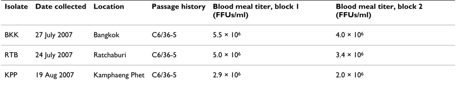
isolate was represented by 28–53 individuals (mean = 37, median = 36) divided into two experimental blocks of 11–28 individuals (mean = 18.5, median = 19). The three DENV-1 isolates used in this study were collected within a one-month period at three locations in Thailand (Bang-kok: BKK, Kamphaeng Phet: KPP, and Ratchaburi: RTB) and had identical passage histories (Table 1). Estimated
virus titers in the blood meals were remarkably similar across isolates and experimental blocks, with all titers ranging within a third of a log unit per ml (Table 1). This allowed us to assume that most of the phenotypic differ-ences observed between isolates were due to genetic dif-ferences. Across families and isolates, 63.1% of females were infected, and 42.9% had a disseminated infection. Isofemale families differed significantly in wing length (S.S. = 0.052, F2,57 = 3.85, P = 0.027), indicating signifi-cant genetic variation in body size. The mean wing length (± SE) was 3.11 ± 0.018, 3.08 ± 0.018, and 3.04 ± 0.018 mm for families A, B, and C, respectively. Corresponding mean age at pupation (± SE) was 7.29 ± 0.035, 7.15 ± 0.037, and 7.28 ± 0.033 days, respectively. Although three data points are not enough to obtain a valid correlation, it is worth noting that the overall proportion of infected mosquitoes by family tended to be negatively correlated with their mean wing length (linear regression: R2 = 0.97,
P = 0.102), supporting the view that larger females are
more resistant to dengue infection than small females [43].
Two factors had a significant influence on end-point mor-tality of adult mosquitoes at 14 days post-infection (dpi). Mortality significantly differed between blocks (S.S. = 0.245, F1,4 = 19.5, P = 0.012), and marginally significantly between virus isolates (S.S. = 0.178, F2,4 = 7.08, P = 0.049). Although 5.8% of females in the first block died before 14 dpi, 16.3% of females in the second block did so, possibly due to the one-day age difference. Across blocks, end-point mortality was 5.3%, 11.1%, and 16.1% for the KPP, BKK, and RTB isolates, respectively. Mortality did not depend on the family-isolate combinations (S.S. = 0.131,
F4,4 = 2.62, P = 0.19).
The proportion of infected mosquitoes strongly depended on the family, the isolate, and most importantly in the context of this study, their interaction (Table 2a; Figure 1a). The percentage of infected mosquitoes ranged from 30.2% to 100% among family-isolate combinations. The interaction effect appeared to be mainly due to the com-bination of the RTB isolate and mosquito family B for which the proportion of infected mosquitoes was lower
Table 1: Description of DENV-1 isolates used in this study
Isolate Date collected Location Passage history Blood meal titer, block 1 (FFUs/ml)
Blood meal titer, block 2 (FFUs/ml)
BKK 27 July 2007 Bangkok C6/36-5 5.5 × 10⁶ 4.0 × 10⁶
RTB 24 July 2007 Ratchaburi C6/36-5 5.0 × 10⁶ 3.4 × 10⁶
KPP 19 Aug 2007 Kamphaeng Phet C6/36-5 2.9 × 10⁶ 2.0 × 10⁶
For each isolate, the date and location of collection, passage history, and blood meal titers for both experimental blocks are indicated. Blood meal titers were estimated by fluorescent focus assay (FFA) in C6/36 cells.
than would have been expected from examination of the other combinations (Figure 1a). Because the family × iso-late × block three-way interaction was not statistically sig-nificant, our results indicated that the effect of the family × isolate interaction was consistent across blocks (Table 2a). In other words, small differences in mosquito age or blood meal titer between blocks did not affect the proba-bility of infection in a given mosquito-virus combination.
In contrast, the proportion of mosquitoes with a dissemi-nated infection among those successfully infected was only significantly influenced by the virus isolate (Table 1b). More than 90% mosquitoes infected by the RTB iso-late had a disseminated infection whereas this percentage ranged from 40% to 70% for the BKK and KPP isolates (Figure 1b). Despite some degree of interaction indicated by crossing lines (Figure 1b), the family × isolate
interac-Table 2: Test statistics of categorical vector competence indices
(a) Proportion infected (b) Proportion infected with disseminated infection
Source of variation d.f. L-R χ2 P-value L-R χ2 P-value
Family 2 16.2 0.0003 2.34 0.3107 Isolate 2 29.4 <0.0001 38.8 <0.0001 Family*Isolate 4 14.8 0.0051 6.28 0.1793 Block 1 0 1.0 0 0.9882 Family*Block 2 0.64 0.7247 0.84 0.6574 Isolate*Block 2 0.64 0.7262 0.27 0.8753 Family*Isolate*Block 4 1.29 0.8634 11.9 0.0183
The table shows the nominal logistic regression for the proportion of (a) mosquitoes with detectable viral RNA in their bodies (thorax+abdomen) and (b) infected mosquitoes (excluding uninfected) with a disseminated infection in their head/legs (determined by FFA in Vero cells) as a function of mosquito isofemale families, virus isolates, experimental blocks, and their interactions.
Effect of family × isolate interactions on virus infection and dissemination
Figure 1
Effect of family × isolate interactions on virus infection and dissemination. (a) The proportion of mosquitoes with a midgut infection and (b) proportion of infected mosquitoes with a disseminated infection as a function of mosquito families and virus isolates. In both panels, three isofemale families from a Ratchaburi population (A, B, and C) are ranked on the x-axis according to the mean proportion of infected mosquitoes across isolates. Each line represents a single virus isolate (BKK: Bangkok; KPP: Kamphaeng Phet; RTB: Ratchaburi). Vertical bars show the confidence intervals of the proportions. Crossing lines give an indication of family × isolate interactions.
A B C 0 0.1 0.2 0.3 0.4 0.5 0.6 0.7 0.8 0.9 1 Proportion infected Isofemale family BKK KPP RTB A B C 0 0.1 0.2 0.3 0.4 0.5 0.6 0.7 0.8 0.9 1
Proportion with disseminated infection
Isofemale family
tion was not statistically significant, possibly due to varia-tion between experimental blocks as indicated by the significant family × isolate × block three-way interaction (Table 1b). Moreover, exclusion of the 123 uninfected mosquitoes reduced statistical power of this analysis. Viral RNA concentrations in the body of infected mosqui-toes and the number of fluorescent focus units (FFUs) in the head/legs of mosquitoes with a disseminated infec-tion varied substantially among family-isolate combina-tions, with varying ranking orders of viral isolates for each isofemale family (Figure 2). Both variables were signifi-cantly influenced by the family × isolate interaction, with no significant difference between experimental blocks (Table 3). Regardless of their ranking order in a given mosquito family, the general profile of each isolate shared some degree of similarity between viral RNA concentra-tion and numbers of disseminated FFUs (Figure 2), possi-bly reflecting differing viral growth capacities in different mosquito genetic backgrounds. Viral RNA concentration in infected mosquitoes did not appear to be correlated to the proportion of infected mosquitoes or the proportion of mosquitoes with a disseminated infection, indicating that the probability of these events may be largely inde-pendent of the efficiency of viral genome replication.
Discussion
Results of our experiment strongly support the hypothesis that vector competence of Ae. aegypti for DENV-1 is gov-erned by G × G interactions. With the exception of virus dissemination success, all of the indices of vector
compe-tence we examined (midgut infection success, viral RNA concentration in bodies, virus titer in head/legs) were at least partly determined by the specific combination of mosquito family and virus isolate. The use of a rand-omized complete block design allowed us to rule out the possibility of environmental bias by confirming the con-sistency of family × isolate interactions effect across exper-imental blocks.
Our conclusions are based on the premise that family × isolate interactions reliably approximate G × G interac-tions [34]; i.e., in our design observed phenotypic differ-ences truly reflect the underlying genetic differdiffer-ences between mosquitoes and viruses. The use of isofemale families to assess the genetic basis of a trait assumes that the ratio of among- to within-families variations is pro-portionate to the heritable variation for that trait [44]. This assumption is reasonable as long as environmental variation in rearing conditions or parental effects do not differ between families. We ensured homogeneity by maintaining the F0-F2 parental generations under stand-ard laboratory conditions and rearing the F3 individuals under strictly identical conditions. Non-genetic differ-ences in virus infectivity were minimized by growing the viruses simultaneously under the same conditions and exposing mosquitoes to very similar blood meal titers (Table 1). Uncontrolled, slight differences in blood meal titers did not appear to influence results. For instance, the RTB isolate produced the highest average values across families for all vector competence indices, although the BKK isolate had the highest blood meal titers in both
Table 3: Test statistics of continuous vector competence indices
(a) Viral RNA concentration in body (b) Mean virus titer in head/legs
Source of variation d.f. S.S. F P-value d.f. S.S. F P-value
Family 2 0.63 2.01 0.1374 2 1.53 2.22 0.1127 Isolate 2 0.59 1.88 0.1550 2 1.45 2.10 0.1266 Family*Isolate 4 2.09 3.33 0.0115 4 3.58 2.60 0.0394 Block 1 0.02 0.14 0.7126 1 0.83 2.42 0.1223 Family*Block 2 0.01 0.03 0.9668 2 0.44 0.63 0.5328 Isolate*Block 2 0.29 0.93 0.3949 2 0.83 1.20 0.3053 Family*Isolate*Block 4 0.87 1.38 0.2417 4 1.27 0.92 0.4530 Error 192 30.1 125 43.1
Results from analysis of variance of (a) viral RNA concentration in the bodies (thorax+abdomen) of infected mosquitoes and (b) mean virus titer (determined by FFA in Vero cells) in the head/legs of mosquitoes with a disseminated infection as a function of mosquito isofemale families, virus isolates, experimental blocks, and their interactions.
experimental blocks. Although virus amplification in cell culture may have imposed a selective pressure on the orig-inal viruses, the low number of passages allowed us to assume that we still had isolates representative of wild-type viral genetic diversity. Overall, it seemed reasonable to conclude that our approach provided a reliable esti-mate of G × G interactions in this system.
It is remarkable that we were able to detect G × G interac-tions using a very restricted experimental design (three mosquito families × three virus isolates). Observing a highly significant effect of the interaction term with lim-ited statistical power indicates that G × G interactions likely represent a strong determinant of vector compe-tence in natural Ae. aegypti populations, which display a much higher level of genetic polymorphism than was cap-tured by our three isofemale families sampled at a single location. We expect G × G interactions to be even more prominent when mosquito genotypes from more geneti-cally distinct populations are examined. Indeed, a nested analysis of Ae. aegypti mitochondrial haplotype frequen-cies in Thailand showed that while 57% of the total varia-tion was found within collecvaria-tions, 18% and 25% of that variation were found among samples collected within a distance of 25 km and more than 100 km apart, respec-tively [25]. Moreover, because the three DENV-1 isolates used in this study were all collected in Thailand in 2007, it is likely that they are closely genetically related. We
expect that G × G interactions will occur to a greater extent when the analysis includes more distantly related viral lin-eages or even different serotypes.
A practical implication of our results is that the conclu-sions drawn from one particular vector-virus combination (such as one mosquito population challenged with a ref-erence DENV isolate) are not likely to be representative of other combinations. This challenges the broad-scale rele-vance of genetic loci associated with vector competence that were identified in laboratory-tractable systems. A recent meta-analysis showed that quantitative trait loci (QTL) controlling host resistance against one pathogen isolate were on average recovered in only 24% of the cases where infections with different isolates were investigated; i.e., each particular host-pathogen combination was based on a different set of QTL and epistatic interactions [45]. G × G interactions may help to explain why the QTL controlling midgut infection by a Jamaican DENV-2 iso-late in highly selected lines of Ae. aegypti [46,47] did not correlate with those previously identified in field-derived
Ae. aegypti challenged with a Puerto Rican DENV-2 isolate
[48]. Likewise, it sheds a new light on a study where DENV-2 isolates of the SA genotype performed better than isolates of the AM genotype in two field-derived Ae.
aegypti populations, but not in a laboratory-adapted
col-ony [21].
Effect of family × isolate interactions on viral RNA concentration in mosquito bodies and virus titer in heads/legs
Figure 2
Effect of family × isolate interactions on viral RNA concentration in mosquito bodies and virus titer in heads/ legs. (a) The log-transformed viral RNA copy number per μl of homogenized body (thorax+abdomen) in infected mosquitoes and (b) log-transformed mean number of fluorescent focus units (FFUs) in the head/legs of mosquitoes with a disseminated infection as a function of mosquito families and virus isolates. In both panels, three isofemale families from a Ratchaburi popula-tion (A, B, and C) are ranked on the x-axis according to the mean proporpopula-tion of infected mosquitoes across isolates (consist-ently with Figure 1). Each line represents a single virus isolate (BKK: Bangkok; KPP: Kamphaeng Phet; RTB: Ratchaburi). Each point represents the mean and vertical bars are the standard errors of the means. Crossing lines give an indication of family × isolate interactions. A B C 3.4 3.5 3.6 3.7 3.8 3.9 4
Log viral RNA concentration in body
Isofemale family BKK KPP RTB A B C 1.2 1.4 1.6 1.8 2 2.2 2.4
Log mean disseminated FFUs
Isofemale family
Although additional studies are warranted to confirm the impact of G × G interactions on actual virus transmission through an infectious bite, their occurrence during DENV incubation in Ae. aegypti provides important insights into the mechanisms shaping DENV population structure and evolution. It helps to explain why despite strong genetic bottlenecks during mosquito transmission, major DENV variants are generally conserved in both vertebrate and arthropod hosts [49,50]. Indeed, G × G interactions indi-cate that elimination of viruses that are incompatible with local mosquito genotypes may counteract the effect of genetic drift in the form of purifying selection at the pop-ulation level. Likewise, the observed spatial clustering of DENV lineages on a restricted spatial scale [8] may in fact be promoted by location-specific vector-driven selection. Over time, genetic specificity of vector-virus compatibility would combine with the genetic structure of Ae. aegypti populations to drive the adaptation of DENV to increased compatibility with local mosquito genotypes [9]. Interest-ingly, it is worth noting that in our experiment, while the three isofemale vector families were derived from samples collected in Ratchaburi, all of the vector competence indi-ces were highest, on average, for the viral isolate from that same location (RTB). Although this does not constitute firm evidence for local adaptation of DENV, it is consist-ent with the hypothesis that DENV transmission may be achieved with greater success by sympatric than by allo-patric vector-virus pairs. Inclusion of mosquito families from different populations in a similar design will help to more definitively address this question. Future research using multiple replicates of sympatric and allopatric pairs will determine the extent of local adaptation in this sys-tem. It is clear that factors other than vector-mediated population structure contribute to the evolution of certain DENV lineages; i.e., 'cosmopolitan' genotypes that are found across broad geographical distributions. We specu-late, however, that to some extent the spatial organization of DENV populations may reflect the geographical distri-bution of Ae. aegypti genotypes, which would be similar to what was demonstrated (at the species level) for Mexican populations of Plasmodium vivax and their Anopheles vec-tors [30].
Conclusion
We demonstrate that the vector competence of Ae. aegypti for DENV-1 is determined by G × G interactions in which potential for mosquito infection and virus transmission depends on the specific combination of vector and virus genotypes. This observation challenges the general rele-vance of genetic loci controlling vector competence that were identified using a single combination of mosquito and virus genotypes. The combination of G × G interac-tions and spatial genetic structure of vector populainterac-tions is consistent with the potential for DENV adaptation to local vectors. Mosquito vector-driven selection may play a more
important role in DENV microevolution than previously thought.
Methods
General design
Because we wanted to (i) use a representative sample of a natural mosquito population and (ii) minimize parental effects due to the variability in environmental conditions [51], we used F3 isofemale families of Ae. aegypti generated from field-sampled immatures (larvae and pupae) raised for two generations in the laboratory. Isofemale families consist of the progeny of individual females. They are clas-sically used in studies of quantitative genetics to investi-gate the genetic basis of a trait (e.g., [52]). Although Ae.
aegypti females can mate multiple times, they are thought
to be monandrous due to male accessory gland proteins that are transferred during copulation and induce subse-quent sexual refractoriness [53]. Based on the assumption that sibs are genetically more similar than non-sibs, one can estimate the extent to which a trait has a genetic basis by comparing the phenotypic variations observed within and between families [44]. Using this approach genetic effects are confounded with potential maternal effects, which can be controlled by rearing the parental genera-tions under standard homogenous condigenera-tions. Our exper-iment was based on a reciprocal cross-infection design involving three Ae. aegypti isofemale families and three DENV-1 isolates. The experiment was duplicated in two separate experimental blocks by splitting F3 mosquitoes from the same batch into two groups and infecting each group on two successive days with the same viral culture harvested one day apart. This procedure allowed us to assess the repeatability of results while controlling for nui-sance factors introduced by duplication of the experiment (e.g., mosquito age, virus titer). We measured G × G inter-actions with the statistical effect of the family × isolate two-way interaction on vector competence [34]. We esti-mated the repeatability of the results with the statistical significance of the family × isolate × block three-way inter-action as an indication of the degree of consistency of the two-way interaction effect across blocks.
Mosquitoes
The F0 generation was initiated with a large number
(>1,500) of immatures collected from multiple (3–6) arti-ficial containers in each of 10–12 households in Don Thako, Muang district, Ratchaburi (Thailand) during Sep-tember 2007. Adults were allowed to emerge in the labo-ratory, mate randomly, and feed on anesthetized hamsters as a blood source. Eggs were collected and stored on dry pieces of paper towel maintained under high humidity. Mosquitoes in F1-F3 generations were raised under stand-ard insectary conditions at 27 ± 1°C, high humidity, and under 12:12 hour light:dark cycle. Eggs were hatched syn-chronously by placing them under low pressure for 30
min and larvae were reared in 25 × 40 cm plastic trays filled with 1.5 l of deionized water at a density of approx-imately 200 individuals per tray. F1-F2 larvae were fed on a 1:1 mix of ground puppy chow and bovine liver powder (0.05 g/tray on days 0, 1, and 2; 0.1 g/tray on day 3; 0.2 g/ tray on day 4; 0.3 g/tray on day 5; and 0.2/tray on days 6 and 7). Adults were housed in large cages with permanent access to 10% sucrose. F1 females were fed on defibrinated sheep blood (Quad Five, Ryegate, MT) through pieces of desalted porcine intestine stretched over water-jacketed glass feeders. F2 females were allowed to mate randomly for three days and then housed individually. They were daily offered a blood meal on a human arm and allowed to lay eggs on wet filter papers. In order to obtain a large number of F3 females in each family, egg batches from
multiple (4–6) gonotrophic cycles were combined and hatched simultaneously. F3 larvae were reared at a low density (approximately 125 per tray) to maximize their survival. Larvae of each family were reared in several trays that were moved daily at random between the shelves of the insectary to minimize any uncontrolled environmen-tal variation. They were fed on a 1:1 mix of ground puppy chow and bovine liver powder (0.02 g/tray on day 0; 0.04 g/tray on day 1; 0.06 g/tray on day 2; 0.08 g/tray on day 3; 0.1 g/tray on day 4; 0.2 g/tray on day 5; and 0.1/tray on days 6 and 7). Female body size was estimated for each F3 isofemale family by measuring the wing lengths of a ran-dom sample of 20 individuals per family. Wings were measured from the tip (excluding the fringe) to the distal end of the alula with a precision of ± 0.01 mm using a dis-secting microscope equipped with a micrometer. All experimental infections were conducted using F3 females. Infection
We used three DENV-1 isolates that were recovered during July-August 2007 as part of routine surveillance diagnostic procedures at the Armed Forces Research Institute of Med-ical Sciences (AFRIMS) Bangkok laboratory from the serum of clinically ill dengue patients attending hospitals in Bangkok, Ratchaburi, and Kamphaeng Phet. Each diag-nostic isolate underwent a strictly identical passage his-tory (Table 1). Five passages in cell culture was the minimum number required to obtain a titer sufficiently high to infect mosquitoes orally using an artificial blood meal. Confluent cultures of Ae. albopictus cells (C6/36, ATCC #CRL-1660) in 6-well plates were inoculated with virus at a multiplicity of infection of 0.01 and incubated at 28°C under 5% CO2. Half of the supernatant was replaced with fresh medium 7 dpi. Cells and medium were harvested at 11 and 12 dpi to prepare the infectious blood meal of experimental blocks 1 and 2, respectively. The blood meal consisted of 1:1 mix of defibrinated sheep blood (Quad Five) and virus suspension. Three- to six-day-old (block 1) and four- to seven-six-day-old adult mos-quitoes (block 2) deprived of sucrose for 24 hours were
offered an infectious blood meal for 30 min via mem-brane feeding as described above. Samples of the blood meal were saved for subsequent viral titration by fluores-cent focus assay (FFA) in C6/36 cells [54]. Preliminary tri-als showed that there was no detectable decrease in blood meal titer over a period of 30 min. After feeding, mosqui-toes were briefly knocked-down with CO2 and fully
engorged females were transferred to paper cups and held in a Precision incubator at 27 ± 1°C and under 12:12 hour light:dark cycle and supplied with 10% sucrose ad libitum. High humidity in the incubator was maintained with con-tainers filled with water.
Vector competence
The intrinsic ability of mosquitoes to transmit DENV was assessed 14 dpi with two standard phenotypes used in vector competence studies: (i) midgut infection and (ii) viral dissemination from the midgut to other tissues. Mid-gut infection was determined by detecting the presence of virus in mosquito bodies (thorax and abdomen). Viral dissemination was determined by detection of virus in mosquito legs and heads. Virus titers in the head and the legs of the same individual were strongly correlated among infected mosquitoes (linear regression: R2 = 0.522,
P < 0.0001). We, therefore, used their average in the
quan-titative analyses of virus dissemination. We used both cat-egorical (infection and dissemination status) and continuous measures (viral RNA concentration in bodies, virus titer in heads/legs) of vector competence compo-nents. We also recorded the proportion of dead mosqui-toes at 14 dpi to account for any differential mortality between experimental groups. Mosquitoes were anesthe-tized with triethylamine (Sigma-Aldrich, St. Louis, MO), and their legs and heads were removed individually and transferred separately into 0.5 ml of mosquito diluent (MD), consisting of 20% heat-inactivated fetal bovine serum (FBS) in Dulbecco's phosphate-buffered saline (PBS) with 50 μg/ml penicillin/streptomycin, 50 μg/ml gentamicin, and 2.5 μg/ml fungizone. The remainder of the mosquito bodies were placed individually into 0.7 ml of MD, and all samples were stored at -80°C before processing. Samples were thawed on ice and homoge-nized in a mixer mill (Qiagen, Valencia, CA) at 24 cycles/ sec for 2 min. Infected bodies were screened by TaqMan®
quantitative RT-PCR (qRT-PCR). Although viral RNA mol-ecules are not equivalent to infectious virions, our con-servative qRT-PCR detection threshold (104 viral RNA
copies per sample, see below) allowed us to consider that positive bodies contained >1 infectious virion because viral RNA copy numbers are 100- to 10,000-fold higher than virus titers [21,55]. Disseminated infections in the heads and legs of mosquitoes whose body was positive by qRT-PCR was determined by FFA in green monkey kidney cells (Vero cells, ATCC #CCL-81) as described in [54]. Mosquitoes whose bodies were negative by qRT-PCR were
considered uninfected and their legs and heads were not processed further. We verified the validity of the screening scheme by confirming that the bodies, legs, and heads of a random sub-sample of individuals negative by qRT-PCR were also negative by FFA.
Quantitative RT-PCR
Viral RNA was quantified in mosquito bodies by a sero-type-specific, one-step TaqMan® qRT-PCR method
modi-fied from [56]. Briefly, RNA was extracted and purimodi-fied using a semi-automated Prism 6100 Nucleic Acid Prepsta-tion (Applied Biosystems, Foster City, CA). A sample of 300 μl of each mosquito homogenate was pre-filtered and lysed with 300 μl of 2× Nucleic Acid Purification Solution, and 500 μl of the resulting lysate was deposited in a Puri-fication Tray. Isolated RNA was washed once with 500 μl of Wash Buffer 1, once with 500 μl of Wash Buffer 2, twice with 300 μl of Wash Buffer 2, and finally eluted in 60 μl of Elution Solution. qRT-PCR was carried out in 96-well plates with a 7900HT Fast Real-Time PCR system (Applied Biosystems) using a TaqMan® One-Step RT-PCR Master
Mix Reagents kit (Applied Biosystems). Each sample was assayed in a 50 μl reaction containing 10 μl of RNA tem-plate, 0.5 μM of forward primer, 1 μM of reverse primer, and 0.25 μM of fluorogenic DENV-1 specific probe. For-ward and reverse primer sequences NS5F and NS5R reported in [56] were used to generate an amplicon of 104 bp within the NS5 gene. The dual-labeled DENV-1 spe-cific probe sequence used in this study (5'-[6-FAM]-CTCA-GAGACATATCAAAGATTCCAGC-[BHQ1]-3') was slightly modified from the DSQ1 sequence reported in [56] so that the target region of the NS5 gene was strictly identical among our three isolates (as confirmed by sequencing). The thermal profile consisted of an RT step at 48°C for 30 min, 10 min of Taq polymerase activation at 95°C, fol-lowed by 40 cycles of PCR with 30 sec of denaturation at 95°C and 1 min of annealing/extension at 60°C. RNA solutions of known concentration were synthesized by in
vitro transcription (IVT) and used as RNA standards for
absolute quantification across plates [56]. A standard RT-PCR was carried out on viral genomic RNA using a for-ward primer including a T7 promoter sequence. The T7 forward primer sequence was the IVT NS5F sequence reported in [56] and the reverse primer sequence was the cFD4 sequence reported in [57]. PCR products were cleaned up using the QIAquick PCR Purification kit (Qia-gen) and 1 μg of DNA was subjected to IVT using the T7 MAXIscript IVT kit (Ambion, Austin, TX) to generate a transcript of 480 bp containing the target NS5 region. IVT products were treated with TurboDNase (Ambion) at 37°C for 15 min and RNA was cleaned using the RNeasy MinElute kit (Qiagen). RNA was re-suspended in DEPC-treated water and molecular concentration was calculated by converting the optical density at 260 nm into the molecular copy number [56]. Solutions of concentrations
ranging from 107 to 101 RNA copies/μl were aliquoted and
stored at -80°C until used to generate a standard curve for each TaqMan® plate. Baseline fluorescence was set
auto-matically to account for differing starting RNA quantities between samples. The threshold level of fluorescence for threshold cycle (Ct) determination was optimized manu-ally so that the slope of the standard curve was as close as possible to the theoretical value -3.32 which corresponds to 100% PCR efficiency. Positive and negative controls were included on each plate. For all plates the standard curve had an R2 > 0.985 and the detection limit was 103
RNA copies/μl; i.e., 104 RNA copies per 10 μl of RNA
tem-plate.
Fluorescent focus assay
Virus titers were quantified using a tissue-culture assay based on immuno-fluorescent detection of infectious foci developing in cell monolayers [54]. Blood meal titers were assayed in C6/36 cells to provide a relevant estimate of viral infectivity to mosquito cells, whereas virus titers in mosquito heads and legs were assayed in Vero cells because we were interested in the eventual infectivity to a mammalian host. Cells were grown in 75-cm2 flasks in
Dulbecco's Modified Eagle Medium with 4,500 mg/l of D-glucose and L-glutamine (DMEM, Invitrogen) supple-mented with 10% FBS, 1.5 g/l sodium bicarbonate, 100 U/ml penicillin, and 100 μg/ml streptomycin. Eight-well chambered slides were seeded with Vero or C6/36 cells at a density of 2.5 × 10⁵ or 5.0 × 10⁶ cells/well, respectively,
and incubated for 24 hours at 37°C (Vero) or 28°C (C6/ 36) under 5% CO2, to produce a confluent monolayer.
Ten-fold serial dilutions of blood meal samples or head/ legs homogenates were inoculated onto cell monolayers in a final volume of 50 μl/well. Viral adsorption was allowed to proceed for one hour at 37°C (Vero) or 28°C (C6/36) under 5% CO2, rocking the slides every 15 min. At the conclusion of the adsorption, an overlay of growth medium with 5% FBS, and 0.8% carboxymethylcellulose (CMC, Sigma-Aldrich) was added in a final volume of 0.5 ml/well. DENV infectious foci develop faster in Vero than in C6/36 cells [54]. After an incubation of 48 hours at 37°C (Vero) or 72 hours at 28°C (C6/36) under 5% CO2, the overlay medium was removed from the infected mon-olayers and the cells were washed twice with cold PBS. Infected monolayers were fixed for 10 min in ice-cold absolute methanol (Sigma-Aldrich) and washed once with PBS. Slides were incubated for one hour with a mouse anti-dengue complex primary antibody clone D3-2H2-9-21 (Millipore, Temecula, CA) diluted 1:200 in PBS containing 0.2% bovine serum albumin (PBS-BSA 0.2%). After three washes in PBS-BSA 0.2%, slides were incubated for 30 min with a goat anti-mouse fluorescein-conjugated secondary antibody (Millipore) diluted 1:50 in PBS-BSA 0.2%, followed by three washes in PBS-BSA 0.2% and a final wash in distilled water. Cells were mounted in
Vectashield anti-fading medium (Vector Laboratories, Burlingame, CA) and observed under a 20× objective on an Olympus Provis fluorescence microscope equipped with a FITC filter. The total number of fluorescent foci in each well was visually counted and virus titers were calcu-lated as fluorescent focus units (FFUs) per ml. Reading was done blindly by number-coding and randomizing samples on the slides.
Data analysis
We first analyzed differences in body size between isofe-male families by comparing wing lengths using a one-way analysis of variance (ANOVA). Then, because our experi-ment fulfilled the characteristics of a randomized com-plete block design [58], we used full-factorial analyses including the factors mosquito family, virus isolate, exper-imental block, and all their interactions. Categorical vari-ables (infection and dissemination status) were analyzed with multi-way logistic nominal regressions, whereas con-tinuous variables (viral RNA concentration in bodies, virus titer in heads/legs, mortality) were analyzed with multi-way ANOVAs. In order to satisfy the assumptions of the statistical tests (in particular, normality of the residu-als), we used a logarithmic transformation of viral RNA concentrations and virus titers and an arcsine-transforma-tion of mortalities. The proporarcsine-transforma-tion of mosquitoes with a disseminated infection and concentration of viral RNA in bodies were analyzed among infected mosquitoes (i.e., excluding uninfected mosquitoes). The average virus titer of heads and legs was analyzed among mosquitoes with a disseminated infection (i.e., excluding mosquitoes that did not have a disseminated infection). Differences were considered statistically significant at P < 0.05. All statisti-cal analyses were performed with the software JMP ver-sion 5.1.2 http://www.jmpdiscovery.com.
Authors' contributions
LL conceived the study, performed the research, analyzed the data, and wrote the manuscript. CC helped to design the study and draft the manuscript. RGA participated in the vector competence assays. BT carried out the isolation and amplification of the viruses. JHR coordinated the field collection of mosquitoes. RGJ coordinated the col-lection of virus isolates. TWS participated in the design and coordination of the study, and helped to draft the manuscript. All authors read and approved the final man-uscript. The opinions or assertions contained herein are the private views of the authors and are not to be con-strued as reflecting the official views of the United States Army, Royal Thai Army, or the United States Department of Defense.
Acknowledgements
The authors thank Jittawadee Murphy, Prasan Kankaew, Somporn Changi-mongkol, Thanyalak Fansiri, Siriporn Phasomkusolsil, Yossasin Kertmanee, Kanchana Pantuwatana, and Jaruwan Tawong for technical assistance during
mosquito collections. We are grateful to William Reisen and Aaron Brault for their continuous support through access to the Center for Vector-borne Disease Research. Thanks to Ying Fang, Sandy Garcia, Keira Sim-mons, Stanley Langevin and Rajeev Vaidyanathan for their help with cell cul-ture and virological assays, and to two anonymous reviewers for valuable comments on an earlier version of the manuscript. LL is supported by a post-doctoral Marie Curie Outgoing International Fellowship from the 6th
Framework Program of the European Commission. This work was partially funded by the French CNRS program 'Maladies Infectieuses Emergentes'.
References
1. Holmes EC, Burch SS: The causes and consequences of genetic
variation in dengue virus. Trends Microbiol 2000, 8(2):74-77.
2. Rico-Hesse R: Microevolution and virulence of dengue viruses.
Adv Virus Res 2003, 59:315-341.
3. Zanotto PM, Gould EA, Gao GF, Harvey PH, Holmes EC:
Popula-tion dynamics of flaviviruses revealed by molecular phyloge-nies. Proc Natl Acad Sci USA 1996, 93(2):548-553.
4. Gubler DJ: Dengue and dengue hemorrhagic fever. Clin
Micro-biol Rev 1998, 11(3):480-496.
5. Guzman MG, Kouri G: Dengue: an update. Lancet Infect Dis 2002,
2(1):33-42.
6. Gubler DJ: Epidemic dengue/dengue hemorrhagic fever as a
public health, social and economic problem in the 21st cen-tury. Trends Microbiol 2002, 10(2):100-103.
7. Holmes EC, Twiddy SS: The origin, emergence and
evolution-ary genetics of dengue virus. Infect Genet Evol 2003, 3(1):19-28.
8. Jarman RG, Holmes EC, Rodpradit P, Klungthong C, Gibbons RV, Nis-alak A, Rothman AL, Libraty DH, Ennis FA, Mammen MP Jr, et al.:
Microevolution of Dengue viruses circulating among pri-mary school children in Kamphaeng Phet, Thailand. J Virol
2008, 82(11):5494-5500.
9. Chevillon C, Failloux AB: Questions on viral population biology
to complete dengue puzzle. Trends Microbiol 2003, 11(9):415-421.
10. Diaz FJ, Black WC 4th, Farfan-Ale JA, Lorono-Pino MA, Olson KE, Beaty BJ: Dengue virus circulation and evolution in Mexico: a
phylogenetic perspective. Arch Med Res 2006, 37(6):760-773.
11. Myat Thu H, Lowry K, Jiang L, Hlaing T, Holmes EC, Aaskov J:
Line-age extinction and replacement in dengue type 1 virus pop-ulations are due to stochastic events rather than to natural selection. Virology 2005, 336(2):163-172.
12. Bennett SN, Holmes EC, Chirivella M, Rodriguez DM, Beltran M, Vorndam V, Gubler DJ, McMillan WO: Selection-driven evolution
of emergent dengue virus. Mol Biol Evol 2003, 20(10):1650-1658.
13. Bennett SN, Holmes EC, Chirivella M, Rodriguez DM, Beltran M, Vorndam V, Gubler DJ, McMillan WO: Molecular evolution of
dengue 2 virus in Puerto Rico: positive selection in the viral envelope accompanies clade reintroduction. J Gen Virol 2006, 87(Pt 4):885-893.
14. Twiddy SS, Farrar JJ, Vinh Chau N, Wills B, Gould EA, Gritsun T, Lloyd G, Holmes EC: Phylogenetic relationships and
differen-tial selection pressures among genotypes of dengue-2 virus.
Virology 2002, 298(1):63-72.
15. Twiddy SS, Woelk CH, Holmes EC: Phylogenetic evidence for
adaptive evolution of dengue viruses in nature. J Gen Virol
2002, 83(Pt 7):1679-1689.
16. Zhang C, Mammen MP Jr, Chinnawirotpisan P, Klungthong C, Rodpra-dit P, Nisalak A, Vaughn DW, Nimmannitya S, Kalayanarooj S, Holmes EC: Structure and age of genetic diversity of dengue virus
type 2 in Thailand. J Gen Virol 2006, 87(Pt 4):873-883.
17. Tsetsarkin KA, Vanlandingham DL, McGee CE, Higgs S: A Single
Mutation in Chikungunya Virus Affects Vector Specificity and Epidemic Potential. PLoS Pathog 2007, 3(12):e201.
18. Vazeille M, Moutailler S, Coudrier D, Rousseaux C, Khun H, Huerre M, Thiria J, Dehecq JS, Fontenille D, Schuffenecker I, et al.: Two
Chikungunya isolates from the outbreak of La Reunion (Indian Ocean) exhibit different patterns of infection in the mosquito, Aedes albopictus. PLoS ONE 2007, 2(11):e1168.
19. Moudy RM, Meola MA, Morin LL, Ebel GD, Kramer LD: A newly
emergent genotype of West Nile virus is transmitted earlier and more efficiently by Culex mosquitoes. Am J Trop Med Hyg
Publish with BioMed Central and every scientist can read your work free of charge
"BioMed Central will be the most significant development for disseminating the results of biomedical researc h in our lifetime."
Sir Paul Nurse, Cancer Research UK
Your research papers will be:
available free of charge to the entire biomedical community peer reviewed and published immediately upon acceptance cited in PubMed and archived on PubMed Central yours — you keep the copyright
Submit your manuscript here:
http://www.biomedcentral.com/info/publishing_adv.asp
BioMedcentral 20. Anderson JR, Rico-Hesse R: Aedes aegypti vectorial capacity is
determined by the infecting genotype of dengue virus. Am J
Trop Med Hyg 2006, 75(5):886-892.
21. Armstrong PM, Rico-Hesse R: Differential susceptibility of
Aedes aegypti to infection by the American and Southeast Asian genotypes of dengue type 2 virus. Vector Borne Zoonotic
Dis 2001, 1(2):159-168.
22. Armstrong PM, Rico-Hesse R: Efficiency of dengue serotype 2
virus strains to infect and disseminate in Aedes aegypti. Am
J Trop Med Hyg 2003, 68(5):539-544.
23. Hanley KA, Nelson JT, Schirtzinger EE, Whitehead SS, Hanson CT:
Superior infectivity for mosquito vectors contributes to competitive displacement among strains of dengue virus.
BMC Ecol 2008, 8:1.
24. Apostol BL, Black WC 4th, Reiter P, Miller BR: Population genetics
with RAPD-PCR markers: the breeding structure of Aedes aegypti in Puerto Rico. Heredity 1996, 76(Pt 4):325-334.
25. Bosio CF, Harrington LC, Jones JW, Sithiprasasna R, Norris DE, Scott TW: Genetic structure of Aedes aegypti populations in
Thai-land using mitochondrial DNA. Am J Trop Med Hyg 2005, 72(4):434-442.
26. Garcia-Franco F, de Lourdes Munoz M, Lozano-Fuentes S, Fernandez-Salas I, Garcia-Rejon J, Beaty BJ, Black WC 4th: Large genetic
dis-tances among Aedes aegypti populations along the South Pacific coast of Mexico. Am J Trop Med Hyg 2002, 66(5):594-598.
27. Huber K, Le Loan L, Hoang TH, Ravel S, Rodhain F, Failloux AB:
Genetic differentiation of the dengue vector, Aedes aegypti (Ho Chi Minh City, Vietnam) using microsatellite markers.
Mol Ecol 2002, 11(9):1629-1635.
28. Black WC 4th, Bennett KE, Gorrochotegui-Escalante N, Barillas-Mury CV, Fernandez-Salas I, de Lourdes Munoz M, Farfan-Ale JA, Olson KE, Beaty BJ: Flavivirus susceptibility in Aedes aegypti. Arch Med
Res 2002, 33(4):379-388.
29. Greischar MA, Koskella B: A synthesis of experimental work on
parasite local adaptation. Ecol Lett 2007, 10(5):418-434.
30. Joy DA, Gonzalez-Ceron L, Carlton JM, Gueye A, Fay M, McCutchan TF, Su XZ: Local adaptation and vector-mediated population
structure in Plasmodium vivax malaria. Mol Biol Evol 2008, 25(6):1245-1252.
31. Gandon S: Local adaptation and host-parasite interactions.
Trends Ecol Evol 1998, 13(6):214-216.
32. Kaltz O, Shykoff JA: Local adaptation in host-parasite systems.
Heredity 1998, 81:361-370.
33. Kawecki TJ, Ebert D: Conceptual issues in local adaptation. Ecol
Lett 2004, 7(12):1225-1241.
34. Lambrechts L, Fellous S, Koella JC: Coevolutionary interactions
between host and parasite genotypes. Trends Parasitol 2006, 22(1):12-16.
35. Bennett KE, Olson KE, de Lourdes Munoz M, Fernandez-Salas I, Far-fan-Ale JA, Higgs S, Black WC 4th, Beaty BJ: Variation in vector
competence for dengue 2 virus among 24 collections of Aedes aegypti from Mexico and the United States. Am J Trop
Med Hyg 2002, 67(1):85-92.
36. Gubler DJ, Nalim S, Tan R, Saipan H, Sulianti Saroso J: Variation in
susceptibility to oral infection with dengue viruses among geographic strains of Aedes aegypti. Am J Trop Med Hyg 1979, 28(6):1045-1052.
37. Tardieux I, Poupel O, Lapchin L, Rodhain F: Variation among
strains of Aedes aegypti in susceptibility to oral infection with dengue virus type 2. Am J Trop Med Hyg 1990, 43(3):308-313.
38. Vazeille-Falcoz M, Mousson L, Rodhain F, Chungue E, Failloux AB:
Variation in oral susceptibility to dengue type 2 virus of pop-ulations of Aedes aegypti from the islands of Tahiti and Moo-rea, French Polynesia. Am J Trop Med Hyg 1999, 60(2):292-299.
39. Mousson L, Vazeille M, Chawprom S, Prajakwong S, Rodhain F, Fail-loux AB: Genetic structure of Aedes aegypti populations in
Chiang Mai (Thailand) and relation with dengue transmis-sion. Trop Med Int Health 2002, 7(10):865-872.
40. Tran TK, Vazeille-Falcoz M, Mousson L, Hoang TH, Rodhain F, Huong NT, Failloux AB: Aedes aegypti in Ho Chi Minh City (Viet
Nam): susceptibility to dengue 2 virus and genetic differenti-ation. Trans Roy Soc Trop Med Hyg 1999, 93(6):581-586.
41. Rosen L, Roseboom LE, Gubler DJ, Lien JC, Chaniotis BN:
Compar-ative susceptibility of mosquito species and strains to oral
and parenteral infection with dengue and Japanese encepha-litis viruses. Am J Trop Med Hyg 1985, 34(3):603-615.
42. Beerntsen BT, James AA, Christensen BM: Genetics of mosquito
vector competence. Microbiol Mol Biol Rev 2000, 64(1):115-137.
43. Alto BW, Reiskind MH, Lounibos LP: Size alters susceptibility of
vectors to dengue virus infection and dissemination. Am J
Trop Med Hyg 2008, 79(5):688-695.
44. Lynch M, Walsh B: Genetics and Analysis of Quantitative
Traits. Sunderland, Massachusetts: Sinauer Associates; 1998.
45. Wilfert L, Schmid-Hempel P: The genetic architecture of
suscep-tibility to parasites. BMC Evol Biol 2008, 8:187.
46. Gomez-Machorro C, Bennett KE, de Lourdes Munoz M, Black WC 4th: Quantitative trait loci affecting dengue midgut infection
barriers in an advanced intercross line of Aedes aegypti.
Insect Mol Biol 2004, 13(6):637-648.
47. Bennett KE, Flick D, Fleming KH, Jochim R, Beaty BJ, Black WC 4th:
Quantitative trait loci that control dengue-2 virus dissemina-tion in the mosquito Aedes aegypti. Genetics 2005, 170(1):185-194.
48. Bosio CF, Fulton RE, Salasek ML, Beaty BJ, Black WC 4th:
Quantita-tive trait loci that control vector competence for dengue-2 virus in the mosquito Aedes aegypti. Genetics 2000, 156(2):687-698.
49. Aaskov J, Buzacott K, Thu HM, Lowry K, Holmes EC: Long-term
transmission of defective RNA viruses in humans and Aedes mosquitoes. Science 2006, 311(5758):236-238.
50. Lin SR, Hsieh SC, Yueh YY, Lin TH, Chao DY, Chen WJ, King CC, Wang WK: Study of sequence variation of dengue type 3 virus
in naturally infected mosquitoes and human hosts: implica-tions for transmission and evolution. J Virol 2004, 78(22):12717-12721.
51. Grech K, Maung LA, Read AF: The effect of parental rearing
con-ditions on offspring life history in Anopheles stephensi. Malar
J 2007, 6:130.
52. Niaré O, Markianos K, Volz J, Oduol F, Touré A, Bagayoko M, Sangaré D, Traoré SF, Wang R, Blass C, et al.: Genetic loci affecting
resist-ance to human malaria parasites in a West African mosquito vector population. Science 2002, 298(5591):213-216.
53. Craig GB Jr: Mosquitoes: female monogamy induced by male
accessory gland substance. Science 1967, 156(781):1499-1501.
54. Payne AF, Binduga-Gajewska I, Kauffman EB, Kramer LD:
Quantita-tion of flaviviruses by fluorescent focus assay. J Virol Methods
2006, 134(1–2):183-189.
55. Richardson J, Molina-Cruz A, Salazar MI, Black W 4th: Quantitative
analysis of dengue-2 virus RNA during the extrinsic incuba-tion period in individual Aedes aegypti. Am J Trop Med Hyg
2006, 74(1):132-141.
56. Kong YY, Thay CH, Tin TC, Devi S: Rapid detection, serotyping
and quantitation of dengue viruses by TaqMan real-time one-step RT-PCR. J Virol Methods 2006, 138(1–2):123-130.
57. Kuno G, Chang GJ, Tsuchiya KR, Karabatsos N, Cropp CB:
Phylog-eny of the genus Flavivirus. J Virol 1998, 72(1):73-83.
58. Sokal RR, Rohlf FJ: Biometry: the principles and practice of
sta-tistics in biological research. 3rd edition. New York: W. H.