ORIGINAL ARTICLE
Effect of ibandronate on spontaneous osteonecrosis
of the knee: a randomized, double-blind,
placebo-controlled trial
C. Meier&C. Kraenzlin&N. F. Friederich&T. Wischer&
L. Grize&C. R. Meier&M. E. Kraenzlin
Received: 5 September 2013 / Accepted: 12 November 2013 / Published online: 22 November 2013 # International Osteoporosis Foundation and National Osteoporosis Foundation 2013
Abstract
Summary Based on this double-blind, placebo-controlled study, ibandronate has no beneficial effect on clinical and radiological outcome in patients with spontaneous osteonecrosis of the knee over and above anti-inflammatory medication.
Introduction Observational studies suggest beneficial effects of bisphosphonates in spontaneous osteonecrosis (ON) of the knee. We investigated whether ibandronate would improve clinical and radiological outcome in newly diagnosed ON. Methods In this randomized, double-blind, placebo-controlled trial, 30 patients (mean age, 57.3±10.7 years) with ON of the knee were assigned to receive either ibandronate
(cumulative dose, 13.5 mg) or placebo intravenously (divided into five doses 12 weeks). All subjects received additional treatment with oral diclofenac (70 mg) and supplementation with calcium carbonate (500 mg) and vitamin D (400 IU) to be taken daily for 12 weeks. Patients were followed for 48 weeks. The primary outcome was the change in pain score after 12 weeks. Secondary endpoints included changes in pain score, mobility, and radiological outcome (MRI) after 48 weeks.
Results At baseline, both treatment groups (IBN, n =14; pla-cebo, n =16) were comparable in relation to pain score and radiological grading (bone marrow edema, ON). After 12 weeks, mean pain score was reduced in both ibandronate-(mean change,−2.98; 95 % CI, −4.34 to −1.62) and placebo-(−3.59; 95 % CI, −5.07 to −2.12) treated subjects (between-group comparison adjusted for age, sex, and osteonecrosis type, p =ns). Except for significant decrease in bone resorp-tion marker (CTX) in ibandronate-treated subjects (p <0.01), adjusted mean changes in all functional and radiological out-come measures were comparable between treatment groups after 24 and 48 weeks.
Conclusions In patients with spontaneous osteonecrosis of the knee, bisphosphonate treatment (i.e., IV ibandronate) has no beneficial effect over and above anti-inflammatory medication.
Keywords Bisphosphonate . Bone marrow edema . Ibandronate . Osteonecrosis
Introduction
Osteonecrosis (ON) of the knee occurs as a localized inflam-matory disease which may progress to subchondral bone collapse and disabling osteoarthritis. Based on its pathophys-iological mechanism, ON is generally classified in three
C. Meier (*)
:
M. E. KraenzlinDivision of Endocrinology, Diabetes and Metabolism, University Hospital Basel, Missionsstrasse 24, CH-4055 Basel, Switzerland e-mail: [email protected]
C. Meier
:
C. Kraenzlin:
M. E. KraenzlinEndocrine Clinic and Laboratory, Basel, Switzerland N. F. Friederich
Orthoklinik, Dornach, Switzerland T. Wischer
Department of Radiology, Merian Iselin Hospital, Basel, Switzerland L. Grize
Biostatistics Unit, Swiss Tropical and Public Health Institute Basel, Basel, Switzerland
L. Grize
University of Basel, Basel, Switzerland C. R. Meier
Division of Clinical Pharmacy and Epidemiology, Department of Pharmaceutical Sciences, University of Basel, Basel, Switzerland C. R. Meier
distinct entities: spontaneous ON of the knee, postarthroscopic ON, and secondary ON in relation to systemic disease (e.g., corticosteroid use, alcoholism, myeloproliferative disorders)
[1–4]. Spontaneous ON of the knee (SONK, Morbus
Ahlbäck) is characterized by a sudden onset of knee pain, affects predominantly women over 55 years, and mostly in-volves the medial femoral condylar epiphysis [5,6]. Specific risk factors for ON are absent in this type of condition; however, vascular insufficiency with compromised micro-circulation leading to infarction of bone and minor unrec-ognized traumatic insults producing microfractures in the subchondral bone have been discussed [7]. Most reports on postarthroscopic ON occur at the medial femoral condyle [8]. Its etiology is thought to be related to occult damage to the cartilage and meniscus during arthroscopy; however, prearthroscopic meniscal tears or subchondral fracture with altered biomechanics may predispose to tissue injury after arthroscopy [7].
Although early stage spontaneous ON of the knee may respond favorably to nonsurgical management, progressive disease has poor prognosis requiring osteotomy or arthroplasty [5]. Bisphosphonates inhibit bone resorption and are widely used in metabolic bone diseases character-ized by increased osteoclastic activity (i.e., osteoporosis and Paget’s disease of bone). The rationale for the use of bisphosphonates in the treatment of early, precollapse stage ON is based on the assumption that the structural bone failure is the result of resorption of necrotic bone during revascularization before new bone has been formed. It can be hypothesized that if accelerated bone resorption could be reduced during the revascularization process until sufficient new bone has been formed, it would appear that structural failure could be avoided. In animal studies, it has been shown that bisphosphonates prevent resorption of necrotic bone during revascularization and ischemic necrosis with preservation of the histologic and macroscopic bone archi-tecture [9–11]. In humans, bisphosphonate treatment has been used in bone marrow edema [12] or avascular necrosis of the femoral head [13–16]. Specifically, favorable results of alendronate and pamidronate treatment, particularly in diminishing pain, improving mobility, and lowering the incidence of articular collapse in avascular necrosis of the hip, have been reported [17].
Studies investigating the effect of bisphosphonates in pa-tients with spontaneous or postarthroscopic ON of the knee are limited. Nevertheless, a case report [18] and three obser-vational studies [19–21] suggest a beneficial effect of various bisphosphonate regimens on clinical and radiological out-come. All studies, however, are limited by small sample size, short duration, and/or uncontrolled study design.
Based on this apparent beneficial effect of bisphosphonates in the treatment of osteonecrosis of the knee, we aimed to investigate the efficacy of ibandronate on clinical (pain score
and mobility) and radiological (MRI) outcome in patients with newly diagnosed spontaneous or postarthroscopic ON of the femoral condyles using a randomized, double-blind, placebo-controlled study design.
Materials and methods Study design and participants
This study was a 1-year randomized, double-blind, placebo-controlled trial of ibandronate in patients with newly diag-nosed spontaneous or postarthroscopic osteonecrosis of the knee. Participants were randomized to receive 13.5 mg ibandronate (Bonviva IV; Roche Pharma AG, Switzerland) or placebo intravenously divided in four injections within 2 weeks (once 1.5 mg then 3 mg per injection) and followed by a fifth injection after 3 months (3 mg). In addition, all patients received daily calcium (500 mg) and vitamin D (400 IU) throughout the study and diclofenac (50 mg/day) for the first 3 months. Weight-bearing was not restricted. Patients, investigator staff, persons performing the assess-ment, and data analysts remained blinded from the time of randomization until database lock.
All participants provided written informed consent before any study procedures were performed. The study was conducted in accordance with the principles of GCP and was approved by the local Ethics Committee for Human Studies. The study was registered at ClinicalTrials.gov (NCT00532220).
All study participants were recruited and followed at one single outpatient clinic. Eligible patients were women and men, aged between 20 and 75 years, in satisfactory general health and with newly diagnosed osteonecrosis in the medial or lateral femoral condyles. Osteonecrosis and/or bone mar-row edema were confirmed by magnetic resonance imaging (MRI) scan. Exclusion criteria included the inability to pro-vide informed consent, presence of renal insufficiency (calcu-lated creatinine clearance <50 ml/min), or hypocalcemia (S-calcium <2.0 mmol/l) prior treatment with bisphosphonates or calcitonin (2 years before study entry), premenopausal women without adequate contraception, and hypersensitivity to bisphosphonates. Furthermore, patients were excluded when reporting conditions that might preclude the participant from adhering to the protocol or completing the trial per protocol. We enrolled participants between December 2007 and June 2010.
Study procedures
After a 4-week run-in phase (screening procedures), eligible participants provided written informed consent were random-ized to receive ibandronate or placebo and were followed for
48 weeks. At baseline information on demographics, health history and medication use was obtained. Clinical evaluations [including visual analogue pain scale (VAS), Western Ontario and McMaster Universities physical function and stiffness score (WOMAC), and International Knee Documentation Committee subjective knee form (IKDC) questionnaires] were performed at baseline and at weeks 2, 4, 12, 24, and 48. Morning blood samples were drawn after an overnight fast including routine chemistry and biochemical markers of bone turnover [alkaline phosphatase (ALP), N-terminal propeptide of type I procollagen (PINP), and C-terminal type I collagen telopeptide (CTX)] at baseline and at weeks 4, 12, 24, and 48. MRI scans were performed at baseline and after 12 and 48 weeks. Clinical and functional outcome was assessed by VAS, WOMAC, and IKDC [22–27].
Calcium, creatinine, and total alkaline phosphatase were analyzed by standard method on an autoanalyzer (Hitachi System 704 analyzer; Roche Diagnostics, Switzerland). β-CrossLaps (CTX) and PINP were measured in serum with an electrochemiluminescence immunoassay on the automated analyzer Elecsys 2010 (Roche Diagnostics, Switzerland) [28]. The intra- and interassay variations were 2.4 and 7.2 % for CTX and 1.7 and 4.0 % for PINP.
All magnetic resonance images were interpreted by one single MSK trained radiologist blinded to clinical data (TW). MR imaging of the knee was performed on a 3 Tesla MRI System (Verio, Siemens Medical Systems, Germany) using a standard knee protocol with a dedicated 15 channel knee coil. Sagittal and coronal proton density (PD) sequences, coronal T2-weighted images with fat saturation (FS), and sagittal PD FS images were obtained. For each patient and visit, the following parameters were visually assessed and graded accordingly:
Bone marrow edema Bone marrow edema (BME) was de-fined by areas of decreased signal intensity of subchondral bone on PD-weighted MRI and increased signal intensity on T2-weighted images. BME was graded using the Whole Or-gan Magnetic Resonance Imaging Score (WORMS) method [29]. WORMS is a validated research tool for semiquantita-tive assessment of knee OA. In WORMS, subchondral BME is scored from 0 to 3 based on the extent of subregional involvement (0, none; 1, <25 % of the subregion; 2, 25– 50 %; 3, >50 %).
ON ON was graded according to the Ficat Classification adapted to MRI (stage 0: normal, stage I: edema and mild cortical irregularity, stage II: geographic defect of cortical bone, stage III: crescent sign and cortical collapse, stage IV: evidence of secondary degenerative change such as subchondral cyst formation) [30]. All areas of ON were localized using a com-partmental approach (medial vs. lateral femoral condyle, central location vs. anterior vs. posterior location).
Objective and endpoints
The primary objective of the study was to evaluate the change in pain score (VAS) after 12 weeks treatment with ibandronate or placebo. Secondary objectives included evaluation of the changes in mobility (WOMAC and IKDC questionnaires) and biochemical markers of bone turnover after 12 weeks, radio-logical outcome (MRI) after 24 and 48 weeks, and dropout rate after 12 weeks. A decrease in VAS score by 50 %, a decrease in WOMAC questionnaire by 26 % [31], and a gain in IKDC score by more than 24 points [32] would be consid-ered clinically significant. At enrollment, patients were in-formed that in case of dropout after 12 weeks due to persistent pain, salvage therapy (ibandronate as per protocol) will be offered to patients experiencing persistent disease randomized to the placebo group. In case of dropout, study protocol allowed to unblind individual randomization codes while ascertaining strict blinding of randomization to all other par-ticipants continuing the study.
We hypothesized that there was a significant difference between the two treatment groups; hence, ibandronate was assumed to be superior in reducing pain, improving functional mobility, and improving radiological changes.
Statistical analysis
The sample size calculation considered the primary objective of the study to show superiority of ibandronate relative to placebo for the beneficial effect on knee pain (VAS score). The choice of the sample size was based on the conservative assumption that ibandronate will produce a 50 % reduction in average pain score from VAS 8.2 to 4.1 (based on data from our previously published observational study) [19] whereas the corresponding reduction under placebo was 30 % (i.e., VAS from 8.2 to 5.7). We further assumed that the standard deviation of individual pain scores after treatment equals 1 unit. Under these assumptions, it was followed that 10 patients per group would guarantee a 90 % probability of observing an effect difference that is statistically significant at the 5 % level. Based on this sample size calculation and adjusting for a dropout rate of approximately 30 % in each treatment group, the required total sample size of 30 was assumed to evaluate the primary efficacy objective.
Continuous variables were summarized as mean ± SD and as median and interquartile ranges, respectively. Categorical variables were summarized as counts and proportions. Base-line characteristics of patients in the treatment and placebo groups were compared using the Fisher’s exact test or the Mann Whitney U test. For intention-to-treat analyses, gener-alized linear model regressions were performed to calculate the outcome difference between the two groups adjusted by age, sex, and osteonecrosis type. The Dunnet-Hsu adjustment for multiple comparisons was applied. p < 0.05 was
considered statistically significant. Analyses were done using SAS software version 9.2 (SAS Institute Inc., USA).
Results
From December 2007 to June 2010, 30 consecutive patients were enrolled in this study and randomized to ibandronate (n = 14) or placebo (n = 16) treatment. The duration of osteonecrosis was defined as the period of time the patients had experienced pain before entering the study (4.7±3.5 vs. 4.1±2.3 months, p =0.91). The time between radiological diagnosis (MRI) and study entry was 1.5±0.8 months for patients randomized to ibandronate and 1.6±1.1 months for patients randomized to placebo (p =0.85).
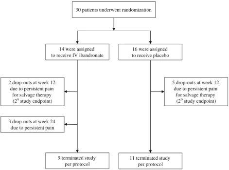
Of the 30 patients who underwent randomization, 10 par-ticipants (five in each treatment group) terminated the study prior as scheduled (Fig.1). A secondary objective of the study was the dropout rate after the first 12 weeks due to persistent pain in affected knee. A total of seven patients (ibandronate, n =2; placebo, n =5) terminated the study at week 12. De-crease in pain score in these patients was significantly less pronounced as compared to participants continuing the study after 12 weeks (mean percent change in VAS, −12.8 vs. −68.6 %, p <0.01). Randomization to either ibandronate or placebo did not influence the dropout rate with similar pro-portions of early study termination in both ibandronate- and placebo-treated subjects (Fisher’s exact test, p =0.39). After 24 weeks, another three patients (all randomized to ibandronate) dropped out in all cases due to persistent pain. Patients were referred to orthopedic surgeons for total knee replacement. Overall, a total number of 23 participants were
followed until week 12 (ibandronate, n =12; placebo, n =11), whereas 20 patients completed the study per protocol (ibandronate, n =9; placebo, n =11; Fig.1).
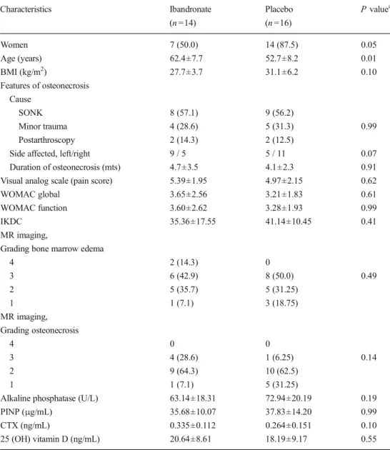
Baseline characteristics
Characteristics of the study population at baseline are displayed in Table1. Except for older age in patients random-ized to ibandronate treatment (p =0.01 vs. placebo), there were no statistically significant differences between the two groups in terms of demographic, clinical, and biochemical character-istics. Specifically, pain score (VAS score) and physical func-tion (assessed by WOMAC and IKDC quesfunc-tionnaires) were similar at baseline. Both treatment groups were comparable in the distribution of baseline severity of osteonecrosis in medial or lateral femoral condyles. All patients had unilateral affection (left knee, n =14, right knee, n =16) with osteonecrotic chang-es either at the medial or lateral femoral condylchang-es.
Clinical, functional, and biochemical outcome measures After 12 weeks of treatment, significant decrease in pain score (VAS) was observed in both ibandronate- and placebo-treated patients. After adjustment for age, sex, and osteonecrosis type, VAS score decreased by −2.98 (95 % CI, −4.34 to −1.62) points in the ibandronate group and by−3.59 (95 % CI, −5.07 to−2.12) points in the placebo group (Table2, Fig.2). Treat-ment effect was not different between the groups (p =0.54); our results rejected the null hypothesis for the primary study endpoint.
Whereas pain score did not change between weeks 12 and 48 in ibandronate-treated patients (p =0.37), placebo-treated
14 were assigned to receive IV ibandronate
16 were assigned to receive placebo
2 drop-outs at week 12 due to persistent pain
for salvage therapy (2°study endpoint) 9 terminated study per protocol 11 terminated study per protocol 3 drop-outs at week 24
due to persistent pain
5 drop-outs at week 12 due to persistent pain
for salvage therapy (2°study endpoint) 30 patients underwent randomization
Fig. 1 Randomization and follow-up
Table 1 Baseline characteristics of the study population by treat-ment group
Data are given as mean±SD or n (%)
WOMAC Western Ontario and McMaster Universities osteoar-thritis index, IKDC International Knee Documentation Committee
aMann Whitney U test or Fisher’s
exact test Characteristics Ibandronate (n =14) Placebo (n =16) P valuea Women 7 (50.0) 14 (87.5) 0.05 Age (years) 62.4±7.7 52.7±8.2 0.01 BMI (kg/m2) 27.7±3.7 31.1±6.2 0.10 Features of osteonecrosis Cause SONK 8 (57.1) 9 (56.2) Minor trauma 4 (28.6) 5 (31.3) 0.99 Postarthroscopy 2 (14.3) 2 (12.5)
Side affected, left/right 9 / 5 5 / 11 0.07
Duration of osteonecrosis (mts) 4.7±3.5 4.1±2.3 0.91 Visual analog scale (pain score) 5.39±1.95 4.97±2.15 0.62
WOMAC global 3.65±2.56 3.21±1.83 0.61
WOMAC function 3.60±2.62 3.28±1.93 0.99
IKDC 35.36±17.55 41.14±10.45 0.41
MR imaging,
Grading bone marrow edema
4 2 (14.3) 0 3 6 (42.9) 8 (50.0) 0.49 2 5 (35.7) 5 (31.25) 1 1 (7.1) 3 (18.75) MR imaging, Grading osteonecrosis 4 0 0 3 4 (28.6) 1 (6.25) 0.14 2 9 (64.3) 10 (62.5) 1 1 (7.1) 5 (31.25)
Alkaline phosphatase (U/L) 63.14±18.31 72.94±20.19 0.19
PINP (μg/mL) 35.68±10.07 37.83±14.20 0.99
CTX (ng/mL) 0.335±0.112 0.264±0.151 0.10
25 (OH) vitamin D (ng/mL) 20.64±8.61 18.19±9.17 0.55
Table 2 Change in clinical and biochemical measures during treatment (adjusted outcome change from baseline to week 12). Adjustments were made for age, sex, and type of osteonecrosis
WOMAC Western Ontario and McMaster Universities osteoar-thritis index, IKDC International Knee Documentation Committee
a
Dunnett-Hsu adjustment
Outcome Treatment Adjusted mean change 95 % CI p valuea Δ VAS (pain score) Placebo −3.59 −5.07 to −2.12 0.54
Ibandronate −2.98 −4.34 to −1.62
Δ WOMAC function Placebo −2.64 −4.57 to −0.72 0.29 Ibandronate −1.31 −2.86 to 0.24
Δ WOMAC global Placebo −2.66 −4.50 to −0.84 0.24 Ibandronate −1.27 −2.74 to 0.19
Δ IKDC Placebo 28.58 13.78 to 43.38 0.13
Ibandronate 10.99 −4.04 to 26.03
Δ ALP (U/L) Placebo −7.97 −16.35 to 0.43 0.71
Ibandronate −10.10 −17.83 to −2.38
Δ PINP (μg/mL) Placebo −4.19 −11.53 to 3.14 0.06 Ibandronate −13.72 −20.62 to −6.81
Δ CTX (ng/mL) Placebo 0.042 −0.044 to 0.129 <0.01 Ibandronate −0.143 −0.223 to −0.064
subjects showed significant further decrease in VAS score between weeks 12 and 48 (p =0.01). At the end of the study after 48 weeks, however, no significant between-group differ-ence in pain score (VAS) was observed (Fig.2).
Functional outcome as assessed by the WOMAC score and the IKDC form improved after 12 weeks in the placebo group, but not in ibandronate-treated patients (Table2). The between-group difference in all function scores was not significant (WOMAC function, p = 0.29; WOMAC global, p = 0.24; IKDC, p =0.13).
Changes in serum CTX, PINP, and alkaline phosphatase after 12 weeks are shown on Table2. Ibandronate treatment resulted in significant decreases in serum levels of CTX (adjust-ed mean change,−0.143 ng/mL; 95 % CI, −0.223 to −0.064), PINP (adjusted mean change,−13.72 μg/mL; 95 % CI, −20.62 to−6.81) and ALP (adjusted mean change, −10.10 U/L; 95 % CI,−17.83 to −2.38), whereas biochemical markers of bone turnover were not significantly altered in placebo-treated sub-jects. The between-group difference was significant for CTX (p <0.01), but not for PINP (p =0.06) and ALP (p =0.71). Radiological outcome measures
MRI evaluation revealed significant changes in the degree of bone marrow edema and degree of osteonecrosis in both ibandronate- and placebo-treated patients. Specifically, after 48 weeks, adjusted mean change in degree of bone marrow edema was−2.05 (95 % CI, −3.17to −0.95) after ibandronate treatment and−1.52 (95 % CI, −2.73 to −0.31) after placebo treatment. Similarly, adjusted mean change in degree of osteonecrosis was −0.83 (95 % CI, −1.33 to −0.34) after ibandronate treatment and−0.92 (95 % CI, −1.45 to −0.39) after placebo treatment. Treatment effects were not different between the groups after 12 and 48 weeks for both degree in
bone marrow edema (p =0.97 and p =0.48, respectively) and degree of osteonecrosis (p =0.20 and p =0.79, respectively).
Discussion
Our results do not support the hypothesized superiority of bisphosphonate treatment in patients with spontaneous or postarthroscopic osteonecrosis of the knee. Specifically, intra-venous ibandronate had no beneficial effect on clinical, func-tional, and radiological outcomes of osteonecrosis in the fem-oral condyles over and above anti-inflammatory medication.
The results of this randomized, double-blind, and placebo-controlled study are in contrast to previous observational stud-ies [19–21] and case reports [18] suggesting a favorable effect of bisphosphonate treatment. In a consecutive case series of five patients suffering from spontaneous osteonecrosis of the knee, intravenous application of ibandronate resulted in early clinical improvement and remission of bone marrow edema in all patients [20]. A larger case series including 17 patients with osteonecrosis of the knee found that alendronate treatment over 6 months resulted in complete radiographic (MRI) recovery in 59 % of patients as compared to a historical untreated control group in which only 12 % of patients showed radiological improvement [21]. Importantly, all these studies and case series are limited by small sample size, short duration, and the lack of a control group which does not allow comparing definitively beneficial effects of bisphosphonates against the natural course of disease.
Our study is the first randomized, placebo-controlled trial to assess the effect of bisphosphonates in patients with osteonecrosis of the knee. After 12 weeks of treatment, signif-icant decrease in VAS was observed in both ibandronate- and placebo-treated patients. After adjustment for age, sex, and
-1 0 1 2 3 4 5 6 7 0 2 4 12 24 48 Pain V isual Analog Scale (points) Time (weeks) Placebo Ibandronate Fig. 2 Change in pain score
(VAS) during treatment. Adjusted means for placebo (black up-pointing triangle) and
ibandronate (square) at different time points. Whiskers show the 95 % confidence intervals. Adjustments were made for age, sex, and type of osteonecrosis
osteonecrosis type, VAS score decreased by−2.98 (95 % CI, −4.34 to −1.62) points in the ibandronate group and by −3.59 (95 % CI,−5.07 to −2.12) points in the placebo group. Impor-tantly, treatment effect was not different between the groups rejecting the null hypothesis for the primary study endpoint. After 6 months, VAS score decreased by 67 % in ibandronate-treated patients and by 75 % in placebo-ibandronate-treated subjects. Inter-estingly, the decrease of pain score after 6 months was to a similar extent as what we have observed in the aforementioned uncontrolled study using pamidronate and alendronate (decrease by 80 %) [19].
Changes of questionnaires assessing functionality were comparable in both treatment groups without superiority of ibandronate treatment. Similarly, treatment effects in radiolog-ical outcome were not different between the groups. MRI evaluation revealed significant changes in the degree of bone marrow edema and degree of osteonecrosis in both ibandronate- and placebo-treated patients. Finally, randomiza-tion to either ibandronate or placebo did not influence the dropout rate with similar proportions of early study termina-tion in both treatment groups.
Overall, our results indicate that bisphosphonates have no major influence on clinical outcome in the course of disease. Changes observed for primary and secondary endpoints in both ibandronate- and placebo-treated patients were mostly within the magnitude which would be considered clinically significant; hence, in most patients, osteonecrosis of the knee may be self-limited. This is strengthened by the fact that as a result of antiresorptive treatment with ibandronate bone turn-over markers (i.e., serum CTX) was significantly suppressed whereas biochemical markers of bone turnover were not al-tered in placebo-treated subjects. As bisphosphonates prefer-entially concentrate at skeletal sites with active remodeling [33], ibandronate treatment must have ensued significant con-centrations in affected femoral condyles to provoke beneficial clinical effects during healing process. However, the degree of suppression of bone turnover with the treatment regimen was modest; hence, whether other antiresorptive agents with more potent suppression of bone remodeling (i.e., zoledronic acid or denosumab) may have exerted a stronger effect on outcome measures remains unclear.
In experimental animal studies, it has been shown that alendronate and more recently zoledronate treatment prevents resorption of necrotic bone during revascularization [9,34]. There are several reports of successful bisphosphonate treat-ment of the transient osteoporosis of the hip or osteonecrosis of the femoral head [14, 17, 35–37]. In one of the few randomized-controlled trial with alendronate treatment in 40 patients with atraumatic osteonecrosis of the femoral head with an involvement of at least 30 % of the head, only one patient in the alendronate group needed a total hip arthroplasty whereas 16 subjects in the control group underwent total hip arthroplasty over an observation period of 28 months [14].
Recently, data from a randomized, double-blind, placebo-controlled study investigating the effect of alendronate in the prevention of collapse of the femoral head in 52 patients with nontraumatic osteonecrosis have been published [38]. In con-trast to all non-placebo-controlled studies, Chen et al. ob-served no significant difference in clinical and radiographic outcomes and disease progression between alendronate- and placebo-treated subjects. Hence, based on randomized, placebo-controlled trials, clinical significant effects of bisphosphonates in osteonecrosis of the femoral head as well as the knee have to be questioned.
This present study’s findings should be interpreted within the context of its strengths and limitations. Our study using a randomized, placebo-controlled study design suggests for the first time that bisphosphonates, i.e., intravenous ibandronate have no beneficial effect on this disease outcome. The study is limited by short follow-up. Longer follow-up time, however, is unlikely to have modulated our findings as nearly all pa-tients presented with major reduction in pain score after 48 weeks (VAS score 0–2). Secondly, the sample size was small, although the number of enrolled subjects reached the target based on the power estimate. We adjusted for imbal-ances in baseline characteristics (age, sex distribution, and type of osteonecrosis); nevertheless, due to small sample size, residual confounding due to differences in these variables cannot be completely ruled out. Thirdly, treatment was initi-ated with some delay after osteonecrosis was diagnosed and patients were not randomized for the size of their lesions. Clearly, our findings are not generalizable to secondary forms of osteonecrosis of the knee or to osteonecrosis of other localizations. Further randomized studies are needed to con-firm our findings and explore alternative bisphosphonates with selective higher affinity to bone surfaces to crystallize optimal nonsurgical management and surgical indications for spontaneous or postarthroscopic osteonecrosis of the knee.
We conclude that in patients with spontaneous osteonecrosis of the knee, intravenous ibandronate has no beneficial effect over and above anti-inflammatory medication. Hence, bisphos-phonate therapy for the use of osteonecrosis of the knee should be used with caution.
Acknowledgments This study was supported by an unrestricted edu-cational grant from Roche Pharma (Switzerland) AG.
Conflicts of interest None.
References
1. Assouline-Dayan Y, Chang C, Greenspan A, Shoenfeld Y, Gershwin ME (2002) Pathogenesis and natural history of osteonecrosis. Semin Arthritis Rheum 32:94–124
Osteonecrosis of the knee: differences among idiopathic and second-ary types. Rheumatology (Oxford) 39:982–989
3. Hofmann S, Kramer J, Vakil-Adli A, Aigner N, Breitenseher M (2004) Painful bone marrow edema of the knee: differential diagnosis and therapeutic concepts. Orthop Clin North Am 35:321–333, ix 4. Zywiel MG, McGrath MS, Seyler TM, Marker DR, Bonutti PM,
Mont MA (2009) Osteonecrosis of the knee: a review of three disorders. Orthop Clin North Am 40:193–211
5. Strauss EJ, Kang R, Bush-Joseph C, Bach BR Jr (2011) The diagno-sis and management of spontaneous and post-arthroscopy osteonecrosis of the knee. Bull NYU Hosp Jt Dis 69:320–330 6. Peters KM, Sachse A (2012) Osteonekrosen der unteren
Extremitäten. Osteologie 21:253–260
7. Mont MA, Marker DR, Zywiel MG, Carrino JA (2011) Osteonecrosis of the knee and related conditions. J Am Acad Orthop Surg 19:482–494
8. DeFalco RA, Ricci AR, Balduini FC (2003) Osteonecrosis of the knee after arthroscopic meniscectomy and chondroplasty: a case report and literature review. Am J Sports Med 31:1013–1016 9. Astrand J, Aspenberg P (2002) Systemic alendronate prevents
re-sorption of necrotic bone during revascularization. A bone chamber study in rats. BMC Musculoskelet Disord 3:19
10. Kim HK, Randall TS, Bian H, Jenkins J, Garces A, Bauss F (2005) Ibandronate for prevention of femoral head deformity after ischemic necrosis of the capital femoral epiphysis in immature pigs. J Bone Joint Surg Am 87:550–557
11. Little DG, Peat RA, McEvoy A, Williams PR, Smith EJ, Baldock PA (2003) Zoledronic acid treatment results in retention of femoral head structure after traumatic osteonecrosis in young Wistar rats. J Bone Miner Res 18:2016–2022
12. Ringe JD, Dorst A, Faber H (2005) Effective and rapid treatment of painful localized transient osteoporosis (bone marrow edema) with intravenous ibandronate. Osteoporos Int 16:2063–2068
13. Agarwala S, Jain D, Joshi VR, Sule A (2005) Efficacy of alendronate, a bisphosphonate, in the treatment of AVN of the hip. A prospective open-label study. Rheumatology (Oxford) 44:352–359
14. Lai KA, Shen WJ, Yang CY, Shao CJ, Hsu JT, Lin RM (2005) The use of alendronate to prevent early collapse of the femoral head in patients with nontraumatic osteonecrosis. A randomized clinical study. J Bone Joint Surg Am 87:2155–2159
15. Nishii T, Sugano N, Miki H, Hashimoto J, Yoshikawa H (2006) Does alendronate prevent collapse in osteonecrosis of the femoral head? Clin Orthop Relat Res 443:273–279
16. La Montagna G, Malesci D, Tirri R, Valentini G (2005) Successful neridronate therapy in transient osteoporosis of the hip. Clin Rheumatol 24:67–69
17. Cardozo JB, Andrade DM, Santiago MB (2008) The use of bisphos-phonate in the treatment of avascular necrosis: a systematic review. Clin Rheumatol 27:685–688
18. Corrado A, Quarta L, Errico S, Cantatore FP (2007) Successful treatment of avascular bone necrosis of the knee with neridronate: a case report. Rheumatol Int 27:891–893
19. Kraenzlin ME, Graf C, Meier C, Kraenzlin C, Friedrich NF (2010) Possible beneficial effect of bisphosphonates in osteonecrosis of the knee. Knee Surg Sports Traumatol Arthrosc 18:1638–1644 20. Breer S, Oheim R, Krause M, Marshall RP, Amling M, Barvencik F
(2012) Spontaneous osteonecrosis of the knee (SONK). Knee Surg Sports Traumatol Arthrosc 21(2):340–5
21. Jureus J, Lindstrand A, Geijer M, Roberts D, Tagil M (2012) Treatment of spontaneous osteonecrosis of the knee (SPONK) by a bisphosphonate. Acta Orthop 83:511–514
22. Bellamy N, Buchanan WW, Goldsmith CH, Campbell J, Stitt LW (1988) Validation study of WOMAC: a health status instrument for
measuring clinically important patient relevant outcomes to antirheu-matic drug therapy in patients with osteoarthritis of the hip or knee. J Rheumatol 15:1833–1840
23. Stucki G, Meier D, Stucki S, Michel BA, Tyndall AG, Dick W, Theiler R (1996) Evaluation of a German version of WOMAC (Western Ontario and McMaster Universities) Arthrosis Index. Z Rheumatol 55:40–49
24. Stucki G, Sangha O, Stucki S, Michel BA, Tyndall A, Dick W, Theiler R (1998) Comparison of the WOMAC (Western Ontario and McMaster Universities) osteoarthritis index and a self-report format of the self-administered Lequesne-Algofunctional index in patients with knee and hip osteoarthritis. Osteoarthritis Cartilage 6:79–86 25. Irrgang JJ, Anderson AF, Boland AL, Harner CD, Kurosaka M,
Neyret P, Richmond JC, Shelborne KD (2001) Development and validation of the international knee documentation committee sub-jective knee form. Am J Sports Med 29:600–613
26. Anderson AF, Irrgang JJ, Kocher MS, Mann BJ, Harrast JJ (2006) The International Knee Documentation Committee Subjective Knee Evaluation Form: normative data. Am J Sports Med 34:128–135 27. Irrgang JJ, Anderson AF, Boland AL, Harner CD, Neyret P,
Richmond JC, Shelbourne KD (2006) Responsiveness of the International Knee Documentation Committee Subjective Knee Form. Am J Sports Med 34:1567–1573
28. Garnero P, Borel O, Delmas PD (2001) Evaluation of a fully auto-mated serum assay for C-terminal cross- linking telopeptide of type I collagen in osteoporosis. Clin Chem 47:694–702
29. Peterfy CG, Guermazi A, Zaim S et al (2004) Whole-Organ Magnetic Resonance Imaging Score (WORMS) of the knee in osteoarthritis. Osteoarthritis Cartilage 12:177–190
30. Ficat RP, Arlet J (1980) Necrosis of the femoral head. In: Hungerford DS (ed) Ischemia and necrosis of bone. Williams & Wilkins, Baltimore, pp 171–182
31. White DK, Keysor JJ, Lavalley MP, Lewis CE, Torner JC, Nevitt MC, Felson DT (2010) Clinically important improvement in function is common in people with or at high risk of knee OA: the MOST study. J Rheumatol 37:1244–1251
32. Suter LG, Fraenkel L, Losina E, Katz JN, Gomoll AH, Paltiel AD (2009) Medical decision making in patients with knee pain, meniscal tear, and osteoarthritis. Arthritis Rheum 61:1531–1538
33. Khan SA, Kanis JA, Vasikaran S et al (1997) Elimination and biochemical responses to intravenous alendronate in postmenopausal osteoporosis. J Bone Miner Res 12:1700–1707
34. Astrand J, Harding AK, Aspenberg P, Tagil M (2006) Systemic zoledronate treatment both prevents resorption of allograft bone and increases the retention of new formed bone during revascularization and remodelling. A bone chamber study in rats. BMC Musculoskelet Disord 7:63
35. Agarwala S, Shah S, Joshi VR (2009) The use of alendronate in the treatment of avascular necrosis of the femoral head: follow-up to eight years. J Bone Joint Surg Br 91:1013–1018
36. Ramachandran M, Ward K, Brown RR, Munns CF, Cowell CT, Little DG (2007) Intravenous bisphosphonate therapy for traumatic osteonecrosis of the femoral head in adolescents. J Bone Joint Surg Am 89:1727–1734
37. Wang CJ, Wang FS, Yang KD, Huang CC, Lee MS, Chan YS, Wang JW, Ko JY (2008) Treatment of osteonecrosis of the hip: comparison of extracorporeal shockwave with shockwave and alendronate. Arch Orthop Trauma Surg 128:901–908
38. Chen CH, Chang JK, Lai KA, Hou SM, Chang CH, Wang GJ (2012) Alendronate in the prevention of collapse of the femoral head in nontraumatic osteonecrosis: a two-year multicenter, prospective, ran-domized, double-blind, placebo-controlled study. Arthritis Rheum 64:1572–1578