Cell Reports, Volume 26
Supplemental Information
Vectorial Import via a Metastable Disulfide-Linked
Complex Allows for a Quality Control Step and
Import by the Mitochondrial Disulfide Relay
Markus Habich, Silja Lucia Salscheider, Lena Maria Murschall, Michaela Nicole Hoehne, Manuel Fischer, Fabian Schorn, Carmelina Petrungaro, Muna Ali, Alican J. Erdogan, Shadi Abou-Eid, Hamid Kashkar, Joern Dengjel, and Jan Riemer
SUPPLEMENTAL TABLES
Table S1: Proteomic data: Refer to enclosed Excel file, Related to Figure 3
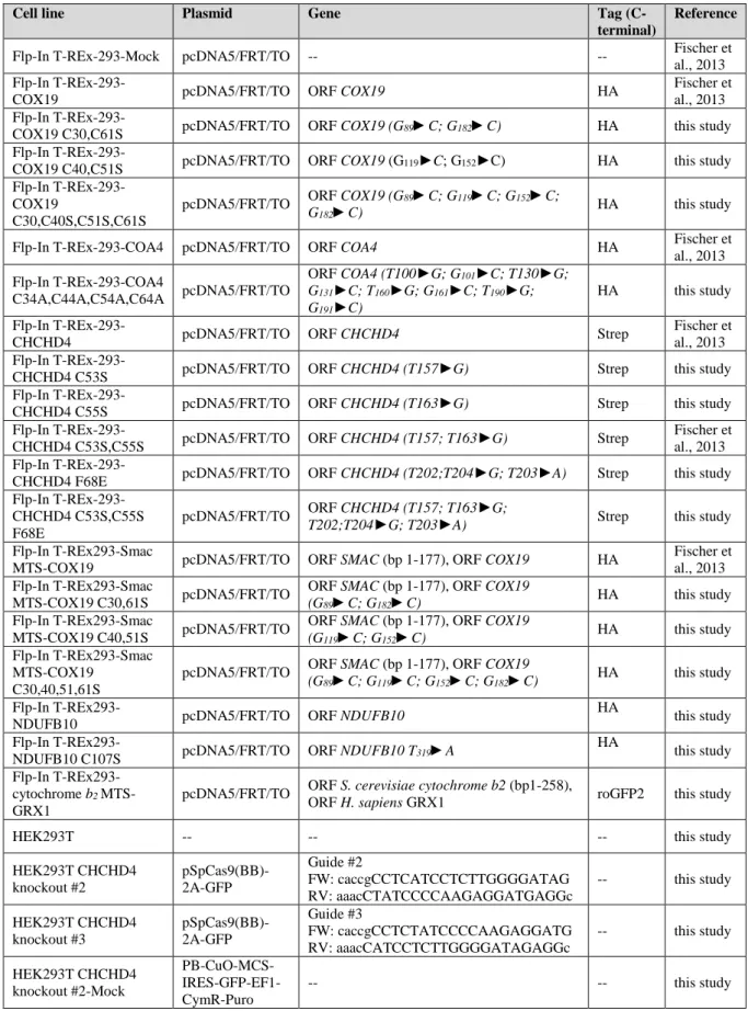
Table S2: Plasmids and cell lines used in this study, Related to Star methods:
Cell line Plasmid Gene Tag
(C-terminal)
Reference
Flp-In T-REx-293-Mock pcDNA5/FRT/TO -- -- Fischer et al., 2013 Flp-In
T-REx-293-COX19 pcDNA5/FRT/TO ORF COX19 HA
Fischer et al., 2013 Flp-In
T-REx-293-COX19 C30,C61S pcDNA5/FRT/TO ORF COX19 (G89►C; G182►C) HA this study Flp-In
T-REx-293-COX19 C40,C51S pcDNA5/FRT/TO ORF COX19 (G119►C; G152►C) HA this study Flp-In
T-REx-293-COX19
C30,C40S,C51S,C61S
pcDNA5/FRT/TO ORF COX19 (G89►C; G119►C; G152►C;
G182►C) HA this study
Flp-In T-REx-293-COA4 pcDNA5/FRT/TO ORF COA4 HA Fischer et al., 2013 Flp-In T-REx-293-COA4 C34A,C44A,C54A,C64A pcDNA5/FRT/TO ORF COA4 (T100►G; G101►C; T130►G; G131►C; T160►G; G161►C; T190►G; G191►C) HA this study Flp-In
T-REx-293-CHCHD4 pcDNA5/FRT/TO ORF CHCHD4 Strep
Fischer et al., 2013 Flp-In
T-REx-293-CHCHD4 C53S pcDNA5/FRT/TO ORF CHCHD4 (T157►G) Strep this study Flp-In
T-REx-293-CHCHD4 C55S pcDNA5/FRT/TO ORF CHCHD4 (T163►G) Strep this study Flp-In
T-REx-293-CHCHD4 C53S,C55S pcDNA5/FRT/TO ORF CHCHD4 (T157; T163►G) Strep
Fischer et al., 2013 Flp-In
T-REx-293-CHCHD4 F68E pcDNA5/FRT/TO ORF CHCHD4 (T202;T204►G; T203►A) Strep this study Flp-In
T-REx-293-CHCHD4 C53S,C55S F68E
pcDNA5/FRT/TO ORF CHCHD4 (T157; T163►G;
T202;T204►G; T203►A) Strep this study
Flp-In T-REx293-Smac
MTS-COX19 pcDNA5/FRT/TO ORF SMAC (bp 1-177), ORF COX19 HA
Fischer et al., 2013 Flp-In T-REx293-Smac
MTS-COX19 C30,61S pcDNA5/FRT/TO
ORF SMAC (bp 1-177), ORF COX19
(G89►C; G182►C) HA this study
Flp-In T-REx293-Smac
MTS-COX19 C40,51S pcDNA5/FRT/TO
ORF SMAC (bp 1-177), ORF COX19
(G119►C; G152►C) HA this study
Flp-In T-REx293-Smac MTS-COX19
C30,40,51,61S
pcDNA5/FRT/TO ORF SMAC (bp 1-177), ORF COX19
(G89►C; G119►C; G152►C; G182►C) HA this study
Flp-In
T-REx293-NDUFB10 pcDNA5/FRT/TO ORF NDUFB10
HA
this study Flp-In
T-REx293-NDUFB10 C107S pcDNA5/FRT/TO ORF NDUFB10 T319►A
HA
this study Flp-In T-REx293-
cytochrome b2
MTS-GRX1
pcDNA5/FRT/TO ORF S. cerevisiae cytochrome b2 (bp1-258),
ORF H. sapiens GRX1 roGFP2 this study
HEK293T -- -- -- this study
HEK293T CHCHD4 knockout #2 pSpCas9(BB)-2A-GFP Guide #2 FW: caccgCCTCATCCTCTTGGGGATAG RV: aaacCTATCCCCAAGAGGATGAGGc -- this study HEK293T CHCHD4 knockout #3 pSpCas9(BB)-2A-GFP Guide #3 FW: caccgCCTCTATCCCCAAGAGGATG RV: aaacCATCCTCTTGGGGATAGAGGc -- this study HEK293T CHCHD4 knockout #2-Mock PB-CuO-MCS- IRES-GFP-EF1-CymR-Puro -- -- this study
HEK293T CHCHD4 knockout #2-CHCHD4
PB-CuO-MCS- IRES-GFP-EF1-CymR-Puro
ORF CHCHD4 -- this study
HEK293T CHCHD4 knockout #2-C53S
PB-CuO-MCS- IRES-GFP-EF1-CymR-Puro
ORF CHCHD4 (T157►G) -- this study HEK293T CHCHD4
knockout #2-C53S, C55S
PB-CuO-MCS- IRES-GFP-EF1-CymR-Puro
ORF CHCHD4 (T157; T163►G) -- this study HEK293T CHCHD4 knockout #2-CHCHD4 F68E PB-CuO-MCS- IRES-GFP-EF1-CymR-Puro
ORF CHCHD4 (T202;T204►G; T203►A) -- this study HEK293T CHCHD4 knockout #3-Mock PB-CuO-MCS- IRES-GFP-EF1-CymR-Puro -- -- this study HEK293T CHCHD4 knockout #3-CHCHD4 PB-CuO-MCS- IRES-GFP-EF1-CymR-Puro
ORF CHCHD4 -- this study
HEK293T CHCHD4 knockout #3-C53S
PB-CuO-MCS- IRES-GFP-EF1-CymR-Puro
ORF CHCHD4 (T157►G) -- this study HEK293T CHCHD4
knockout #3-C53S, C55S
PB-CuO-MCS- IRES-GFP-EF1-CymR-Puro
ORF CHCHD4 (T157; T163►G) -- this study HEK293T CHCHD4 knockout #3-CHCHD4 F68E PB-CuO-MCS- IRES-GFP-EF1-CymR-Puro
SUPPLEMENTAL FIGURES
Supplementary Figure 1. CHCHD4 interacts with and oxidizes COX19 cysteine variants lacking the inner or the outer disulphide bond in vitro (related to Figure 1)
A) Structure of COX19, NDUFB10 and COA4 as well as the respective mutated variants used
in this study. The structure of human COX19 (Uniprot: Q49B96) was built on the solution structure of CHCHD7 (pdb: 2LQT) with SWISS-MODEL. The structure of human NDUFB10 pdb: 5XTC). NDUFB10C107 represents a human disease mutant. The structure of human COA4 (Uniprot: Q9NYJ1) was built on the solution structure of CHCHD7 (pdb: 2LQT) with
SWISS-MODEL. Colour-coding represents hydrophobicity of the amino acids according to the Tanford hydrophobicity scale.
B) In cell CHCHD4-substrate interaction assay for NDUFB10 and a patient variant of the
protein (NDUFB10C107S). Performed as described in Figure 1A. 1% of the IP against CHCHD4
was loaded as control. Expression was induced during the 20 min pulse with 1 µg/ml doxycycline. CHCHD4 interacts covalently with wildtype NDUFB10 and with the patient mutant NDUFB10C107S in vivo. red, reducing SDS-PAGE; non-red, non-reducing SDS-PAGE.
red arrow, disulphide-linked CHCHD4-NDUFB10 dimer; blue arrow, NDUFB10. hash tag,
disulphide-linked CHCHD4-NDUFB10 dimer possibly with truncated CHCHD4; plus sign, NDUFB10 not in disulphide-linked complex. (n=2 biological replicates)
C) Twin CX9C proteins contain multiple potential CHCHD4 interaction sites. Amino acids in
twin CX9C motifs were scored for hydrophobicity using the Tanford scale. Data was grouped
using hierarchical clustering (metric: euclidean distance; linkage method: average) and plotted in a heatmap using Morpheus (https://software.broadinstitute.org/morpheus/). Both helices of most substrates (depicted in light and dark grey, respectively) are sufficiently amphipathic to allow initial hydrophobic interaction with CHCHD4 that is required for formation of the disulphide-linked metastable complex. This is also reflected by the overall hydrophobicity as outlined in the graph.
D) In vitro oxidation kinetics assay. Recombinant expressed and purified human CHCHD4 was
added to 35S-labeled COX19 variants to allow disulphide exchange reactions. The reaction was
stopped by rapid acidification via addition of trichloroacetic acid (TCA). Lysates were treated with mmPEG24 to determine protein redox states, followed by SDS-PAGE and autoradiography. Reduced COX19 is modified with four mmPEG24, whereas oxidized COX19 remains unmodified.
E) Control experiment. Wildtype COX19 does not become oxidized in the absence of
CHCHD4. Small amounts of in vitro translated radioactive COX19 were incubated with buffer for the indicated times, TCA-precipitated, and incubated with 15 mM mmPEG24 for 1 hr at 25°C. Protein was analysed by non-reducing SDS-PAGE and autoradiography. Reduced and oxidized forms as well as semi-oxidized intermediates (*) of COX19 are indicated. (n=2 biological replicates)
F-H) Oxidation kinetics of COX19 cysteine variants. CHCHD4 oxidizes COX19 variants
lacking the cysteines for the outer and inner disulphide bond, respectively. Performed as described in (D), except that in vitro translated radioactive COX19 was incubated with 30 μM purified CHCHD4 for the indicated times. Reduced and oxidized forms of COX19 as well as COX19-CHCHD4 complexes are indicated. Apparently, oxidation of COX19WT can follow two routes: 1) In the COX19WT-CHCHD4 complex, COX19 forms one disulphide bond over time and is then released as fully oxidized protein (two bands for the complex), 2) COX19 disulphide bonds are serially introduced. Occurrence of multiple semi-oxidized intermediates (*) might indicate off-target intermediates that also appear to persist over the duration of the experiment. (n=2 biological replicates)
Supplementary Figure 2. COA4-HA cysteine variants do not accumulate in mitochondria.
(related to Figure 2)
A) Localization of COA4-HA cysteine variant.. HEK293 cells stably expressing the indicated
COA4-HA variants were incubated for 24 hours with 1 μg/ml doxycycline to induce expression. Cells were fixated, permeabilized and stained using primary antibodies against the HA epitope (αHA; green) and cytochrome c (αcyt c; red). Cells were analysed by fluorescence microscopy. While COA4WT-HA accumulated in mitochondria, the cysteine variant did not. Instead, it remained in the cytosol. Scale bar = 10 µm. (WT: n=6 cells, C34A,C44A,C54A,C64A: n=10 cells, technical replicates)
B) Pulse chase fractionation experiment of COA4-HA cysteine variants expressed in HEK293
cells. Expression was induced by doxycycline treatment for one hour before the pulse chase experiment. Cells were first pulse labelled for 5 min with 35S-methionine and then chased with
cold methionine for 0 or 20 min. At the end of the chase, cells were transferred to ice and incubated with low concentration of digitonin for 10 min to selectively permeabilize the plasma membrane and leave the OMM intact. To separate the mitochondrial from the cytosolic fraction, lysates were centrifuged and COA4-HA was immunoprecipitated (IP). Eluates were analysed by SDS–PAGE and autoradiography. While COA4WT-HA accumulated in mitochondria, the cysteine variant did not. Instead, it remained in the cytosol. (n=2 biological replicates)
Supplementary Figure 3. Parameters of CHCHD4-substrate and CHCHD4-ALR interaction (related to Figure 3)
A) CHCHD4-substrate interaction is transient. HEK293 cells were pulse-labeled with 35 S-methionine for 1 hour. Then, thiol-exchange reactions were inhibited by addition of NEM to preserve mixed disulphides. Cells were lysed in 2% SDS. Then, lysates were subjected to IP against CHCHD4, analysed by non-reducing or reducing SDS-PAGE and autoradiography. Disulphide-linked substrate-CHCHD4 complexes and CHCHD4 substrates are indicated. (n=2 biological replicates)
B) CHCHD4WT and CHCHD4C53S interact with ALR. HEK293 cells stably expressing different CHCHD4 cysteine variants were treated with NEM to block thiol-disulphide exchange. Then, denaturing IP against the Strep-tag was performed. Total sample (T) and eluates (E) were analysed by SDS-PAGE and immunoblot against the indicated proteins. (n=1)
C) Schematic inverse shift assay for the determination of the CHCHD4 redox state. To
determine the redox state of CHCHD4 at steady state, intact cells were treated with NEM to stop all thiol-disulphide exchange reactions and trap the redox state of CHCHD4. Then, cells were lysed and lysates were treated at 4 °C with TCEP. Previously oxidized cysteines in the CPC motif are now accessible to modification with mmPEG24. As controls, unmodified samples (min) and samples without NEM pretreatment (max) were loaded.
D) Redox state of F68E variants of CHCHD4. These variants are almost completely reduced at
steady state. Performed as described in (C). (n=2 biological replicates)
E-H) Interaction partners of CHCHD4 cysteine variants and the F68E mutant from native and
denaturing affinity purification (AP). Cells were grown in SILAC medium. Cells were treated with NEM, mixed and lysed under native or denaturing conditions. CHCHD4-Strep variants were enriched using strep-tactin beads and eluates were analysed by mass spectrometry. The experiment was reproduced with inverted isotope labelling. Results from both experiments are plotted against each other. Proteins were counted as hits if at least two peptides per protein were enriched more than 2-fold. (n=2 biological replicates)
Supplementary Figure 4. Reproduction of data shown in Figure 4 with a second CRISPR clone. (related to Figure 4). Findings obtained with clone #3 are analogues to data obtained
Supplementary Figure 5. Properties of ‘redox’ and ‘chaperone’ mutants of CHCHD4.
(related to Figure 5)
A) Steady state levels of CHCHD4 substrates in HEK293 cells stably expressing different F68E
variants of CHCHD4. Expression of CHCHD4 variants was induced for 5 days in glucose-containing medium. Cells were lysed and protein levels were analysed by SDS-PAGE and immunoblot against the indicated proteins. CHCHD4F68E variants exhibit no deleterious effect for substrate levels. (n=2-3 biological replicates)
B) Quantification of (A) shows substrate-specific differences in substrate levels.
C) Viability of cells upon overexpression of CHCHD4 F68E variants. Expression of CHCHD4
variants was induced for 4 and 7 days in galactose-containing medium. Cells were analysed by PrestoBlue Cell Viability Reagent. CHCHD4F68E variants exhibit no deleterious effect on growth. Fold viability is presented as the viability data of induced cells divided by the data of non-induced cells of the same cell line. (n=3 biological replicates)
D-F) Only CHCHD4WT can oxidize COX19. Small amounts of in vitro translated radioactive COX19 were incubated with buffer for the indicated times, TCA-precipitated, and incubated with 15 mM mmPEG24 for 1 hr at 25°C. Protein was analysed by non-reducing SDS-PAGE and autoradiography. Reduced and oxidized forms of COX19 are indicated. (n=1)
G) Redox state of cysteine variants of CHCHD4. C53S and C55S are reduced at steady state.
Performed as described in Figure S3C. This indicates that in thiol-disulphide-exchange reactions these variants would execute nucleophilic attacks. (n=2 biological replicates)
Supplementary Figure 6. Proteasome and IMS redox influence CHCHD4 substrates.
(related to Figure 6)
A) Localization of MTS-COX19 cysteine variants in the presence and absence of proteasomal
inhibitor. Experiment was performed as described in Figure 1E except that cells were cultured in the presence or absence of the proteasomal inhibitor MG132. Like the four-fold cysteine mutant of COX19 (Figure 6B), the mutants lacking the two cysteines required for formation of the inner or outer disulphide bonds, respectively accumulate in the cytosol upon MG132 treatment. MitoTrackerRed was used to stain mitochondria (mito). Scale bar = 10 µm. (C30,61S: n=37, C40,51S: 27 cells, technical replicates)
B) Pulse chase stability assay of COA4 cysteine variants. Cysteine variants but not the wildtype
are stabilized by MG132 treatment. Performed as described in Figure 6C. For proteasome inhibition 5 µM MG132 was present throughout the pulse chase experiment. (n=2 biological replicates)
C) Steady state levels of COA4 wild type and cysteine variant. The COA4 cysteine variant is
stabilized by MG132 treatment. For proteasome inhibition, cells were treated with 5 µM MG132 for 4 h prior to cell lysis. (n=3 biological replicates)
D) Steady state levels of NDUFB10 wild type and cysteine variant (NDUFB10C107S). NDUFB10C107S is stabilized by MG132 treatment. For proteasome inhibition, cells were treated with 5 µM MG132 for 4 h prior to cell lysis. (n=1)
E) Levels of endogenous CHCHD4 substrate proteins in HEK293 cells upon treatment with
MG132. HEK293 cells were treated with 5 µM MG132 for 4 h or remained untreated. Cells were fractionated and separated by centrifugation into mitochondrial and cytosolic fractions. Cellular fractions were separated by SDS-PAGE and analyzed by immunoblot using antibodies against the indicated proteins. (n=1)
F) Steady state levels of endogenous CHCHD4 substrates upon overexpression of
IMS-localized GRX1. Expression of MTS-GRX1 was induced for 5 days in glucose-containing medium. Cells were lysed and protein levels were analysed by SDS-PAGE and immunoblot against the indicated proteins. Endogenous COX17 levels are decreased upon increased levels of IMS-localized GRX1. This is in line for a potential role of GRX1 in modulating import and oxidation of selected CHCHD4 substrates. (n=4 biological replicates)