HAL Id: hal-02996688
https://hal.archives-ouvertes.fr/hal-02996688
Submitted on 13 Nov 2020
HAL is a multi-disciplinary open access
archive for the deposit and dissemination of
sci-entific research documents, whether they are
pub-lished or not. The documents may come from
teaching and research institutions in France or
abroad, or from public or private research centers.
L’archive ouverte pluridisciplinaire HAL, est
destinée au dépôt et à la diffusion de documents
scientifiques de niveau recherche, publiés ou non,
émanant des établissements d’enseignement et de
recherche français ou étrangers, des laboratoires
publics ou privés.
Distributed under a Creative Commons Attribution - NonCommercial| 4.0 International
Claire Leveau, Tania Gajardo, Marie-Thérèse El-Daher, Nicolas Cagnard,
Alain Fischer, Geneviève de Saint Basile, Fernando Sepulveda Garrido
To cite this version:
Claire Leveau, Tania Gajardo, Marie-Thérèse El-Daher, Nicolas Cagnard, Alain Fischer, et al.. Ttc7a
regulates hematopoietic stem cell functions while controlling the stress-induced response.
Haema-tologica, Ferrata Storti Foundation, 2020, 105 (1), pp.59-70. �10.3324/haematol.2018.207100�.
�hal-02996688�
Received: September 18, 2018. Accepted: April 17, 2019. Pre-published: April 19, 2019.
©2020 Ferrata Storti Foundation
Material published in Haematologica is covered by copyright. All rights are reserved to the Ferrata Storti Foundation. Use of published material is allowed under the following terms and conditions:
https://creativecommons.org/licenses/by-nc/4.0/legalcode. Copies of published material are allowed for personal or inter-nal use. Sharing published material for non-commercial pur-poses is subject to the following conditions:
https://creativecommons.org/licenses/by-nc/4.0/legalcode, sect. 3. Reproducing and sharing published material for com-mercial purposes is not allowed without permission in writing from the publisher.
Correspondence:
GENEVIÈVE DE SAINT BASILE [email protected] FERNANDO E. SEPULVEDA [email protected]
Haematologica
2020
Volume 105(1):59-70
doi:10.3324/haematol.2018.207100 Check the online version for the most updated information on this article, online supplements, and information on authorship & disclosures: www.haematologica.org/content/105/1/59Ferrata Storti Foundation
T
he molecular machinery that regulates the balance between
self-renewal and differentiation properties of hematopoietic stem cells
(HSC) has yet to be characterized in detail. Here we found that the
tetratricopeptide repeat domain 7 A (Ttc7a) protein, a putative scaffold
pro-tein expressed by HSC, acts as an intrinsic regulator of the proliferative
response and the self-renewal potential of murine HSC in vivo. Loss of Ttc7a
consistently enhanced the competitive repopulating ability of HSC and
their intrinsic capacity to replenish the hematopoietic system after serial cell
transplantations, relative to wildtype cells. Ttc7a-deficient HSC exhibit a
different transcriptomic profile for a set of genes controlling the cellular
response to stress, which was associated with increased proliferation in
response to chemically induced stress in vitro and myeloablative stress in
vivo. Our results therefore revealed a previously unrecognized role of Ttc7a
as a critical regulator of HSC stemness. This role is related, at least in part,
to regulation of the endoplasmic reticulum stress response.
Introduction
In flaky skin (fsn) mice, the spontaneous insertion of early transposon into the gene for tetratricopeptide repeat domain 7 A (Ttc7a) is known to impair Ttc7a pro-tein expression.1,2 Consequently, fsn mice develop a proliferative lymphoid and
myeloid disorder, with hyperplasia of the spleen and lymph nodes, elevated mono-cyte, granulocyte and lymphoid cell counts,3-6and severe anemia.7Moreover, fsn
mice have a reduced lifespan and changes in the skin (epidermal hyperplasia and inflammation)8,9and the intestinal tract (gastric papillomas).10The marked
pheno-typic alterations in fsn mice suggest that Ttc7a protein has one or more major reg-ulatory roles in the hematopoietic system, and, potentially, in other tissues of epithelial origin.
Ttc7a is a putative scaffolding protein as it contains nine tetratricopeptide repeats (TPR) domains that are predicted to interact with proteins containing their own TPR or other motifs.11These TPR-containing proteins are involved in a variety of
biological processes, including cell cycle control, protein trafficking, secretion and protein quality control. Indeed, TPR-containing proteins have been shown to bind chaperones such as Hsp90 and Hsp70, controlling their activity.12-14Thus, Ttc7a is
likely to be involved in a broad range of protein complexes and hence functions. In
vitro studies have shown that the loss of Ttc7a causes inappropriate activation of
RhoA-dependent effectors and thus disrupts cytoskeletal dynamics.15,16
Ttc7a regulates hematopoietic stem cell
functions while controlling the stress-induced
response
Claire Leveau,1,2Tania Gajardo,1,2Marie-Thérèse El-Daher,1,2
Nicolas Cagnard,2,3,4Alain Fischer,2,5,6,7Geneviève de Saint Basile1,2,8,*
and Fernando E. Sepulveda1,2,9,*
1Laboratory of Normal and Pathological Homeostasis of the Immune System, INSERM
UMR 1163, Imagine Institute, Paris; 2Université Paris Descartes -Sorbonne Paris Cité,
Imagine Institute, Paris; 3Bioinformatic Platform, INSERM UMR 1163, Université Paris
Descartes-Sorbonne Paris Cité, Imagine Institute, Paris; 4Structure Fédérative de
Recherche (SFR) Necker, INSERM US24/CNRS UMS 3633, Paris; 5Assistance
Publique-Hôpitaux de Paris, Hôpital Necker-Enfants Malades Immunology and Pediatric Hematology Department, Paris; 6Collège de France, Paris, France; 7INSERM UMR1163, Paris;
8Assistance Publique-Hôpitaux de Paris, Hôpital Necker-Enfants Malades, Centre d'Etudes
des Déficits Immunitaires, Paris and 9Centre National de la Recherche Scientifique –
CNRS, Paris, France
* These authors contributed equally to this work ABSTRACT
Furthermore, TTC7A reportedly interacts with EFR3 homolog B and phosphatidylinositol 4-kinase alpha, which is known to catalyze the production of phos-phatidylinositol 4-phosphate at the plasma membrane in yeast and human cells.17,18 This observation emphasizes
the conservation, at least in part, of the functions of Ttc7a during evolution. However, data on TTC7A’s biological function(s) are still scarce.
Inadequate proliferation of peripheral hematopoietic lineages has been reported in several modified murine models; this impairment is ultimately associated with the exhaustion of the hematopoietic stem cell (HSC) pool.19
Indeed, the production of blood cells requires HSC to leave their quiescent state and differentiate into functional progeny. An excessive requirement for hematopoietic cell production biases HSC function toward differentiation, at the expense of self-renewal.20Various intrinsic and
extrin-sic factors influence HSC fate, i.e. quiescence or prolifera-tion. Endoplasmic reticulum (ER) stress has recently been highlighted as an important regulator of HSC function.21
This stress is triggered by various stimuli and leads to the accumulation of unfolded proteins in the lumen of the ER, and induction of the unfolded protein response (UPR). The chaperone BIP (Hspa5/GRP78) is the main inducer of the UPR.22This response results in enhanced expression of
chaperone proteins (heat shock proteins, Hsp), phospho-diesterase (Pdi), and other proteins such as calreticulin that, together with BIP, boost protein folding capacities. Depending on the intensity of the ER stress, UPR activa-tion can lead to apoptosis or survival.23
In the present study, we found that Ttc7a regulates murine HSC self-renewal and hematopoietic reconstitu-tion potential and controls the sensitivity of these cells to stress. Loss of Ttc7a consistently enhanced HSC stemness, since Ttc7a-deficient HSC displayed a greater proliferation capacity than control counterparts in response to ER stress
in vitro, and after myeloablative stress in vivo. Hence, our
results reveal a new role for Ttc7a as a regulator of self-renewal and response to stress in HSC.
Methods
Mice
Heterozygous Balb/cByJ fsn (CByJ.A-Ttc7fsn/J) mice and Balb/cByJ CD45.1 (CByJ.SJL(B6)-Ptprca/J) mice were obtained from the Jackson Laboratory. All mice were maintained in specific pathogen-free conditions and handled according to national and institutional guidelines.
Repopulations assays
Bone marrow (BM) cells were transferred into CD45.1+control
recipient mice upon irradiation and then 30,000 Lin-Sca1+cKit+
(LSK) donor cells were injected into the irradiated recipient mice. For serial transplantations, recipients were reconstituted with 107
BM cells. To perform competitive repopulation assays, 1,000 LSK cells were injected with 2 x 10⁶unfractionated CD45.1+BM cells.
Twelve weeks after transfer, mice were treated with a single dose of 5-fluorouracil (5-FU, 150 mg/kg).
Flow cytometry and isolation of hematopoietic stem
cells
Splenocytes and peripheral blood cells were incubated with conjugated antibodies and viability exclusion dyes. The antibodies used are listed in Online Supplementary Table S2. Stained cells were
quantified using a Gallios flow cytometer (Beckman Coulter), and analyzed with FlowJo software (Treestar). HSC and LSK cells were isolated by depleting Lin+ cells using the Lineage Cell
Depletion Kit according to the manufacturer’s protocol (Miltenyi Biotec), stained with a Lin- antibody cocktail, and antibodies
against CD117, Sca-1, CD150 and CD48, and sorted with FACS AriaTM(BD Biosciences).
Cell culture
Lin- cells were cultured in StemSpan medium (StemCell
Technologies) supplemented with 5% fetal bovine serum, 1% penicillin/streptomycin, recombinant human thrombopoietin (100 mg/mL), recombinant murine stem cell factor (100 mg/mL) and recombinant murine FLT3 ligand (100 mg/mL). Tunicamycin (Cayman Chemical) was added (0.6 or 1.2 mg/mL) for 24 or 48 h.
RNA-sequencing
RNA was extracted using the ZR-RNA MicroPrepTMisolation kit
(Proteinegene). cDNA libraries were generated using the Ovation SoLo RNA-seq system (NuGEN). The libraries were controlled with a High Sensitivity DNA Analysis Kit and Bioanalyzer (Agilent). NextSeq 500 (Illumina) was used for sequencing. FASTQ files were mapped to the ENSEMBL MM38 reference using Hisat2 and counts were produced with feature Counts. Read count nor-malization and group comparisons were performed by DESeq2, edgeR, and LimmaVoom. Heatmaps were made with R and imaged by Java Treeview software. Differentially expressed genes were examined with gene set enrichment analysis (GSEA) for functional enrichment in gene ontology (GO) terms using normal-ized expression values of LimmaVoom.
Western blot
Lin-cells were cultured for 3 days and HSC were sorted directly
into 10% trichloroacetic acid. Proteins were extracted and solubi-lized as previously described.24
Statistical analysis
Data were analyzed with GraphPad Prism 6 software. Statistical analyses were performed using two-tailed Student t-test. Differences were considered to be statistically significant when P<0.05 (indicated as *P<0.05, **P<0.01, ***P<0.001 and ****P<0.0001).
Data availability
The data are available at the Sequence Read Analysis (SRA) database under accession number SRA139913.
Results
Ttc7a is required for the maintenance of immune
homeostasis
It has previously been shown that adult Ttc7a-deficient (fsn) mice (aged 8 to 10 weeks) develop an imbalance in hematopoiesis, characterized by leukocytosis and anemia.7To gain insight into the change over time in the
fsn mice’s pathology, we analyzed the different
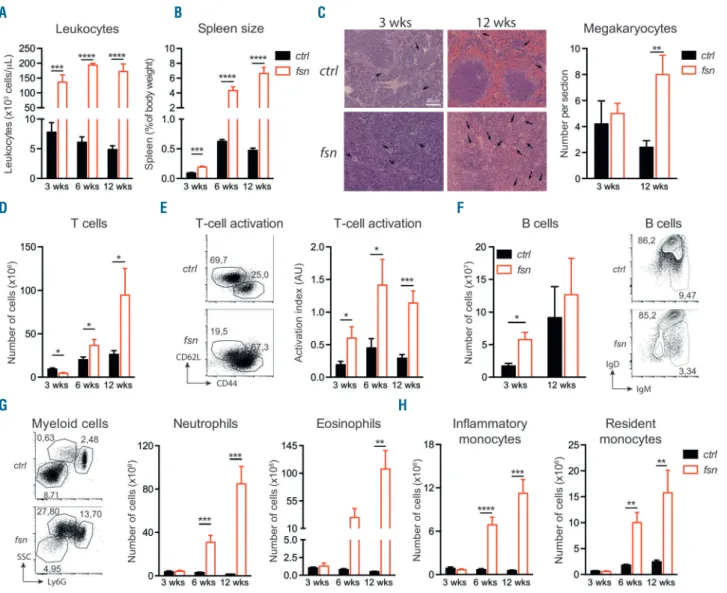
hematopoietic lineages in the blood and the spleen at 3, 6 and 12 weeks of age. Fsn mice had a considerably higher circulating leukocyte count than control littermates (ctrl) at all time points (Figure 1A). The spleen was much larger in
fsn mice than in ctrl mice, twice as large at 3 weeks and ten
times larger at 12 weeks (Figure 1B). The splenic architec-ture in fsn mice became increasingly disorganized, with an age-related expansion of red and white pulp (Figure 1C).
Furthermore, histological assessment of splenic sections revealed extramedullary hematopoiesis as evidenced by elevated counts of megakaryocytes (Figure 1C) and of hematopoietic stem and progenitor cells (HSPC) (Online
Supplementary Figure S1). Relative to ctrl mice, the absolute
splenic T-cell count in fsn mice was slightly lower at 3 weeks of age but higher at 6 and 12 weeks of age (Figure 1D). A large proportion of Ttc7a-deficient T lymphocytes had an effector memory phenotype (CD44+ CD62L-)
(Figure 1E). Splenic B-cell counts were slightly elevated, and B cells presented the impaired maturation phenotype previously described in fsn mice6 (Figure 1F). The
lym-phoid alterations were accompanied by massive myelo-proliferation, with an increase over time in the numbers of
splenic granulocytes (both neutrophils and eosinophils) and resident and inflammatory monocytes (Figure 1G, H). Thus, Ttc7a-deficient mice displayed a number of persist-ent hematopoietic alterations (i.e., leukocytosis, T-lym-phocyte activation and anemia) at a very early age, where-as other manifestations appeared later in life and/or were exacerbated with age (i.e., myeloproliferation and elevat-ed T-cell counts).
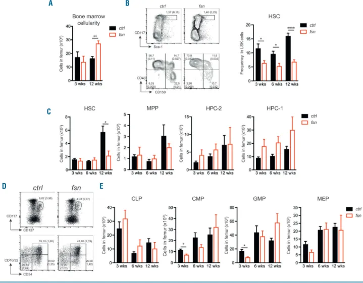
Since all the peripheral hematopoietic lineages were affected in fsn mice, we next looked at whether the HSPC compartment was also altered. BM cellularity in fsn mice, in contrast to ctrl mice, increased between 3 and 12 weeks of age (Figure 2A). The LSK stem cell population was slightly higher in fsn mice than in ctrl mice at all the time
Figure 1. Ttc7a-deficiency perturbs homeostasis of all immune populations. Control littermates (ctrl – black bars) and Ttc7a-deficient (fsn – red bars) mice were analyzed at 3, 6 and 12 weeks of age (mean ± standard error of mean) *P<0.05; **P<0.01; ***P<0.001; ****P<0.0001 (two-tailed t-test). (A) White blood cell count (n≥6). (B) Spleen size determined as percent of body weight (n≥6). (C) Histological sections of spleen stained with hematoxylin and eosin showing megakaryo -cytes (black arrow) (left panel) and quantification of spleen megakaryo-cytes (right panel). (D) Total number of T cells in the spleen (n≥7). (E) Flow cytometry repre-sentative of T-cell activation (according to CD44 and CD62L expression) at 12 weeks (left panel), and activation index of T cells [ratio of effector memory T cells (CD44+CD62L-) to naïve T cells (CD44-CD62L+)] (right panel) of fsn and ctrl mice (n≥7). Numbers adjacent to the outlined areas indicate percent cells in the parent
gate (mean). (F) Total number of B cells in the spleen (n=4) (left panel) and flow cytometry representative of B-cell maturation (according to IgM and IgD expression) (right panel). (G) Representative flow cytometry at 12 weeks (left panel) and total number of neutrophils (CD11b+Ly6Ghi) and eosinophils (CD11b+Ly6GintSSChi). (H)
Total number of inflammatory (CD11b+Ly6G-Ly6C+) and resident monocytes (CD11b+Ly6G-Ly6C-) (n≥6). Numbers adjacent to the outlined areas indicate percent
cells among leukocytes in the spleen (mean). A
D E F
G H
points analyzed (Online Supplementary Figure S2A). The proportion of HSC (Lin-Sca1+cKit+CD150+CD48-)25was
decreased in fsn mice, whereas the proportion of more mature hematopoietic progenitor cells (HPC-1: Lin-Sca1+
cKit+CD150-CD48+) was increased, compared to the
pro-portions in ctrl mice (Figure 2B and Online Supplementary
Figure S2B). At 12 weeks of age, the HSC progenitor
count was significantly lower in fsn mice than in ctrl mice, while the numbers of multipotent progenitors (MPP: Lin
-Sca1+cKit+CD150-CD48-) were unchanged and those of
HPC-2 (Lin-Sca1+cKit+CD150+CD48+) and HPC-1 were
slightly higher (Figure 2C). Within the committed progen-itor compartment, the numbers of common myeloid pro-genitors (CMP: Lin- Sca1- cKit+ CD34+ CD16/32low) and
granulocyte-monocyte progenitors (GMP: Lin-Sca1-cKit+
CD34+CD16/32+) were lower than ctrl values at 3 weeks
of age, although the differences disappeared with time (Figure 2D, E). There were no significant differences in fsn
vs. ctrl values in the numbers of common lymphoid
pro-genitors (CLP: Lin- Sca1- cKitint) or
megakaryocyte-ery-throcyte progenitors (MEP: Lin- Sca1- cKit+ CD34
-CD16/32-) (Figure 2D, E). We confirmed previous reports
that the profound anemia observed in fsn mice (Online
Supplementary Figure S3A) is peripheral in nature and does
not result from a decreased number of early erythroid progenitors but rather from a defect in the last step of ery-thropoiesis (Online Supplementary Figure S3B, C).7
Erythropoiesis and enucleation processes have been shown to involve chromatin compaction26 and actin
cytoskeleton dynamics.27 Interestingly, we previously
showed that Ttc7a plays a role in actin dynamics15,16as
well as in chromatin compaction and genomic stability.28
Hence, it is tempting to speculate that altered actin dynamics and chromatin organization, as a consequence of Ttc7a-deficiency, contribute to defective erythrocyte generation in fsn mice. A high splenic erythroblast count
Figure 2. Ttc7a-deficiency alters the hematopoietic stem and progenitor cell compartment. The hematopoietic stem and progenitor cell compartment was analyzed in the bone marrow (BM) of 3-, 6-, and 12-week old control (ctrl - black bars) and Ttc7a-deficient (fsn - red bars) mice (mean ± standard error of mean) *P<0.05; **P<0.01; ****P<0.0001 (two-tailed t-test). (A) Quantification of total femoral BM cells (n≥6). (B) Representative flow cytometry at 12 weeks (left panel) and per-centage of hematopoietic stem cells (HSC: Lin-Sca1+cKit+CD150+CD48-) among LSK (Lin-Sca1+cKit+) cells (right panel) (n≥7). (C) Quantification of LSK cell
popu-lations, HSC, multipotent progenitors (MPP: Lin-Sca1+cKit+CD150-CD48-), HPC-2 (Lin-Sca1+cKit+CD150+CD48+) and HPC-1 (Lin-Sca1+cKit+CD150-CD48+). (D)
Representative flow cytometry at 12 weeks and (E) quantification of common lymphoid progenitors (CLP: Lin-Sca1-cKitintCD127+), common myeloid progenitors (CMP:
Lin-Sca1-cKit+CD34+CD16/32-), granulocyte-monocyte progenitors (GMP: Lin-Sca1-cKit+CD34+ CD16/32+) and megakaryocyte-erythroid progenitors (MEP: Lin
-Sca1-cKit+CD34-CD16/32-) (n≥7). (B-D) Numbers adjacent to outlined areas indicate percent cells in the parent gate (mean). Numbers in parentheses indicate
per-centage among leukocytes in the BM (mean). A
D E
B
suggested the presence of stress erythropoiesis as a possi-ble attempt to compensate for the peripheral anemia (Online Supplementary Figure S3B).
Thus, our present results show that the absence of Ttc7a in fsn mice is associated with deregulation of the homeostatic balance between hematopoietic lineages, from the HSC stage onwards, and a tendency of all leuko-cyte subsets to expand over time.
Ttc7a has an intrinsic role in the fate of progenitor
cells
Since Ttc7a is broadly expressed, it was not possible to distinguish the respective involvements of hematopoietic factors (i.e., HSC) and non-hematopoietic factors (e.g., BM niches and the thymic epithelium) in the generation of the
fsn phenotype. In a previous study we found that the skin
barrier is impaired in fsn mice;9this defect may enhance
antigen sensitization and thus induce immune system activation. Therefore, in order to determine the Ttc7a-deficient hematopoietic cells’ intrinsic contribution to fsn-associated hematologic manifestations, we generated chimeric mice by reconstituting lethally irradiated control recipients with LSK cells purified from either ctrl or fsn mice. Hereafter, these chimeric mice are respectively referred to as Ctrlctrland Ctrlfsn. Three-week old mice were
chosen as donors so that we could use a similar LSK graft inoculum in both control and fsn samples, and thus mini-mize the potential immune consequences caused by the altered fsn skin barrier. We monitored the hematologic reconstitution over time by collecting blood samples from the recipient mice every 2 weeks. As observed in native
fsn mice, white blood cell counts were higher in Ctrlfsn mice
than in Ctrlctrlmice, and a difference was observed as early
as 4 weeks after transplantation (Figure 3A). The Ctrlfsn
mice were also anemic (Figure 3B) and developed splenomegaly (Figure 3C), although the latter was less pronounced than in native fsn mice (Figure 1B). The total body weight of Ctrlctrl and Ctrlfsnmice was not different.
The distribution of the splenic myeloid, T- and B-cell pop-ulations was the same as in ctrl mice (Figure 3D). As observed in fsn mice, BM cellularity was higher in Ctrlfsn
mice than in Ctrlctrl mice (Figure 3E), whereas the LSK
counts were slightly increased (Figure 3F). The distribu-tion of the HSC, MPP, HPC-2 and HPC-1 populadistribu-tions was similar in Ctrlfsn and Ctrlctrl mice, suggesting that the low
HSC count observed in 12-week old native fsn mice (Figure 2C) is primarily caused by external (i.e., non-hematopoietic) factors. Taken as a whole, these data sug-gest that Ttc7a has an intrinsic role in hematopoietic cells; the absence of Ttc7a in hematopoietic progenitors results in the over-proliferation of the various cell lineages as seen in native fsn mice.
Loss of Ttc7a enhances the reconstitution potential
of hematopoietic stem cells
Next, we sought to determine the impact of Ttc7a loss on the reconstitution potential of HSC in a controlled in
vivo environment. Using lethally irradiated congenic
recip-ients, we transferred equal numbers of LSK cells purified from 3-week old ctrl (LSKctrl)- or fsn (LSKfsn)-(CD45.2+) mice
together with competitor wildtype-(CD45.1+) BM cells
(i.e., Ctrl-LSKctrl or Ctrl-LSKfsn). We then assessed the
Figure 3. Transferred Ttc7a-deficient LSK cells reproduce fsn manifestations in a control environment. Control irradiated mice were transferred with LSK cells puri-fied from 3-week old ctrl (Ctrlctrl– black bars) or Ttc7a-deficient mice (Ctrlfsn– red bars). (mean ± standard error of mean) *P<0.05; **P<0.01 (two-tailed t-test). (A,
B) Monitoring of the number of leukocytes (A) and red blood cells, hematocrit and hemoglobin (B) during hematopoietic reconstitution. (C) Spleen size determined as percent of body weight 10 weeks after bone marrow (BM) transfer. (D) Relative contribution of myeloid compared to T cells and B cells in the spleen. (E, F) BM cellularity (E) and absolute number of LSK cells (F) 10 weeks after BM transfer. RBC: red blood cells.
A
D E F
B
respective contributions of cells originating from ctrl or fsn LSK donors during hematopoietic reconstitution. As early as 2 weeks after transfer, the proportion of LSKfsn
donor-derived leukocytes in the recipients’ blood was higher than that of LSKctrl. These differences persisted 14 weeks
after transfer (Figure 4A). The proportions of cells originat-ing from donor LSK in the recipients’ organs, particularly the thymus and BM, were higher in Ctrl-LSKfsnmice than
in Ctrl-LSKctrlmice (Figure 4B). In the spleen, the
reconsti-tution advantage of LSKfsn donor-derived cells led to the
expansion of neutrophil, eosinophil and monocyte lineag-es and to a llineag-esser extent, T-cell lineaglineag-es (Figure 4C). To fur-ther evaluate the effect of Ttc7a loss on long-term recon-stitution, total BM cells from primary recipient mice were transplanted into secondary recipients. The competitive advantage of Ttc7a-deficient LSK donor cells with regards to reconstitution was maintained and even enhanced upon secondary and tertiary transplantation (Figure 4D). Thus, our results show that a defect in Ttc7a improves the competitive fitness of HSC following transplantation.
Loss of Ttc7a increases the long-term self-renewal
potential of hematopoietic stem cells
In view of the elevated proliferative capacity of Ttc7a-deficient hematopoietic cells, we next sought to assess the
properties of HSC that could modify their reconstitution potential (i.e., quiescence and self-renewal capacity). To evaluate the impact of Ttc7a loss on the quiescence of reconstituting HSC, we measured bromodeoxyuridine (BrdU) incorporation in control and Ttc7a-deficient HSPC before and after BM transplantation. Upon 24 h of BrdU treatment, we observed similar percentages of BrdU-posi-tive (BrdU+) HSC, HPC-1 and HPC-2 cells purified from
control and fsn mice (Online Supplementary Figure S4A). Similar results were obtained when comparing cell cycle progression in control and Ttc7a-deficient HSC upon transplantation of irradiated recipients (Online
Supplementary Figure S4B, C). Altogether, these results
sug-gest that the increased repopulation capacity of fsn HSC was not caused by a disturbed quiescent state.
We then looked at whether the long-term self-renewal ability of HSC was altered in this context. We therefore performed serial BM transplants from irradiated mice hav-ing received whole BM from 3-week old ctrl or fsn mice. Unexpectedly, Ttc7a-deficient cells successfully sustained BM reconstitution longer than ctrl cells did. The ctrl HSC sustained six rounds of transplantation (Figure 5A) but all the Ctrlctrlmice died during the seventh round of BM
trans-fer (Figure 5A, B). In contrast, most Ctrlfsnmice survived 6
weeks after the seventh round and were able to undergo
Figure 4. Ttc7a-deficient hematopoietic stem cells have a higher repopulation capacity. (A-C) Lethally irradiated CD45.1 mice were reconstituted with a mix of con-trol whole bone marrow (BM) and sorted LSK cells purified from ctrl (Ctrl-LSKctrl– black bars and lines) or Ttc7a-deficient mice (Ctrl-LSKfsn– red bars and lines) (mean
± standard error of mean) *P<0.05; **P<0.01; ***P<0.001; ****P<0.0001 (two-tailed t-test). These data are representative of three independent experiments. Proportion of LSK donor-derived leukocytes in the blood over time (A), in lymphoid organs (15 weeks after transfer) (B) and in the spleen for the different types of leukocytes (C) (n=8). (D) BM cells from first and then second round recipient mice of each group were pulled and transplanted to secondary and tertiary ctrl CD45.1 recipients, respectively. The proportions of LSK donor-derived leukocytes of Ctrl-LSKctrland Ctrl-LSKfsnmice were determined in each round (n=14 for control and n=16
for Ttc7a-reconstituted mice). A
D B
an additional round of transplantation before dying after the eighth round (Figure 5A, B). Similar results were obtained in experiments with 12-week old donors (Online
Supplementary Figure S5A, B). We next determined the
abil-ity of ctrl and Ttc7a-deficient BM cells from 3-week old mice to properly reconstitute hematopoiesis over the dif-ferent rounds of transplantation. During the first five rounds, the same phenotype was always observed. Ctrlfsn
mice displayed anemia (Figure 5C), together with an ele-vated leukocyte count up until the third round (Figure 5D). The spleen was larger in Ctrlfsn mice than in Ctrlctrl mice
until the fifth round (Figure 5E). However, differences between Ctrlfsnand Ctrlctrlmice were no longer observed in
the sixth round; this was probably caused by exhausted donor cells that failed to properly reconstitute recipient mice at the end of the reconstitution process (Figure 5C-E). Interestingly, the distribution of splenic leukocyte sub-sets in the Ctrlfsn mice was progressively biased toward
myeloid populations at the expense of B cells, and to a lesser extent, T cells, as notably observed for the fifth
round (Figure 5F). In Ctrlfsn mice, this bias became
detectable in the third round and persisted until the sev-enth round (Online Supplementary Figure S5C). In summary, these data show that Ttc7a-deficient HSC have a greater ability to self-renew and to induce myeloid cell expansion.
Ttc7a-deficiency perturbs the transcriptomic profile
of the endoplasmic reticulum stress response in
hematopoietic stem cells
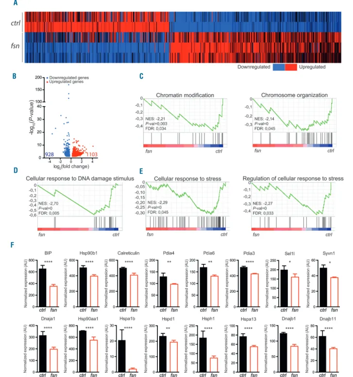
In order to gain further mechanistic insight into the Ttc7a-related regulation of HSC homeostasis, we carried out a transcriptomic analysis of HSC isolated from Ttc7a-deficient and control BM (from 4-week old mice). A two-way hierarchical clustering analysis of differentially expressed genes (P value ≤0.05, fold change ≥1.2) revealed a clear-cut separation between Ttc7a-deficient and control HSC samples (Figure 6A). We found that 1,103 genes were significantly upregulated and 928 significantly downregu-lated in Ttc7a-deficient HSC relative to the expression lev-els in control HSC (Figure 6B). Among the differentially
Figure 5. Ttc7a-deficiency promotes self-renewal ability of hematopoietic stem cells. Lethally irradiated mice were serially transplanted with 3-week old ctrl (Ctrlctrl
– black bars, dots and lines) or Ttc7a-deficient (Ctrlfsn– red bars, dots and lines) donor bone marrow (BM) cells (mean ± standard error of mean) *P<0.05; **P<0.01;
***P<0.001; ****P<0.0001 (two-tailed t-test). These data are representative of four independent experiments with at least four mice in each round of transplan-tation. (A) Percent survival of recipient mice across the seven transplantation cycles. (B) Survival of Ctrlctrland Ctrlfsnmice during the seventh round and Ctrlfsnmice
during the eighth over time (n=4 for control- and n=10 for Ttc7a-reconstituted mice). (C-F) Red blood cell count, hematocrit, and hemoglobin level (C), leukocytes count (D) and spleen weight (E) across the transplantation cycles. (F) Relative contribution of myeloid compared to T- and B-lymphoid cells in the spleen of mice transplanted with BM cells that had undergone five transplantation cycles. RBC: red blood cells; Htc: hematocrit; Hgb: hemoglobin.
A
D E F
B
expressed genes, the most statistically significant differ-ences were observed in the group of downregulated genes (Figure 6B). To determine the functional profile of the dif-ferentially expressed genes, we performed GSEA for genes with a fold change ≥1.5. GO analysis of the identified gene signature revealed a significant enrichment of genes
in three main categories. Two categories are related to chromatin organization/modification and DNA damage repair; this observation fits with our recent finding that Ttc7a is a chromatin-binding nuclear factor involved in chromatin compaction and nuclear organization28(Figure
6C, D). Another category corresponds to genes involved
Figure 6. Ttc7a deficiency results in reduced expression of endoplasmic reticulum stress response genes in hematopoietic stem cells. RNA sequencing was per-formed on 3-week old control (ctrl – black bars) and Ttc7a-deficient (fsn – red bars) hematopoietic stem cell (HSC) transcripts. (A) Heatmap of relative expression of differentially expressed genes (fold change ≥1.2) in Ttc7a-deficient HSC compared to control. (B) Volcano plot of differentially expressed genes (fold change ≥1.2) in Ttc7a-deficient HSC compared to control HSC showing the adjusted P-value (-log10) vs. fold change (log2). Upregulated and downregulated genes are shown in red and
blue, respectively. Total numbers in each group are indicated in red and blue, respectively. (C-E) Enriched gene sets in Ttc7a-deficient HSC compared to control HSC, as determined by gene set enrichment analysis of differentially expressed genes (fold change ≥1.5). (F) Normalized expression of endoplasmic reticulum stress response genes downregulated in Ttc7a-deficient HSC (fold change ≥1,2) *P<0.05; **P<0.01; ****P<0.0001 (LimmaVoom analysis). NES: normalized enrichment score; FDR: false discovery rate; AU: arbitrary units.
A
D E
F
in cellular response to stress (Figure 6E). Our transcriptom-ic analysis also highlighted high expression levels of Ttc7a in HSC (Online Supplementary Figure S6). A growing body of evidence suggests that ER stress regulates the function of the HSC pool.21In particular, a recent study highlighted
a link between ER stress perturbation in HSC and an ele-vated reconstitution capacity following BM transplanta-tion.29Accordingly, we found that several effectors of the
ER stress response were significantly downregulated in Ttc7a-deficient HSC, including the UPR master regulator Bip (Hspa5/GRP78), calreticulin, Pdia3, Pdia4, Pdia6,
sev-eral Hsp, as well as Sel1l and Syvn1 which have been shown to regulate an ER-associated protein degradation (ERAD) pathway30,31 (Figure 6F and Online Supplementary
Table S1). Overall, our data suggest that Ttc7a loss affects
the cellular response to ER stress in HSC.
Ttc7a controls the response to stress in hematopoietic
stem cells
ER stress is mainly triggered in response to altered pro-tein homeostasis leading to pro-apoptotic or pro-survival responses. Notably, Ttc7a-deficient HSC had reduced
lev-Figure 7. Ttc7a controls the response to stress in hematopoietic stem cells. (A) Representative histograms of protein aggregation level of 3-week old ctrl (black line) and fsn (red line) hematopoietic stem cells (HSC). (B) Protein expression of Bip in ctrl and fsn HSC after 3 days of in vitro expansion. (C) Proliferation index (calculated as the ratio between the number of cells at 48 h and 24 h) of HSC after Lin-cells were sorted from ctrl (black bars) and Ttc7a-deficient (fsn – red bars) mice and
cultured for 2 days with or without tunicamycin. **P<0.01 (two-tailed t-test). (D-I) Ctrlctrl(black line and bars) and Ctrlfsn(red line and bars) mice were analyzed after
they had received a single intraperitoneal injection of 150 mg/Kg of 5-fluorouracil (5-FU), 12 weeks after bone marrow transfer. White blood cell count over time (D). Spleen size (E) and absolute number of lymphoid (F) and myeloid (G) cells in the spleen 15 days after 5-FU injection (n=9). Percentage bromodeoxyuridine (BrdU) incorporation in LSK subpopulations (H) and representative flow cytometry histograms of BrdU incorporation in LK (Lin-Kit+) populations (I) 7 days after 5-FU injection.
(n=10 for control- and n=12 for Ttc7a-reconstituted mice) *P<0.05; **P<0.01; ***P<0.001 (two-tailed t-test). AU: arbitrary units; LSK: Lin-Sca1+cKit+cells; MPP:
multipotent progenitors; HPC: hematopoietic progenitor cells; CMP: common myeloid progenitors; GMP: granulocyte-monocyte progenitors; MEP: megakaryocyte-ery-throid progenitors; CLP: common lymphoid progenitors.
A
D E F
G H I
els of protein aggregation compared to control HSC (Figure 7A). In keeping with this, in vitro-expanded fsn HSC had an elevated level of BIP protein (Figure 7B). These results suggest that Ttc7a loss could modify HSC suscep-tibility to ER stress. Therefore, to determine the impact of Ttc7a loss in the response of HSC to ER stress, we ana-lyzed the proliferative capacity of Ttc7a-deficient HSC and progenitor cells (i.e., HSC, MPP, HPC-2 and HPC-1) upon chemical induction of ER stress in vitro. To do so, lin-eage-negative cells from 4-week old fsn and ctrl mice were cultured for 48 h in the presence or absence of tuni-camycin, which blocks the synthesis of N-linked glyco-proteins, leading to an accumulation of unfolded proteins and the induction of ER stress.32 As expected, on day 2,
tunicamycin treatment reduced the proliferation ability of control cells in a dose-dependent manner (Figure 7C). In contrast, at a low dose of tunicamycin, the proliferative capacity of Ttc7a-deficient HSC was significantly greater than that of control HSC (Figure 7C). These differences were particular to HSC, as Ttc7a-deficient MPP, HPC-2 and HPC-1 subsets had a similar response to tunicamycin as their control counterparts (Online Supplementary Figure
S7A). The alterations in the ER stress response in
Ttc7a-deficient HSC were not due to protein aggregation (Figure 7A and Online Supplementary Figure S7B), nor to low expression of the ER stress sensors and effectors (Ire1α, Perk, Atf6, etc.), as no differences were observed in our transcriptomic analysis (Online Supplementary Figure S7C and Online Supplementary Table S1). Surprisingly, the reduction in cell proliferation of ctrl HSC in response to tunicamycin was independent of apoptosis, in contrast to that of other progenitor cells. Apoptosis of Ttc7a-deficient HSC was reduced compared to that of unstimulated ctrl cells, and remained unchanged upon tunicamycin treat-ment (Online Suppletreat-mentary Figure S7D). No differences were observed in other progenitor populations (Online
Supplementary Figure S7D). Altogether, these data suggest
that ex vivo purified Ttc7a-deficient HSC had a higher level of ER stress compared to their control counterparts.
Interestingly, we observed that the expression of Hsp70, a chaperone protein associated with broad cellular stress-es, was also higher in fsn HSC than in controls (Online
Supplementary Figure S7E). In order to determine whether
Ttc7a regulates the cellular response to stress in vivo, we monitored the proliferative response of Ttc7a-deficient cells following the induction of stress by 5-FU. The deple-tion of cycling cells by 5-FU stimulates HSC to replenish peripheral leukocytes,33,34inducing a broad stress response
in HSC, not limited to ER stress (e.g., oxidative stress, pro-liferative stress). We injected 5-FU into Ctrlfsn and Ctrlctrl
mice 3 months after BM transplantation and monitored the replenishment of peripheral leukocytes for 19 days. The Ttc7a-deficient and ctrl leukocyte counts fell until day 9 post-injection, and then increased. On day 15, Ttc7a-deficient leukocytes were growing significantly more strongly than ctrl cells, with a peak on day 16 (Figure 7D). Interestingly, the spleen of Ctrlfsn mice enlarged further
after 5-FU treatment (Figure 7E, compared with Figure 3D), with higher lymphoid and myeloid counts (Figure 7F-G). To assess the proliferative response of Ttc7a-deficient HSC following stress injury, we assayed BrdU uptake by LSK subsets between day 6 and day 7 after 5-FU injection. The greater BrdU uptake in Ttc7a-deficient HSC, MPP, HPC-2 and HPC-1 (Figure 7H), suggested that Ttc7a con-trols the cell cycle progression of HSC under stress
condi-tions. Strikingly, BrdU uptake did not differ in committed progenitors, CLP, CMP, GMP and MEP (Figure 7I). These results suggest that Ttc7a is involved in the regulation of the proliferative response of HSC under stress conditions but not in that of committed progenitor cells.
Discussion
The present study revealed a previously unrecognized role for Ttc7a in the negative regulation of HSC function. Using murine transplantation models and Ttc7a-deficient HSC, we found that Ttc7a intrinsically regulates the main-tenance and proliferation of HSC in vivo, and the subse-quent homeostasis of downstream cell populations. We also found that Ttc7a expression in HSC is closely associ-ated with the transcriptional response to ER stress.
HSC are the only cells capable of self-renewing and dif-ferentiating into all mature blood lineages. The quiescence of HSC must be tightly regulated in order to control pro-liferation, maintain normal homeostasis, and prevent stem cell exhaustion.35,36Various intrinsic and cell-extrinsic
regu-latory factors of the HSC cell cycle have been described, such as phosphatase and tensin homologue (Pten) signal-ing, Wnt signaling and cytokine signaling.37 Indeed, in
mice that lack growth factor independent 1 (Gfi1),38Pten,
forkhead box proteins 1, 3, 4, or M139or other proteins,20,36
excessive HSC proliferation is associated with stem cell exhaustion and the loss of self-renewal. In contrast, we found that mice reconstituted with Ttc7a-deficient pro-genitors exhibit a characteristic phenotype with enhanced HSC function, higher HSC-derived peripheral blood cell counts and no evidence of stem cell exhaustion when compared with Ttc7a-proficient HSC. Indeed, Ttc7a-defi-cient HSC were able to repopulate the hematopoietic sys-tem better in serial transplantation experiments, indicating that the self-renewal of Ttc7a-deficient HSC is not com-promised by repeated rounds of proliferation. Although this situation clearly differs from the above-mentioned knock-out mice, a few similar observations have been reported after the deletion of the cyclin-dependent kinase 4 inhibitor C (CDKN2C),40the ubiquitin-mediated protein
degradation Cbl41and Itch,41and the transcription factors
Hif1a42and Egr1.43The loss of these proteins enabled the
maintenance of HSC, despite an increase in the cells’ pro-liferative capacity. However, the specific mechanisms by which these proteins regulate HSC function remain large-ly unknown.
Our findings support a role for the ER stress response in the enhanced function of Ttc7a-deficient HSC. Since long-lived HSC are particularly sensitive to stress stimuli, their response must be tightly controlled in order to prevent either a loss of function or the clonal persistence of onco-genic mutations. It has been shown that HSC are enriched in components of the UPR pathway. Upon exposure to acute stress in vitro, HSC are more prone to apoptosis, via upregulation of the canonical UPR genes, than related pro-genitors that have lost their self-renewal capacity.21,44
Along these lines, the ectopic expression of developmental pluripotency-associated 5 (Dppa5) was associated with enhanced HSC function, via suppression of the ER stress response (by downregulating the expression of ER stress chaperones) and the subsequent apoptotic signals.29
However, UPR activation can also have an anti-apoptotic outcome in HSC. It has been shown that stimulation of
estrogen receptor α (ERα) confers HSC resistance to pro-teotoxic stress by activating the Ire1α-Xbp1 branch of the UPR and promoting the cells’ reconstituting potential.24In
Ttc7a deficient HSC, expression levels of ER stress response genes were abnormally low, whereas the level of the Bip chaperone protein was increased. Furthermore, Ttc7a deficient HSC were less sensitive to stress induction than Ttc7a-proficient-HSC both in vitro and in vivo. A sim-ilar phenomenon was observed in mouse liver in which mild chronic ER stress decreases the mRNA level of Bip while maintaining its protein level. This response allows hepatocytes to avoid the overproduction of UPR effectors that could lead to apoptosis.45It is, therefore, tempting to
speculate that Ttc7a deficiency could be associated with mild chronic stress. In this context, the increased resist-ance of fsn HSC to tunicamycin could be caused by a cel-lular adaptive response aiming to increase the threshold of ER stress sensitivity, and ensure cell survival. Interestingly, HSC exposed to other sources of persistent cellular stress develop mechanisms of stress resistance resulting in increased self-renewal capacity and reconstitution poten-tial.46Knowing that TTC7A stabilizes several interacting
proteins, the role of additional components, altered as a consequence of Ttc7a deficiency, cannot be excluded. Ttc7a could represent a pivotal connection between ER stress regulation and the maintenance of HSC functions.
Along with an abnormally proliferative hematopoietic system, fsn mice develop hyperplasia of the epidermis and the gastric epithelium. Notably, stem cells from other tis-sues can similarly sense ER stress and activate the UPR
pathway to control self-renewal and differentiation. This has been shown for the intestinal epithelium in particular, and several lines of evidences support the concept where-by ER stress and UPR activity regulate the differentiation of intestinal stem cells.47An attractive hypothesis would
be that the other phenotypic manifestations that charac-terized Ttc7a deficiency might be due to perturbation of the ER stress response.
In summary, our results show that Ttc7a has a critical but previously unrecognized role as a regulator of HSC homeostasis and function through the regulation of the ER stress response.
Acknowledgments
We thank Gaël Ménasché, Annarita Miccio and Isabelle Andre-Schmutz for helpful comments and guidance; Olivier Pellé for help in cell sorting; the Necker histology and morphol-ogy facility [Structure Fédérative de Recherche (SFR) Necker] and the Cochin Genomic Platform for their services in histologi-cal and transcriptomic studies, respectively. This work was sup-ported by The French National Institutes of Health and Medical Research (INSERM), state funding from the Agence Nationale de la Recherche “Investissements d’avenir” program, la Fondation pour la Recherche Médicale (FRM project DEQ20150734354), and the Imagine Foundation. CL was supported by a fellowship from the Ministry of Education and FRM and MTED by a fellowship from the ANR, the FRM and the European Research Council (ERC). TG is a fellow of the International PhD program of the Imagine Institute funded by the Bettencourt Schueller Foundation.
References
1. Helms C, Pelsue S, Cao L, et al. The tetratri-copeptide repeat domain 7 gene is mutated in flaky skin mice: a model for psoriasis, autoimmunity, and anemia. Exp Biol Med (Maywood). 2005;230(9):659-667. 2. Lees JA, Zhang Y, Oh MS, et al. Architecture
of the human PI4KIIIalpha lipid kinase com-plex. Proc Natl Acad Sci U S A. 2017;114(52):13720-13725.
3. Pelsue SC, Schweitzer PA, Schweitzer IB, et al. Lymphadenopathy, elevated serum IgE levels, autoimmunity, and mast cell accumu-lation in flaky skin mutant mice. Eur J Immunol. 1998;28(4):1379-1388.
4. Abernethy NJ, Hagan C, Tan PL, Birchall NM, Watson JD. The peripheral lymphoid compartment is disrupted in flaky skin mice. Immunol Cell Biol. 2000;78(1):5-12. 5. Abernethy NJ, Hagan C, Tan PL, Watson JD.
Dysregulated expression of CD69 and IL-2 receptor alpha and beta chains on CD8+ T lymphocytes in flaky skin mice. Immunol Cell Biol. 2000;78(6):596-602.
6. Welner R, Hastings W, Hill BL, Pelsue SC. Hyperactivation and proliferation of lym-phocytes from the spleens of flaky skin (fsn) mutant mice. Autoimmunity. 2004;37(3): 227-235.
7. Beamer WG, Pelsue SC, Shultz LD, Sundberg JP, Barker JE. The flaky skin (fsn) mutation in mice: map location and descrip-tion of the anemia. Blood. 1995;86(8):3220-3226.
8. Schon M, Denzer D, Kubitza RC, Ruzicka T, Schon MP. Critical role of neutrophils for the generation of psoriasiform skin lesions in flaky skin mice. J Invest Dermatol. 2000;114(5):976-983.
9. Leclerc-Mercier S, Lemoine R, Bigorgne AE, et al. Ichthyosis as the dermatological phe-notype associated with TTC7A mutations. Br J Dermatol. 2016;175(5):1061-1064. 10. Sundberg JP, Kenty GA, Beamer WG,
Adkison DL. Forestomach papillomas in flaky skin and steel-Dickie mutant mice. J Vet Diagn Invest. 1992;4(3):312-317. 11. Scheufler C, Brinker A, Bourenkov G, et al.
Structure of TPR domain-peptide complex-es: critical elements in the assembly of the Hsp70-Hsp90 multichaperone machine. Cell. 2000;101(2):199-210.
12. D'Andrea LD, Regan L. TPR proteins: the versatile helix. Trends Biochem Sci. 2003;28(12):655-662.
13. Assimon VA, Southworth DR, Gestwicki JE. Specific binding of tetratricopeptide repeat proteins to heat shock protein 70 (Hsp70) and heat shock protein 90 (Hsp90) is regulat-ed by affinity and phosphorylation. Biochemistry. 2015;54(48):7120-7131. 14. Taipale M, Tucker G, Peng J, et al. A
quanti-tative chaperone interaction network reveals the architecture of cellular protein homeo -stasis pathways. Cell. 2014;158(2):434-448. 15. Bigorgne AE, Farin HF, Lemoine R, et al.
TTC7A mutations disrupt intestinal epithe-lial apicobasal polarity. J Clin Invest. 2014;124(1):328-337.
16. Lemoine R, Pachlopnik-Schmid J, Farin HF, et al. Immune deficiency-related enteropa-thy-lymphocytopenia-alopecia syndrome results from tetratricopeptide repeat domain 7A deficiency. J Allergy Clin Immunol. 2014;134(6):1354-1364 e1356.
17. Avitzur Y, Guo C, Mastropaolo LA, et al. Mutations in tetratricopeptide repeat domain 7A result in a severe form of very early onset inflammatory bowel disease. Gastroenterology. 2014;146(4):1028-1039. 18. Baird D, Stefan C, Audhya A, Weys S, Emr
SD. Assembly of the PtdIns 4-kinase Stt4 complex at the plasma membrane requires Ypp1 and Efr3. J Cell Biol. 2008;183(6):1061-1074.
19. Rossi L, Lin KK, Boles NC, et al. Less is more: unveiling the functional core of hematopoietic stem cells through knockout mice. Cell Stem Cell. 2012;11(3):302-317. 20. Orford KW, Scadden DT. Deconstructing
stem cell self-renewal: genetic insights into cell-cycle regulation. Nat Rev Genet. 2008;9(2):115-128.
21. van Galen P, Kreso A, Mbong N, et al. The unfolded protein response governs integrity of the haematopoietic stem-cell pool during stress. Nature. 2014;510(7504):268-272. 22. Rutkowski DT, Kaufman RJ. A trip to the
ER: coping with stress. Trends Cell Biol. 2004;14(1):20-28.
23. Grootjans J, Kaser A, Kaufman RJ, Blumberg RS. The unfolded protein response in immu-nity and inflammation. Nat Rev Immunol. 2016;16(8):469-484.
promotes murine hematopoietic regenera-tion through the Ire1alpha-mediated unfold-ed protein response. Elife. 2018;7:e31159 25. Oguro H, Ding L, Morrison SJ. SLAM family
markers resolve functionally distinct sub-populations of hematopoietic stem cells and multipotent progenitors. Cell Stem Cell. 2013;13(1):102-116.
26. Zhao B, Yang J, Ji P. Chromatin condensation during terminal erythropoiesis. Nucleus. 2016;7(5):425-429.
27. Konstantinidis DG, Pushkaran S, Johnson JF, et al. Signaling and cytoskeletal require-ments in erythroblast enucleation. Blood. 2012;119(25):6118-6127.
28. El-Daher MT, Cagnard N, Gil M, et al. Tetratricopeptide repeat domain 7A is a nuclear factor that modulates transcription and chromatin structure. Cell Discov. 2018;4:61.
29. Miharada K, Sigurdsson V, Karlsson S. Dppa5 improves hematopoietic stem cell activity by reducing endoplasmic reticulum stress. Cell Rep. 2014;7(5):1381-1392. 30. Kikkert M, Doolman R, Dai M, et al. Human
HRD1 is an E3 ubiquitin ligase involved in degradation of proteins from the endoplas-mic reticulum. J Biol Chem. 2004;279(5): 3525-3534.
31. Sun S, Shi G, Han X, et al. Sel1L is indispen-sable for mammalian endoplasmic reticu-lum-associated degradation, endoplasmic reticulum homeostasis, and survival. Proc Natl Acad Sci U S A. 2014;111(5):E582-591. 32. DuRose JB, Tam AB, Niwa M. Intrinsic
unfolded protein response to sense alternate forms of endoplasmic reticulum stress. Mol Biol Cell. 2006;17(7):3095-3107.
33. Randall TD, Weissman IL. Phenotypic and functional changes induced at the clonal level in hematopoietic stem cells after 5-flu-orouracil treatment. Blood. 1997;89(10): 3596-3606.
34. Venezia TA, Merchant AA, Ramos CA, et al. Molecular signatures of proliferation and quiescence in hematopoietic stem cells. PLoS Biol. 2004;2(10):e301.
35. Wilson A, Laurenti E, Oser G, et al. Hematopoietic stem cells reversibly switch from dormancy to self-renewal during homeostasis and repair. Cell. 2008;135(6): 1118-1129.
36. Trumpp A, Essers M, Wilson A. Awakening dormant haematopoietic stem cells. Nat Rev Immunol. 2010;10(3):201-209.
37. Walasek MA, van Os R, de Haan G. Hematopoietic stem cell expansion: chal-lenges and opportunities. Ann N Y Acad Sci. 2012;1266:138-150.
38. Hock H, Hamblen MJ, Rooke HM, et al. Gfi-1 restricts proliferation and preserves func-tional integrity of haematopoietic stem cells. Nature. 2004;431(7011):1002-1007. 39. Hou Y, Li W, Sheng Y, et al. The
transcrip-tion factor Foxm1 is essential for the quies-cence and maintenance of hematopoietic stem cells. Nat Immunol. 2015;16(8):810-818.
40. Yuan Y, Shen H, Franklin DS, Scadden DT, Cheng T. In vivo self-renewing divisions of
the absence of the early G1-phase inhibitor, p18INK4C. Nat Cell Biol. 2004;6(5):436-442. 41. Rathinam C, Matesic LE, Flavell RA. The E3 ligase Itch is a negative regulator of the homeostasis and function of hematopoietic stem cells. Nat Immunol. 2011;12(5):399-407.
42. Takubo K, Goda N, Yamada W, et al. Regulation of the HIF-1alpha level is essen-tial for hematopoietic stem cells. Cell Stem Cell. 2010;7(3):391-402.
43. Min IM, Pietramaggiori G, Kim FS, Passegue E, Stevenson KE, Wagers AJ. The transcrip-tion factor EGR1 controls both the prolifera-tion and localizaprolifera-tion of hematopoietic stem cells. Cell Stem Cell. 2008;2(4):380-391. 44. Laurenti E, Doulatov S, Zandi S, et al. The
transcriptional architecture of early human hematopoiesis identifies multilevel control of lymphoid commitment. Nat Immunol. 2013;14(7):756-763.
45. Gomez JA, Rutkowski DT. Experimental reconstitution of chronic ER stress in the liver reveals feedback suppression of BiP mRNA expression. Elife. 2016;5:e20390 46. Cheng CW, Adams GB, Perin L, et al.
Prolonged fasting reduces IGF-1/PKA to pro-mote hematopoietic-stem-cell-based regen-eration and reverse immunosuppression. Cell Stem Cell. 2014;14(6):810-823. 47. Heijmans J, van Lidth de Jeude JF, Koo BK, et
al. ER stress causes rapid loss of intestinal epithelial stemness through activation of the unfolded protein response. Cell Rep. 2013;3(4):1128-1139.