HAL Id: hal-03064377
https://hal.archives-ouvertes.fr/hal-03064377
Submitted on 6 Jan 2021HAL is a multi-disciplinary open access
archive for the deposit and dissemination of sci-entific research documents, whether they are pub-lished or not. The documents may come from teaching and research institutions in France or abroad, or from public or private research centers.
L’archive ouverte pluridisciplinaire HAL, est destinée au dépôt et à la diffusion de documents scientifiques de niveau recherche, publiés ou non, émanant des établissements d’enseignement et de recherche français ou étrangers, des laboratoires publics ou privés.
The regulation of HAD-like phosphatases by signaling
pathways modulates cellular resistance to the metabolic
inhibitor, 2-deoxyglucose
Quentin Defenouillère, Agathe Verraes, Clotilde Laussel, Anne Friedrich,
Joseph Schacherer, Sébastien Léon
To cite this version:
Quentin Defenouillère, Agathe Verraes, Clotilde Laussel, Anne Friedrich, Joseph Schacherer, et al.. The regulation of HAD-like phosphatases by signaling pathways modulates cellular resistance to the metabolic inhibitor, 2-deoxyglucose. Science Signaling, American Association for the Advancement of Science, 2019, 12 (597), pp.eaaw8000. �10.1126/scisignal.aaw8000�. �hal-03064377�
The induction of HAD-like phosphatases by multiple signaling pathways confers
resistance to the metabolic inhibitor 2-deoxyglucose
Quentin Defenouillère
1,2, Agathe Verraes
1,2, Clotilde Laussel
1, Anne Friedrich
3, Joseph
Schacherer
3& Sébastien Léon
1,41: Institut Jacques Monod, UMR 7592 Centre National de la Recherche Scientifique/Université Paris-Diderot, Sorbonne Paris Cité, 75205 Paris cedex 13, France
2: These authors contributed equally to this work
3: Université de Strasbourg, CNRS, GMGM UMR 7156, 67000 Strasbourg, France 4: Author for correspondence:
Sébastien Léon, PhD Institut Jacques Monod 15 Rue Hélène Brion
75205 Paris Cedex 13, France.
Abstract
Anti-cancer strategies that target the glycolytic metabolism of tumors have been proposed. The glucose analog 2-deoxyglucose (2DG) is imported into cells and after phosphorylation becomes 2DG-6-phosphate, a toxic by-product that inhibits glycolysis. Using yeast as a model, we performed an unbiased mass spectrometry-based approach to probe the cellular effects of 2DG on the proteome and study resistance mechanisms to 2DG. We found that two phosphatases that target 2DG-6-phosphate were induced upon exposure to 2DG and participated in 2DG detoxication. Dog1 and Dog2 are HAD (haloacid dehalogenase)-like phosphatases, which are evolutionarily conserved. 2DG induced Dog2 by activating several signaling pathways, such as the stress response pathway mediated by the p38 MAPK ortholog Hog1, the unfolded protein response (UPR) triggered by 2DG-induced ER stress, and the cell wall integrity (CWI) pathway mediated by the MAPK Slt2. Loss of the UPR or CWI pathways led to 2DG hypersensitivity. In contrast, mutants impaired in the glucose-mediated repression of genes, were 2DG-resistant because glucose availability transcriptionally repressed DOG2 by inhibiting signaling mediated by the AMPK ortholog Snf1. The characterization and genome resequencing of spontaneous 2DG-resistant mutants revealed that DOG2 overexpression was a common strategy underlying 2DG resistance. The human Dog2 homolog HDHD1 displayed 2DG-6-phosphate phosphatase activity in vitro and its overexpression conferred 2DG resistance in HeLa cells, suggesting that this 2DG phosphatase could interfere with 2DG-based chemotherapies. These results show that HAD-like phosphatases are evolutionarily conserved regulators of 2DG resistance.
Introduction
1
Most cancer cells display an altered metabolism, with an increased glucose consumption to
2
support their proliferative metabolism that is based on aerobic glycolysis (Warburg effect) (1, 2).
3
Inhibiting glycolysis has been proposed as a strategy to target cancer cells and various metabolic
4
inhibitors have been considered (3, 4).
5
2-deoxy-D-glucose (2DG) is a derivative of D-glucose that is imported by glucose transporters
6
and is phosphorylated by hexokinase into 2-deoxy-D-glucose-6-phosphate (2DG6P), but cannot be
7
further metabolized due to the 2-deoxy substitution, triggering a decrease in cellular ATP content in
8
tumors (5). Mechanistically, 2DG6P accumulation hampers glycolysis by inhibiting hexokinase activity
9
in a non-competitive manner (6, 7), as well as by inhibiting phospho-glucose isomerase activity in a
10
competitive manner (8). Because cancer cells rely on an increased glycolysis rate for proliferation, 2DG
11
has been of interest for cancer therapy, particularly in combination with radiotherapy or other
12
metabolic inhibitors (9-11). These features led to a phase I clinical trial using 2DG in combination with
13
other drugs to treat solid tumors (12). Its derivative 18Fluoro-2DG is also used in cancer imaging (PET
14
scans) because it preferentially accumulates in tumor cells due to their increased glucose uptake (13).
15
Additionally, due to its structural similarity to mannose, 2DG (which could also be referred as to
2-16
deoxymannose, because mannose is the C2 epimer of glucose) also interferes with N-linked
17
glycosylation and causes ER stress (14-16), which has been proposed to be the main mechanism by
18
which 2DG kills normoxic cells (17). 2DG toxicity has also been linked to the depletion of phosphate
19
pools following 2DG phosphorylation (18). Finally, interference of 2DG with lipid metabolism and
20
calcium homeostasis through unknown underlying mechanisms have been described (19). Resistance
21
to 2-deoxyglucose has been detected in cell cultures (20).
22
Because these metabolic and signalling pathways are evolutionarily conserved, simpler
23
eukaryotic models such as the budding yeast Saccharomyces cerevisiae can be used to understand the
24
mode of action of 2DG. Moreover, yeast is particularly well-suited for these studies because of its
25
peculiar metabolism (21). Akin to cancer cells, Saccharomyces cerevisiae preferentially consumes
26
glucose through glycolysis over respiration, regardless of the presence of oxygen. This occurs through
1
glucose-mediated repression of genes involved in respiration and alternative carbon metabolism,
2
which operates at the transcriptional level. This glucose-mediated repression mechanism is relieved
3
upon activation of the yeast ortholog of 5’AMP-activated protein kinase (AMPK), Snf1, which
4
phosphorylates the transcriptional repressor Mig1 and leads to its translocation out of the nucleus
(22-5
25). Mutations that render yeast cells more tolerant to 2DG have been identified (26-31). These
6
findings suggest the existence of cellular mechanisms that can modulate 2DG toxicity, which are
7
important to characterize if 2DG is to be used therapeutically.
8
In yeast, 2DG was initially used to identify genes involved in glucose repression because 2DG,
9
like glucose, causes Snf1 inactivation and thus prevents the use of alternative carbon sources (32-34).
10
The characterization of mutants that can grow in 2DG-containing sucrose medium has allowed the
11
identification of components of the glucose repression pathway (26, 29, 35) and has revealed that
12
mutations in HXK2, which encodes hexokinase II, also render yeast cells more tolerant to 2DG, perhaps
13
by limiting 2DG phosphorylation and 2DG6P accumulation (29-31, 36, 37). Finally, several
2DG-14
resistant mutants display increased 2DG6P phosphatase activity, which could detoxify the cells of this
15
metabolite and dampen its negative effects on cellular physiology (38, 39). Indeed, two 2DG6P
16
phosphatases named DOG1 and DOG2 have been cloned, and their overexpression triggers 2DG
17
resistance and prevents 2DG-mediated repression of genes (33, 40, 41).
18
The toxicity of 2DG has also been studied in the context of cells grown in glucose-containing
19
media, which may be more relevant for the understanding its mode of action in mammalian cells.
20
Under these conditions, 2DG toxicity is independent of its effect on the glucose repression of genes
21
and involves different mechanisms, such as a direct inhibition of glycolysis and other cellular pathways
22
(30, 42, 43). Accordingly, several mutations leading to 2DG resistance in cells grown in sucrose medium
23
do not lead to resistance in cells grown in glucose medium (30, 44). Deletion of REG1, which encodes
24
a regulatory subunit of protein phosphatase 1 (PP1) that inhibits Snf1 (45, 46) leads to 2DG resistance
25
(30). The resistance of the reg1∆ mutant depends on the presence of Snf1, and the single deletion of
26
SNF1 also renders yeast hypersensitive to 2DG (31), thus demonstrating that Snf1 activity is crucial for
1
2DG resistance. A model has been proposed in which the 2DG sensitivity displayed by the snf1∆ mutant
2
involves misregulated expression and localization of the low-affinity glucose transporters Hxt1 and
3
Hxt3 (47). Additionally, the deletion of LSM6, which encodes a component of a complex involved in
4
mRNA degradation, also leads to 2DG resistance in a Snf1-dependent manner, but the mechanism by
5
which this occurs is unknown (31). Thus, many aspects of the pathways mediating 2DG sensitivity or
6
resistance remain to be explored.
7
In the present study, our unbiased, mass-spectrometry-based approach in yeast revealed that
8
the main 2DG6P phosphatase, Dog2, was induced upon exposure to 2DG and participated in 2DG
9
detoxification in glucose medium. We found that 2DG induced Dog2 (and Dog1, to a certain extent),
10
by triggering UPR and MAPK-based stress-responsive signaling pathways. Moreover, the expression of
11
DOG2 was additionally regulated by Snf1 and the glucose-repression pathway through the action of
12
downstream transcriptional repressors and contributed to the resistance of glucose-repression
13
mutants to 2DG. The partial characterization of 24 spontaneous 2DG-resistant mutants revealed that
14
most mutants displayed increased DOG2 expression, suggesting a common strategy used to acquire
15
2DG resistance. Particularly, genome resequencing identified that mutations in CYC8, which encodes
16
a transcriptional corepressor, caused 2DG resistance through increased Dog2 expression. The
17
identification of a potential human homolog of the Dog1/2 proteins, HDHD1, which displays
2-18
deoxyglucose-6-phosphate (2DG6P) phosphatase activity in vitro and which caused 2DG resistance in
19
HeLa cells upon overexpression, suggests that HAD-like phosphatases are conserved regulators of 2DG
20
resistance.
21
Results.
1
A proteomic assessment of the cellular response to 2DG reveals increased expression of
2
metabolic enzymes
3
As a first step to characterize the cellular response of yeast cells to 2-deoxyglucose (2DG) treatment
4
in an unbiased and quantitative manner, we performed proteome-wide mass spectrometry-based
5
proteomics on WT cells treated with 0.2% 2DG, a concentration that prevents growth of WT cells on
6
plates (30). Untreated cells were used as a negative control. Overall, 78 proteins were significantly
7
more abundant, whereas 18 proteins were less abundant after 2DG treatment (False Discovery Rate
8
(FDR) of 0.01) (Fig. 1A, Data File S1). Among the up-regulated candidates, proteins involved in various
9
metabolic processes were significantly enriched, including those involved in the metabolism of
10
glucose, glucose-6-phosphate and other carbohydrates (Fig. 1B, fig. S1), in line with 2DG interfering
11
with glycolysis (5).
12
These proteomics data revealed an increase in the abundance of the 2-deoxyglucose-6-phosphate
13
phosphatases Dog1 or Dog2, which could not be discriminated at the mass spectrometry level because
14
of their high sequence identity (92%). Although these phosphatases can promote 2DG resistance (33,
15
40, 41), the increased expression of Dog1 and/or Dog2 in response to 2DG was intriguing because it
16
raised the question of how exposure to this synthetic molecule could trigger an adaptive resistance
17
mechanism in yeast. To first confirm that 2DG induced Dog1 and Dog2, we GFP-tagged each at their
18
chromosomal locus to maintain endogenous regulation. Western blotting revealed that both Dog1 and
19
Dog2 expression levels were increased in the presence of 2DG, and that Dog2-GFP was more abundant
20
than Dog1-GFP (Fig. 1C).
21
To evaluate the contribution of transcription in the regulation of DOG1 and DOG2 genes by 2DG,
22
we fused the corresponding promoters to a b-galactosidase reporter. 2DG increased the activity of the
23
DOG1 and DOG2 promoters (Fig. 1D), suggesting that the expression of Dog1 and Dog2 was at least
24
partially due to increased transcription. The deletion of DOG2 sensitized yeast cells to 2DG, but that of
25
DOG1 had little effect (Fig. 1E), indicating that Dog2 is functionally more important than Dog1, perhaps
due to its higher expression level (Fig. 1C). These results indicate that Dog2 participates in the natural
1
tolerance of WT cells to low concentrations (0.05%) of 2DG.
2
The expression of Dog2, whose endogenous function in yeast metabolism is unknown, is induced
3
by various stresses, such as oxidative and osmotic stresses, through the stress-responsive mitogen
4
activated protein kinase (MAPK) ortholog of p38, Hog1 (48). The deletion of HOG1 prevented the
5
maximal induction of pDOG2-LacZ expression (Fig. 1F) and Dog2 induction (Fig. 1G-H) without affecting
6
the induction of Dog1 (fig. S2A). When we tested the effect of 2DG on Hog1 phosphorylation using an
7
antibody directed against the phosphorylated form of mammalian p38 (49), we found that Hog1
8
appeared partially activated upon 2DG addition, but to a lower extent compared to the activation
9
induced by hyperosmotic shock (fig. S2B). These data show that the stress-activated protein kinase
10
Hog1 participates in Dog2 induction but also that the maximal expression of Dog1 and Dog2 by 2DG
11
involves at least one additional level of regulation.
12
13
The expression of Dog1 and Dog2 is induced by the unfolded protein response pathway through
14
2DG-induced ER stress
15
Exposure of cancer cells to 2DG interferes with N-linked glycosylation, likely because of the
16
structural similarity of 2DG with mannose, a constituent of the N-glycan structures (14). This
17
interference results in endoplasmic reticulum (ER) stress and consequently, in the induction of the
18
unfolded protein response (UPR) pathway in mammalian cells (14). Treatment of yeast with 2DG also
19
induced a defect in the glycosylation of carboxypeptidase Y (CPY) (Fig. 2A), a vacuolar (lysosomal)
20
protease whose membrane-anchored precursor is N-glycosylated in the course of its intracellular
21
trafficking (50). This defect was not as extensive as that observed upon treatment of cells with
22
tunicamycin, an inhibitor of the first step of glycosylation that also causes ER stress and is a strong UPR
23
inducer (51, 52) (Fig. 2A). The addition of exogenous mannose in the medium suppressed the
24
glycosylation defects caused by 2DG, but not those caused by tunicamycin (Fig. 2A). This result
25
supports the idea that 2DG mimics mannose and interferes with its incorporation in N-glycans, as has
1
been proposed in mammalian cells (14, 17).
2
In yeast, the UPR pathway is initiated by a multifunctional ER membrane protein named Ire1. Upon
3
sensing ER stress and after dimerization, Ire1 splices pre-mRNAs encoding the transcription factor
4
Hac1, leading to its translation and the trans-activation of Hac1 targets carrying an UPRE (UPR Element)
5
in their promoter (Fig. 2B
)
(53). To address whether 2DG can induce the UPR pathway in yeast, we6
used an UPRE-driven reporter, UPRE1-LacZ (52). 2DG elicited the expression of this reporter in a
7
manner dependent on Hac1, confirming that 2DG is a bona fide UPR inducer (Fig. 2C).
8
We then tested whether 2DG-mediated induction of Dog2 involved the UPR pathway. First, we
9
found that tunicamycin strongly induced the pDOG2-LacZ reporter (Fig. 2D), suggesting that
10
glycosylation defects and/or the ensuing ER stress promotes DOG2 expression. Moreover, the
11
induction of this reporter by 2DG was reduced by more than 2-fold in hac1∆ mutant cells, suggesting
12
that UPR contributed to this induction (Fig. 2E). Comparable results were obtained for pDOG1-LacZ
13
(fig. S3, A and B). These results were confirmed at the protein level for Dog2 (Fig. 2F-G).
14
UPR-compromised mutants such as hac1∆ and ire1∆ were both hypersensitive to 2DG (Fig. 2H),
15
suggesting that they cannot cope with ER stress caused by 2DG. The addition of exogenous mannose
16
in the medium, which can alleviate the glycosylation defects caused by 2DG (Fig. 2A), restored
2DG-17
tolerance of these mutants to a comparable level as that of WT cells (Fig. 2H) but not that of snf1∆, a
18
2DG-hypersensitive mutant (31). Together, these results confirm that 2DG induces ER stress by
19
interfering with N-glycosylation, that the subsequent activation of the UPR pathway increased Dog1
20
and Dog2 expression, and that UPR mutants display an increased sensitivity to 2DG which correlates
21
with a lower level of Dog1 and Dog2 expression. This was further supported by the ability of Dog2
22
overexpression to restore the growth of hac1∆ or ire1∆ cells at various concentrations of 2DG (Fig. 2I).
23
24
2DG also activates the MAPK-based cell-wall integrity pathway which additionally contributes to
25
Dog1 and Dog2 expression
To address a possible additional contribution of other signaling pathways that would contribute to
1
the 2DG-induced expression of Dog1 and Dog2, we ran a bioinformatics analysis on the proteins that
2
were more abundant upon 2DG treatment (Fig. 1A and Data File S1) using YEASTRACT (54) to evaluate
3
whether the observed variations in protein expression could reveal a potential transcriptional
4
signature. The best hit (corresponding to 35/78 candidates, p-value=0) was the MADS-box
5
transcription factor Rlm1, a downstream target of the cell wall integrity (CWI) signaling pathway (55).
6
The CWI pathway is activated by several stresses such as cell wall damage, and involves plasma
7
membrane-localized sensors, the GTPase Rho1 and its cofactors (guanine nucleotide exchange factors
8
[GEFs] and accessory proteins), protein kinase C, and a cascade leading to the activation of the MAPK
9
Slt2 and downstream transcription factors, such as Rlm1 (Fig. 3A) (55). There are well-established links
10
between CWI signaling and ER stress (56-60); in particular, the CWI pathway is required for viability
11
upon tunicamycin-induced ER stress. Moreover, 2DG causes cell wall defects (43, 61-63) and as such
12
might trigger CWI pathway activation. We found that the deletion of many genes in the CWI pathway
13
that act upstream of Rlm1, such as those encoding the sensors MID2 and WSC1, caused an increase in
14
2DG sensitivity (Fig. 3B). Thus, much like the UPR pathway, the CWI pathway is required for 2DG
15
tolerance. 2DG treatment induced the phosphorylation of the MAPK Slt2 within the first hour of
16
exposure (fig. S4A). 2DG also triggered the induction of a reporter consisting of an Rlm1-regulated
17
promoter fused to LacZ (fig. S4B). Thus, the CWI pathway is activated by 2DG treatment.
18
We thus questioned whether CWI activation by 2DG contributed to Dog1 and Dog2 induction.
19
Indeed, the activity of both promoters was decreased in the slt2∆ mutant (Fig. 3C and S4C), which was
20
confirmed for Dog2 by analyzing the expression levels of Dog2-GFP (Fig. 3D-E). Thus, similarly to
21
tunicamycin (58), 2DG induces cell-wall integrity signaling and promotes increased Dog1 and Dog2
22
expression, in addition to inducing the UPR pathway. These effects may in turn contribute to 2DG
23
tolerance. However, the addition of exogenous mannose did not improve tolerance of CWI mutants
24
to 2DG (Fig. 3F). Thus, ER stress relief is not sufficient to suppress 2DG toxicity in these mutants,
25
suggesting that 2DG has other cellular effects beyond triggering ER stress.
26
1
A third pathway inhibits the expression of Dog2, but not Dog1, by glucose availability and
2
participates in the resistance of glucose-repression mutants to 2DG
3
2DG inhibits the activity of hexokinase and phosphoglucose isomerase (6-8), thereby impairing
4
glycolysis and leading to energetic stress. Accordingly, 2DG treatment activates AMPK in mammals
5
(64). Several lines of evidence indicate that the activity of the yeast AMPK orthologue Snf1 is important
6
for 2DG tolerance. Whereas the snf1∆ mutant is hypersensitive to 2DG, the reg1∆ mutant, in which
7
Snf1 is hyperactive, displays increased 2DG resistance that depends on Snf1 activity (31). We
8
questioned whether some of the resistance or sensitivity phenotypes associated with this pathway
9
(Fig. 4A) could be due to an altered level of Dog1 or Dog2 expression, particularly because Snf1
10
promotes Dog2 expression (48). To confirm the effect of Snf1 on Dog1 and Dog2 expression,
glucose-11
grown cells were transferred to a medium containing lactate as a sole carbon source, which should
12
trigger Snf1 activation and consequently, the de-repression of glucose-repressed genes (65) (Fig. 4A).
13
As expected, switching cells to lactate medium induced Snf1 activation, as determined using antibodies
14
directed against the activated (phosphorylated) form of human AMPKα, which cross-reacts with yeast
15
Snf1 (66) (Fig. 4B). The expression of Dog2, but not that of Dog1, was increased under these conditions
16
in a Snf1-dependent manner (Fig. 4B). In line with these data, the pDOG2-LacZ reporter was induced
17
in cells transferred to lactate medium, but this response was decreased in the snf1∆ mutant,
18
suggesting that DOG2 is repressed by glucose and is thus constantly repressed in the snf1∆ mutant
19
(Fig. 4C), which may explain why the snf1∆ mutant is sensitive to 2DG. Although Dog1/2
20
overexpression in a snf1∆ mutant has been reported to not rescue resistance to 2DG (47), we thought
21
that this might be because the authors used a high-copy plasmid in which Dog2 expression was still
22
under the control of its endogenous, Snf1-regulated promoter. Indeed, when overexpressed using a
23
strong and constitutive promoter (pGPD), Dog2 rescued Snf1 growth in 2DG-exposed cells (Fig. 4D).
24
In contrast, Dog2 expression was increased in glucose-repression mutants which display increased
25
Snf1 activity, such as mutants lacking the hexokinase Hxk2 or the PP1 regulatory phosphatase Reg1
26
(44, 67), both at the promoter and the protein levels (Fig. 4E-F). In addition, lack of the Snf1-regulated
1
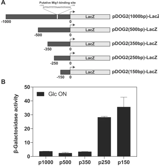
transcriptional repressors Mig1/Mig2 (Fig. 4A) also led to an increase in Dog2 expression (Fig. 4E-F). A
2
regulatory sequence in the promoter that is present between -250 and -350 bp relative to the ATG
3
start codon, combined with a proposed Mig1-binding site located at ~200 bp (40, 48), was critical for
4
this glucose-mediated repression (fig. S5, A and B). The reg1∆ or hxk2∆ mutants, which are more
5
tolerant to 2DG than WT (30, 31), showed increased Dog2 expression (Fig. 4E-F), which was also the
6
case for the double mutant mig1∆ mig2∆ (Fig. 4G). We deleted DOG1 and DOG2 in these mutants to
7
evaluate their contribution to 2DG resistance. The absence of Dog1 and Dog2 sensitized all strains to
8
2DG (Fig. 4G); in particular, the mig1∆ mig2∆ dog1∆ dog2∆ showed a level of sensitivity that was
9
comparable to that of the WT, demonstrating that the resistance of the mig1∆ mig2∆ mutant was due
10
to increased DOG expression. In contrast, the reg1∆ dog1∆ dog2∆ and the hxk2∆ dog1∆ dog2∆ strains
11
remained more resistant to 2DG than the WT, suggesting additional mechanisms of resistance. Finally,
12
we observed that the snf1∆ mig1∆ mig2∆ mutant was resistant to 2DG despite the absence of Snf1, in
13
line with the idea that snf1∆ is 2DG-sensitive because of the constitutive repression of Mig1/Mig2
14
target genes, such as Dog2 and possibly other genes (Fig. 4F). Overall, we conclude that Dog2 is also
15
regulated by glucose availability through Snf1 activity, which contributes to the resistance of
glucose-16
repression mutants to 2DG.
17
18
Increased expression of DOG2 is frequently observed in spontaneous 2DG-resistant clones
19
Previous screens have identified 2DG-resistant mutants (26, 29, 35). The initial purpose of these
20
screens was to identify mutants that were insensitive to the repressive effect of 2DG, or that were
21
impaired for glucose phosphorylation (because only 2DG6P is toxic to cells), and consequently, were
22
performed on media containing other carbon sources than glucose. The mechanisms involved in 2DG
23
resistance on glucose medium were tackled later by screening of the deletion library (30), which led
24
to the identification of resistant mutants, some of which were subsequently confirmed (31). In the
25
course of our experiments, we often found that 2DG-resistant clones could spontaneously arise from
26
WT or even 2DG-susceptible strains (Fig. 1E). Yeast acquires 2DG resistance at a high frequency, which
1
can be accompanied by an increase in 2DG-6-phosphatase activity (28, 38). To study whether Dog2,
2
which is functionally critical for 2DG resistance, was upregulated during the emergence of spontaneous
3
2DG-resistant mutants, we spread ~2x10⁶ cells on glucose-based medium containing 0.2% 2DG and
4
selected clones that had appeared after 6 days of growth. In total, 24 clones were obtained whose
5
resistance to 2DG was confirmed (fig. S6A). Using the pDOG2-LacZ reporter, we found that 13 clones
6
displayed significantly increased DOG2 promoter activity as compared to WT strains (Fig. 5A),
7
suggesting that Dog2 overexpression is a frequent feature of 2DG-resistant clones. Among those, we
8
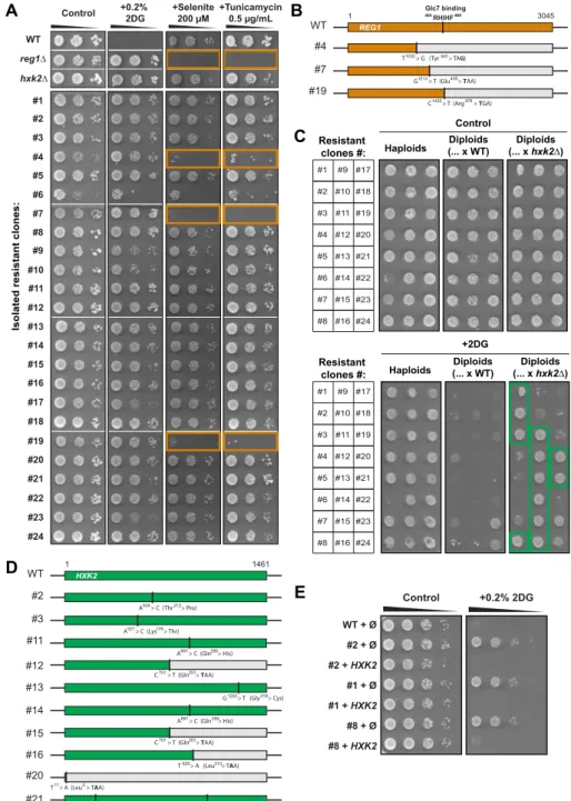
expected to isolate reg1 and hxk2 mutants because both are resistant to 2DG in glucose medium (See
9
Figure 5G; 30, 31). To identify them, we first tested whether some of the isolated resistant clones
10
displayed phenotypes typical of reg1 mutants, such as sensitivity to tunicamycin (68, 69) or selenite
11
(which enters the cell through the glucose-repressed transporter Jen1: 70, 71). Indeed, three of the
12
isolated clones displayed these phenotypes (fig. S6A). Sequencing of the REG1 coding sequence in
13
these clones revealed the presence of nonsense mutations which likely explain Reg1 loss of function
14
(fig. S6B). To identify potential hxk2 mutants, we performed a complementation test by crossing the
15
2DG-resistant clones with either a WT strain or an hxk2∆ strain and tested the ability of the resulting
16
diploids to grow on 2DG. All diploids generated by the cross with the WT starin lost their ability to grow
17
on 2DG, except for two (clones #23 and #24) (fig. S6C), indicating that 2DG resistance was generally
18
caused by a recessive mutation(s). In contrast, 12 diploids obtained by the cross with the hxk2∆ strain
19
maintained their ability to grow on 2DG (fig. S6C), suggesting that 2DG resistance of the initial strains
20
was caused by a deficiency in HXK2 function. Sequencing of the HXK2 ORF in these 12 mutants (fig.
21
S6D) revealed that two did not display any mutation in the HXK2 ORF (clones #1 and #8) and may
22
represent regulatory mutants in cis, possibly in regions that were not sequenced. In favor of this
23
hypothesis, we found that their 2DG sensitivity was restored by re-expression of HXK2 using a
24
multicopy, genomic clone (fig. S6E). The other 10 mutants carried at least one mutation in the HXK2
25
coding sequence (fig. S6D). Four mutants acquired nonsense mutations (clones #12, #15, #16 and #20),
26
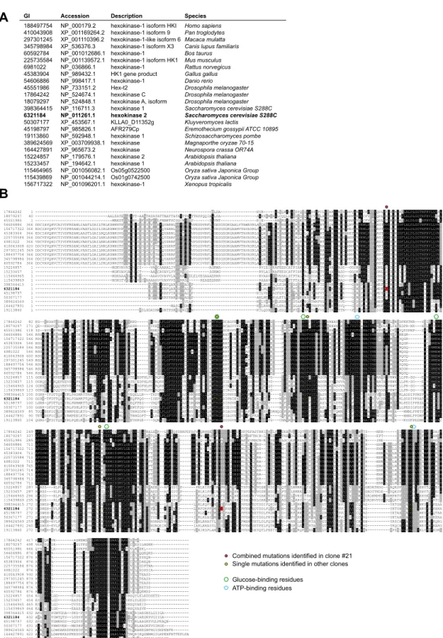
5 mutants (#2, #3, #11, #13 and #14) carried a missense mutation in residues conserved across all
1
hexokinase sequences tested (fig. S7, alignment) and one mutant (#21) bore two missense mutations.
2
All of these missense mutations occurred in residues involved in glucose or ATP binding or were located
3
near such residues (fig. S7) (72-74).
4
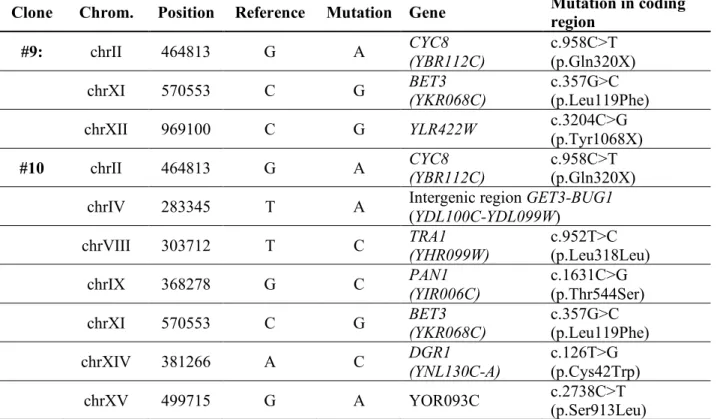
Of the 2DG-resistant clones which expressed pDOG2-LacZ at a high level, 2 clones (#9 and #10)
5
lacked mutations in either REG1 or HXK2, suggesting that their resistance involved a mechanism that
6
did not involve these proteins. Whole genome resequencing of the genomic DNA isolated from these
7
clones and comparison with that of the parent strain revealed several SNPs (Table S1) including a
8
nonsense mutation in CYC8 (C958>T) that was identified in both strains, causing a premature stop codon
9
at position 320 (Fig. 5B). The CYC8 gene is also known as SSN6 (Suppressor of snf1) and mutations in
10
this gene lead to constitutive expression of the glucose-repressed gene encoding invertase (SUC2),
11
even in a snf1 mutant (75, 76). Indeed, CYC8 encodes a transcriptional co-repressor which controls the
12
expression of glucose-regulated genes (77). Because the repression of DOG2 expression by glucose is
13
controlled by Snf1 and Mig1/Mig2 (Fig. 4, E and F), we hypothesized that CYC8 could also take part in
14
DOG2 regulation, such that a mutation in CYC8 could lead to increased 2DG resistance through DOG2
15
overexpression. We observed that indeed, mutants #9 and #10 expressed invertase even in when
16
grown in glucose medium (repressive conditions), in agreement with a mutation in CYC8 (Fig. 5C). We
17
also observed that these mutants displayed increased adhesion, or persistence of colony structures
18
upon washing the plate (78) that is often associated with mutants that are prone to flocculation, such
19
as cyc8 mutants (79, 80) (Fig. 5D). Introduction of a low copy (centromeric) plasmid containing CYC8
20
under the control of its endogenous promoter suppressed the 2DG resistance of mutants #9 and #10
21
as well as their adhesion to agar plates (Fig. 5D). This was not the case when mutants #9 and #10
22
expressed a truncated version of CYC8 identified in this screen (Fig. 5D), although these mutants did
23
not display slow growth in contrast to the cyc8∆ mutant, suggesting that it is at least partially active
24
for other functions. Finally, we tested whether the strong activity of the DOG2 promoter observed in
25
mutants #9 and #10 (Fig. 5A) was also due to the lack of a functional CYC8. Indeed, the re-introduction
26
of low-copy vector containing CYC8 led to a decreased reporter expression in these mutants (Fig. 5E).
1
This result was confirmed by examining Dog2-GFP expression at the protein level (Fig. 5F-G).
2
Therefore, DOG2 expression is repressed by Cyc8 and spontaneous cyc8 mutants display an increased
3
Dog2 expression. The increased resistance of these mutants to 2DG also depended on the expression
4
of Dog1 and Dog2 as the deletion of DOG1 and DOG2 restored their sensitivity to 2DG (Fig. 5H).
5
Together, this initial characterization revealed that DOG2 overexpression is a common phenomenon
6
within spontaneous 2DG-resistant clones, both in known 2DG-resistant mutants (reg1 and hxk2) and
7
in the 2DG-resistant cyc8 mutants that we isolated.
8
9
HDHD1, a human member of the HAD-like phosphatase family, is a 2DG-6-P phosphatase
10
involved in 2DG resistance
11
The increase in Dog2 expression in various spontaneously 2DG-resistant mutants reminded us of
12
an earlier study in HeLa cells showing increased 2DG6P phosphatase activity in isolated 2DG resistant
13
clones (20). Dog1 and Dog2 belong to the family of HAD (haloacid dehalogenase)-like phosphatases
14
which are conserved from bacteria to human (Fig. 6A). The bacterial homolog of Dog1/Dog2, named
15
YniC, can also dephosphorylate 2DG6P in vitro (81) and we found that the expression of YniC in the
16
double dog1∆ dog2∆ yeast mutant also restored 2DG resistance (Fig. 6B). We used this phenotype to
17
identify potential human homologs (Fig. 6B). A PSI-BLAST analysis (82) on the human proteome using
18
the Dog2 protein sequence retrieved HDHD1-isoform a (NP_036212.3) as the candidate with highest
19
(39%) homology. HDHD1 (for Haloacid Dehalogenase-like Hydrolase Domain containing 1; also named
20
PUDP for pseudouridine-5'-phosphatase) is a HAD-like phosphatase with in vitro activity towards
21
phosphorylated metabolites such as pseudouridine-5'-phosphate (83). When expressed in in the
22
double dog1∆ dog2∆ mutant, HDHD1 partially rescued growth on 2DG containing medium (Fig. 6B).
23
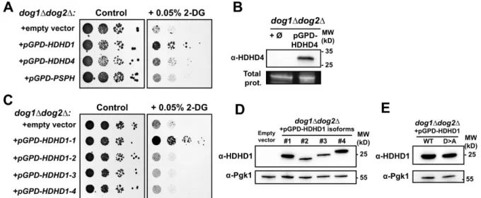
Yeast growth on 2DG was not restored by the expression of HDHD4 (also named NANP, for
N-24
acylneuraminate-9-phosphatase; 84), which belongs to the same subfamily as HDHD1 within the
HAD-25
phosphatase family (37% homology), or of PSPH (phosphoserine phosphatase; 85), another close
26
family member (fig. S8A,B). Moreover, among the four predicted isoforms of HDHD1 (isoforms 1-4),
1
only HDHD1-1 rescued the growth of the dog1∆ dog2∆ on 2DG-containing medium (fig. S8C) although
2
all isoforms were expressed in yeast, as confirmed by Western blotting using HDHD1 antibodies (fig.
3
S8D).
4
The mutation of conserved aspartate residues (Asp12 and Asp14; Fig. 6A) predicted to be essential
5
for the catalytic activity of HAD phosphatases (86) abolished the ability of HDHD1 to restore growth of
6
the dog1∆ dog2∆ mutant on 2DG (Fig. 6C and fig. S8E), suggesting it may act as a 2DG-6P phosphatase.
7
This notion was confirmed by the ability of purified recombinant HDHD1 (Fig. 6D) to dephosphorylate
8
2DG-6P in vitro, an activity that required its putative catalytic residues (Fig. 6E). We then tested
9
whether HDHD1 overexpression in HeLa cells could lead to an increased resistance to 2DG. Low
10
concentrations of 2DG (5 mM) in the presence of glucose (25 mM) were sufficient to inhibit the growth
11
of HeLa cells transfected with an empty vector, whereas those that overexpressed HDHD1 were
12
insensitive to 2DG treatment (Fig. 6F). These results suggest that dysregulated expression of HDHD1
13
could modulate 2DG resistance in human cells.
14
Discussion
1
2
Although the first studies examining the effect of 2DG on glycolysis in yeast and in normal and
3
cancer tissues were published more than 60 years ago (87-89), the mode of action for 2DG is not fully
4
understood. 2DG can be considered to be a general competitor of glucose (90) and a glycolysis inhibitor
5
because of its inhibition of glycolytic enzymes (6-8). However, this view has been challenged because
6
exposure of cancer cells to 2DG interferes with N-linked glycosylation, likely because of the structural
7
similarity of 2DG with mannose (91), resulting in ER stress and UPR induction in mammalian cells (14,
8
16). Therefore, 2DG interferes with other cellular functions beyond glycolysis.
9
In this study, we used an unbiased mass spectrometry approach to analyze the effects of 2DG on
10
the total cellular proteome. We found that the abundance of many glycolytic enzymes was increased,
11
likely because of impaired glycolysis, as well as many genes regulated by the MAPK-based cell wall
12
integrity pathway, suggestive of its activation. Using the list of 2DG-upregulated proteins, we also
13
identified the phosphatases Dog1 and/or Dog2, which have been shown to play a role in 2DG
14
resistance (33, 40, 41). Dog2 induction upon 2DG treatment was intriguing because it was not clear
15
how a synthetic molecule such as 2DG could trigger a resistance mechanism. It should be noted that
16
the cellular functions of Dog1 and Dog2, beyond 2DG dephosphorylation, are unknown. They can
17
dephosphorylate various phosphorylated sugars in vitro (40), in agreement with the ability of other
18
members of the HAD family of phosphatases to accommodate various substrates (81). Therefore, it
19
seemed plausible that Dog1/2 induction was a response to one or more of the cellular consequences
20
of 2DG treatment, rather than to 2DG itself. Our study revealed that the expression of Dog1 and Dog2
21
was actually controlled by multiple signaling pathways, each of which are activated as a response to
22
2DG (Fig. 7).
23
This study also probed the signaling pathways that are activated upon 2DG exposure and
24
determined the causes for these activations. First, we confirmed that 2DG triggered ER stress and the
25
onset of the UPR pathway. This finding was not unexpected given the proposed mode of action of this
26
drug and the available data in the literature on mammalian cells (14, 16), but had not been formally
27
proven in yeast with current tools. The induction of an UPR reporter upon 2DG treatment and the
1
hypersensitivity of UPR mutant strains such as hac1∆ or ire1∆ support this conclusion. The latter
2
phenotype was alleviated by restoring N-glycosylation through the addition of mannose to the culture
3
medium. The consolidation of the links between 2DG and ER stress, notably the identification of UPR
4
mutants as 2DG-hypersensitive mutants suggests that drugs targeting the UPR may synergize with 2DG
5
in a glycolytic cancer background. We also showed that Dog1 and Dog2 are induced by ER stress, in
6
line with high-throughput studies suggesting that Dog1 and Dog2 are UPR-induced genes (92).
7
Second, we showed that 2DG activates the MAPK-based CWI pathway, which mediates the cellular
8
response to cell wall alteration and other stresses. Several studies reported connections between ER
9
stress signaling and the CWI pathway (56-60), and 2DG-elicited N-glycosylation defects and ER stress
10
may have repercussions on the CWI pathway. For instance, CWI pathway mutants are sensitive to
11
tunicamycin because they lack an ER surveillance-pathway which normally prevents the inheritance of
12
stressed ER to the daughter cell during cell division (58). Many CWI mutants were indeed sensitive to
13
low concentrations (0.05%) of 2DG but unlike UPR mutants, their growth was not restored by the
14
addition of exogenous mannose, suggesting that an N-glycosylation defect is not the sole reason why
15
CWI mutants are hypersensitive to 2DG. However, 2DG interferes with the synthesis of structural
16
polysaccharides that make up the yeast cell wall because 2DG acts as an antagonist of mannose and
17
glucose incorporation into these polymers (42, 61-63). 2DG exposure leads to yeast cell lysis at sites of
18
growth, which is where glucan synthesis occurs (43) and where the major glucan synthase, Fks1, is
19
localized (93). 2DG-induced weakening of the cell wall could trigger the CWI pathway (Fig. 7), which
20
would explain why the main sensors responsible for sensing cell wall damage, Wsc1 and Mid2
21
(reviewed in 55), are required for growth on 2DG. In addition, we report that the expression of Dog1
22
and Dog2 was decreased in CWI mutants, which could further sensitize these strains to 2DG. The effect
23
of 2DG on cell wall synthesis suggests an interference of 2DG with UDP-glucose metabolism that is
24
likely to be conserved in metazoans (94), where it may affect metabolic pathways involving these
25
precursors, such as glycogen synthesis (95, 96).
26
Our data also revealed that Dog2 expression was regulated at the transcriptional level by glucose
1
availability through the glucose-repression pathway, which is controlled by the kinase Snf1 and the
2
regulatory subunit of the PP1 phosphatase, Reg1. These findings explain why reg1 mutants have
3
increased 2DG6P phosphatase activity (28, 38, 39), because the glucose-mediated repression of Dog2
4
is defective in these mutants. The negative regulation of Dog2 by this pathway may also contribute to
5
the 2DG resistance of hxk2 and mig1 mutants, which participate in the glucose-repression pathways
6
and have been identified in previous screens (29, 35). Indeed, the 2DG resistance displayed by the
7
reg1∆ and hxk2∆ strains partially depended on increased expression of Dog2, because even when
8
these mutants lacked both DOG genes, they still grew better than a WT strain on 2DG-containing
9
media. In the hxk2∆ mutant, the lack of hexokinase 2 is compensated for by the expression of other
10
glucose-phosphorylating enzymes (such as Hxk1 and the yeast glucokinase homolog, Glk1) (97) which
11
may be less prone to phosphorylate 2DG (36), and thus could lead to a lower accumulation of 2DG6P
12
in the cell. Hxk2 also has non-metabolic roles beyond sugar phosphorylation (reviewed in Ref. 98), and
13
loss of these non-metabolic roles may additionally contribute to this phenotype. However, many of
14
the hxk2 point mutants isolated during the characterization of spontaneous 2DG-resistant clones were
15
affected at or near glucose-binding residues, suggesting a primary metabolic role for Hxk2 in 2DG
16
resistance. Concerning the reg1∆ strain, additional mechanisms of resistance beyond an increased
17
expression of Dog2 also remain to be investigated. Previous work suggested that the resistance of the
18
reg1∆ mutant depends on Snf1 hyperactivity, because the additional deletion of SNF1 in this
19
background restores 2DG sensitivity to the reg1∆ strain (31). Thus, additional Snf1-dependent
20
mechanisms cooperate with the increased expression of DOG2 to allow reg1∆ cells to resist 2DG.
21
After 2DG import and phosphorylation, the amount of 2DG6P in cells has been reported to exceed
22
that of G6P by up to 80-fold (99), although this number should probably be confirmed by more direct
23
and modern methods. Overexpression of Dog2 can revert the repressive effect of 2DG (33), suggesting
24
that it can clear the 2DG6P pool up to a point at which 2DG6P is no longer detected by a yet unknown
25
cellular glucose-sensing mechanism. Thus, Dog2 overexpression appears to be a good strategy for
26
acquiring 2DG resistance, and indeed, Dog2 was overexpressed in the majority of the spontaneously
1
resistant mutants we isolated, including strains with mutations in REG1 and HXK2. Additionally, we
2
identified two mutants carrying a point mutation in the gene encoding the transcriptional repressor
3
Cyc8, leading to a truncated protein at codon 320, within its 8th predicted tetratricopeptide (TPR)
4
repeat. The TPR repeats are involved in the interaction of Cyc8 with its co-repressor Tup1 and in its
5
recruitment to specific promoters, possibly through pathway-specific DNA-binding proteins (100).
6
Point mutations in TPR units 9 and 10 affect Cyc8 function in a similar manner as the mutation we
7
isolated (namely, an effect on glucose repression but not global growth), suggesting that alterations at
8
this region only affect a subset of Cyc8 functions (101). This finding confirms the differential
9
requirement of TPR repeats for the various functions of Cyc8, and in particular the involvement of the
10
TPR repeats 8-10 in glucose repression (102). Together, these data describe DOG2 overexpression as
11
a successful strategy to overcome 2DG toxicity.
12
Based on sequence similarity, we identified HDHD1 as an enzyme displaying in vitro 2DG6P
13
phosphatase activity and whose overexpression in both yeast and HeLa cells allowed resistance to
14
2DG. Whether HDHD1 is responsible for the 2DG resistance previously reported in human cells (20)
15
remains to be investigated.
16
Together, our work shows that 2DG-induced activation of multiple signaling pathways can rewire
17
the expression of endogenous proteins that target 2DG6P to promote 2DG tolerance, and whose
18
increased expression can lead to 2DG resistance. Because of the existence of endogenous genes that
19
can confer 2DG resistance when overexpressed, such as HDHD1, and of the perturbation of several
20
cellular pathways in mammalian cells by 2DG, it is possible that similar resistance strategies occur in
21
human cells. Such strategies likely superimpose on other resistance mechanisms that should be
22
scrutinized in the future.
23
24
25
Material and Methods
1
2
Yeast strain construction and growth conditions
3
All yeast strains used in this study derive from the Saccharomyces cerevisiae BY4741 or BY4742
4
background and are listed in Table S2. Apart from the mutant strains obtained from the yeast deletion
5
collection (Euroscarf) and the fluorescent GFP tagged strains originating from the yeast GFP clone
6
collection (103), all yeast strains were constructed by transformation with the standard lithium
7
acetate-polyethylene glycol protocol using homologous recombination and verified by PCR on genomic
8
DNA prepared with a lithium acetate (200mM) / SDS (0.1%) method (104).
9
Yeast cells were grown in YPD medium (Yeast extract-Peptone-Dextrose 2%) or in synthetic complete
10
medium (SC, containing 1.7g/L yeast nitrogen base (MP Biomedicals), 5g/L ammonium sulfate
(Sigma-11
Aldrich), the appropriate drop-out amino acid preparations (MP Biomedicals) and 2% (w/vol) glucose
12
unless otherwise indicated). Alternatively, SC medium could contain 0.5% lactate as a carbon source
13
(from a 5% stock adjusted to pH=5; Sigma Aldrich). Pre-cultures of 4mL were incubated at 30°C for 8
14
hours and diluted in fresh medium on the evening to 20mL cultures grown overnight with inoculation
15
optical densities (OD600) of 0.0003 for YPD and 0.001 for SC medium, giving a culture at mid-log phase
16
the next morning.
17
For glucose depletion experiments, cultures were centrifuged and resuspended in an equal volume of
18
SC/lactate medium and incubated at 30°C during the indicated times. For 2DG, NaCl and tunicamycin
19
treatments, the compounds were added to mid-log phase yeast cultures grown overnight to respective
20
final concentrations of 0.2% (w/v), 400mM and 1µg/mL and incubated for the indicated times.
2-21
deoxyglucose and tunicamycin were purchased from Sigma-Aldrich. The mannose-supplemented
22
medium (Fig. 2A) consisted of an SC medium that contained all the elements indicated above plus 2%
23
(w/vol) mannose.
24
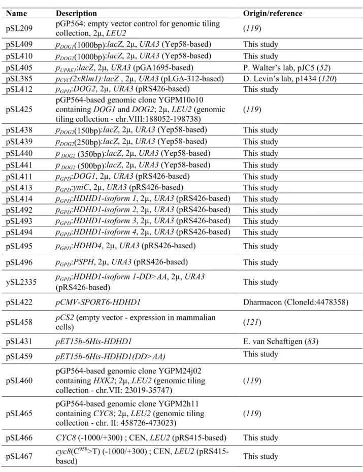
Plasmid construction
25
All the plasmids presented in this study are listed in Table S3 and were directly constructed in yeast
26
using plasmid homologous recombination (105). DNA inserts were amplified by PCR using 70-mer
27
primers containing 50nt homology overhangs and Thermo Fisher Phusion High-fidelity DNA
28
Polymerase and receiver plasmids were digested with restriction enzymes targeting the insertion
29
region. Competent yeast cells rinsed with lithium acetate were incubated for 30 minutes at 30°C with
30
20µL of the PCR product and 1µL of the plasmid digestion product, followed by a heat shock at 42°C
31
for 20 minutes and a recovery phase in rich medium (YPD) for 90 minutes at 30°C and plated on
32
synthetic medium without uracil. The pDOG1/2-LacZ vectors were generated using the pJEN1-LacZ
33
vector obtained from Bernard Guiard (106) and cloning the pDOG1 or pDOG2 promoters (1kb) at BglII
34
and EcoRI sites. The DOG1, DOG2, HDHD1 (all four isoforms), HDHD4, PSPH and yniC overexpression
35
vectors were obtained by digesting a pRS426 vector (2µ, URA3) containing the pGPD
(glyceraldehyde-1
3-phosphate dehydrogenase gene, TDH3) (107) promoter with EcoRI and BamHI enzymes. In parallel,
2
inserts were PCR-amplified using the following DNA templates: DOG1 and DOG2 from yeast genomic
3
DNA preparations (primers: oSL1166/1167 and oSL1141/1142), the yniC ORF from Escherichia coli
4
DH5α cells (oSL1172/1173) and the human gene ORFs (Uniprot identifiers: HDHD1=Q08623-1/2/3/4,
5
HDHD4=Q8TBE9 and PSPH=P78330) from DNA sequences generated by gene synthesis after codon
6
optimization for yeast expression (Eurofins Genomics) (HDHD1: oSL1170/1171 for all isoforms except
7
isoform 3: oSL1170/1216; HDHD4: oSL1214/1215; PSPH: oSL1212/1213). These PCR products were
8
designed to include a 50-bp overlap with the digested plasmid to enable their cloning by homologous
9
recombination in yeast after co-transformation. The pGPD-HDHD1-DDAA vector was obtained by PCR
10
amplification (oSL1155/1171) of the HDHD1 DNA sequence obtained by gene synthesis using a specific
11
5' primer carrying two D>A mutations and insertion of this insert into the pRS426 vector digested with
12
EcoRI and BamHI as described above. This construct was verified by sequencing. The DDAA mutant
13
was PCR amplified (oSL1297/oSL1298) and subcloned at NdeI/BamHI sites into pET15b-6His-HDHD1, a
14
kind gift of Dr. E. Van Schaftingen. The wild-type CYC8 gene and the mutant allele present in mutant
15
#9 were obtained by PCR amplification on the corresponding genomic DNAs with primers
16
(oSL1369/1370) containing a 50-bp overlap with a pRS415 vector (CEN, LEU2). The resulting PCR
17
products were digested with XbaI and BamHI, and co-transformed for cloning by homologous
18
recombination in yeast as described above. The plasmids generated in yeast were rescued by
19
extraction (lithium acetate/SDS method (104)) and electroporation in bacteria, then amplified and
20
sequenced before being re-transformed in the appropriate strains.
21
Mass spectrometry and proteomics analyses
22
Samples used for the proteome-wide analysis of 2-deoxyglucose treatments were prepared from six
23
liquid cultures (WT strain, BY4741) growing overnight at 30°C in 100mL of rich medium with 2% glucose
24
to mid-log phase. On the next morning, 2-deoxyglucose was added to three of the cultures to a final
25
concentration of 0.2% in order to obtain triplicates treated with 2DG and triplicates without drug
26
treatment (negative control). After 2.5 hours of incubation at 30°C, the six cultures were centrifuged
27
at 4000g for 5 minutes at 4°C, resuspended in 500µL of 10% trichloroacetic acid (TCA, Sigma-Aldrich)
28
and lysed by shaking after addition of glass beads (0.4-0.6mm, Sartorius) for 10 minutes at 4°C. Cell
29
lysates were retrieved by piercing under the 1.5mL tubes and brief centrifugation. Precipitated
30
proteins were centrifuged at 16000g for 10 minutes at 4°C, supernatants were discarded and pellets
31
were rinsed 4 times in 1mL of 100% cold acetone.
32
Proteins were then digested overnight at 37°C in 20 μL of 25 mM NH4HCO3 containing
sequencing-33
grade trypsin (12.5 μg/mL; Promega). The resulting peptides were sequentially extracted with 70%
34
acetonitrile, 0.1% formic acid. Digested samples were acidified with 0.1% formic acid. All digests were
35
analyzed by an Orbitrap Fusion equipped with an EASY-Spray nanoelectrospray ion source and coupled
1
to an Easy nano-LC Proxeon 1000 system (all from Thermo Fisher Scientific, San Jose, CA).
2
Chromatographic separation of peptides was performed with the following parameters: Acclaim
3
PepMap100 C18 pre-column (2 cm, 75 μm i.d., 3 μm, 100 Å), Pepmap-RSLC Proxeon C18 column (50
4
cm, 75 μm i.d., 2 μm, 100 Å), 300 nl/min flow, using a gradient rising from 95 % solvent A (water, 0.1
5
% formic acid) to 40 % B (80 % acetonitrile, 0.1% formic acid) in 120 minutes, followed by a column
6
regeneration of 20 min, for a total run of 140 min. Peptides were analyzed in the orbitrap in full-ion
7
scan mode at a resolution of 120,000 (at m/z 200) and with a mass range of m/z 350-1550, and an AGC
8
target of 2x10⁵. Fragments were obtained by higher-energy C-trap dissociation (HCD) activation with
9
a collisional energy of 30 %, and a quadrupole isolation window of 1.6 Da. MS/MS data were acquired
10
in the linear ion trap in a data-dependent mode, in top-speed mode with a total cycle of 3 seconds,
11
with a dynamic exclusion of 50 seconds and an exclusion duration of 60 seconds. The maximum ion
12
accumulation times were set to 250 ms for MS acquisition and 30 ms for MS/MS acquisition in
13
parallelization mode.
14
Raw mass spectrometry data from the Thermo Fisher Orbitrap Fusion were analyzed using the
15
MaxQuant software (108) version 1.5.0.7, which includes the Andromeda peptide search engine (109).
16
Theoretical peptides were created using the Saccharomyces cerevisiae S288C proteome database
17
obtained from Uniprot. Identified spectra were matched to peptides with a main search peptide
18
tolerance of 6ppm. After filtering of contaminants and reverse identifications, the total amount of
19
yeast proteins identified among the six samples was equal to 3425. Protein quantifications were
20
performed using MaxLFQ (110) on proteins identified with a minimum amount of two peptides with a
21
False Discovery Rate threshold of 0.05. LFQ values were then analyzed using Perseus (version 1.5.0.15).
22
For the statistical analysis of yeast proteomes treated with 2-dexogylucose compared to negative
23
control samples, each group of triplicates was gathered into a statistical group in order to perform a
24
Student's t-test. Results are presented in the form of Volcano-plots (111) and significantly up-regulated
25
and down-regulated candidates were determined by setting an FDR of 0.01 and an S0 of 2.
26
GO-term analyses of proteomics data
27
The 79 significantly up-regulated candidates obtained in the proteomics analysis were used as input
28
for the FunSpec web interface (http://funspec.med.utoronto.ca/) with default settings and a p-value
29
cutoff of 0.01 in order to determine the Gene Ontology biological processes that are enriched in this
30
list of 79 genes compared to the total Saccharomyces cerevisiae genome annotation (number of total
31
categories=2062). The complete list of enriched GO biological processes with a p-value<0.01, as well
32
as the genes included in each category, are displayed in Fig. 1B.
33
Protein extracts and immunoblotting
Yeast cultures used for protein extracts were all grown in synthetic complete medium. For each protein
1
sample, 1.4mL of culture was incubated with 100µL of 100% TCA for 10 minutes on ice to precipitate
2
proteins, centrifuged at 16000g at 4°C for 10 minutes and broken for 10 minutes with glass beads, as
3
described for LC-MS/MS sample preparation. Lysates were transferred to another 1.5mL tube and
4
centrifuged 5 minutes at 16000g at 4°C, supernatants were discarded and protein pellets were
5
resuspended in 50µL*(OD600 of the culture) of sample buffer (50 mM Tris-HCl pH6.8, 100 mM DTT, 2%
6
SDS, 0.1% bromophenol blue, 10% glycerol, complemented with 50mM Tris-Base pH8.8). Protein
7
samples were heated at 95°C for 5 minutes and 10µL were loaded on SDS-PAGE gels
(4-20%Mini-8
PROTEAN TGX Stain-Free, BioRad). After electrophoresis, gels were blotted on nitrocellulose
9
membranes for 60 minutes with a liquid transfer system (BioRad), membranes were blocked in 2%
10
milk for 20 minutes and incubated for at least two hours with the corresponding primary antibodies.
11
Primary and secondary antibodies used in this study as well as their dilutions are listed in Table S4.
12
Membranes were washed three times for 10 minutes in Tris-Borate-SDS-Tween20 0.5% buffer and
13
incubated for at least an hour with the corresponding secondary antibody (coupled with Horse Radish
14
Peroxidase). Luminescence signals were acquired with the LAS-4000 imaging system (Fujifilm). Rsp5
15
was used as a loading control; alternatively, total proteins were visualized in gels using a trihalo
16
compound incorporated in SDS–PAGE gels (stain-free TGX gels, 4–20%; Bio-Rad) after 1 min
UV-17
induced photoactivation and imaging using a Gel Doc EZ Imager (Bio-Rad).
18
b-galactosidase assays
19
β-Galactosidase assays were performed using 1mL of mid-log phase yeast cultures carrying the
pDOG1-20
LacZ or pDOG2-LacZ plasmids, grown overnight in SC medium without uracil with glucose 2% and
21
switched to the specified conditions. The OD (600 nm) of the culture was measured, and samples were
22
taken and centrifuged at 16000g at 4°C for 10 minutes. Cell pellets were snap frozen in liquid nitrogen
23
and resuspended in 800µL of Buffer Z (pH=7, 50mM NaH2PO4, 45mM Na2HPO4, 10mM MgSO4, 10mM
24
KCl and 38mM β-mercaptoethanol). After addition of 160µL of 4mg/mL ONPG
(ortho-nitrophenyl-β-D-25
galactopyranoside, Sigma-Aldrich), samples were incubated at 37°C. Enzymatic reactions were
26
stopped in the linear phase (60min incubation for pDOG2-LacZ and 120min incubation for the pDOG1-LacZ
27
plasmid, as per initial tests) by addition of 400µL of Na2CO3, and cell debris were discarded by
28
centrifugation at 16000g. The absorbance of clarified samples was measured with a
29
spectrophotometer set at 420nm. β-Galactosidase activities (arbitrary units, AU) were calculated using
30
the formula 1000*[A420/(A600* t)], where A420 refers to the enzyme activity and A600 is the turbidity
31
of the culture, and t the incubation time. Each enzymatic assay was repeated independently at least
32
three times.
33
Drop tests
1
Yeast cells grown in liquid rich or synthetic complete medium for at least 6 hours at 30°C were adjusted
2
to an optical density (600nm) of 0.5. Serial 10-fold dilutions were prepared in 96-well plates and, using
3
a pin replicator, drops were spotted on plates containing rich or SC medium containing 2% (w/v) agar
4
and when indicated, 2DG (0.05%, or 0.2%, w/vol), sodium selenite (200µM) or tunicamycin (1µg/mL).
5
Mannose plates (Fig 2I and 3F) were prepared as regular SC plates containing 2% mannose (w/vol) in
6
addition to glucose. Plates were incubated at 30°C for 3 to 4 days before scanning. For the adhesion
7
test (Fig. 5D), the plates were scanned and the colonies were then washed with tap water under a
8
constant flow of water for 1 min as previously described (78). Excess water was removed before the
9
plates were scanned again.
10
Isolation of spontaneous mutants and characterization.
11
WT cells transformed with a pDOG2-LacZ plasmid (pSL410) were grown overnight in SC-Ura medium
12
and ca. 2x10(6) cells were spread on SC-Ura plates containing 0.2% 2DG and grown for 6 days at 30°C.
13
The clones obtained (24 clones) were restreaked on SC-Ura to isolate single clones. Resistance to 2DG
14
was confirmed by drop tests (Fig. S6A). b-galactosidase enzyme assays and total protein extracts were
15
performed on cultures grown to the exponential phase. For b-galactosidase assays, the results were
16
statistically tested using an unpaired t-test with equal variance, assuming a normal distribution of the
17
values.
18
Diploids were obtained by crossing each resistant mutant with a WT or hxk2∆ strain of the opposite
19
mating type (BY4742, Matα) and selecting single diploid clones on selective medium (SC-Met-Lys).
20
Sequencing of the REG1 and HXK2 loci were done after PCR amplification on genomic DNA isolated
21
from the corresponding clones. For whole genome sequencing, genomic DNA of the WT, clone 9 and
22
clone 10 was purified using the Qiagen genomic DNA kit (Genomic-tip 20/G) using 30 OD equivalents
23
of material following the manufacturer’s instructions after zymolyase treatment (Seikagaku). A
PCR-24
free library was generated from 10 µg of gDNA and sequenced at the Beijing Genomics Institute (Hong
25
Kong) on Illumina HiSeq 4000. The mutations in each clone were identified through comparative
26
analysis of the variants detected by mapping their reads to the reference genome (BY4741) (112, 113)
27
and those detected by mapping the WT reads to the reference. The differential variants were filtered
28
by quality (vcf QUAL>1000) and manually inspected through IGV for validation (114).
29
Cell culture and transfection.
30
HeLa cells were maintained at 37°C and 5% CO2 in a humidified incubator and grown in Dulbecco’s
31
modified Eagle’s medium (DMEM), supplemented with 10% fetal calf serum. Cells were regularly split
32
using Trypsin-EDTA to maintain exponential growth. HeLa cells were transfected with plasmid