ELSEVIER
FEMS Microbiology Letters 143 (1996) l-6Identification
Ni Chin a,
MICROBIOLOGY LETTERS
of a locus involved in the utilization of iron by
Actinobacillus pleuropneumoniae
Joachim Frey b, Chao-Fu Chang a, Yung-Fu Chang ay*
a Diagnostic Laboratory, College of Veterinary Medicine. Cornell University, Ithaca, NY 14853, USA b Institute for Veterinary Bacteriology, University of Berne. Liinggass-Strasse 122, CH-3012 Berne, SwitzerlandReceived 8 February 1996; revised 4 June 1996; accepted 7 June 1996
Abstract
The cloned afu locus of Actinobacillus pleuropneumoniae restored the ability of an Escherichia coli K-12 mutant (aroB) to grow on iron-limited media. DNA sequence analysis of the fragment showed that there are three genes designated afuA, afuB and afuC (Actinobacillus ferric uptake) that encode products similar to the SfuABC proteins of Serratia marcescens, the
HitABC proteins of Haemophilus injluenzae, the FbpABC proteins of Neisseria gonorrhoeae and the YfuABC proteins of
Yersinia enterocolitica. The three genes encode a periplasmic iron-binding protein (AfuA), a highly hydrophobic integral cytoplasmic membrane protein with two consensus permease motifs (AfuB) and one hydrophilic peripheral cytoplasmic membrane protein with Walker ATP-binding motifs (AfuC), respectively. This system has been shown to constitute a periplasmic binding protein-dependent iron transport system in these organisms. The afuABC operon is locating approximately 200 bp upstream of apxIC gene, but transcribed in opposite direction to the ApxI-toxin genes.
Keywords: Actinobacillus pleuropneumoniae; Iron-uptake operon; ajiiABC; ApxI
1. Introduction
The low concentration of free iron, an essential nutrient for bacteria, on mucous membranes and in tissues is one of the first lines of host defense against bacterial infection. The presence of iron-binding pro- teins in the body fluids, such as transferrin, lactofer- t-in, haem, haemoglobin, and ferritin further serves to maintain low free-Fe concentrations, inhibiting bac- teria growth [l]. To sequester the limited iron from the host, bacteria have evolved several mechanisms, such as the secretion of siderophores and iron chela- tors which compete with lactoferrin and transferrin
* Corresponding author.
for iron. Iron-repressible outer membrane proteins (IROMP) that serve as receptors for iron-sidero- phore complexes are essential for iron uptake have been identified in many pathogenic bacteria [2], in- eluding Actinobacillus pleuropneumoniae [3].
A. pleuropneumoniae obtains iron from haem com- pounds [3] via the production hemolysins [4,5], and membrane-bound transferrin-specific receptors [6].
A. pleuropneumoniae probably binds the iron-loaded transferrin molecule to its surface and then, trans- ports the iron from the transferrin into the cells. However, a mechanism for the transfer of iron from the transferrin to the bacterium has not been elucidated.
In this study, we reported the cloning and se- 0378-1097 /96/$12.00 Copyright 0 1996 Federation of European Microbiological Societies. Published by Elsevier Science B.V.
2 N. Chin et ul. IFEMS Microbiology Letters 143 (1996) I&5
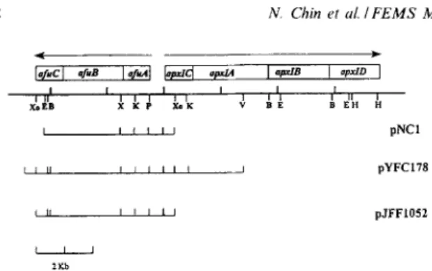
Fig. 1. Partial restriction enzyme map of afir and apxl operons
of A. pleuropneumoniae. The inserted DNA from pNC1 and pJFF1052 were completely sequenced. E, EcoRI; V, EcoRV; B, EgnI; K, KpnI; H, HindIII; P, PstI; X, X&I; Xo, XhoI. Arrows indicate direction of expression.
quence analysis of an iron utilization system in A.
pfeuronpneumoniue that is very similar to the peri- plasmic-binding protein-dependent transport system in Serratia marcescens [7,8], Haemophilus influenzae [9,10], Neisseria gonorrhoeae [l 1,121 and Yersia en- terocolitica (unpublished data). This locus, termed ufu (Actinobacillus ferric uptake) is located upstream of ApxZCABD gene cluster, but is transcribed in op- posite direction to the hemolysin gene (apxZC). Complementation analysis showed that this locus could restore the ability of an E. coli aroB mutant to grow on iron-depleted medium.
2. Materials and methods
2.1. Bacterial strains, plasmids, growth conditions and DNA preparation
A. pleuropneumoniae serotype 1 to 12 reference strains described previously [ 131 were grown in brain-heart infusion broth (BHI, Difco Labora- tories) supplemented with 0.1% NAD or on choco- late agar. The E. coli strains, TBl, araa (lac-
proAB)rpsLWOdlacZ A Ml 5 hsdR17 (r-m+),
H1443, aroD araD139 AlacU169 rpsL1.50 relA1
deoC1 p&t;25 rbsR thiJb5301, and DH5a @80dlac-
Z A Ml5 A (ZacZYA_argF)U169 endA 1 recA 1 hsdR 17(rk_mk+)deoR thi- lsupE44 ygyrA96 relA1 were grown in Luri-Bertani (LB) broth or LB agar. Antibiotics were used as appropriate for selection or maintenance of plasmids, ampicillin 40 @ml, and kanamycin 50 &ml. X-gal (Sbromo-in-
dolyl-P-D-galactopyranoside) was added to agar plates to 40 Fg/ml. Plasmids pYFC126 and pYFC127 contains apxZCA genes and a segment up- stream apxZC from serotype 1 and 5, respectively [14]. pNC1 contains ufuAB and partial afuC genes. Plasmid pYFCl78 contains afuABC genes subcloned from the phage clone, hyfc40 originating from A. pleuropneumoniae serotype 5 (Fig. 1). Plasmid
pJFF1052 contains @ABC in pBS (same direction to Pl,,) (Fig. 1) and plasmid pSZ1 contains sfuABC
in pBR322 [7].
2.2. DNA isolation and construction of a genomic and sub-genomic library of A. pleuropneumoniae DNA in h-dash and screening
A. pleuropneumoniae genomic DNA from different serotypes was prepared as previously described [13]. A lamda-dash library was constructed by using the genomic DNA from a serotype 5 strain as previously described [5]. A subgenomic library from serotype 5 was also constructed using BgZII and PstI digested DNA fragments separated by agarose gel electro- phoresis. The 4.5-6 kbp fragments were ligated into pHG165 digested with BamHI and SaZI. The bacteriophage and subgenomic libraries were screened by hybridization using a probe (a 1033 bp
PstI-XhoI DNA fragment from pYFC126) contan- ing the partial apxZC gene and its upstream region from A. pleuropneumoniue serotype 5 [14].
Table I
The conserved EAA motifs from the periplasmic permeases of A.
pleuropneumoniar, S. marcensens. H. inzuenzae, N. gonorrhoeae
and Y. enterocolitica
Protein” Residue Conserved sequence AfuB (N) 303 EEASYTLRANRYQTFYNIIFP SfuB (N) 167 EDVATSLGSRPPAVFFRVVLP HitB (N) 148 EEVSISLGKSPVYTFWYAIFP YfuB (N) 168 EDAAASLGSTPSAVFFHVVLP FbpB (N) 153 EEVSLSLGKSRLQTFFSAILP AfuB (C) 581 EASLSLKGSSLKTIWFIVFPL SfuB (C) 428 ENVARSLGKSPAQAiWSTTLR HitB (C) 410 EKVGGSLGRNPFYIFRTITLP YfuB (C) 429 ENVARSLGKTPTQAIWSTTMR FbpB (C) 426 EQVGATLGRGHFFIFRTLVLP “The sequence were taken from the following sources: afuB (this study), sfua [7], fbpb [12], hitb [9] and yfuB (unpublished data).
N. Chin et al. IFEMS Microbiology Letters 143 (1996) ld 3 0 mM 2,Y_diwridy( 0.05 mM 2.Z-dipyrldyl 1 8 * 0.1 0.01 0 60 120 180 240 300 300 the [min.] 0.i mM 2,2WpyWyl 1 8 ( 0.1 0.01 0 60 120 180 240 300 360 time [min.] 1 8
u
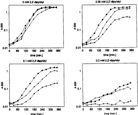
0.1 80 120 180 240 300 300 time [min.] 0.2 mM 2.2’diwrWl 0.01 0 60 120 180 240 300 360 time [min.]Fig. 2. Growth rates of the aroB mutant H1443 and the complemented mutant in LB containing different concentrations of 2,2’-dipyridyl. Growth was monitored with a spectrophotometer at A650. The cultures were inoculated to Asso = 0.01 with fresh precultures. +, H1443 (aroB); 0, H1443 [pJFF1052 (afuA+BfC)]; W, H1443 [pSZl (sfuA+BtP)].
2.3. DNA sequencing and analysis
Plasmid DNA for cycle DNA sequencing was iso- lated with a mini-kit from Qiagen (Chatsworth, CA). The nucleotide sequence was determined by an auto- mated fluorescence procedure based on the Sanger dideoxy chain termination method using a Taq Dye- Deoxy Terminator Cycle Sequencing kit (Applied Biosystem, Inc.). DNA sequences were determined by using double-stranded DNA templates (pNC1, pYFC178 and pJFF1052). Oligonucleotide primers based on the DNA sequence in this study were pre- pared by the Analytical and Synthetic Facility, Cor- nell University. Both strands of the cloned DNA were completely sequenced.
2.4. Trans-complementation of E. coli aroB
E. coli H1443 (aroB mutant) transformed with plasmid pJFF1052 or with plasmid pSZ1 and E. coli C600 (arop) was inoculated in LB broth or in LB broth supplemented with 0.05, 0.1, 0.2, or 0.4 mM 2,2’-dipyridyl (NBD) (Sigma, St Louis, MO) and the growth was monitored by measuring the absortion As50 in a spectrophotometer.
2.5. Southern blotting, hybridization
A PstI and XbaI DNA fragment from pNC1 con- taining the afuA gene was isolated from agarose gel and labeled with [32P]dATP by nick-translation. Fil- ters were hybridized in 45% formamide, 5 X SSC,
4 N. Chin et al. IFEMS Microbiology Letters 143 (1996) l&5 1 2 3 4 5 6 7 8 9 10 11 12
Fig. 3. Southern blotting analysis of 12 serotypes reference strains of A. pleuropneumoniae. The lanes are labeled according to the serotype number. The filter was hybrized and washed as described in Section 2. Molecular mass in kbp as indicated at the right.
thymus DNA for 12 h at 37°C. Filters were then washed twice with 2 x SSC-0.1% SDS and twice with 0.2% SSC-0.1% SDS at room temperature [4,13]. The final wash was with 0.16% SSC-0.1% SDS at 37°C.
3. Results and discussion
3. I. Nucleotide sequences of the afuA, afuB and afuC genes
Plasmid pNC1 and phage clone hyfc40 were se-
lected from subgenomic and a-dash library, respec- tively. Plasmid pNC1 contains a 5.0 kb of the A. pleuropneumoniae serotype 5 chromosome, cloned
into the SalI and BamHI sites of pHG165 [4,14]. hyfc40 was digested with SalI and a 9 kb fragment was ligated into pBluescriptII- SK-, to obtain pYFC178. Plasmid pJFF1052 contains a 5.8 kb chromosomal fragment of A. pleuroneumoniae sero- type 1 (strain 4074) including the 5’-terminal part of the apxIC and a 5.4 kb segment upstream apxIC cloned into the XhoI site of pBluescriptII-SK- (Fig. 1). The sequences of both clones showed three open reading frames with high similarities to the s&ABC gene cluster of S. marcescens [7]. In analogy to sfuABC, the three reading frames were designated afuA, afuB, and afuC, respectively (Fig. 1). The DNA sequence of afuABC genes from A. pleuropneu- moniae serotype 1 and 5 are identical.
The deduced amino acid sequence of AfuA de- duced from the nucleotide sequence of afuA contains a typical signal sequence found in exported proteins. Cleavage of the signal peptide most likely occurs between the A and K residue at positions 27 and 28. AfuB is proposed to function as a cytoplasmic membrane permease and is composed of 663 amino acids, most of which are hydrophobic. Two se- quences that match the consensus permease EAA motifs (EAA---G---I-LP) are found (Table
Table 2
Comparison of non-polar membrane transport proteins containing sequences homologous to nucleotide-binding domains
AfuC (Ap) 136 SfuC (Sm) 136 HiTC (Hi) 146 YfuC (Ye) 136 fBPc (Ng) 138 FecE 139 FhuC 141 BtuD 127 HisP 154 MalK 134 PstB 152 OPPD 1 165 OPPD~ 169 OPPD (St) 167 RbsA (N) 144 Conserved sequence ISGGQQQRVALARALVLK-PK---VLILDEPLSNLDANLRRSMREKIRE LSGGQQQRVALARALSQQ-PR---LMLLDEPFSALDTGLRAATRKAVAE LSGGQQQRVALARALAPN-PE---LILLDEPFSALDEHLRQQIRQEMLQ ISGGQQQRVALARALGQR-PA---LMLLVEPFSTLDTALRASTRKAVAE LSGGQMRVALARALAPD-PE---LILLDEPFSALDEQLRRQIREDMIA LSGGQRQRAFLAMVLAQNTP---VVLLDEPTTYLDINHQVDLMRLMGE LSGGERQRAWIAMLVAQDS-R---CLLLDEPTSALDIAHQVDVLSLVHR LSGGEWQRVRLAAVVLAITPQANPAGQLLLLDEPMNSLDVAQQSALDKILSA LSGGQQQRVSIARAL-AMEPD---VLLFDEPTSALDPELVGEVLRIMQQ LSGGQRQRVAIGRJLVA-EPS---VFLLDEPLSNLDAALRVQMRIEISR LSGGQQQRLCIARGL-AIRPE---VLLLDEPCSALDPISTGRIEELITE FSGGQCQRIGIARAL-ILEPK---LIICDEPVSALDVSIQAQVVNLLQQ FSGGMRQRVMIAMALL-CRPK---LLIADEPTTALDVTVQAQIMTLLNE FSGGMRQRVMIAMALL-CRPK---LLIADEPTTALDVTVQAQIMTLLNE LSIGDQQMVEIAKVLSF-ESK---VIIMDEPTSALTDTETESLFRVIRE
*Proteins were from E. coli unless otherwise indicated. A glycine-glutamine-rich sequence, LSGGQQQ (Linker peptide) is underlined. Ap, A.
pleuropneumoniae (this study); Hi, H. influenzae [9]; Ng, N. gonorrhoeae [12]; Sm, S. marceScens [7]; St, S. tryphimurium; Ye, Y. enterocolitica
N. Chin et al. IFEMS Microbiology Letters 143 (1996) 16 5
1). These two motifs are suggested to be located on cytoplasmic loops that interact with the ATP-bind- ing protein [16,17]. AfuC shows strong similarities to the nucleotide-binding proteins of ABC (ATP Binding Cassette) transporters [18] (Table 2). A com- parison of AfuA, AfuB and AfuC with homologous proteins are presented in Table 3. A. pleuropneumo-
niae together with other pathogenic bacteria possess a siderophore-independent mechanisms for iron se- questration ([7,10,12], this study). In N. gonorrhoeae
and N. meningitidis, two proteins (Tbpl and Tbp2) are responsible for binding transferrin to the cell sur- face [15]. Similarly, the genes for two transferrin binding proteins (Tbpl and Tbp2) have been cloned and sequenced in A. pleuropneumoniae [6]. In Neis- seria species, the iron can be removed from transfer- rin or lactoferrin to the periplasmic space, and car- ried by Fbp to transport the iron molecule into the cells [15]. It has also been suggested that the iron is diffusible through the E. coli porin to the periplasm that is independent of the transferrin receptor [lo]. The mechansim of iron transport from porcine trans- ferrin into A. pleuropneumoniae is unknown. How- ever, the presence of afu operon homologs in H.
influenzae [9,10], S. marcescens [7], and N. gonor-
rhoeae [ 11,121 suggests that the function of this op- eron may be involved in high-affinity iron acquisition from the host environment.
was studied in E. coli aroB mutant strain H1443 carrying cloned afuABC genes. As shown in Fig. 2, the afuABC genes confer the E. coli aroB mutant which is unable to synthesise enterochelin, to grow in iron-limiting medium. In medium supplemented with 0.05 mM or 0.1 mM 2,2’-dipyridyl, the strains containing the cloned ajiiABC genes or the sfuABC
genes grow significantly faster and to a higher den- sity than the non-complemented aroB mutant (Fig. 2). At 0.2 mM 2,2’-dipyridyl, growth of the aroB
mutant was inhibited, but the complemented mutant was able to grow, albeit at a reduced growth rate. The growth rate of E. coli C600 (aroB) was unaf- fected at these concentrations, but was generally higher in this medium compared to H1443. Addition of 0.4 mM 2,2’-dipyridyl also inhibited growth of the complemented transformants of H1443, and also re- duced the growth rate of the control strain E. coli
C600 (aroB+). Supplementation of medium contain- ing 0.2 mM 2.2’-dipyridyl with 2 mM Fe(SO)d re- stored the growth rates of H1443 and the comple- mented H1443 strains. However, only partial restoration of the growth rates was observed in the medium contained 0.4 mM 2,2’-dipyridyl, supple- mented with Fe(S0)4. These results indicated that the importance of the functional afuABC operon for iron acquisition by complementation of the side- rophore-deficient E. coli H1443 to growth on dipyr- idyl-containing medium.
3.2. Complementation of the iron transport negative
E. coli strain 3.3. Identification of the afuA gene by Southern
blotting analysis
To examine the function of the A. pleuropneumo- niae transport genes in iron uptake, iron transport
Table 3
Comparision of AfuA, AfuB and AfuC with homologous pro- teins
Organism Gene A(W) B(N) C(S/I)
S. marcescensb sfu 476123.1 50.5/20.0 5o.lv33.0 H. i&enzaeb hit 45.5125.0 52.1123.9 56.2133.5 Y. enterocolitica YfU 45.7i21.5 49.7/20.0 55.4131.9 N. gonorrhoeaeb fbp 45.6122.8 51.4122.9 55.6132.2 “Percent similar/identical residues (S, similarity; I, identity). Per- cent similar residues assuming that the following amino acid pairs are equivalent; I and V, S and T, E and D, K and R, F and Y. bThe sequence were taken from the following sources: afuABC (this study), sfuABC (7),&(11,12), hit (9,lO) and yfuABC (unpub- lished data).
A PstI and XbaI DNA fragment containing afuA
was purified, labelled with [32P]dATP, and used as a hybridization probe on genomic DNA of A. pleuro- pneumoniae serotypes. The results showed that the
afuA DNA hybridized to one unique fragment in the DNA of the A. pleuropneumoniae serotypes 10 and 11 (8 kbp), 1, 5 and 9 (7.8 kbp), 3 (7.4 kbp) and 2, 4, 7, and 8 (5 kbp), but not to serotype 6 (Fig. 3).
In conclusion, the afuABC operon of A. pleuro-
pneumoniae is sufficient to enable an E. coli
KlZ(aroB) mutant to grow on iron-limited medium (4 mM dipyridyl). The three polypeptides deduced from the DNA sequence were similar to that of SfuABC [7], HitABC [9,10], FbpABC [11,12] and
6 N. Chin et al. IFEMS Microbiology Letters 143 (1996) Id
YfuABC. Based on these data, we hypothesized that the AfuA, AfuB and AfuC polypeptides are involved the transport of ferric iron across the cytoplasmic membrane. An efficient iron acquisition system for these pathogenic bacteria may play an important role in the pathogensis of bacterial infection. 3.4. Nucleotide sequence accession number
The sequence of afuABC-apxICA genes from sero- type 1 and 5 has been submitted to Genbank and assigned accession numbers UO5042 and UO4954, re- spectively.
Acknowledgments
We are grateful to Helen Bell for administration assistance, to V. Braun, Tiibingen, for gift of E. coli strain H1443 and plasmid pSZ1 and to S. High- lander, Houston, TX, for helpful discussions. This work was support by grants from the USDA Animal Health and Disease Research Program, the Pfizer Animal Health Inc. (formerly SmithKline Beecham Animal Health) to Y.F.C. and the Swiss National Science Foundation Grant 3100.39123.93 to J.F.
References
[l] Weinberg, E.D. (1978) Iron and infection. Microbial. Rev. 42, 45-66.
[2] Otto, B.R., Verweij-van Vught, A.M.J.J. and Maclaren, D.M. (1992) Transfer& and heme-compounds as iron sources for pathogenic bacteria. Crit. Rev. Microbial. 18, 217-233. [3] Deneer, H.G., and Potter, A.A. (1989) Effect of iron restric-
tion on the outer membrane proteins of Actinobacillus (Hae- mophilus) pleuropneumoniae. Infect. Immun. 57, 798-804. [4] Chang, Y.F., Young, R. and Struck, D.K. (1989) Cloning and
characterization of a hemolysin gene from Actinobacillus (Haemophilus) pleuropneumoniae. DNA 8, 635-646. [5] Frey, J., Beck, M., Stucki, U. and Nicolet, J. (1993) Analysis
of hemolysin operons in Actinobacillus pleuropneumoniae. Gene 123, 51-58.
[6] Gonzalez, G.C., Yu, R.-H., Eosteck, P.R., Jr. and Schryvers, A.B. (1995) Sequence, genetic analysis, and expression of Ac- tinobacillus pleuropneumoniae transferrin receptor genes. Mi- crobiology 141, 2405-2416.
[7] Angerer, A., Gaisser, S. and Braun, V. (1990) Nucleotide se- quences of the sfuA, sjiiB, and sfiC genes of Serratia marces- tens suggest a periplasmic-binding protein-dependent iron transport mechanism. J. Bacterial. 172, 572-578.
[E] Angerer, A., Klupp, B. and Braun, V. (1992) Iron transport systems of Serratia marcescens. J. Bacterial. 174, 1378-1387. [9] Sanders, J.D., Cope. L.D. and Hansen, E.J. (1994) Identifica-
[lOI I1 11 [I21 [I31 u41 [I51 [I61 [I71 vu
tion of a locus invlved in the utilization of iron by Haemophi- lus influenzae. Infect. Immun. 62, 45154525.
Adhikari, P., Kirby, SD., Nowalk, A.J., Veraldi, K.L., Schry- vers, A.B. and Mietzner, T.A. (1995) Biochemical character- ization of a Haemophilus influenzae periplasmic iron transport operon. J. Biol. Chem. 270, 25142-25149.
Berish, S.A., Mietzner, T.A., Mayer, L.W., Genco, C.A., Hol- loway, B.P. and Morse, S.A. (1992) Molecular cloning and characterization of the structural gene for the major iron-regu- lated protein expressed by Neisseria gonorrhoeae. J. Exp. Med. 171, 1535-1546.
Adhikari, P., Berish, S.A., Nowalk, A.J., Veraldi, K.L., Morse, S.A. and Mietzner, T.A. (1996). The fbpABC locus of Neisseria gonorrhoeae functions in the periplasm-to-cytosol transport of iron. J. Bacterial. 178, 2145-2149.
Chang, Y.F., Shi, J., Ma, D.P., Shin, S.J. and Lein, D.H. (1993) Molecular analysis of the Actinobacillus pleuropneumo- niue RTX toxin-III gene cluster. DNA Cell Biol. 12, 351-362. Chang, Y.F., Wang, Y., Chin, N., Shin, S.J. and Lein, D.H. (1994) Expression and sequence analysis of an RTX-1 toxin determinant from Actinobacillus pleuropneumoniae serotype 5. Virulence mechanisms of bacterial pathogens, international symposium, June 68, 1994. Abstract 38. Iowa State Univer- sity, Ames, 10.
Chen, C.Y., Berish, S.A.. Morse, S.A. and Mietzner, T.A. (1993) The ferric iron-binding protein of pathogenic Nesseria spp. functions as a periplasmic transport protein in iron ac- quisition from human transferrin. Mol. Microbial. 10, 31 l- 318.
Saurin, W., Kaster, W. and Dassa, E. (1994) Bacterial binding protein-dependent permeases: characterization of distinctive signatures for functionally related integral cytoplasmic mem- brane proteins. Mol. Microbial. 12, 993-1004.
Kerppola, R.E. and Ames, G.F.-L. (1992) Topology of the hydrophobic membrane-bound components of the histidine periplasmic permease. J. Biol. Chem. 267, 2329-2336. Higgins, C.F. (1992) ABC transporters: from microorganisms to man. Annu. Rev. Cell Biol. 8, 67-113.