ELSEVIER
FEMS Microbiology Letters I36 (1996) 31-37Cellular localisation by immunolabelling
and transmission
electron microscopy of oxaloacetate decarboxylase or its
individual subunits synthesised in Escherichia coli
Marco Di Berardino, Renk Hermann, Peter Dimroth
*Mikrohiolg~isches Instirut, Eidgenibsische Technische Hochschule. Schmelzhergsrrasse 7, CH-8092 Zibich, Swirzerland Received 21 November 1995; revised 28 November 1995; accepted 29 November 1995
Abstract
The genes oadGAB encoding the oxaloacetate decarboxylase y, LY and /%subunits from Klebsiella pneumoniae were expressed in Escherichia coli. Using different expression vectors, the entire enzyme or its individual subunits were synthesised. The expression was evidenced immunologically in whole cells with polyclonal antibodies raised against the purified oxaloacetate decarboxylase. The expressed o-subunit or a combination of (Y and P-subunits were shown to reside in the cytoplasm, while the entire oxaloacetate decarboxylase or a yew-complex were located mostly in the cytoplasmic membrane. Interestingly, overexpression of the ya-complex or the entire oxaloacetate decarboxylase in E. co/i led to a significant immunogold labelling in the cytoplasm, indicating that the a-subunit was not completely complexed to the membrane-bound y or By-subunits.
Keword.~: Klehsiello pneurnoniae; Oxaloacetate decarboxylase: Sodium pump; Expression of membrane proteins; Immunogold labelling
1. Introduction
The Gram-negative, facultatively anaerobic enter- obacterium Klehsiella pneumoniae synthesises ox- aloacetate decarboxylase during its anaerobic growth on citrate as sole carbon source. This enzyme plays an essential role in the anaerobic citrate fermentation pathway since it takes advantage of the exergonic decarboxylation reaction to maintain an electrochem- ical Naf potential across the membrane. This poten-
z Corresponding author. Tel.: +41 (I) 632 3321; Fax: +41 (1) 632 1148: E-mail: dimroth@micro.biol.ethz.ch.
tial is exploited by the Naf-dependent citrate carrier (CitS) to take up citrate in a co-transport mechanism [ I.21 as well as by the Na+ translocating NADH:ubi- quinone oxidoreductase, which utilises this energy for the formation of reducing equivalents needed for the synthesis of cell materials [3]. Oxaloacetate de- carboxylase consists of three different subunits, (Y, p and y with M, 63 600, 44900 and 8900, respec- tively [4-61. The biotin-containing a-subunit is a water-soluble protein which is associated with the membrane-bound By-subunits.
The decarboxylation of oxaloacetate can be di- vided into two separate half-reactions. In the first half-reaction, the carboxyl group from oxaloacetate 0378.1097/96/$12.00 0 1996 Federation of European Microbiological Societies. All rights reserved
32 A4. LX Brmrdino rt crl. / FEMS Mkrohiolo:~ Lrttrr.v I.36 (iYY6) .?I-37
is transferred to the prosthetic group biotin on the a-subunit. This carboxyltransferase activity is inde- pendent of Na + ions and resides on the isolated a-subunit [7]. However, this reaction is markedly accelerated in the presence of the y-subunit, which was shown to bind the cofactor Zn” [8]. The second half-reaction, i.e. the decarboxylation of the formed carboxybiotin enzyme, requires the presence of Na’ ions and of the /3y-subunits.
The oadGAB genes encoding the oxaloacetate decarboxylase y, (Y and P-subunits represent only a part of the citrate regulon, a large gene cluster harbouring additional genes specifically required for the anaerobic citrate metabolism, i.e. those for citrate lyase (citDEF), citrate lyase ligase (citC) and the Na+-dependent citrate carrier (citS) [9, lo]. The tran- scription of these genes is controlled by a two-com- ponent regulatory system (citAB) whose genes are located on the same regulon, downstream to the
oadB gene [ 111. In a recent work, we expressed the
oxaloacetate decarboxylase genes in Escherichia coli and characterised further the function of the individ- ual subunits [8]. In this report we visualise the expression of the oxaloacetate decarboxylase genes by electron microscopy with immunological tech- niques using polyclonal antibodies which were la- belled with protein A-gold after binding to the anti- genie a-subunit. The results confirm those obtained with the biochemical characterisation.
2. Materials and methods
2. I. Bacterial struins, plasmids and culture condi- tions
Bacterial strains and plasmids used in this work are listed in Table 1. Klebsiella pneumoniae was grown anaerobically in citrate minimal medium as described [12]. For the expression of the entire ox- aloacetate decarboxylase or the ycy-subunits 5 ml LB cultures containing 100 pg ml-’ ampicillin were inoculated with a single colony of freshly trans- formed E. coli DHSLY carrying the expression plas- mid pSK-GAB or pSK-GA2. The cells were grown overnight at 37°C and centrifuged at the late expo- nential growth phase (after about 12 h). E. coli BL2l(DE3)pLysS cells expressing the (Y- or cup- subunits were also inoculated with freshly trans- formed single colonies and grown in 5 ml LB medium containing 100 pg ml-’ ampicillin and 40 pug ml - ’ chloramphenicol. The expression of the oxaloacetate decarboxylase subunits (Y or LY/~ was induced with 0.4 mM IPTG after the cultures reached an OD,,, = 0.6-0.8. The induced cultures were grown for an- other 3 h and harvested by centrifugation. The sedi- mented cells were directly prepared for the subse- quent immunological and microscopical steps.
Table 1
Bacterial strains and plasmids used in this work
Strain/plasmid Genotype/description Source/reference
K. pneumoniae E. coli DH5cx Expression plasmids pSK-GAB pSK-CA2 pT7-AL pn-AB pT7-BXY
supE44 lacU169(801acZh415~ hsdRI7 recA1 endA gyAY6 thi-I t-e/Al
CmR; hsdS gal (kIts857 indl Sam7 nin5
lacUV5-77 gene I), T7 lysozyme gene in pACYC184
Bethesda Research Laboratories
[17,181
ApR (pBluescript); oadGAB l81
ApR (pBluescript); oadGA Is1
ApR (pT7-7); oadA hirA RI
ApR (pT7-7); oadAB @I
ApR (pT7-7); oadB l81
Boehringer Mannheim
2.2 I. Ver$cation
of
the expressionsut wh crc
M. Di Berardino et al. / FEMS Microbiology Letten 136 (19%) 31-37 33
To be sure that the oxaloacetate decarboxylase junits were indeed synthesised in the E. coli cells lich were further investigated by electron mi- )scopy, the expression of the genes was analysed
either by SDS-PAGE according to [ 131 or stitution of the oxaloacetate decarboxylas after recombining the different subunits a:
E. co/i DHSa/pSK-GAB cells expressing plete enzyme complex were disrupted with Press and the decarboxylation activity of
by recon- e activity s follows: the com- a French the crude
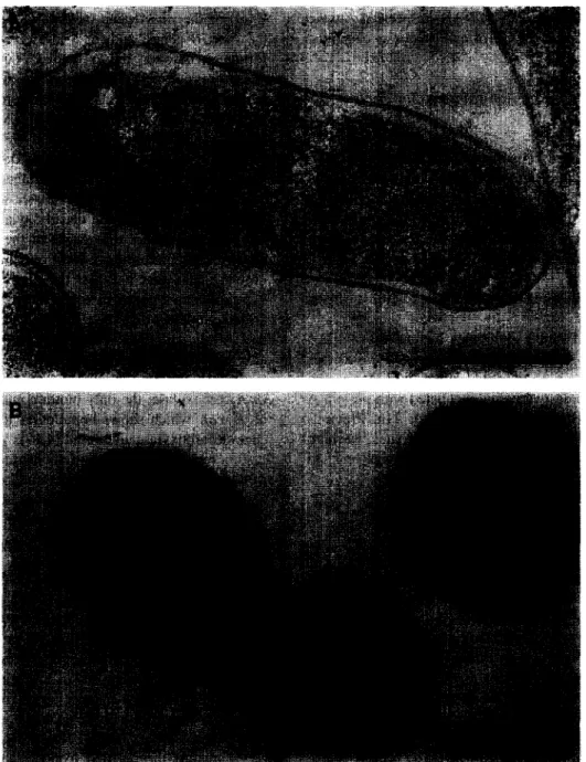
Fig. 1. Immunolabelling and transmission electron microscopy of oxaloacetate decarboxylase expressed in K. pneumoniae (A) or E. coli (B). The black spots indicate the immunogold-labelled enzyme complex. Bars represent 0.5 Wm.
34 M. Di Berardino et al. / FEMS Microbiology Letters 136 (19961 31-37
extract was measured as described [12]. The expres- sion of the ya-subunits was verified by reconstitut- ing oxaloacetate decarboxylation activity after incu- bation of the solubilised membrane vesicles of E.
coli DHSa/pSK-GA2 and E. coli
BL21(DE3)pLysS/pT7-BXY. The expression of the a, P and cup-subunits from E. coli BL21(DE3)pLysS containing the expression plasmid pT7-AL, pT7-BXY and pT7-AB, respectively, were visualised by SDS-PAGE of cell lysates as described
Bl.
2.3. Immunolabelling for transmission electron mi- croscopy
The harvested cells were subjected to high pres- sure freezing in cellulose capillary tubes [ 14,151. The capillary tubes, containing the frozen bacterial sus- pensions, were freeze-substituted in ethanol contain- ing 0.5% uranyl acetate. Samples were kept in the freeze-substitution medium for 9 h at -90°C 6 h at - 60°C 3 h at - 30°C 1 h at 0°C and subsequently Epon/Araldite embedded according to Hohenberg et al. [15].
Polyclonal rabbit antibodies against the purified oxaloacetate decarboxylase were diluted 1:400, ap- plied to the thin-sections and incubated for 2 h. The bound antibodies were then incubated with protein A coupled to 10 nm colloidal gold for 1 h. Previous masking of unspecific protein binding sites, washing and post-staining was performed according to Schwarz and Humbel [16].
3. Results and discussion
Since many years it is known that oxaloacetate decarboxylase from Klebsiella pneumoniae is a membrane-bound enzyme. This location of the pro- tein in the cytoplasmic membrane is shown by im- munogold labelling with antibody that reacted specif- ically with the peripheral membrane-bound u-sub- unit of the decarboxylase (Fig. 1A). Remarkably, almost all of the labelled enzyme is visualised on the surface of the cells. In contrast, after synthesis of the complete enzyme in E. coli (DHSa/pSK-GAB), a moderate part of the labelling is also located in the cytoplasmic volume (Fig. 1B). The specific oxaloac-
etate decarboxylase activity of the cytoplasmic frac- tion from the E. coli expression clone, however, was not higher than the specific activity of the corre- sponding fraction from K. pneumoniae cells (data not shown), indicating that the elevated cytoplasmic labelling probably derived from the isolated cu-sub- unit. A reason for the cytoplasmic labelling could therefore be the higher expression (on translational level) of the oadA gene in E. coli, as compared to the oadGB genes, and/or a less effective complex formation between the cr- and py-subunits due to overexpression and partial degradation of the mem- brane-bound components. Furthermore, it was ob- served that not all E. coli/pSK-GAB cells were labelled, or only weakly labelled by the antibody protein-A-gold complex, in contrast to the K. pneu-
moniae cells which all showed labelling on their cytoplasmic membrane (data not shown). This was indeed expected since the decarboxylase was not an essential enzyme for the growth of the E. coli cells, which may therefore have been overgrown by faster growing cells that had lost the plasmid during the late exponential phase, in which ampicillin is thought to be completely degraded. The fact that there was a part of E. coli/pSK-GAB cells without labelling also indicates that the cytoplasmic immunolabelling was not due to a possible cross-reaction with acetyl- CoA carboxylase, the only biotin-containing enzyme of E. coli which shows homologies to the oxaloac- etate decarboxylase a-subunit [4,19].
In a recent study [8], we demonstrated the expres- sion of the ya and cY/3-subunits in E. coli. We were able to purify ya-complexes from the membrane fraction of the cells, while it was not possible to isolate cYP-complexes neither from the cytoplasmic fraction nor from membrane vesicles. The purifica- tion protocol of the oxaloacetate decarboxylase sub- units involved cell disruption with a French Press. Thereby, we could not exclude that a loosely bound ap-complex dissociated during the preparation. Im- munolabelling and the subsequent analysis by elec- tron microscopy allowed us in this study to localise the expressed products under less harsh conditions. From various immunoblot analysis we knew that only the soluble a-subunit reacted with antibodies raised against the purified oxaloacetate decarboxyl- ase from K. pneumoniae (unpublished results) and therefore renounced to further expand this study for
M. Di Berardino et al. / FEMS Microbiology Letrem 136 f 19%) 31-37 35
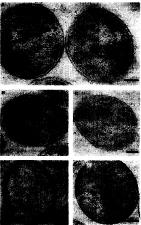
Fig. 2. Expression of the oxaloacetate decarboxylase subunits in E. coli and immunolabelling of the synthesised products. a-Subunit (A), cup-complex before (B) and after (C) induction with IPTG, ay-complex poorly (D) or highly expressed (EL Bars represent 0. I km.
36 M. Di Berardino et al. /FEMS Microbiology Letters 136 (1996) 31-37
the localisation of the separately expressed hy- drophobic p and y-subunits, for which no antibodies are available. In Fig. 2 it is clearly visible that ya-expressing E. coli cells show a part of the
labelling on the surface of the cells (D, E), while a@-expressing cells have the labelling spread ran- domly into the cytoplasmic compartment (B, C), confirming the biochemical data obtained in the pre- vious study [S]. E. coli/pT7-A cells overexpressing the a-subunit of the oxaloacetate decarboxylase complex show, as expected, a labelling only within the cytoplasm (Fig. 2A). The overexpression of this subunit as well as the overexpression of a possible a@-complex from clone E. coZi/pT7-AB using the strong T7 promoter did not lead to the formation of inclusion bodies (Fig. 2B, C), although considerable amounts of the a-subunit (50 mg (1 cell culture)-‘) could be purified [8].
Mutagenesis studies let us assume that the P-sub- unit contains a carboxybiotin binding pocket, which may interact with important residues of the mem- brane-spanning segments catalysing the Na+ translo- cation across the membrane (Di Berardino and Dim- roth, submitted). It was also shown that the overex- pression of the a-subunit led to the synthesis of only partially biotinylated enzyme [8]. Fig. 2B shows that the missing complex formation between the (Y and P-subunit is not due to low biotinylation of the a-subunit in ap-expressing E. coZi/pT7-AB cells, since even not induced cells, in which a complete biotinylation of the moderately expressed a-subunit should be ensured, have the labelling of the epitopic a-subunit located in the cytoplasm. From this result we conclude that there is no sufficient interaction between the (Y- and P-subunits to attain the forma- tion of a complex.
As with E. coli/pSK-GAB cells synthesising the complete decarboxylase, ya-expressing cells (E.
coli/pSK-GA2) also showed inhomogeneous la- belling of the expressed a-subunits due to the differ- ential loss of the dispensable expression plasmid. In cells in which a high expression of the genes was observed, the labelling was distributed over the whole cells, whereas cells expressing only minor amounts of this complex were mostly labelled on the cell surface. These results may indicate that, beside a possible higher expression of the a-subunit alone, the interactions between the y and a-subunit were
only weak and, consequently, that part of the com- plex was in a dissociated state. Moreover, it was not possible to reconstitute the decarboxylation activity of the enzyme by mixing solubilised membranes of P-expressing E. coli (BL2 l(DE3)pLysS/pT7-BXY
with the cytoplasm of E. coli/pSK-GA2 [8]. Since in a similar experiment with a membrane extract from E. coli/pSK-GA2 (containing subunits (Y and y) the catalytically active decarboxylase was recon- stituted, the labelling in the cytoplasm was only due to the a-subunit. From the previous study we must assume that the y-subunit is subjected to proteolytic digestion when expressed alone [8]. The degradation of a significant part of the y-subunit, which is re- quired to bind the a-subunit to the membrane, may be another reason for the substantial labelling of the expressed enzymes in the cytoplasm.
In summary, this study confirms the results of our biochemical experiments. Notable differences were observed by comparing the expression of the com- plete oxaloacetate decarboxylase in E. coli and K.
pneumoniae. The a-subunit in K. pneumoniae re- sides almost entirely at the cytoplasmic membrane, consistent with the existence of a membrane-bound oxaloacetate decarboxylase complex. Overexpression of the enzyme in E. coli, however, also produced moderate amounts of the a-subunit in the cytoplasm. Similarly, E. coli cells expressing the ycu-subunits in
elevated amounts show a significant proportion of the a-subunit in the cytoplasm, whereas under condi- tions of moderate expression most of the a-subunit is bound to the membrane. These results may indi- cate a preferred translation of the oadA gene within the oadGAB cluster and/or that the formation of membrane-bound oxaloacetate decarboxylase or of ~yy subcomplexes is partially impaired under high level expression in E. cob, perhaps because the y-subunit becomes partially degraded under these conditions.
Acknowledgements
Protein A-gold was kindly provided by Dr. Heinz Schwarz, MPI Tiibingen, Germany.
M. Di Berardino et al. / FEMS Microbiology Letters 136 f 1996) 31-37 37
References
[I] Dimroth, P. and Thomer, A. (1986) Citrate transport in Klebsiella pneumoniae. Biol. Chem. Hoppe-Seyler 367, 8 13- 823.
[2] Dimroth, P. and Thomer, A. (1990) Solubilization and recon- stitution of the Na’-dependent citrate carrier of Klebsielkr pneumonias. J. Biol. Chem. 265, 7221-7224.
[3] Pfenninger-Li, X.D. and Dimroth, P. (1992) NADH forma- tion by Na+-coupled reversed electron transfer in Klebsiella pneumoniar. Mol. Microbial. 6, l943- 1948.
.I
[61
[71
Bl
[91
Schwarz, E., Oesterhelt, D., Reinke, H., Beyreuther, K. and Dimroth, P. (1988) The sodium ion translocating oxaloac- etate decarboxylase of K. pneumoniae: sequence of the biotin-containing a-subunit and relationship to other biotin- containing enzymes. .I. Biol. Chem. 263, 9640-9645. LauSermair, E., Schwarz, E.. Oesterhelt, D., Reinke, H., Beyreuther, K. and Dimroth, P. (1989) The sodium ion translocating oxaloacetate decarboxylase of Klebsiella pneu- rnoniae: sequence of the integral membrane-bound subunits /3 and y, J. Biol. Chem. 264, 14710-14715.
Woehlke, G., LauRermair, E., Schwarz, E., Oesterhelt, D., Reinke, H., Beyreuther, K. and Dimroth, P. (1992) Sequence of the P-subunit of oxaloacetate decarboxylase from Kleb- tie//o pneumoniae: a correction of the C-terminal part. J. Biol. Chem. 267, 22804-22805.
Dimroth. P. and Thomer, A. (1983) Subunit composition of oxaloacetate decarboxylase and characterization of the LY chain as carboxyltransferase. Eur. J. Biochem. 137, 107- 112. Di Berardino, M. and Dimroth, P. (1995) Synthesis of the oxaloacetate decarboxylase Na+ pump and its individual subunits in Escherirhia co/i and analysis of their function. Eur. J. Biochem. 231. 790-801.
Van der Rest. M., Siewe, R.M., Abee, T., Schwarr, E., Oesterhelt. D. and Konings, W.N. (1992) Nucleotide se-
1101 [Ill
I121
[I31 I41 t151I161
[l711181
[l91quence and functional properties of a sodium-dependent cit- rate transport system from Hebsiella pneumoniae. J. Biol. Chem. 267, 897 l-8976.
Bott, M. and Dimroth, P. (1994) Klebsiello pneumonitre genes for citrate lyase and citrate lyase ligase: localization, sequencing, and expression. Mol. Microbial. 14, 347-356. Bott, M., Meyer, M. and Dimroth. P. (1995) Regulation of anaerobic citrate metabolism in K/eb.~ir//a pneumoniae. Mol. Microbial. 18. in press.
Dimroth, P. (1986) Preparation, characterization, and recon- stitution of oxaloacetate decarboxylase from K/ebsie//u orro- genes, a sodium pump. Methods Enzymol. 125, 530-540. Schlgger. H. and von Jagow. G. (1987) Tricine-sodium dodecyl sulfate-polyacrylamide gel electrophoresis for the separation of proteins in the range from I to 100 kDa. Anal. Biochem. 166, 368-379.
Miller, M. and Moor, H. (1984) Cryofixation of suspensions and tissues by propane-jet freezing and high-pressure freez- ing. Proc. 42nd Ann. Meet. Electron Microsc. Am., pp. 6-9. Hohenberg, H., Mannweiler, K. and Miiller. M. (1994) High-pressure freezing of cell suspensions in cellulose capil- lary tubes. J. Microsc. 175, 34-43.
Schwarz, H. and Humbel, B. (1989) Influence of fixatives and embedding media on immunolabeling of freeze-sub- stituted cells. Scanning Microsc. Suppl. 3. 57-64.
Dunn, J.J. and Studier, F.W. (1983) Complete nucleotide sequence of bacteriophage T7 DNA and the location of T7 genetic elements. J. Mol. Biol. 219, 45-59.
Studier, F.W. and Moffatt, B.A. (1986) Use of bacteriophage T7 RNA polymerase to direct selective high-level expression of cloned genes. J. Mol. Biol. 189, 113-130.
Muramatsu, S. and Mizuno, T. (1989) Nucleotide sequence of the ,fabE gene and flanking regions containing a bent DNA sequence of Escherichia co/i. Nucleic Acids Res. 17, 3982.