REVIEW
Microextraction techniques combined with capillary
electrophoresis in bioanalysis
Isabelle Kohler&Julie Schappler&Serge Rudaz
Received: 4 July 2012 / Revised: 14 August 2012 / Accepted: 19 August 2012 / Published online: 11 September 2012 # Springer-Verlag 2012
Abstract Over the past two decades, many environmen-tally sustainable sample-preparation techniques have been proposed, with the objective of reducing the use of toxic organic solvents or substituting these with environmentally friendly alternatives. Microextraction techniques (MEs), in which only a small amount of organic solvent is used, have several advantages, includ-ing reduced sample volume, analysis time, and operat-ing costs. Thus, MEs are well adapted in bioanalysis, in which sample preparation is mandatory because of the complexity of a sample that is available in small
quan-tities (mL or even μL only). Capillary electrophoresis
(CE) is a powerful and efficient separation technique in which no organic solvents are required for analysis. Combination of CE with MEs is regarded as a very attractive environmentally sustainable analytical tool, and numerous applications have been reported over the last few decades for bioanalysis of low-molecular-weight compounds or for peptide analysis. In this paper we review the use of MEs combined with CE in bio-analysis. The review is divided into two sections: liquid and solid-based MEs. A brief practical and theoretical description of each ME is given, and the techniques are illustrated by relevant applications.
Keywords Bioanalysis . Capillary electrophoresis . Environmentally sustainable chemistry . Green chemistry . Microextraction . Sample preparation
Abbreviations
μ-SLM Micro-supported liquid membrane
BGE Background electrolyte
C4D Capacitively-coupled contactless
conductivity detector
CB-ICE Chip-based immunoaffinity capillary
electrophoresis
CE Capillary electrophoresis
CME Centrifuge microextraction
CM-LPME Carrier-mediated liquid-phase
microextraction
CM-SDME Carrier-mediated single-drop
microextraction
CZE Capillary zone electrophoresis
DEHP bis(2-Ethylhexyl) phosphate
DI-SPME Direct-immersion solid-phase
microextraction
DLLME Dispersive liquid–liquid microextraction
DMD-LPME Droplet–membrane–droplet liquid-phase
microextraction
DSDME Directly suspended droplet microextraction
EK Electrokinetic injection
EME Electro membrane extraction
ENB 1-Ethyl-2-nitrobenzene
ESI Electrospray ionization
FASI Field-amplified sample injection
GC Gas chromatography
HS Headspace
i-PrOH Isopropanol
ILBE In-line back-extraction
IT Ion trap
LC Liquid chromatography
LIF Laser-induced fluorescence
I. Kohler
:
J. Schappler:
S. Rudaz (*)School of Pharmaceutical Sciences,
University of Geneva, University of Lausanne, Bd d’Yvoy 20,
1211 Geneva 4, Switzerland e-mail: [email protected]
I. Kohler
:
J. Schappler:
S. RudazSwiss Centre for Applied Human Toxicology, University of Geneva, CMU,
Rue Michel-Servet 1, 1211 Geneva 4, Switzerland
Anal Bioanal Chem (2013) 405:125–141 DOI 10.1007/s00216-012-6367-y
LLE Liquid–liquid extraction
LLLME Liquid–liquid–liquid microextraction
LOD Limit of detection
LVSS Large-volume sample stacking
MCE Microchip capillary electrophoresis
ME Microextraction
MeCN Acetonitrile
MEKC Micellar electrokinetic chromatography
MeOH Methanol
MEPS Microextraction by packed sorbent
MIP Molecularly imprinted polymer
NACE Non-aqueous capillary electrophoresis
NPOE 2-Nitrophenyl octyl ether
NSAIDs Non-steroidal anti-inflammatory drugs
OLBE On-line back-extraction
PAH Polycyclic aromatic hydrocarbons
PF Preconcentration factor
PP Protein precipitation
RAM Restricted-access material
SBSE Stir-bar-sorptive extraction
SL Sheath liquid
SPE Solid-phase extraction
THF Tetrahydrofuran
tITP Transient isotachophoresis
TOF Time-of-flight
UHPLC Ultra-high-pressure liquid chromatography
USAEME Ultrasound-assisted emulsification
microextraction
Introduction
The overall analytical procedure includes several consecu-tive steps—sampling, sample storage, sample preparation, separation of target analytes, detection, and data treatment. For analysis of complex samples and matrices, for example biological, environmental, or food analysis, the sample preparation is of utmost importance for obtaining the analy-tes of interest in a suitable injection solution able to provide reliable and accurate results. Sample preparation has sub-stantial objectives before sample injection, including: 1. reducing or eliminating matrix interferents or undesired
endogenous compounds;
2. increasing selectivity for targeted analyte(s);
3. preconcentrating the sample to enhance sensitivity; and 4. stabilizing the sample by reconstituting it in an inert
solvent.
Although great improvements have been made in the development of fast separation techniques, sample pretreat-ment remains the most time-consuming step, accounting for
ca two thirds of the entire analytical procedure [1]. In
addition, because of the lack of automation of several offline
procedures, sample preparation is also regarded as a primary source of analytical errors that can significantly affect the
throughput [2].
Sample preparation can be based either on selective methods, e.g., the widely used solid-phase extraction
(SPE) and liquid–liquid extraction (LLE), or non-selective
methods, e.g., using membrane techniques or protein pre-cipitation (PP). A common feature of all these conventional sample-preparation techniques is the relatively high con-sumption of solvents that are environmentally hazardous and health risks for humans. The advent of the concept of “green chemistry” at the beginning of the 1990s emphasized the need for non-toxic and environmentally friendly analyt-ical procedures. The concept also promoted the use of environmentally sustainable sample-preparation methods with the development of solvent-free or miniaturized
extrac-tion methods [3,4]. Different approaches can be envisaged
when developing environmentally sustainable sample preparation:
1. solventless procedures [5,6];
2. substitution of organic solvents with less-toxic alterna-tives, for example use of supercritical-fluid extraction, cloud-point extraction, subcritical water extraction, or
extraction with ionic liquids [7,8]; or
3. use of microextraction techniques (MEs), in which min-iaturization of the extraction procedure not only mini-mizes the use of organic solvents but also the sample volume required.
MEs are defined as non-exhaustive procedures that use very small volumes of the extracting phase and for which the volume of sample is relatively large compared with that
of the extracting phase [9]. MEs reduce or eliminate the
consumption of solvents while simultaneously reducing
sample volume, analysis time, and operating costs [10].
Many techniques have been developed over the last few decades for a variety of applications, i.e., in environmental
analysis (pesticides, hormones) [11–13], food analysis [11,
13–15], and bioanalysis [16,17] for clinical, toxicological
and forensic purposes [18,19] or doping analysis [20]. In
bioanalysis, often only small amounts of the sample are available, typically in the mL range for urine and in the μL range for serum or plasma or alternative matrices, for example sweat, saliva, or tears. Because of the complexity of theses matrices and the low concentrations of the target analytes compared with endogenous interferents, sample preparation is mandatory, and MEs are particularly well adapted for this purpose.
A variety of analytical techniques, including separation-based approaches, can be implemented in combination with MEs in bioanalysis. Non-polar and volatile compounds are conveniently analyzed by gas chromatography (GC), where-as liquid chromatography (LC), including
ultra-high-pressure liquid chromatography (UHPLC), is extensively used in bioanalysis for both quantitative and qualitative purposes, because of its wide applicability to a large number of compounds with different physicochemical properties. Capillary electrophoresis (CE) is another powerful separa-tion technique that is often used in bioanalysis, because of its high separation efficiency. As very small amounts of (μL range) or no organic solvents are required for CE analysis, its use in combination with ME techniques is regarded an attractive, environmentally sustainable analytical tool. Extracts can be directly injected for analysis, or evaporated and reconstituted in a very small volume. Because a few nL of sample is injected in CE, very high preconcentration factors (PFs) can be achieved, enhancing the overall sensi-tivity, which is a disadvantage of the capillary format.
Applications of ME techniques before to CE analysis have been reported over the past few decades in bioanalysis of low-molecular-weight compounds or small peptides. In this paper we review the MEs used in bioanalysis and combined with CE. It is divided into two sections: liquid and solid-based MEs. MEs are classified according to their extraction principle and improvement of extraction perfor-mance. A brief description and the theoretical concepts of each ME technique are introduced and discussed, and illus-trated by relevant applications.
Liquid-based microextraction techniques
LLE, which involves partition of analytes between an aque-ous sample and water-immiscible organic solvent, has been widely used in bioanalysis because of its simplicity and ease of implementation. LLE suffers from major drawbacks, for example emulsion formation at the interface of the immis-cible phases, lack of selectivity (co-extraction of endoge-nous interferents), lack of automation, and use of large sample volumes and large amounts of toxic organic solvents that are environmentally harmful (up to 10 mL per mL of
sample) [10,16,17,21].
New methods based on the LLE principle or with original set-ups have been developed during the last two decades to overcome these drawbacks. Miniaturization of LLE has led to several new liquid-based ME techniques in which the total volume of organic solvent required has been reduced to the sub-mL level.
In 1996, Cantwell and co-workers [22] and Dasgupta and
co-workers [23] were the first to propose the use of a solvent
drop in theμL range as extractant, laying the foundation for
liquid-phase microextraction (LPME). Cantwell and
co-workers used an 8-μL drop of n-octane held at the end of a
Teflon rod to extract 4-methylacetone from water [22],
where-as Dwhere-asgupta and co-workers extracted sodium dodecyl sulfate
(SDS) from a water sample with only 1.3μL chloroform [23].
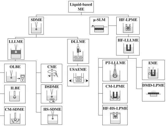
A variety of liquid MEs based on LPME were subsequently developed, leading to a large selection of miniaturized techni-ques that are still evolving. A schematic diagram of these techniques, based on their principle of extraction, is given in
Fig.1. All of the bioanalytical applications that use
LPME-based techniques before CE are listed in Table1.
Liquid-based ME techniques are derived either from single-drop microextraction (SDME), in which a single drop of water-immiscible solvent suspended from the tip of a syringe is immersed in the aqueous sample, or hollow-fiber liquid-phase microextraction (HF-LPME), in which a hollow polymeric fiber is used as a support for the acceptor (aqueous or organic) phase.
Single-drop microextraction (SDME)
SDME was introduced in 1997 by Jeannot et al. and He et
al. [24,25]. In the first study, a 1-μL drop of n-octane was
suspended in a stirred aqueous sample from the tip of a microsyringe needle. After a few minutes, the drop was retracted into the needle and injected directly for gas
chro-matographic (GC) analysis [24]. He and Lee used the same
method with a 1-μL drop of toluene that was immersed in the aqueous sample for 15 min before retraction and
injec-tion [25]. SDME uses very small amounts of organic
ex-traction solvents, which enables important PFs to be achieved. The main problems with this method are lack of droplet stability at high stirring speeds and the high manual dexterity required. Moreover, SDME is only suitable for relatively non-polar analytes and suffers from low recovery and repeatability. Therefore, SMDE was regarded as be not suitable for biological matrices, in which an extra filtration
step is necessary [2,11,26]. Many derived techniques based
on SDME were thus proposed (Fig. 1), including liquid–
liquid–liquid microextraction (LLLME) or dispersive liq-uid–liquid microextraction (DLLME), and used in combi-nation with CE to obtain sufficient selectivity, sensitivity, and repeatability in bioanalysis.
Liquid-liquid-liquid microextraction (LLLME)
LLLME, also referred to as LPME by back-extraction, was
first introduced in 1998 by Ma and Cantwell [27] and is
particularly suitable for water-soluble analytes, for example ionizable compounds. In LLLME, the targeted analytes are first extracted from the aqueous sample (donor) into a water-immiscible organic phase (acceptor I) and then back-extracted into a separate aqueous phase (acceptor II). The transfer occurs by manipulating the pH in the donor and acceptor phases. LLLME is particularly suitable for CE analysis, because of the direct injection of the aqueous acceptor phase into the system.
Extraction improvement
The particular configuration of CE enables on-line or in-line back-extraction to be performed. On-line back-extraction (OLBE) with field-amplified sample injection (FASI) was de-veloped for analysis of cocaine and thebaine in urine samples
[28]. Eight milliliters of urine were placed in a vial, and a 2-μL
drop of chloroform was generated at the tip of a syringe and immersed in the sample. After extraction for 5 min, with stirring, the chloroform drop was retracted and transferred to
another vial that was sealed with 40 μL acidified water for
back-extraction. OLBE was performed by carefully immersing the capillary tip in the water plug. During high-voltage appli-cation, FASI occurred, and charged analytes moved rapidly from the organic phase to the capillary, stacked at the boundary with the high-conductivity background electrolyte (BGE). MeCN (20 %, v/v) was also added to the water plug to reduce conductivity, thus substantially enhancing sensitivity.
In-line back-extraction (ILBE) was performed with a
wa-ter–organic drop hanging at the tip of the capillary [29]. In this
case, the capillary was filled with acidic BGE (acceptor phase), and then 13 nL octanol was injected. After injection, the tip of the capillary was immersed in the urine sample, which had previously been made alkaline, and a backpressure was applied from the outlet to the inlet, forming a small drop of the acceptor phase that was covered with a thin organic layer hanging at the tip. After extraction, the acceptor phase was injected into CE. This configuration is well adapted to
saline samples, for example urine; however, it is hardly
achievable on a commercial CE instrument [26].
To enhance the transfer of analytes between the sample and organic phase, use of carriers with LLLME was envisaged by Choi et al. in 2011, in so-called carrier-mediated single-drop
microextraction (CM-SDME) [30]. Amino acids were extracted
from urine by use of nonane-1-sulfonic acid as carrier. Addition of this negatively charged carrier at a low pH with positively charged amino acids enabled the formation of a neutral ion pair that could be extracted into the organic phase. Octanol was chosen as the extracting phase because of its capacity to form hydrogen bonds with the ion pair. CM-SDME enabled 120-fold sensitivity improvement compared with CZE without SDME. Drop stability improvement
Although the above-mentioned LLLME technique [28] has
been shown to be fast, simple, inexpensive, and sensitive, it clearly suffers from drop instability. Therefore, in the same year, Fang et al. developed centrifuge microextraction (CME), which combines desalting, preconcentration, and removal of macro-molecular contaminants and other interfering components in a
single step [31]. After pH adjustment and addition of NaCl for
the salting-out effect, 1 mL urine was mixed with 50μL toluene
and centrifuged at 10,000 rpm for 10 min. A lower-density water-immiscible solvent was chosen so the acceptor phase was at the top of the sample. During centrifugation, the centrif-ugal force applied by the rotor led to sedimentation of
Liquid-based ME SDME HF-LPME LLLME DLLME OLBE ILBE CM-SDME USAEME µ-SLM HF-LLLME EME DMD-LPME PT-LLLME CM-LPME HF-HS-LPME CME DSDME HS-SDME + -
Fig. 1 Classification of liquid-based microextractions used in combination with CE. Light gray, aqueous phase; dark gray, organic phase; cross hatched, membrane or fiber
T able 1 Liquid-based microextraction techniques used in combination with CE in bioanalysis Analyte(s) Matrix ME Sample volume Or ganic solvent(s) Or ganic solvent(s) volume Analysis PF/LOD Ref. Cocaine Urine OLBE 8 m L CHCl 3 4 μ L F ASI –CZE –UV 120 –340/2 –10 ng mL − 1 [ 28 ] Thebaine Basic amines Urine ILBE 1.8 mL n -octanol 13 nL CZE –UV 1,000/0.5 ng mL − 1 [ 29 ] Alkaloids Urine LLLME 4 m L n -octanol 350 μ L MEKC –UV 1,583 –3,556/0.2 –1.5 ng mL − 1 [ 128 ] Cyanide Urine, saliva HS-SDME 4.5 mL –– CZE –UV after derivatization 58/0.08 μ mol/L [ 37 ] Doping agents Horse urine HS-SDME 10 mL CHCl 3 –MeOH 90:10 5 μ LO T –CEC –UV 38 –102/0.9 –17.6 ng mL − 1 [ 38 ] T oxic drugs Urine DLLME 4 m L i-PrOH –CH 2 Cl 2 70:30 2 m L CZE –ESI –TO F M S 7 5– 100/0.1 –10 ng mL − 1 [ 40 ] 20 μ L( i-PrOH, SL) Serotonin Urine USAEME 5 m L Ethyl acetate 500 μ L CZE –UV 360/7.9 nmol L − 1and 13.3 μ mol L − 1 [ 43 ] Creatinine Ephedrine derivatives Urine, serum CME 1 m L (urine) T oluene 50 μ L F ASI –CZE –UV 3,800/0.15 –0.25 ng mL − 1 [ 31 ] 20 μ L (serum) Steroid hormones Urine CME 1.3 mL Cyclohexane 100 μ L MEKC –UV 500/5 –15 ng mL − 1 [ 32 ] Alkaloids Urine DSDME 3.5 mL n -octanol < 6 0 μ L CZE –UV 231 –524/8.1 –14.1 ng mL − 1 [ 34 ] Amino acids Urine CM-SDME 20 mL n -octanol 24 nL CZE –UV 120/70 –500 nmol L − 1 [ 30 ] NSAIDs Urine μ -SLM 4 m L Dihexyl ether and MeOH n.d. MEKC –UV 1.2 –1.7 μ gm L − 1 [ 129 ] Nitroimidazoles Pig liver tissues μ -SLM 0.5-5 g 2-Phenylpropane n.d. MEKC –UV 0.01 –0.99 μ gm L − 1 [ 47 ] Bambuterol Plasma μ -SLM 500 μ L MeOH n.d. CZE –UV n.d. [ 130 ] Bambuterol Plasma μ -SLM 350 μ L MeOH n.d. CZE –UV 14 [ 46 ] Amino acids Serum, plasma μ -SLM 12.5 μ L ENB –DEHP 2.5 μ L CZE –C 4D 0.75 –2.5 μ mol L − 1 [ 48 ] Methamphetamine Urine, serum HF-LLLME 2.5 mL 1-octanol n.d. CZE –UV 75/5 ng mL − 1 [ 49 ] Org anomercury Hair HF-LLLME 12 mL Bromobenzene n.d. L VSS –CZE –UV 2,610 –4,580/0.03 –0.14 μ gm L − 1 [ 57 ] NSAIDs Urine HF-LLLME 2.5 mL Dihexyl ether 25 μ L CZE –UV 75 –100/1 ng mL − 1 [ 52 ] Methamphetamine Urine, plasma HF-LLLME 4.0 mL (urine) n -octanol n.d. CZE –UV 30 –125 [ 50 ] Naproxen 2.5 mL (plasma) Citalopram Plasma HF-LLLME 1 m L Hexyl ether n.d. CZE –UV 25 –30/5 and 5.5 ng mL − 1 [ 54 ] Desmethylcitalopram Mianserin Plasma HF-LLLME 0.5 mL Di-n -hexyl ether n.d. CZE –UV 4 n g m L − 1 [ 131 ] Citalopram Plasma HF-LLLME 1 m L Dodecyl acetate n.d. CZE –UV 19 –31/1.4 –3.4 ng mL − 1 [ 53 ] Desmethylcitalopram Methamphetamine Urine, plasma HF-LLLME 2.5 mL Hexyl ether n.d. CZE –UV 95 –145/2 ng mL − 1 [ 51 ] Citalopram Whole blood Basic drugs Plasma HF-LLLME 250 μ L Dihexyl ether and MeOH n.d. CZE –UV <20 ng mL − 1 [ 55 ] Antidepressants Human milk HF-LLLME 500 μ L Polyphenylmethylsiloxane n.d. CZE –UV 14 –23/<50 ng mL − 1 [ 56 ] Rosiglitazone Plasma, urine HF-LLLME 5.0 mL (urine) Dihexyl ether n.d. CZE –UV 280/2.83 ng mL − 1 [ 132 ] 2.5 mL (plasma) T ricyclic antidepressants Plasma HF-LLLME 250 μ L Dihexyl ether 10 μ L CZE –UV 0.02 –0.03 μ mol L − 1 [ 133 ]
T able 1 (continued) Analyte(s) Matrix ME Sample volume Or ganic solvent(s) Or ganic solvent(s) volume Analysis PF/LOD Ref. Cyanide Urine, saliva H F -H S -LPME 4.5 mL –– CZE –UV 0.01 μ mol L − 1 [ 134 ] Polar drugs Plasma, urine CM-LPME 100 μ L 1-octanol n.d. CZE –UV 10/< 1 μ gm L − 1 [ 59 ] Basic drugs Plasma CM-LPME 50 μ L n -octanol n.d. CZE –UV <0.5 μ gm L − 1 [ 60 ] Basic drugs Plasma, urine EME 100 μ L NPOE ~15 μ L CZE –UV n.d. [ 62 ] Basic drugs Plasma, whole blood EME 500 μ L 1-ethyl-2-benzene n.d. CZE –UV <0.5 μ gm L − 1 [ 64 ] Angiotensins Plasma EME 500 μ L 1-octanol –DEHP n.d. CZE –UV <2.5 ng mL − 1 [ 66 ] Basic drugs Plasma, urine EME 1 m L 1-isopropyl-4- nitrobenzene n.d. CZE –UV Up to 22 [ 65 ] Human milk Amino acids Serum, plasma EME 36 μ L ENB –DEHP n.d. CZE –C 4D 0.15 –10 μ mol L − 1 [ 67 ] Whole blood, Lithium Urine EME 35 μ L 1-octanol n.d. CZE –C 4D 9 nmol L − 1 [ 68 ] Serum, plasma Whole blood Basic drugs Urine, serum EME 875 μ L (urine) ENB/DEHP n.d. CZE –UV 1– 4n g m L − 1(bases) [ 135 ] Amino acids 145 μ L (serum) 0.6 –3 μ mol L − 1(acids) Amlodipine Plasma, urine EME 1 m L (serum) NPOE n.d. CZE –UV Up to 150/3 ng mL − 1 [ 69 ] 1.5 mL (urine) T rimipramine Plasma, urine EME 1.25 mL (serum) NPOE n.d. CZE –UV 120 –149/8 –10 ng mL − 1 [ 70 ] 2.5 mL (urine) Basic drugs Urine EME 10 μ L NPOE 1 μ L CZE –UV < 1 μ gm L − 1 [ 71 ] Inorganic and or ganic mercury Hair PT -LLLME 0.25 g T oluene 200 μ L (T oluene) L VSS –CZE –UV 12,138 [ 58 ] MeCN Acetone 225 μ L (MeOH) Basic and acidic drugs Urine DMD-LPME 15 μ L 1-octanol 1 μ L MCE –LIF 2 [ 72 ] T ramadol, paracetamol and metabolites Urine DMD-LPME 4 μ L 1-octanol ~1 μ L MCE –ESI –MS 2/9.3 nmol L − 1for model compound [ 73 ] n.d., not defined LOD is determined at a signal-to-noise ratio of 3
macromolecules whereas diffusion enabled transfer of the tar-geted compounds to the acceptor phase. The supernatant was
directly injected in CE–UV with FASI, leading to a limit of
detection (LOD) of 0.15 ngmL−1. CME has also been used for
analysis of steroids (e.g., testosterone and progesterone) in urine
by micellar electrokinetic chromatography (MEKC) [32].
In 2006, Yangcheng et al. proposed directly suspended droplet microextraction (DSDME) to further improve drop stability. Here, the microdroplet of solvent is suspended at the
top in the center of the aqueous sample before sampling [33]. A
symmetrical rotated flow field is created by a stirring bar that is placed on the bottom of the cylindrical sample cell to ensure the droplet suspension. This rotation also intensifies transfer of analytes to the inside of the droplet. DSDME has been com-bined with single-drop back-extraction and CE for analysis of
alkaloids in urine samples [34]. After the first extraction in a
large microdrop (approx. 60μL) of n-octanol, alkaloids were
back-extracted in a 1-μL aqueous drop that was immersed in the organic phase droplet. PFs of greater than 500 were achieved with lower solvent consumption and shorter extrac-tion time than those of LLLME.
Introduced by Theis et al. [35] in 2001, headspace
single-drop microextraction (HS-SDME) has excellent extraction and preconcentration performance for volatile compounds. With a suspended drop in the gaseous phase (headspace), this method enables rapid stirring of an aqueous sample, for a shorter anal-ysis time, without affecting drop stability. Moreover,
non-volatile matrix interferences are reduced or eliminated [19,26,
36]. HS-SDME has also been used in combination with CE
analysis with in-drop derivatization. Free cyanide was solvent-lessly extracted from smoker and non-smoker urine and saliva
[37], using water to extract volatile and water-soluble
com-pounds. An aqueous 5-μL drop containing Ni(II)–NH3as
de-rivatization agent for CE analysis was used for the extraction. In
the basic acceptor phase, cyanide reacted with Ni2+to form a
stable Ni(CN4)2− complex analyzed by CE–UV at 257 nm.
Water-based HS-SDME was very selective, despite the rather universal detection wavelength, because the non-volatile inter-ferents remained unaffected in the sample. HS-SDME with a chloroform–MeOH mixture as extracting drop has also been used to extract seven toxic compounds from horse urine samples at room temperature, before analysis by open tubular capillary
electrochromatography (OT-CEC) [38].
Dispersive liquid–liquid microextraction (DLLME) In DLLME, which was first introduced by Rezaee et al. in 2006, the extracting solvent is mixed with a dispersing solvent that is miscible both with the former and with the
aqueous sample [39]. The mixture is rapidly injected into
the sample with a syringe, producing high turbulence that leads to the formation of tiny droplets. Because of the large surface area between the extracting droplets and sample, the
extraction time is drastically reduced. After centrifugation, the sedimented phase at the bottom of the tube is collected and either injected directly or evaporated to dryness before reconstitution and injection.
DLLME combined with CE and time-of-flight mass spectrometry (TOF/MS) was used for qualitative
toxicolog-ical screening of urine samples [40]. An experimental
de-sign strategy was used to increase the extraction efficiency.
CH2Cl2and i-PrOH were selected as extracting and
dispers-ing solvents, respectively, with a total volume of 2 mL. Because of a high PF (more than 130) and the high sensi-tivity and selecsensi-tivity of CE–TOF/MS, LODs down to the
sub-ngmL−1 range were obtained for more than 30 toxic
basic compounds and their main metabolites and confirmed by real case analysis.
Extraction improvement
One of the main disadvantages of DLLME is the need to use a dispersing solvent to create an emulsion, which can reduce the partition coefficient of the analytes in the extracting phase and increase total solvent consumption. The dispersing solvent can be substituted by using ultrasound to achieve
ultrasound-assisted emulsification-microextraction (USAEME) [41].
Based on previous work on ultrasound-assisted sample
prepa-ration [42], USAEME is beneficial for promoting emulsion
formation, extending the contact surface between both phases, and reversing the potential coalescence effect. Increasing the
temperature also enables efficient and fast extraction [41]. A
serial USAEME procedure was developed for analysis of
cre-atinine and serotonin in urine samples [43]. Five hundred
microliters of ethyl acetate was added to 5 mL urine and the sample was immersed in an ultrasonic bath for 5 min at 40 Hz. The emulsion was centrifuged, and the organic supernatant
mixed with 25 μL 0.1 molL−1 HCl. Back-extraction was
performed by 3-min ultrasonication at 40 Hz. After centrifuga-tion, the sedimented acceptor phase was collected and injected by use of a pH-mediated stacking procedure. With serial USAEME and sample stacking, a PF of 360 was obtained for serotonin.
Hollow-fiber-based liquid-phase microextraction (HF-LPME)
The chemical principle of HF-LPME is derived from supported-liquid membrane (SLM) extraction, which was
previously developed by Jönsson and coworkers [44]. In
SLM, analytes are extracted through a flat porous polymeric membrane sheet with continuous sample pumping. SLM was first miniaturized (“μ-SLM”) in 1996 by Jönsson and
coworkers [45] and applied to the analysis of bambuterol in
SLM–CZE [46]. An in-line SLM approach with a Teflon micromembrane unit glued to a plastic microtube integrated in the CE vial was developed by Nozal et al. for analysis of nitroimidazoles in pig liver tissues homogenized in water
[47]. Very recently, Kuban and Bocek proposed on-line
μ-SLM-CE with a planar SLM screwed between two PTFE
blocks to determine amino acids in plasma or serum [48].
This home-built set-up did not require additional pumps.
In contrast to μ-SLM, HF-LPME is performed without
any pumping device. It was introduced in 1999 by
Pedersen-Bjergaard and Rasmussen [49]. In HF-LPME, the extracting
phase is placed inside the lumen of a porous polypropylene
fiber (pore size 0.2 μm) of minimal dimensions used in
different configurations, e.g., U-shaped, rod-like, 96-well,
or directly connected to a microsyringe [21,44]. The
poly-meric fiber, which is compatible with a broad range of organic solvents, enables use of a larger extraction volume compared with SDME and acts as a physical barrier between phases, avoiding undesirable emulsions and enhancing
cleanup efficiency [19]. HF-LPME can be performed in
either two or three-phase systems. In three-phase systems,
referred to as hollow-fiber-based liquid–liquid–liquid
microextraction (HF-LLLME), supported liquid membrane microextraction (SLMME), or, rather improperly, LPME, the analytes are extracted from the aqueous sample through the organic film (a few microliters) that is present in the pores of the aqueous acceptor phase in the lumen of the hollow fiber. HF-LLLME is well suited to extraction of polar or ionizable compounds and particularly suitable for CE analysis.
Hollow-fiber-based liquid–liquid–liquid microextraction (HF-LLLME)
Pedersen-Bjergaard and Rasmussen with co-workers have developed many applications of HF-LLLME in combination
with CE. Methamphetamine [49–51], non-steroidal
anti-inflammatory drugs (NSAIDs) [52], naproxen [50],
citalo-pram and metabolites [51, 53, 54], and a variety of basic
drugs [55] have been successfully extracted from urine and
serum or plasma. HF-LLLME has also been used to extract
antidepressants from human milk [56]. Human milk is
char-acterized by high protein, fat, and carbohydrate content, which can affect the recovery and repeatability of the ex-traction procedure. Because of interaction of antidepressants with fat and proteins, recovery from milk was lower than from water. Thus, PP was implemented, with addition of hydrochloric acid to the sample before centrifugation and extraction to remove the fat-rich layer and release unbound
drugs, leading to recovery of 50–70 %. Li et al. used
HF-LLLME for extraction of organomercury from human hair,
with the fiber pores impregnated with bromobenzene [57].
Hair samples were first rinsed with detergent and acetone,
and air-dried before cutting and leaching. The leached solu-tion was centrifuged, and the supernatant collected for
HF-LLLME. An aqueous acceptor phase containingL-cysteine
for organomercury complexation was injected with large volume sample stacking (LVSS), enabling enrichment of more than 4,000.
Extraction improvement
A new LPME-based technique referred to as phase-transfer-based liquid–liquid–liquid microextraction (PT-LLLME) was developed in 2011 by Li et al. for extraction of organic
and inorganic mercury from hair [58]. In this homemade
set-up, a porous, hydrophilic, nylon-membrane-supported
ex-traction tip was built and used with 15μL aqueous acceptor
phase. MeCN and dodecylamine were added to the sample before extraction as intermediate solvent and complexing reagent, respectively. MeCN improved the dispersion of water-immiscible dodecylamine in the aqueous sample to ensure maximum contact with the mercury. Compared with mercury extraction by HF-LLLME, PT-LLLME provided the potential for simultaneous speciation of inorganic and organic mercury and improved the sensitivity with enhanced extraction efficiency.
Use of carriers, also used in SDME (section “Liquid–
liquid–liquid microextraction (LLLME)”, subsection “Ex-traction improvement”), was first introduced in 2003 by Ho et al. in the so-called carrier-mediated liquid-phase micro-extraction (CM-LPME) to enhance micro-extraction recovery of
polar or ionic analytes [59]. The carriers form lipophilic
complexes with the target analytes, promoting the transport of the analytes through the organic membrane. Polar basic compounds could be extracted from plasma and urine sam-ples, through the 1-octanol layer into the aqueous acceptor phase, with good recovery, after addition of sodium octa-noate (ion-pair reagent) to the sample. The pH of the sample had to be adjusted so the analytes and carrier were ionized in such a way to enable the formation of ion-pair complexes that could diffuse through the membrane. Numerous car-riers, including organic borates, phosphates, sulfates, and carboxylic acids, were investigated at different concentra-tions with a special emphasis on their compatibility with
plasma samples [60]. Bromothymol blue (sulfate carrier)
resulted in the best recovery from the plasma samples. Interestingly, recovery was enhanced when sodium sulfate was added to the sample to reduce matrix effects.
In 2005, Lee and coworkers developed hollow-fiber-protected headspace liquid-phase microextraction (HF-HS-LPME), in which the hollow fiber protected and held the
extractant droplet in the headspace [61]. The surface area
between the organic and acceptor phases was dramatically
enhanced compared with HS-SDME (section “Liquid–
stability improvement”), increasing the extraction efficiency. HS-HF-LPME was used to extract free cyanide from urine and saliva with a simultaneous in-fiber derivatization to form a
stable Ni(CN)42−complex. Lower LODs (0.01μmolL−1
ver-sus 0.08μmolL−1) and similar recovery (90–105 %) were
obtained compared with HS-SDME [37]. HF-HS-LPME is an
effective alternative to HS-SDME for quantitative analysis of volatile compounds.
Throughput improvement
In 2006, Pedersen-Bjergaard and Rasmussen proposed use of an electrically-driven force to aid extraction of charged
compounds and to speed HF-LLLME [62,63]. This
tech-nique was first referred to as“electro membrane isolation”
(EMI) and was later termed electro membrane extraction (EME). Two platinum electrodes are placed in the sample solution and in the aqueous acceptor phase in the lumen of the fiber. A potential (typically 300 V) is applied, and charged analytes migrate through the membrane toward the oppositely charged electrode in the acceptor solution in less than 5 min. Interesting clean-up, enrichment, and isola-tion of basic compounds with 2-nitrophenyl octyl ether (NPOE) as organic solvent were observed with high extrac-tion recovery (>70 %) from plasma and urine.
Pedersen-Bjergaard, Rasmussen, and co-workers showed the benefits of EME for analysis of basic drugs (e.g., anal-gesics, antidepressants, and antiepileptics) in plasma and
whole blood [64], or urine and human milk [65]. They also
evaluated the potential of EME as a fast and effective extraction technique for peptides (angiotensin as the model
peptide) in plasma [66].
Other groups evaluated EME for a variety of
applica-tions, for example extraction of amino acids [67], lithium
[68], amlodipine enantiomers [69], and trimipramine
enan-tiomers [70] from urine, plasma or serum, or whole blood.
A miniaturized form of EME, termed drop-to-drop LPME, has been proposed for extraction of basic drugs from
urine and plasma [71]. A small well with a volume of 15μL
was pressed into 5-cm2 aluminium foil connected to the
power supply’s positive outlet. The well, containing 10 μL sample, was covered with the membrane and a 10-μL ac-ceptor droplet. Recovery of 33–47 % was obtained with excellent clean-up, short extraction time, and very low sol-vent and sample consumption.
In 2010, this miniaturization was built upon with the
de-velopment of on-line droplet–membrane–droplet LPME
(DMD-LPME) [72]. The extraction set-up was the same as
in Ref. [71] and was combined on-line with microchip
capil-lary electrophoresis (MCE) with fluorescence detection. DMD-LPME was directly compatible with MCE because of the very low acceptor phase volume. After 5 min, analysis of two model analytes spiked in blank urine led to recovery of 15
and 25 %, which was lower than from aqueous standards. However, DMD-LPME was found to be competitive for high-throughput analysis, because of the high extraction speed and its feasibility for coupling with rapid microfluidic analysis. DMD-LPME has also been combined with MCE for drug metabolism studies with ESI-triple quadrupole MS detection
[73]. Compared with SPE, DMD-LPME enabled faster
anal-ysis and higher selectivity for phase I metabolites.
Solid-based microextraction techniques
SPE is the most widely used technique for clean-up, precon-centration, and selective extraction. Over the last few decades, a large variety of commercial silica-based or polymeric sorbents (e.g., normal-phase, reversed-phase, ion-exchange mode, mixed-mode, and, more recently, molecularly imprinted poly-mers (MIP), monoliths, and restricted-access media, RAM) have been developed to enable extraction of a variety of analy-tes with divergent chemical structure and polarity, with careful attention to higher loading capacity and efficiency. SPE can be automated easily, furnishes high recovery, and is claimed to be
highly selective in relation to matrix interferences [2, 17].
However, conventional SPE has some limitations, for example relatively high solvent consumption and batch-to-batch
vari-ability [16,17]. A significant amount of progress has been
made with SPE to substantially reduce solvent consumption and increase sample throughput, for example the advent of column-switching systems with on-line extraction, or the multi-well plate format. Another substantial step was achieved with SPE miniaturization and the development of new micro-extraction techniques (based MEs), for example solid-phase microextraction (SPME), microextraction by packed sor-bent (MEPS), and stir-bar-sorptive extraction (SBSE), all of which have several advantages and result in significantly im-proved sample preparation. The solid-based MEs used in
com-bination with CE are presented in Fig. 2, and all the
bioanalytical applications are listed in Table2.
Solid-phase microextraction (SPME)
SPME was introduced in 1990 by Arthur and Pawliszyn
[74]. A small amount of sorptive, homogenous, non-porous
extracting phase dispersed on the surface of or inside a solid support is exposed to the sample for a specific period of time
until equilibrium is reached [75,76]. The main
commercial-ly used sorbents are pocommercial-lydimethylsiloxane (PDMS) for rath-er non-polar or volatile compounds and polyacrylate (PA),
PDMS–divinylbenzene (PDMS–DVB), or Carbowax–
divinylbenzene (CW–DVB) for polar compounds. Extrac-tion can be performed in two main formats: fiber SPME and in-tube SPME.
Fiber solid-phase microextraction (fiber SPME)
In fiber SPME, the sorbent (variable film thickness) is coated on the external surface of a fused-silica fiber tip as an appropriate polymeric stationary phase. The device, a modified syringe, consists of a fiber assembly with the built-in fiber inside the needle and an assembly holder. A plunger is used to move the coated fiber inside or outside the
needle [19, 77]. Two extraction modes can be used with
fiber SPME: direct immersion of the fiber in the aqueous sample (DI-SPME) or headspace extraction (HS-SPME),
which was first described in 1993 [78].
Direct-immersion fiber solid-phase microextraction (DI-SPME)
DI-SPME entails direct immersion of the fiber into the aqueous sample with consequent stirring, enabling transfer
of non-volatile analytes into the coating [76]. Barbiturates
and benzodiazepines have been extracted by use of a PA-coated fiber that was immersed in 10 mL urine for 2 h at 60 °C. After extraction, the targeted drugs were desorbed
into 20μL MeCN for 30 min and analyzed by MEKC on
neutral polyacrylamide-coated capillaries [79,80].
Headspace fiber solid-phase microextraction (HS-SPME) HS-SPME has been shown to be advantageous, mainly for volatile compounds, because of its higher speed, higher
recovery, greater selectivity, longer fiber lifetime, and lower fiber contamination than for DI-SPME, but it is only
suit-able for highly volatile compounds [76,81]. Instead of using
a conventional PDMS, PA, or poly(vinyl chloride) fiber, Zeng and coworkers developed HS-SPME with a calix{4}
arene fiber [82], for propranolol determination, and co-poly
(butyl methacrylate–hydroxy-terminated silicone oil), using
a sol–gel coating, for extraction of ephedrine derivatives in
urine [83]. After a second back-extraction step in a MeCN–
water solution (less than 20μL MeCN), the analytes were
injected with the FASI stacking method, leading to impor-tant PFs.
In-tube SPME
In-tube SPME, which was introduced in 1997, was primarily developed to overcome the inherent problems of fiber SPME, i.e., fiber fragility, low sorption capacity, and bleeding of fiber
coatings, and to provide an automation option [84]. In this
method, targeted compounds are directly extracted into the internally coated stationary phase of a fused-silica capillary,
enabling on-line coupling with CE [85]. In-tube SPME is a
type of so-called capillary MEs, which also include open-tubular trapping, wire-in-tube SPME, fiber in-tube SPME, sorbent-packed capillary in-tube SPME, and monolithic
cap-illary in-tube SPME [75,84,86]. Capillary MEs are
distin-guished from the composition of the extraction stationary
phase (fiber, polymer, sorbent) and its packing [86] and can
be used on-line with CE.
Solid-based ME
SPME MEPS SBSE
On/in-line Off-line Fiber SPME In-tube SPME DI-SPME HS-SPME On-line SPE-CE In-line SPE-CE
Fig. 2 Classification of solid-based microextractions used in combination with CE. Light gray, aqueous phase; cross hatched, solid support
T able 2 Solid-based microextraction techniques used in combination with CE in bioanalysis Analyte(s) Matrix ME Sample volume Or ganic solvent(s) Or ganic solvent(s) volume Analysis PF/LOD Ref. Ephedrine Urine, serum MIP-SPME 5 m L T oluene 5 m L CZE –UV 0.96 and 1.1 ng mL − 1(water sample) [ 136 ] Pseudoephedrine Ethanol 7 m L MeOH n.d. Ephedrine derivatives Urine HS-SPME 5 m L MeCN 16 μ L F ASI –CZE –UV 3– 5n g m L − 1 [ 83 ] Propranolol enantiomers Urine HS-SPME 5 m L MeCN 10 μ L F ASI –CZE –UV 8– 10 ng mL − 1 [ 82 ] Barbiturates Human urine, bovine serum DI-SPME 3.5 mL –– CZE –UV Up to 60/0.1 –0.3 μ gm L − 1 (urine) and 1 μ gm L − 1(serum) [ 137 ] Barbiturates, benzodiazepines Urine DI-SPME 10 mL MeCN 20 μ L MEKC –UV <1 μ gm L − 1 [ 79 , 80 ] Amphetamines Urine Monolithic in-tube SPME 40 μ L MeOH n.d. EK –CZE –UV 25 –34 μ gm L − 1 [ 88 ] Opiates Urine Monolithic in-tube SPME 1 m L MeOH 325 μ LE K –CZE –UV 6.6 –19.5 ng mL − 1 [ 89 ] Ephedrine Urine, plasma Monolithic in-tube SPME 1 m L MeOH 300 μ L (MeOH) L VSS –CZE –UV 5.3 –8.4 ng mL − 1 [ 90 ] Pseudoephedrine MeCN ~100 μ L (MeCN) Angiotensin II receptor antagonists Urine Monolithic in-tube SPME 2 m L MeCN 500 μ L + BGE CZE –UV 15 –20 ng mL − 1 [ 91 ] Propranolol enantiomers Urine In-tube SPME 1 m L MeOH 35 μ L (MeOH) CEC –UV 4 and 7 n g m L − 1 [ 138 ] MeCN 100 μ L (MeCN) T ricyclic antidepressants Urine Fiber -in-tube SPME 1 m L MeCN 1.8-2.2 μ L + BGE CZE –UV >100/44 –153 ng mL − 1 [ 87 ] Caf feine, paracetamol, acetylsalicylic acid Bovine plasma RAM capillary in-tube SPME <10 μ L MeOH n.d. CZE –UV 0.3 –1.9 ng mL − 1 [ 92 ] P AHs Fish bile SBSE 0.3 g MeCN 150 μ L MEKC –UV 2– 11 μ gm L − 1 [ 99 ] Fluoroquinolones Urine MEPS 48 μ L MeCN n.d. NACE –ESI –MS 6.3 –10.6 μ gm L − 1 [ 96 ] MeOH Anesthetic drugs Plasma MEPS 200 μ L MeCN n.d. NACE –ESI –MS 10.4 –15.2 μ gL − 1(free) [ 95 ] MeOH 0.6 –1.6 ng mL − 1(total) Opioids Urine In-line SPE ~60 μ L MeOH ~30 nL (MeOH) CZE –ESI –MS 0.013 –0.210 ng mL − 1 [ 139 ] i-PrOH ~45 μ L( i-PrOH) Methionine encephalin Cerebrospinal fluid In-line SPE 3.2 μ L MeCN 40 nL + conditioning (MeCN) CZE –ESI –MS 40/1 ng mL − 1 [ 11 7 ] MeOH ~90 μ L (MeOH, SL) Enkephalin peptides Cerebrospinal fluid On-line SPE 100 μ L MeCN 1.7 μ L (elution and rinse) CZE –ESI –IT/ MS 1,000/1.5 –3n g m L − 1 [ 103 ] ~20 μ L (SL) Cephalosporins Cow plasma On-line SPE 50 μ L MeCN 1.8 μ L CZE –UV 100 ng mL − 1 [ 104 ] Opioid peptides Plasma In-line SPE 200 μ L MeOH ~40 nL (MeOH, elution) CZE –ESI –IT/ MS 100 –10,000/0.1 –10 ng mL − 1 [ 11 2 ] i-PrOH ~30 μ L( i-PrOH, SL) MeCN 1200 μ L (MeCN, PP) Neuropeptides Plasma In-line SPE 200 μ L MeOH ~40 nL (MeOH, elution) CZE –ESI –IT/ MS 100 –10,000/0.1 –10 ng mL − 1 [ 111 ] i-PrOH ~50 μ L( i-PrOH, SL) MeCN 1400 μ L (MeCN, PP) Opioid peptides Plasma In-line SPE 200 μ L MeOH ~40 nL (MeOH, elution) tITP –CZE –TOF / MS Up to 5,000/0.1 ng mL − 1 [ 11 3 ] i-PrOH ~10 μ L( i-PrOH, SL)
T able 2 (continued) Analyte(s) Matrix ME Sample volume Or ganic solvent(s) Or ganic solvent(s) volume Analysis PF/LOD Ref. MeCN 1400 μ L (MeCN, PP) Opioid peptides Plasma In-line SPE 200 μ L MeOH <100 nL (MeOH, elution) CZE –ESI –IT/ MS 10 –100/0.1 –1n g m L − 1 [ 11 0 ] i-PrOH <70 μ L( i-PrOH, SL) MeCN 1400 μ L (MeCN, PP) Endormorphins Plasma In-line SPE 200 μ L i-PrOH ~20 μ L( i-PrOH, SL) CZE –ESI –T OF/ MS 100/1 ng mL − 1 [ 11 4 ] MeCN 1400 μ L (MeCN, PP) Neurotransmitters Urine In-line SPE <1 μ L -CZE –UV Up to 462/3.7 –4.3 ng mL − 1 [ 11 8 ] Caf feine Urine In-line SPE <1 μ L -CZE –UV 1,500 –1,900/0.5 –0.7 ng mL − 1 [ 11 9 ] Escitalopram Urine In-line SPE <2.25 μ L MeCN n.d. (MeCN, BGE and elution) CZE –ESI –T OF/ MS 10 ng mL − 1 [ 140 ] MeOH ~25 μ L (MeOH, SL) Sulfonamides Urine, serum On-line SPE 1 m L THF 600 μ L (THF , elution) CZE –UV 0.05 –0.1 μ gm L − 1(urine) [ 100 ] MeCN 200 μ L (MeCN, PP) 0.05 –0.3 μ gm L − 1(serum) NSAIDs Urine, serum On-line SPE 1 m L MeCN <1 mL (PP and elution) CZE –UV 0.05 –0.1 μ gm L − 1(urine) [ 101 ] 0.1 –1 μ gm L − 1(serum) T ricyclic antidepressants Urine, serum On-line SPE 0.5 mL MeOH <1 mL (elution, wash, BGE composition) NACE –UV 40 –80 ng mL − 1 (urine) [ 102 ] MeCN 60 –100 ng mL − 1(serum) Endogenous biomarkers Urine On-line SPE 2.5 mL MeCN <1 mL (elution, wash, BGE composition) CZE –UV 0.14 –4.50 μ gm L − 1 [ 141 ] MeOH 3-Nitrotyrosine Rat urine In-line SPE <200 μ L MeCN n.d. CZE –UV 100/4.4 μ mol L − 1 [ 142 ] MeOH T riazine herbicides Urine In-line SPE 1 m L MeCN ~30 nL (MeCN, elution) CZE –UV 0.2 –0.6 μ gm L − 1 [ 11 5 ] MeOH ~15 μ L (MeOH, rinse) n.d., not defined LOD is determined at a signal-to-noise ratio of 3
Fiber in-tube SPME has been used for analysis of four
tricyclic antidepressant drugs (TCAs) in urine [87]. A
10-mm-long Zylon fiber filling a capillary placed inside a 0.25-mm i.d. Teflon tube was connected on-line to the CE sys-tem. After continuous pumping of the sample, TCAs were desorbed with a few microliters of MeCN, directly trans-ferred to a cross connector, and separated by CE, leading to 100-fold greater sensitivity.
Feng and co-workers used monolith capillary in-tube SPME with poly(methacrylic acid–ethylene glycol
dimetha-crylate) for extraction of amphetamines [88], opiates [89],
ephedrine and pseudoephedrine [90], and angiotensin II
re-ceptor antagonists [91] from urine and plasma samples. Some
of these applications were performed with an adapted device composed of a regular plastic syringe and a monolithic capil-lary connected by a pinhead (polymer monolith
microextrac-tion, PMME) [89,91]. In monolith capillary in-tube SPME, a
single piece of monolith with a double-pore structure enables use of high flow rates with a low generated pressure through
the capillary, leading to high throughput [84,86]. An
alterna-tive approach is the use of continuous bed RAM in-tube SPME, which enabled simultaneous protein separation from
the matrix while directly extracting target analytes [92].
Microextraction by packed sorbent (MEPS)
MEPS was developed in 1993 and consists of a 100 to 250-μL syringe containing 1 to 4 mg of packed sorbent (inserted into the barrel of the syringe as a plug or between the barrel and needle as a cartridge). The sorbents are miniaturized to work with microliter bed volumes, enabling use of sample
and elution volumes as low as 10μL. All the commercially
available SPE sorbents, including RAM and MIP, can be
used in MEPS [17,77,93,94].
Recently, Morales-Cid et al. used at-line and on-line coupled MEPS with CE–MS for determination of anesthetic drugs in
plasma [95] and fluoroquinolones in urine [96]. In the first
study, MEPS was performed with a 200-μL syringe containing
4 mg C18packing. A microdialysis probe was connected to the
needle of the MEPS syringe and the method was fully
auto-mated. Using 200μL plasma and non-aqueous CE (NACE)
analysis coupled with MS, LODs as low as 10 ngmL−1were
reported for the free anesthetic drugs [95].
In the second study, the extraction step was directly integrat-ed into a commercial CE system. The barrel insert and neintegrat-edle
containing 4 mg C18packing were fitted to the outlet position of
the CE–MS cartridge and connected to a Teflon tube inside the
cartridge working as a reservoir (300 μL) for conditioning,
preconcentration, and elution. Using CE equipment pressures, samples were preconcentrated and extracted on-line before
sep-aration. Only 48μL urine and 140 μL MeOH were required for
conditioning and elution. The eluates were analyzed by
NACE-MS to increase resolution and sensitivity. With this configura-tion, absolute recovery from urine ranged from 70 to 109 % with
LODs of less than 10 ngmL−1[96].
Stir-bar-sorptive extraction (SBSE)
Based on the same extraction principle as SPME, SBSE was first developed in 1999 by Baltussen et al. to overcome the
limited amount of extraction sorbent used in SPME [97,98].
In SBSE, the extraction sorptive phase is coated (0.5 to 1-mm layer) on to magnetic stir bars (1 to 4 cm in length) composed of a magnetic rod surrounded by a glass jacket. During stirring of the aqueous sample (typically 30 to 240 min), analytes are extracted in accordance with their partition coefficients. De-sorption can be performed thermally or by liquid deDe-sorption by organic solvent back-extraction.
There have been few applications of SBSE in combination
with CE. Do Rosario et al. developed an SBSE–MEKC
meth-od for determination of polynuclear aromatic hydrocarbons
(PAHs) in fish bile [99], but no applications in human
bio-analysis have been found. Nonetheless, SBSE could be used for extraction of urine samples, because of the relatively large volumes of urine available and the long detection times re-quired to achieve very low LODs of metabolites. However, commercial coated stir bars (Twisters; Gerstel, Mühlheim, Germany) are still limited to PDMS and PDMS–ethylene glycol phases, which are better suited to extraction of non-polar compounds.
SPE-CE
Off-line SPE is largely used in combination with CE, because of its ease of implementation. Over the past two decades, new set-ups have been developed to automate this process, increase sample throughput, and reduce solvent consumption. At-line coupling of SPE with CE is performed with a robotic arm interface or a modification of the replenishment system. Despite increased throughput, the same solvent quantities are used for sample preparation. More advantageous techniques are on-line and in-line SPE-CE, in which the liquid stream is shared be-tween SPE and CE and analysis can therefore be achieved with relatively small volumes of organic solvents.
On-line SPE-CE
In on-line SPE-CE, an interface (vial, valve, or T-piece type) is used to directly connect the stream from the SPE part and the CE capillary. Because the SPE process is performed independently of CE analysis, no adsorption of the matrix components on to the capillary wall is observed, nor any perturbation of the electrophoretic process. However, peak broadening can be
observed because the desorption volume generally larger than the CE injection volume.
Veraart et al. developed on-line dialysis SPE–CE for analysis
of sulfonamides [100] and NSAIDs [101] in urine and serum.
The system comprised a dialysis unit, four switching valves, four high-pressure pumps, and a polymer-based SPE column. When dialysis SPE had been performed, a signal was sent to the CE system to transfer the analytes that could be analyzed. A THF–water mixture could be used for analyte desorption to
avoid bubble formation [100] or a MeCN–water mixture could
be used to ensure a good stacking effect during injection [101].
For serum analysis, a PP with MeCN and decanoic acid was first performed to disrupt drug–protein bonding. LODs in the ng
mL−1range were reported for urine samples analyzed by CE–
UV. This dialysis set-up, with NACE, was also used for analysis
of tricyclic antidepressants in urine and serum [102].
More recently, de Jong and co-workers proposed use of on-line SPE-CE with ion-trap (IT) MS detection for analysis
of peptides in cerebrospinal fluid [103]. Enkephalin peptides
were extracted on C18 sorbent from diluted cerebrospinal
fluid and introduced into the CE system via a valve
inter-face. Less than 2 μL MeCN was necessary for analyte
desorption. The sensitivity was 1,000-fold better than that obtained by conventional CE–MS. This technique was ap-plied to a tryptic digest of cytochrome c, and LODs were as
low as 20 nmol L−1, indicating the potential for proteomics.
An alternative on-line SPE–CE–UV procedure was pro-posed for analysis of antibiotics (cephalosporins) in cow
plasma with a T-split interface [104]. Part of the SPE eluate
was injected and the rest of the sample was flushed to waste
(split ratio 1:40). Before SPE–CE, PP with 10 % perchloric
acid was performed for plasma samples, avoiding the use of organic solvent, which would reduce the breakthrough
vol-ume on the C18 SPE column or increase the total analysis
time, because of evaporation and reconstitution. With these
conditions, LODs were in the 50–100-ngmL−1range,
sim-ilar to those reported for other LC–UV methods. In-line SPE-CE
In in-line SPE-CE, the SPE material is part of the CE capillary, and the potential is applied on the entire system during separa-tion using either an open tubular capillary coated with SPE sorbent, a packed-bed sorbent retained with frits, silica- or polymer-based monoliths, or an impregnated membrane. Re-cently, carbon nanotubes, magnetic particles, or antibodies for immunoaffinity recognition have been successfully investigated. The overall SPE eluate is analyzed by CE, resulting in good recovery. Nevertheless, the latter greatly depends on the nature and volume of the elution solvent. Furthermore, because of direct transfer of the extraction eluate, adsorption of matrix components on to the capillary wall can affect the separation
or clog the capillary [105–109].
Sanz-Nebot, Barbosa, and co-workers developed several applications of CE–ESI–MS with in-line SPE microcartridges
[110–114]. In their homemade set-up, a CE capillary is cut
into two pieces to enable insertion of an SPE microcartridge. Its body is coupled to both parts with a 0.5-cm polyethylene sleeve and equipped with 0.1-cm polyethylene frits after sor-bent filling. The tight junction obtained means no adhesive sealing is necessary, and the modified capillary is fitted into commercial CE cartridges. This approach has been
success-fully applied to the analysis of opioid peptides [110,112,113]
and neuropeptides [111] in plasma samples with C18or other
sorbents [110]. In a recent study, use of an immunoaffinity
sorbent for the analysis of endormorphins in plasma by in-line
SPE–CE–ESI-MS was also evaluated [114]. In this case, the
previously developed microcartridge contained the immu-noaffinity sorbent consisting of anti-endorphin antibodies that were covalently attached to activated hydrazide silica particles via carbohydrate groups. Immunoaffinity sorbents resulted in improved selectivity and extraction efficiency with a larger
introduced sample volume. LODs as low as 1 ngmL−1 in
standard solutions were achieved with a 100-fold PF
com-pared with CE–MS, and LODs as low as 100 ngmL−1were
achieved for plasma after PP and filtration. However, some cross-reactivity against dynorphin, because of non-specific binding, was also observed.
MIPs, also, are regarded as highly selective synthetic materials with recognition sites that can specifically bind target analytes. Molecularly imprinted solid-phase extrac-tion (MISPE) has been evaluated as an in-line SPE-CE technique for monitoring of triazine herbicides in urine,
and compared with use of HLB sorbent [115]. MIPs have
several advantages, for example physical robustness, rigid-ity, resistance to elevated temperature or pressure, and in-ertness toward organic solvents. The concentrator was constructed from a 2-mm capillary filled with MIP sorbent
(particle size 55 μm) by use of a vacuum pump and then
introduced into a 1.5-cm piece of PTFE tubing that fitted the outer diameter of the capillary. No frits were necessary to retain the sorbent. The results obtained for MIPs were su-perior to those for HLB sorbent.
Finally, during the last five years, increasing attention has been paid to the use of monoliths as sorbent in in-line SPE-CE. Monoliths are rapidly synthesized in one step and are character-ized by low backpressure and chemical stability over a wide range of pH. Silica-based (prepared by use of sol–gel technolo-gy) and polymer-based (prepared by in-situ polymerization of monomers and cross-linkers) monoliths can be easily fixed at
the end of a capillary by chemical modification [105, 116].
Several in-line SPE–CE applications with a variety of monolith materials have recently been proposed for analysis of, for ex-ample, methionine enkephalin in deproteinated cerebrospinal
fluid [117], neurotransmitters (e.g., dopamine, adrenaline,
Conclusions and future trends
Sample preparation is recognized as the most critical step in bioanalysis if good accuracy, selectivity, sensitivity, and robustness are to be achieved. Over the past few decades significant efforts have been devoted to reducing time, cost, manual handling, and consumption of solvents and samples. MEs have been shown to be very attractive compared with conventional LLE or SPE, and numerous innovative devel-opments in respect of the liquid phase or miniaturized solid devices have been proposed. The combination of miniatur-ized sample preparation with CE has significant potential in bioanalysis, with only a few microliters of solvent required for the entire analytical process. Several MEs with CE analysis have been emphasized in this review, with a variety of bioapplications. A suitable approach should be selected considering the physicochemical properties of the analyte, the nature and volume of the biological matrix, the concen-tration range of the targeted analyte(s), the selectivity and sensitivity required, and the possibility of at-line, on-line and in-line automation.
Future developments will, hopefully, enable CE anal-ysis to be used with the most recent sample pretreat-ments, which have already attracted attention in combination with LC or GC. As examples, disposable pipette extraction (DPX), in which a loose SPE sorbent
is placed inside a pipette tip [120], was first proposed in
2008 and has already been successfully applied to a variety of applications with LC or GC analysis and is also fully adapted to CE analysis. In vivo SPME, in which sample preparation encompasses less-invasive sam-pling with direct exposure to human or animal living systems, could be of great interest in combination with
CE for pre-clinical studies or clinical purposes [121].
Dried-blood spot sampling (DBS) has been shown to be not only a biofluid support but also a sample pre-treatment with use of a small amount of solvents,
induc-ing “on support” PP and selective desorption [122–124].
Moreover, recent microextraction techniques (e.g., SDME, HS-SDME, DLLME, and HF-LPME) substituting organic extraction solvents with non-toxic ionic liquids
[125, 126] or natural oils [127] combined with CE could
lead to powerful and solvent-free analytical procedures. Surprisingly, especially in combination with liquid-based MEs, very few applications have revealed the potential of CE hyphenation with MS detection to substantially increase
both sensitivity and selectivity. However, CE–MS is now
easily implemented with dedicated interfaces, either with addition of a sheath liquid or in the sheathless configuration, and should undoubtedly be considered in combination with MEs for all bioanalytical applications to achieve the desired
sensitivity (sub-ngmL−1range) and provide the possibility
of compound identification.
References
1. Hyotylainen T (2009) Anal Bioanal Chem 394(3):743–758
2. Ramos L (2012) J Chromatogr A 1221:84–98
3. Anastas P, Eghbali N (2010) Chem Soc Rev 39(1):301–312
4. Anastas PT, Kirchhoff MM (2002) Acc Chem Res 35(9):686–694
5. Urbanowicz M, Zabiegala B, Namiesnik J (2011) Anal Bioanal
Chem 399(1):277–300
6. Nerin C, Salafranca J, Aznar M, Batlle R (2009) Anal Bioanal
Chem 393(3):809–833
7. Tobiszewski M, Mechlinska A, Zygmunt B, Namiesnik J (2009) Trends Anal Chem 28(8):943
8. Tobiszewski M, Mechlinska A, Namiesnik J (2010) Chem Soc Rev 39(8):2869–2878
9. Pawliszyn J, Pedersen-Bjergaard S (2006) J Chromatogr Sci 44
(6):291–307
10. Novakova L, Vlckova H (2009) Anal Chim Acta 656(1–2):8–35
11. Lambropoulou DA, Albanis TA (2007) J Biochem Biophys
Meth-ods 70(2):195–228
12. Aufartova J, Mahugo-Santana C, Sosa-Ferrera Z, Santana-Rodriguez JJ, Novakova L, Solich P (2011) Anal Chim Acta
704(1–2):33–46
13. Pico Y, Fernandez M, Ruiz MJ, Font G (2007) J Biochem
Bio-phys Methods 70(2):117–131
14. Ridgway K, Lalljie SP, Smith RM (2007) J Chromatogr A
1153(1–2):36–53
15. Asensio-Ramos M, Ravelo-Perez LM, Gonzalez-Curbelo MA,
Hernandez-Borges J (2011) J Chromatogr A 1218(42):7415–
7437
16. Kole PL, Venkatesh G, Kotecha J, Sheshala R (2011) Biomed Chromatogr 25(1–2):199–217
17. Ashri NY, Abdel-Rehim M (2011) Bioanalysis 3(17):2003–2018 18. Samanidou V, Kovatsi L, Fragou D, Rentifis K (2011)
Bioanal-ysis 3(17):2019–2046
19. Kataoka H (2010) Anal Bioanal Chem 396(1):339–364
20. Badoud F, Guillarme D, Boccard J, Grata E, Saugy M, Rudaz S,
Veuthey JL (2011) Forensic Sci Int 213(1–3):49–61
21. Lucena R, Cruz-Vera M, Cardenas S, Valcarcel M (2009)
Bioanal-ysis 1(1):135–149
22. Jeannot MA, Cantwell FF (1996) Anal Chem 68(13):2236–2240
23. Liu H, Dasgupta PK (1996) Anal Chem 68(11):1817–1821
24. Jeannot MA, Cantwell FF (1997) Anal Chem 69:235–239
25. He Y, Lee HK (1997) Anal Chem 69:4634–4640
26. Xu L, Basheer C, Lee HK (2007) J Chromatogr A 1152(1–2):184–
192
27. Ma M, Cantwell FF (1998) Anal Chem 70(3912–3919):3912
28. Fang H, Zeng Z, Liu L, Pang D (2006) Anal Chem 78(4):1257– 1263
29. Choi K, Kim J, Jang YO, Chung DS (2009) Electrophoresis 30 (16):2905–2911
30. Choi J, Choi K, Kim J, Ahmed AY, Al-Othman ZA, Chung DS
(2011) J Chromatogr A 1218(41):7227–7233
31. Fang H, Zeng Z, Liu L (2006) Anal Chem 78(17):6043–6049
32. Fang H, Yang F, Sun J, Tian Y, Zeng Z, Xu Y (2011) Talanta 85
(4):2148–2153
33. Yangcheng L, Quan L, Guangsheng L, Youyuan D (2006) Ana
Chim Acta 566:25–264
34. Gao W, Chen G, Chen T, Zhang X, Chen Y, Hu Z (2011) Talanta
83(5):1673–1679
35. Theis AL, Waldack AJ, Hansen SM, Jeannot MA (2001) Anal
Chem 73(23):5651–5654
36. Pena-Pereira F, Lavilla I, Bendicho C (2010) Trends Anal Chem
29(7):617–628
37. Jermak S, Pranaityte B, Padarauskas A (2006) Electrophoresis 27 (22):4538–4544
38. Stege PW, Lapierre AV, Martinez LD, Messina GA, Sombra LL
(2011) Talanta 86:278–283
39. Rezaee M, Assadi Y, Milani Hosseini MR, Aghaee E, Ahmadi F,
Berijani S (2006) J Chromatogr A 1116(1–2):1–9
40. Kohler I, Schappler J, Sierro T, Rudaz S (2012) J Pharm Biomed
Anal. doi:10.1016/j.jpba.2012.03.036
41. Regueiro J, Llompart M, Garcia-Jares C, Garcia-Monteagudo JC, Cela R (2008) J Chromatogr A 1190(1–2):27–38
42. Luque de Castro MD, Priego-Capote F (2007) Talanta 72(2):321– 334
43. Huang H, Chen Z, Yan X (2012) J Sep Sci 35(3):436–444
44. Pedersen-Bjergaard S, Rasmussen KE (2008) J Chromatogr A
1184(1–2):132–142
45. Thordarson E, Palmarsdottir S, Mathiasson L, Jonsson JA (1996)
Anal Chem 68(15):2559–2563
46. Palmarsdottir S, Thordarson E, Edholm LE, Jonsson JA, Mathiasson
L (1997) Anal Chem 69(9):1732–1737
47. Nozal L, Arce L, Simonet BM, Rios A, Valcarcel M (2006)
Electrophoresis 27(15):3075–3085
48. Kuban P, Bocek P (2012) J Chromatogr A 1234:2–8
49. Pedersen-Bjergaard S, Rasmussen KE (1999) Anal Chem 71 (14):2650–2656
50. Rasmussen KE, Pedersen-Bjergaard S, Krogh M, Ugland HG, Gronhaug T (2000) J Chromatogr A 873(1):3–11
51. Halvorsen TG, Pedersen-Bjergaard S, Rasmussen KE (2001) J Chromatogr B 760:219–226
52. Pedersen-Bjergaard S, Rasmussen KE (2000) Electrophoresis 21
(3):579–585
53. Andersen S, Halvorsen TG, Pedersen-Bjergaard S, Rasmussen KE,
Tanum L, Refsum H (2003) J Pharm Biomed Anal 33(2):263–273
54. Halvorsen TG, Pedersen-Bjergaard S, Rasmussen KE (2001) J
Chromatogr A 909(1):87–93
55. Ho TS, Pedersen-Bjergaard S, Rasmussen KE (2002) Analyst
127(5):608–613
56. Bjorhovde A, Halvorsen TG, Rasmussen KE, Pedersen-Bjergaard
S (2003) Anal Chim Acta 491:155–161
57. Li P, Duan J, Hu B (2008) Electrophoresis 29(14):3081–3089
58. Li P, Zhang X, Hu B (2011) J Chromatogr A 1218(52):9414–9421
59. Ho TS, Halvorsen TG, Pedersen-Bjergaard S, Rasmussen KE (2003) J Chromatogr A 998(1–2):61–72
60. Ho TS, Pedersen-Bjergaard S, Rasmussen KE (2006) J Chromatogr Sci 44(6):308–316
61. Jiang X, Basheer C, Zhang J, Lee HK (2005) J Chromatogr A 1087(1–2):289–294
62. Pedersen-Bjergaard S, Rasmussen KE (2006) J Chromatogr A
1109(2):183–190
63. Petersen NJ, Rasmussen KE, Pedersen-Bjergaard S, Gjelstad A
(2011) Anal Sci 27(10):965–972
64. Gjelstad A, Rasmussen KE, Pedersen-Bjergaard S (2009) Anal
Bioanal Chem 393(3):921–928
65. Kjelsen IJ, Gjelstad A, Rasmussen KE, Pedersen-Bjergaard S
(2008) J Chromatogr A 1180(1–2):1–9
66. Balchen M, Halvorsen TG, Reubsaet L, Pedersen-Bjergaard S
(2009) J Chromatogr A 1216(41):6900–6905
67. Strieglerova L, Kuban P, Bocek P (2011) J Chromatogr A 1218
(37):6248–6255
68. Strieglerova L, Kuban P, Bocek P (2011) Electrophoresis 32 (10):1182–1189
69. Nojavan S, Fakhari AR (2010) J Sep Sci 33(20):3231–3238 70. Fakhari AR, Tabani H, Nojavan S, Abedi H (2012) Electrophoresis
33(3):506–515
71. Petersen NJ, Jensen H, Hansen SH, Rasmussen KE,
Pedersen-Bjergaard S (2009) J Chromatogr A 1216(9):1496–1502
72. Sikanen T, Pedersen-Bjergaard S, Jensen H, Kostiainen R,
Rasmussen KE, Kotiaho T (2010) Anal Chim Acta 658(2):133–140
73. Nordman N, Sikanen T, Moilanen ME, Aura S, Kotiaho T, Franssila
S, Kostiainen R (2011) J Chromatogr A 1218(5):739–745
74. Arthur CL, Pawliszyn J (1990) Anal Chem 62(19):2145–2148
75. Baltussen E, Cramers CA, Sandra PJ (2002) Anal Bioanal Chem 373(1–2):3–22
76. Theodoridis G, Koster EH, de Jong GJ (2000) J Chromatogr B: Biomed Sci Appl 745(1):49–82
77. Kataoka H (2005) Curr Pharm Anal 1:65–84
78. Zhang Z, Pawliszyn J (1993) Anal Chem 65:1843–1852 79. Jinno K, Han Y, Sawada H, Taniguchi M (1997) Chromatographia
46(5/6):309–314
80. Jinno K, Sawada H, Han Y (1998) Biomed Chromatogr 12
(3):126–127
81. Snow NH (2000) J Chromatogr A 885(1–2):445–455
82. Zhou X, Li X, Zeng Z (2006) J Chromatogr A 1104(1–2):359–365
83. Fang H, Liu M, Zeng Z (2006) Talanta 68(3):979–986
84. Kataoka H, Saito K (2011) J Pharm Biomed Anal 54(5):926–950
85. Eisert R, Pawliszyn J (1997) Anal Chem 69:3140–3147
86. Kataoka H, Ishizaki A, Nonaka Y, Saito K (2009) Anal Chim
Acta 655(1–2):8–29
87. Jinno K, Kawazoe M, Saito Y, Takeichi T, Hayashida M (2001) Electrophoresis 22(17):3785–3790
88. Wei F, Fan Y, Zhang M, Feng YQ (2005) Electrophoresis 26 (16):3141–3150
89. Wei F, Zhang M, Feng YQ (2006) Electrophoresis 27(10):1939–1948 90. Wei F, Zhang M, Feng YQ (2007) J Chromatogr B 850(1–2):38–44 91. Zhang M, Wei F, Zhang YF, Nie J, Feng YQ (2006) J Chromatogr
A 1102(1–2):294–301
92. Jarmalaviciene R, Szumski M, Kornysova O, Klodzinska E, Westerlund D, Krawczyk S, Mickevicius D, Buszewski B,
Maruska A (2008) Electrophoresis 29(8):1753–1760
93. Abdel-Rehim M (2010) J Chromatogr A 1217(16):2569–2580
94. Abdel-Rehim M (2011) Anal Chim Acta 701(2):119–128
95. Morales-Cid G, Cardenas S, Simonet BM, Valcarcel M (2009)
Electrophoresis 30(10):1684–1691
96. Morales-Cid G, Cardenas S, Simonet BM, Valcarcel M (2009)
Anal Chem 81(8):3188–3193
97. Baltussen E, Sandra P, David F, Cramers CA (1999) J Microcol Sep 11(10):737–747
98. David F, Sandra P (2007) J Chromatogr A 1152(1–2):54–69 99. do Rosario PM, Nogueira JM (2006) Electrophoresis 27
(23):4694–4702
100. Veraart JR, van Hekezen J, Groot MC, Gooijer C, Lingeman H, Velthorst NH, Brinkman UA (1998) Electrophoresis 19(16–
17):2944–2949
101. Veraart JR, Groot MC, Gooijer C, Lingeman H, Velthorst NH,
Brinkman UA (1999) Analyst 124(2):115–118
102. Veraart JR, Brinkman UA (2001) J Chromatogr A 922(1–2):339–
346
103. Tempels FW, Underberg WJ, Somsen GW, de Jong GJ (2007)
Electrophoresis 28(9):1319–1326
104. Puig P, Tempels FW, Borrull F, Calull M, Aguilar C, Somsen
GW, de Jong GJ (2007) J Chromatogr B 856(1–2):365–370
105. Puig P, Borrull F, Calull M, Aguilar C (2008) Anal Chim Acta
616(1):1–18
106. Ramautar R, Jong GJ, Somsen GW (2012) Electrophoresis 33
(1):243–250
107. Ramautar R, Somsen GW, de Jong GJ (2010) Electrophoresis 31 (1):44–54
108. Tempels FW, Underberg WJ, Somsen GW, de Jong GJ (2008) Electrophoresis 29(1):108–128
109. Saavedra L, Barbas C (2007) J Biochem Biophys Methods 70
(2):289–297
110. Benavente F, Medina-Casanellas S, Barbosa J, Sanz-Nebot V