169
Characterization of a 14 kDa oocyst wall protein of
Eimeria tenella and E. acervulina
K.-H. ESCHENBACHER1, P. EGGLI2, M. WALLACH3 and R. BRAUN1*
1
Institut fiir Allgemeine Mikrobiologie, Universitdt Bern, Baltzerstrasse 4, CH-3012 Bern, Switzerland
* Anatomisches Institut, Medizinische Fakultdt, Universitdt Bern, Buhlstrasse 26, CH-3012 Bern, Switzerland
3
ABIC Ltd Pharmaceutical and Chemical Industries, Industrial Zone, Kiryat Nordau, Natanya, P.O.B. 8077, Israel
(Received 19 April 1995; revised 16 August 1995; accepted 16 August 1995)
SUMMARY
We have extracted a protein of 14 kDa from purified oocyst walls of several Eimeria species. Polyclonal antibodies were raised in rats against the 14 kDa proteins oiE. acervulina and E. tenella. On immunoblots these antisera reacted in a highly specific manner with the homologous 14 kDa antigens, but not with heterologous antigens. In addition, specific binding of the two antisera to oocyst wall fragments of E. acervulina and E. tenella was demonstrated by immunofluorescence. Partial amino-terminal sequences comprising 20 amino acid residues were obtained from the 14 kDa oocyst wall proteins off. acervulina and E. tenella. They are characterized by an abundance of amino acids containing hydroxyl groups in their side chains (serine, tyrosine, threonine). Binding of the oocyst wall protein of E. tenella by peanut agglutinin indicates the presence of O-linked carbohydrates.
Key words: Apicomplexa, Eimeria, oocyst wall protein, polyclonal antibodies, N-terminal sequence, peanut agglutinin.
INTRODUCTION
Members of the apicomplexan genus Eimeria cause disease in poultry, sheep, cattle, and other farm animals. Transmission of Eimeria spp. occurs by oocysts which are excreted with the faeces to become ingested by another host animal. The oocysts are ruptured mechanically (or enzymatically) and the sporocysts are set free. Under the influence of bile and of pancreatic enzymes, the infective sporozoites are released from the sporocysts (Levine, 1982). The shape, dimensions, and colour of the oocysts are useful diagnostic characteristics in describing dif-ferent species of Eimeria. However, it is generally not possible to rely on these characters alone for species identification, since they can be affected by several factors (Jeffers & Shirley, 1982; Long & Joyner, 1984).
The wall surrounding Eimeria oocysts has some remarkable features. It provides a protective barrier which enhances the chances for survival under adverse conditions. The oocyst wall is resistant to several harsh chemicals, such as treatment with sulfuric acid, sodium hydroxide, sodium hypo-chlorite and potassium dichromate. It is, however, permeable to many small, uncharged and hydro-phobic molecules, e.g. gases or organic solvents (Wang, 1982). A chemical analysis of purified oocyst • Reprint requests to Dr R. Braun, Institut fur Allgemeine Mikrobiologie, Universitat Bern, Baltzerstrasse 4, CH-3012 Bern, Switzerland.
walls of Eimeria tenella indicated that they are composed of 67% peptide, 14% lipid, and 19% carbohydrate. All carbohydrate was found to be associated with the protein. Electron microscopy revealed that oocyst walls have an electron-dense outer layer (about 10 nm thick) and a less dense inner layer (90 nm thick). It was proposed that the lipid fraction, which contains long-chain alcohols, phospholipids, sterols and triglycerides, constitutes the outer layer, whereas the inner layer consists of glycoprotein (Stotish, Wang & Meyenhofer, 1978; Wang, 1982).
Little is known about the biosynthesis of this complex wall matrix. Electron microscopical obser-vations indicate that it is derived from 2 distinct types of granules, called wall-forming bodies 1 and 2 (WF 1, WF 2), which are present in the macro-gametocytes. The WF 1 contain electron-dense material and are thought to give rise to the outer layer of the oocyst wall. Correspondingly, the WF 2, which appear to contain proteinaceous material, are apparently involved in the formation of the inner layer of the oocyst wall (Chobotar & Scholtyseck, 1982; Wang, 1982).
Stotish et al. (1978) demonstrated the presence of one or several polypeptides of about 10 kDa in oocyst walls of Eimeria tenella after solubilization by boiling in 8 M urea for several hours. In this study, we have used comparatively mild conditions to extract a protein of low molecular weight from purified oocysts walls. In this way we were able to characterize wall proteins of 2 Eimeria species, E.
K.-H. Eschenbacher and others 170
acervulina and E, tenella, by N-terminal amino acid
sequencing, and by raising polyclonal antibodies.
MATERIALS AND METHODS
Parasites
Eimeria oocysts were obtained from the following
sources: Hoffmann-La Roche Company, Basel (E.
acervulina, E. tenella); BIOPHARM Research
In-stitute of Biopharmacy and Veterinary Drugs, Prague, Czech Republic {E. maxima, E. media, E.
stiedai); Institute for Animal Health, Compton,
England (E. tenella, Houghton strain); Institut fur Parasitologie, Universitat Zurich, Switzerland {E.
tenella, Houghton strain). Oocysts were purified
from chicken faeces by salt flotation and sodium hypochlorite treatment. The oocysts were fully sporulated; in the case of E. tenella, unsporulated oocysts were also used.
Purification of oocyst walls
The protocol described by Stotish et al. (1978) was
used in a slightly modified form. About 108-109
oocysts were pelleted in a 50 ml conical plastic centrifuge tube and washed twice with water. Centrifugation was done at 2000 g for 5 min at room temperature using a swinging bucket rotor. The final pellet was resuspended in a small volume (ca. 1 ml) of wash solution (1 mM EDTA, pH 8, 01 % Triton X-100). An equal volume of glass beads was added (0-75—1 mm in diameter), and the mixture was vortexed in a glass centrifuge tube at full speed for 2-3 min until most of the oocysts were disrupted, as seen by microscopical examination. By repeatedly adding wash solution and swirling, the suspension was transferred to a 50 ml conical tube, leaving the glass beads behind. For further disruption of the oocysts, the suspension was sonicated for 1 min in intervals of 10 s (MSE Soniprep, 3 mm tip, power setting at 18 microns). The sonicate was centrifuged as before, and washed twice with 50 ml of wash buffer. The pellet was resuspended in 50 ml of 1 M sucrose (in wash solution) and centrifuged for 10 min at 2500^ in a swing-out rotor. This sucrose centri-fugation step removes amylopectin granules and debris, and it was repeated until the supernatant was clear. After discarding the supernatant, the pellet containing the oocyst walls was washed 3 times with wash solution. The purified oocysts walls were either stored as frozen pellets, or resuspended directly in sample buffer.
Protein electrophoresis
Sodium dodecyl sulfate-polyacrylamide gel electro-phoresis (SDS-PAGE) was performed by standard
procedures using 12-5% or 8-15% gradient gels. The gel dimensions were 7-5 x 14x0-75 cm. The pelleted oocysts walls were resuspended in a double volume of SDS sample buffer ( 1 5 % SDS, 2-5 % fi-mercaptoethanol, 31 mM Tris, pH 6-8, 5% glycerol, 0-01 % bromophenol blue) and incubated in a boiling water bath for 3 min. Aliquots containing approxi-mately 5x 10* oocyst walls were loaded onto each 6 mm wide gel slot. For isolating larger amounts of protein for immunization, preparative gels (1-5 mm thick) were run and stained without fixation. For protein sequence analysis, samples were run on 13 % gels using a Tris/Tricine buffer system (Schagger & von Jagow, 1987).
Protein blotting
Electrophoretic transfer of proteins onto nitrocel-lulose membranes was accomplished by semi-dry blotting using a Multiphor II electrophoresis unit (LKB Pharmacia). The transfer buffer contained 25 mM Tris, 192 mM glycine, 20% methanol, 005 % SDS, and was pH unadjusted. The presence of SDS markedly enhanced the effectiveness of the transfer of the 14 kDa protein. Transfer was performed for
30 min at 2 mA/cm2. Protein bands were stained on
the blots with Ponceau S. For immunodetection, blots were blocked for 30 min in Tris-buffered saline (TBS; 10 mM Tris pH 7-4,150 mM NaCl) containing 5 % powdered skimmed milk and then incubated with the first antibody (diluted 1:10000 in blocking buffer) for at least 1 h, followed by 3 washes (10 min each) with TBS-T 0 1 % (Tween 20 in TBS). Peroxidase-conjugated rabbit anti-rat antibody (dil. 1:2000) was used as second antibody (30 min). After repeated washing, bands were visualized on X-ray films by chemiluminescence (ECL detection system, Amersham). Exposure times were between 10 and 30 s. All incubations were at room temperature.
Amino acid sequencing
Proteins were transferred from SDS-PAGE/Tricine gels to a polyvinylidene difluoride (PVDF) mem-brane (ProBlott, Applied Biosystems) as described above, except that a solution containing 50 mM borate, 20% methanol, 0 0 5 % SDS (at pH 90) was used as transfer buffer. Transfer time was 1 h at 2 mA/cm*. After transfer, the proteins were stained on the blot by immersing it in Coommassie blue solution (0-25 % in 45 % methanol, 9 % acetic acid) for 2 min, followed by destaining in 40% methanol,
10% acetic acid. Membrane pieces containing the 14 kDa band doublet were cut out and subjected to Edman degradation using a model 477 A protein sequencer (Applied Biosystems). Protein sequencing was performed by a service unit of the Biochemistry Department of the University of Berne.
Eimeria oocyst wall protein 171
Immunizations
Bands containing the 14kDa oocyst wall protein were cut out from preparative gels. The protein was recovered from the gel by electrocution and emulsi-fied with adjuvant using a glass-Teflon homogenizes A BIOTRAP elution chamber (Schleicher und Schuell) was used according to the supplied proto-cols. Three rats were immunized with the E. acervuUna antigen, and 2 with the E. tenella antigen. Freund's complete adjuvant was used for primary immunizations, and Freund's incomplete adjuvant for the 2 booster injections. Each immunization dose contained about 10 fig of protein. The injections were administered intramuscularly at time-intervals of 2 weeks. The animals were bled 1 week after the second boost and serum was prepared (Harlow & Lane, 1988).
Scanning electron microscopy
Oocyst suspensions were processed as described by Kirschner (1978). Formvar-coated aluminum sup-port disks were placed on moist filter paper in Petri dishes. One drop of oocyst suspension (approx. 25 fi\) was placed onto each disk at 4 °C and allowed to settle for 2 min. The samples were fixed by adding 50 fi\ of cold glutaraldehyde (1 % in 015 M sodium cacodylate buffer, pH 7-3,425 mOsm). Immediately, glutaraldehyde and oocyst suspension were gently mixed and fixation was continued for 17 h at 4°C. After washing in phosphate-buffered saline, the samples were dehydrated in a graded series of ethanol, dried in a critical-point drier, coated with a layer of gold and examined in a Philips XL 30 FEG scanning electron microscope.
Indirect immunofluorescence
Purified oocyst walls were air-dried onto microscope slides and processed with or without fixation with acetone/methanol (1:1). Samples were blocked with bovine serum albumin (3% in T B S - T ) and then incubated for 1 h with the primary antibody, which was diluted 1:300 (E. acervuUna) or 1:100 (E. tenella) in blocking buffer. Fluorescein isothio-cyanate (FITC)-labelled rabbit anti-rat IgG (DAKO) was used as second antibody (diluted 1:200). Each antibody-binding step was followed by 3 washes with T B S - T (10 min each). The specimens were mounted in a 2-5% solution of 1,4-diazo-bicyclo[2,2,2]octane (DABCO) in glycerol. The slides were observed under a Zeiss Axiovert 135 fluorescence microscope equipped with filter set no. 23. Colour slide films (Kodak EPH 1600X) or negative films (Kodak Ektar 1000) were used for photography (Harlow & Lane, 1988).
Lectin binding
A Glycan Differentiation Kit (Boehringer Mann-heim) was used according to the manufacturer's protocol. This method employs a set of digoxigenin-labelled lectins for on-blot detection of carbohydrate moieties of glycoproteins. The lectins are Galanthus nivalis agglutinin (GNA), Sambucus nigra agglutinin (SNA), Maackia amurensis agglutinin (MAA), pea-nut agglutinin (PNA) and Datura stramonium agglu-tinin (DSA). Bound lectins were visualized by an alkaline phosphatase-conjugated anti-digoxigenin antibody and subsequent colour reaction with nitro-blue tetrazolium (NBT) and 5-bromo-4-chloro-3-indolyl phosphate (BCIP) as substrates. For com-petition experiments, D-galactose or lactose (both from Merck, Darmstadt) were added to the lectin incubation solution at a concentration of 100 mM.
RESULTS
The purification protocol yielded oocyst wall prepar-ations which contained only small amounts of contaminating particles, as judged by light mi-croscopy. No intact oocysts, sporocysts, or sporo-zoites were present. The disrupted oocyst walls had a needle-like appearance (Fig. 1 B). Scanning elec-tron microscopy revealed that the broken oocyst walls rolled up into tube-like structures (Fig. 1 C). Triangular or spread-out shapes were also observed (Figs 1 B and 4B).
When purified oocyst walls were subjected to SDS-PAGE under reducing conditions, a major protein of 14 kDa was observed, which migrated as a doublet on most gels. Proteins of this size were present in oocyst walls of E. acervuUna, E. tenella (2 strains), E. maxima (Fig. 2), as well as in E. praecox, E. media and E. stiedai (not shown). No difference was seen when samples from unsporulated and sporulated oocysts were compared (Fig. 2, lane Tx
and T3). The 14 kDa protein was not present in supernatants that contained soluble material released from the disrupted oocysts, sporocysts and sporo-zoites (Fig. 2, lane T/s). An additional minor protein species migrated in the 30 kDa range. This band was missing in extracts obtained from unsporulated oocysts (Fig. 2, lane T3). It was found that this protein has a high content of proline (about 20 % of all residues; data not shown) but it has not been analysed further.
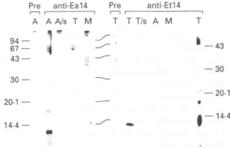
Polyclonal antibodies were generated against the 14 kDa protein isolated from oocyst walls of E. acervuUna and E. tenella. Since resolution of the doublet bands could not be achieved with preparative gels, the antigen preparations used for the immun-ization contained both bands of the doublet. The produced antisera are referred to as anti-Eal4 (E. acervuUna) and anti-Etl4 (E. tenella), respectively. On Western blots, these antisera produced strong
K.-H. Eschenbacher and others L 72
Fig. 1. (A) Intact sporulated oocvsts of Etmeria tenella (phase contrast). (B) Broken oocvst walls of E. tenella after purification (phase contrast), (C) Same, scanning electron micrograph.
T1 T2 A M T/s T3 94 — 67 — 4 3 — -— 67 — 43 94 67 43 Pre A — A
1
4
anti-Ea14 A/s T M """ w Pre T /-anti T T/s A -Et14 M T i 20-1 — 14.4 __ 30 — 20-1 14-4 Fig. 2. SDS-PAGE of oocysts wall proteins under reducing conditions using an 8-15% gradient gel (lanes T j - T / s ) or a 12-5% gel (lane T3). Samples in lanesT , - T / s were from sporulated oocysts. Tx, Eimerta
tenella (Houghton strain); T2, E, tenella (Roche strain);
A, E. acervulina; M, E, maxima; T / s , supernatant of E. tenella (Roche strain) oocysts after disruption; T3, E.
tenella, unsporulated oocysts (Houghton strain). The amount of protein loaded onto each lane corresponds to about 5 x 10s oocysts. Numbers indicate the apparent molecular weights in kDa.
signals with the homologous 14 kDa antigens (Fig. 3). When the antiserum was used at a higher concentration, additional weaker bands appeared at 17 kDa and at about 50 kDa (Fig. 3, lane T on the extreme right side). The anti-Eal4 serum did not react with oocvst wall proteins of E. tenella, and vice
30 20-1 14-4 — 43 — 30 20-1
t
14-4Fig. 3. Western blot of oocvst wall proteins probed with anti-Eal4 and anti-Etl4 sera. Lanes labelled ' P r e ' were incubated with the corresponding pre-immune sera. (A) Eimerta acervulina; T, E. tenella; M, E. maxima; A/s, T / s , supematants from disrupted oocysts of E,
acervulina and E. tenella, respectively. Numbers indicate apparent molecular weights in kDa. Antisera were used at a dilution of 1 :10000, except for lane T on the extreme right side (1 :5000).
versa. Neither of the two antisera reacted with oocyst wall proteins of another species, E. maxima. In addition, protein supematants of disrupted oocysts from E. tenella and E. acervulina did not react with the respective antisera (Fig. 3, lane A/s and T/s).
Oocyst wall fragments could be stained by im-munofluorescence using the antibodies directed against the 14 kDa proteins (Fig. 4). It was possible to distinguish between oocyst walls of E. acervulina (Fig. 4 A) and E. tenella (Fig. 4B) by specific binding
Eirnena oocyst wall protein 173
\\n. 4. Iiimiunotluorescent staining of oocyst wall fragments of Eirnena acervulma (A) and /•„'. tenella (B) by anti-Eal4 (A) and anti-Etl4 (B), The corresponding phase-contrast images are shown in (C) and (D). With pre-immune serum only autofluorescence was observed. This was also the case when E. acervulina oocysts were incubated with anti-Etl4 as primary antibody, and vice versa (not shown).
1 11
A) GWASPYTRYY SSYSYYTPYS
1
11
B) FSSSFYPAFH PYTYSSSYAL
Fig. 5. Amino-terrninal ammo acid sequences of the 14 kDa oocyst wall protein of Eimeria acervulina (A) and E. tenella (B). The E. tenella sequence was determined twice from 2 different strains (Houghton and La Roche) and was found to be identical in both cases.
to anti-Eal4 and anti-Etl4, respectively. Intact oocysts or sporocysts did not stain (not shown). The staining of the oocyst walls was essentially uniform, although it was somewhat stronger at the edges of the folded structures. Fixation of the oocyst wall fragments with acetone/methanol prior to the anti-body incubation did not affect the staining pattern (not shown).
Amino-terminal sequencing of the 14 kDa oocyst wall proteins of E. acervulina and E. tenella resulted
in the identification of the first 20 amino acid residues of each protein (Fig. 5). Both N-terminal sequences contain large amounts of amino acids with hydroxyl groups in their side-chains. These 3 amino acids (serine, tyrosine, threonine) constitute 7 0 % of all amino acids in the E. acervulina partial protein sequence, and 5 5 % in the E. tenella sequence. Aromatic amino acids (phenylalanine, tyrosine, tryptophan) amount to 4 0 % (E. acervulina) or 3 5 % (E. tenella) of all residues. Remarkably, a sequence motif consisting of 3 serine residues flanked by aromatic amino acids (F/Y-S-S-S-F/Y) is repeated twice within the sequence of the E. tenella protein.
To determine if the 14 kDa protein of E. tenella is glycosylated, we performed a lectin-binding test on the blotted protein band. A positive signal was obtained with peanut agglutinin (PNA; Fig. 6, lane 1). None of the 4 other lectins tested (GNA, SNA, MAA, DSA; see Materials and Methods section) reacted with the 14 kDa band (not shown). PNA did not bind to bacterial proteins, which were used as a negative control (Fig. 6, lane 3).
K.-H. Eschenbacher and others 174 1 2 3 94-67 43
- I
30 20-1 14-4Fig. 6. Western blots probed with digoxigenin-Iabelled peanut agglutinin (PNA). Lane 1, oocyst wall proteins of Eimeria tenella. Lane 2, desialylated fetuin as a positive control protein. Lane 3, Escherichia coli (XL1-Blue, Stratagene) lysate proteins as a negative control" Numbers on the left indicate size markers in kDa.
DISCUSSION
By following a protocol originally described by Stotish et al. (1978), we were able to obtain highly purified fractions of disrupted oocyst walls. Light and electron microscopy reveals that these walls tend to fold up into tube-like structures. This could be attributed to tension forces which are present within the wall matrix of the intact oocyst and which make it collapse when mechanical stress is exerted.
SDS-PAGE analysis of purified oocyst wall fragments led to the identification of a major protein of 14 kDa and only a few additional minor bands. Likewise, in the related coccidian parasite,
Crypto-sporidium parvum, only a limited number of protein
bands were found when fractions enriched for oocyst shells were analysed (Lally et al. 1992). The 14 kDa band was found in all Eimeria species so far, including 2 species of rabbit Eimeria (E. media, E.
stiedai; not shown), and the proteins obtained from
the various Eimeria species show only very little variation in size. This suggests that the occurrence of this small protein species is probably common to all Eimerian oocyst walls.
Based on its presence in oocyst walls and on its apparent molecular weight, we assume that the 14 kDa protein described here might be related to the 10 kDa fraction that has been reported earlier by Stotish et al. (1978). However, this fraction appeared as a rather broad band on an SDS-PAGE gel and
probably contained degradation products as well, due to prolonged solubilization in hot urea (Stotish
et al. 1978). We preferred the more gentle
solubil-ization by SDS (for only 3 min), although it is less efficient (Stotish et al. 1978). To avoid possible contamination with sporocyst walls, we used un-sporulated oocysts in our initial experiments. Since the 14 kDa protein was present in oocyst wall extracts from both unsporulated and sporulated oocysts, however, we decided to use sporulated oocysts which are easier to obtain.
The isolation of the 14 kDa protein from pure fractions of oocyst walls, and its absence from sporozoite extracts, suggested that it is a component of the wall. This could clearly be demonstrated by immunofluorescence using oocyst wall fragments and antisera raised against the 14 kDa proteins of E.
acervulina and E. tenella. Intact oocysts could not be
stained, which could be due to the outer lipid layer which might render the antigenic sites inaccessible to antibodies. Immunofluorescence microscopy was complicated by the strong autofluorescence pro-duced by the oocyst walls (Russel & Sinden, 1981). However, this problem could be overcome by using a filter set which makes the emitted autofluorescence appear red in colour so that it can be distinguished from the green fluorescence of FITC.
Western blot analysis showed that the anti-Eal4 and anti-Etl4 antibodies recognized the homologous antigens in a highly specific manner. This species-specific recognition could also be confirmed by immunofluorescent labelling of oocyst wall frag-ments. Since the N-terminal sequences of E.
acer-vulina and E. tenella were completely different, it can
be argued that differences in the primary sequences are responsible for the highly specific recognition by antibodies. Antigenic differences between oocyst wall proteins of the various Eimeria species may be exploited for the development of species-specific diagnostic tests. The identification of Eimeria species still remains a laborious and difficult task (Long & Joyner, 1984; MacPherson & Gajadhar, 1993; Stucki, Braun & Roditi, 1993).
The binding of peanut agglutinin to the 14 kDa oocyst wall protein of E. tenella (and also of E.
acervulina; not shown) suggests the presence of
O-linked carbohydrate residues. PNA recognizes the disaccharide galactose-/?( 1 -> 3)iV-acetylgalactos-amine which usually forms the core unit of O-glycans. However, complex-type glycan moieties often contain sialic acid residues in their terminal regions (Alberts et al. 1989) which prevent PNA from binding unless they are removed by treatment with neuraminidase (e.g. in the case of fetuin which was used as a positive control in Fig. 6). Since binding of PNA to the 14 kDa protein occurred without pre-treatment with neuraminidase or exo-glycosidases, the carbohydrate moiety is probably not of the complex type (Lotan et al. 1975; Sueyoshi,
Eimeria oocyst wall protein 175 Tsuji & Osawa, 1988; Boehringer Mannheim DIG
Glycan Differentiation Kit, product information). Since PNA might also have other binding speci-ficities, further experiments will be needed to determine the nature of the carbohydrate residue(s). Binding of PNA to the 14 kDa protein was inhibited in the presence of the disaccharide lactose, and - less efficiently - the monosaccharide galactose (data not shown). This observation is in agreement with the data reported by Lotan et al. (1975) and Russel & Sinden (1981). The notion that the 14 kDa protein might be O-glycosylated is also supported by the finding that the N-terminal sequences are rich in serine and threonine, which are target sites for this type of modification. However, since excessive glycosylation would have interfered with Edman degradation, we assume that the degree of modi-fication is probably low, at least in the N-terminal region. Post-translational modifications of various kinds could also be responsible for the appearance of the 14 kDa protein as a band doublet. Microhetero-geneity was also found to occur in the 10 kDa oocyst wall protein described by Stotish et al. (1978).
The observation that FITC-labelled PNA binds to the surface of glutaraldehyde-fixed oocysts of E.
acervulina and E. tenella (Russel & Sinden, 1981) is
also in agreement with the results presented here. In addition, PNA-binding glycoproteins have also been found in sporulated oocysts and sporozoites of E.
tenella, suggesting that O-glycosylation is quite
frequent in Eimeria spp. (Michalski et al. 1993). The nature of the carbohydrate component of the oocyst wall proteins, as well as its function in the wall matrix, remain to be determined. It has been suggested that mannitol or mannitol phosphate might become incorporated into the oocyst wall (Schmatz, 1989).
At present, only 1 genomic sequence coding for a coccidian oocyst wall protein has been reported (of
C. parvum; Lally et al. 1992; Ranucci et al. 1993).
The putative amino acid sequence of this
Crypto-sporidium protein, however, bears no similarities to
the partial N-terminal sequences we obtained for
Eimeria oocyst wall proteins. The N-terminal
sequ-ences of the 14 kDa proteins of E. tenella and E.
acervulina constitute only about 18% of the total
length of the polypeptide (assuming a molecular weight of 14 kDa), and interpretation of these data is therefore speculative.
A striking feature of the partial amino acid sequences is the high frequency of amino acids which contain hydroxyl groups in their side chains. By cross-linking individual proteins via these hy-droxyl groups, a complex polymeric wall matrix may be formed. For example, cross-linking by tyrosine residues is involved in the hardening of the fertil-ization envelope of sea urchin eggs (Hall, 1978) and in the synthesis of the yeast ascospore wall (Briza et
al. 1986). Tanning processes utilizing tyrosines and
other phenolic compounds also play a crucial role in insect cuticle sclerotization (reviewed by Hopkins & Kramer, 1992) and in eggshell formation in
Schisto-soma mansoni (Seed & Bennett, 1980), Fasciola hepatica (Waite & Rice-Ficht, 1987) and Trichuris suis (Fetterer & Hill, 1994). The putative amino acid
sequence of an eggshell protein of S. mansoni shows a high content of tyrosine residues (Johnson, Taylor & Cordingley, 1987). Monne & Honig (1954) proposed that the formation of the coccidian oocyst wall could occur by a tanning process. The ob-servation that the anti-14 antisera weakly recognize bands in the higher molecular weight range could be explained by cross-links between protein monomers. The 14 kDa protein would then represent the monomer. It should also be noted that additional polypeptide species might participate in the for-mation of the wall like, for example, the 30 kDa proline-rich protein which was not recognized by the anti-14 antibodies. In addition, the identification of a mannitol cycle in E. tenella has led to the speculation that during oocyst formation mannitol could create an osmotic pressure within the oocyst until the hardening of the shell is completed (Schmatz, 1989).
It remains to be established whether the 14 kDa wall proteins can be localized within the wall-forming bodies of the gametocytes. Since gametocyte antigens have been reported to induce a protective immune response in chickens (Wallach et al. 1989), the 14 kDa proteins might also be useful for vaccination trials.
We wish to thank J. Eckert (University of Zurich), P. Bcdrnik (BIOPHARM, Prague), G. Weber (Hoffmann-La Roche, Basel) and F. Tomley (Institute for Animal Health, Compton) for providing oocysts, and I. Roditi for helpful discussions and for critically reading the manu-script. This work was supported by the Bundesamt fur Bildung und Wissenschaft, Bern (Project: Coccidiosis, COST 89 & 820) and the Swiss National Science Foundation.
R E F E R E N C E S
ALBERTS, B . , BRAY, D . , LEWIS, J . , RAFF, M . , ROBERTS, K., WATSON, j . D. (1989). Molecular Biology of the Cell,
2nd edn. New York: Garland Publishing.
BRIZA, P., WINKLER, C , KALCHHAUSER, H. & BREITENBACH,
M. (1986). Dityrosine is a prominent component of the yeast ascospore wall. Journal of Biological Chemistry 261, 4288-94.
CHOBOTAR, B. 4 SCHOLTYSECK, E. (1982). Ultrastructure.
In The Biology of the Coccidia (ed. Long, P. L.), pp. 101-165. London: University Park Press.
FETTERER, R. H. k HILL, D. E. (1994). Localization of
phenol oxidase in female Trichuris suis. Journal of Parasitology 80, 952-9.
HALL, H. G. (1978). Hardening of the sea urchin
fertilization envelope by peroxidase-catalyzed phenolic coupling of tyrosines. Cell 15, 343-55.
K.-H. Eschenbacher and others 176
HARLOW, E. & LANE, D. (1988). Antibodies. A Laboratory
Manual. Cold Spring Harbor Laboratory. New York:
Cold Spring Harbor Laboratory Press.
HOPKINS, T. L. & KRAMER, K. j . (1992). Insect cuticle
sclerotization. Annual Review of Entomology 37,
273-302.
JEFFERS, T. K. & SHIRLEY, M. w. (1982). Genetics, specific
and infraspecific variation. In The Biology of the
Coccidia (ed. Long, P. L.), pp. 63-100. London:
University Park Press.
JOHNSON, K. S., TAYLOR, D. W. & CORDINGLEY, J. S. ( 1 9 8 7 ) .
Possible eggshell protein gene from Schistosoma
mansoni. Molecular and Biochemical Parasitology 22,
89-100.
KIRSCHNER, R. H. (1978). High resolution scanning
electron microscopy of isolated cell organdies. In
Principles and Techniques of Scanning Electron
Microscopy (ed. Hayat, M. A. ), pp. 278-296. New
York: Van Nostrand Reinhold Company.
LALLY, N . C , BAIRD, C. D . , M c Q U A Y , S. J . , W R I G H T , F. &
OLIVER, j . j . (1992). A 2359-base pair DNA fragment
from Cryptosporidium parvum encoding a repetitive
oocyst protein. Molecular and Biochemical Parasitology
56, 69-78.
LEVINE, N. D. (1982). Taxonomy and life cycles of
coccidia. In The Biology of the Coccidia (ed. Long, P.
L.), pp. 1-33. London: University Park Press.
LONG, p. L. 4 JOYNER, L. P. (1984). Problems in the
identification of species of Eimeria. Journal of
Protozoology 31, 535-41.
LOTAN, R., SKUTELSKI, E., DANON, D. 4 SHARON, N. ( 1 9 7 5 ) .
The purification, composition and specifity of the
anti-T lectin from peanut (Arachis hypogaea). Journal of Biological Chemistry 250, 8518-23.
M A C P H E R S O N , J. M. 4 GAJADHAR, A. A. ( 1 9 9 3 ) .
Differentiation of seven Eimeria species by random amplified polymorphic DNA. Veterinary Parasitology 45, 257-66.
M I C H A L S K I , W . P . , PROWSE, S. J . , BACIC, A. 4 F A H E Y , K. J.
(1993). Molecular characterization of peanut
agglutinin binding glycoproteins from Eimeria tenella.
International Journal for Parasitology 23, 985-95.
MONNE, L. 4 H6NIG, G. (1954). On the properties of the
shells of the coccidian oocysts. Arkiv for Zoologi (Uppsala) 7, 251-6.
RANUCCI, L. R., MULLER, H. M., LAROSA, G. L., RECKMANN, I. R., GOMEZ MORALES, M. G., POZIO, E. P., SPANO, F. S. 4 CRISANTI, A. A. (1993). Characterization and
immunolocalization of a Cryptosporidium protein containing repeated amino acid motifs. Infection and
Immunity 61, 2347-56.
RUSSEL, D. G. & SINDEN, R. E. (1981). The role of the
cytoskeleton in the motility of coccidian sporozoites.
Journal of Cell Science 50, 345-59.
SCHAGGER, H. 4 VON JACOW, c. (1987). Tricine-sodium dodecyl sulfate-polyacrylamide gel electrophoresis for the separation of proteins in the range from 1 to
100 kDa. Analytical Biochemistry 166, 368-79.
SCHMATZ, D. M. (1989). The mannitol cycle - a new
metabolic pathway in the coccidia. Parasitology Today 5, 205-8.
SEED, j . L. 4 BENNETT, j . L. (1980). Schistosoma mansoni:
phenol oxidase's role in eggshell formation.
Experimental Parasitology 49, 430-41.
STOTISH, R. L., WANG, C. C. 4 MEYENHOFER, M. ( 1 9 7 8 ) .
Structure and composition of the oocyst wall of
Eimeria tenella. Journal of Parasitology 64, 1074-81.
STUCKI, v., BRAUN, R. 4 RODITI, I. (1993). Eimeria tenella: Characterization of a 5S ribosomal RNA repeat unit and its use as a species-specific probe. Experimental
Parasitology 76, 68 75.
SUEYOSHI, s., Tsuji, T. 4 OSAWA, T. (1988). Carbohydrate-binding specificities of five lectins that bind to O-glycosyl-linked carbohydrate chains. Quantitative analysis by frontal-affinity chromatography.
Carbohydrate Research 178, 213-24.
WAITE, j . H. 4 RICE-FICHT, A. c. (1987). Presclerotized
eggshell protein from the liver fluke Fasciola hepatica.
Biochemistry 26, 7819-25.
WALLACH, M., MENCHER, D . , YARUS, S., PILLEMER, G., HALABI, A. 4 PUGATSCH, T. (1989). Eimeria maxima:
identification of gametocyte protein antigens and their possible role in protective immunity. Experimental
Parasitology 68, 49-56.
WANG, c. c. (1982). Biochemistry and physiology of the
coccidia. In The Biology of the Coccidia (ed. Long, P.