Characterization of hypovirulent isolates of the chestnut
blight fungus,
Cryphonectria parasitica from the Marmara
and Black Sea regions of Turkey
Seçil Akıllı&Çiğdem Ulubaş Serçe&Yakup Zekai Katırcıoğlu&Salih Maden&
Daniel Rigling
Accepted: 10 September 2012 / Published online: 21 September 2012 # KNPV 2012
Abstract Chestnut blight caused by the introduced fungus Cryphonectria parasitica has been responsible for the decline of Castanea sativa in Turkey since the 1960s. In this study, 72 C. parasitica isolates were recovered from the Marmara and Black Sea regions of Turkey showing white or cream-coloured culture mor-phology and were subjected to various tests to deter-mine if they were infected by Cryphonectria hypovirus 1 (CHV-1). The vast majority of the isolates (69 out of 72) were vc type EU-1. Both mating types were found among a subsample of the isolates. The hypovirus was detected in 55 isolates by dsRNA ex-traction and/or virus specific RT-PCR on total RNA
extracts. All but one isolates showed no or only weak phenol oxidase activity on agar medium containing tannic acid, typical of CHV-1 infected isolates. Through sequencing of a specific region of the hypo-virus genome, we found that 24 hypohypo-virus isolates belonged to the CHV-1 subtype I and six to the CHV-1 subtype F2. The distribution of the two CHV-1 subtypes in Turkey showed a clear geographic pattern. CHV-1 subtype I was only detected in the Marmara and western Black Sea region, whereas sub-type F2 was restricted to the eastern part of the Black Sea region. The effectiveness of 23 hypovirulent iso-lates was tested against a virulent isolate on 2–3 years old chestnut sprouts. Ten hypovirulent isolates, all infected by CHV-1 subtype I, prevented canker devel-opment by more than 80 % suggesting that they might be suitable for biological control of chestnut blight in Turkey.
Keywords Cryphonectria hypovirus . CHV-1 . RT-PCR detection . Sequence variation . Biological control . Castanea sativa
Introduction
Chestnut blight, caused by the fungus Cryphonec-tria parasitica (Murrill) Barr was first observed in the US in the early 20th century, when the Amer-ican chestnut (Castanea dentata [Marsh.] Borkh) was devastated by the introduced pathogen DOI 10.1007/s10658-012-0089-z
S. Akıllı
Faculty of Science, Department of Biology, Çankırı Karatekin University,
Çankırı, Turkey Ç. Ulubaş Serçe
Agricultural Faculty, Department of Plant Protection, Mustafa Kemal University,
31034 Hatay, Turkey Y. Z. Katırcıoğlu
:
S. MadenAgricultural Faculty, Department of Plant Protection, Ankara University,
06110 Dışkapı, Ankara, Turkey D. Rigling (*)
Swiss Federal Institute for Forest, Snow and Landscape Research (WSL),
Zürcherstrasse 111,
8903 Birmensdorf, Switzerland e-mail: daniel.rigling@wsl.ch
(Anagnostakis 1988). The disease was first recorded on European chestnut (Castanea sativa Mill.) in Italy in 1938. Chestnut blight spread rapidly and, with few exceptions (e.g. UK), all chestnut growing areas in Europe have become infected by C. parasitica (Robin and Heiniger2001). However, many European chestnut stands began to recover from the disease as indicated by the occurrence of superficial non-lethal chestnut blight cankers. This recovery has been attrib-uted to hypovirulence, a phenomenon in which fungal viruses infect C. parasitica and significantly reduce its virulence and sporulation capacity (Grente1965; Choi and Nuss1992; Heiniger and Rigling1994; Milgroom and Cortesi 2004). Because the viruses are causing hypovirulence in C. parasitica they have been named Cryphonectria hypoviruses (CHV) and were assigned to the virus genus Hypovirus (Hillman et al. 2000). Al-though the hypoviruses mostly occur as double-stranded (ds) RNA replicative forms in their host, they essentially are single-strand RNA viruses with only one strand employed in transcription (Choi and Nuss1992).
Four members of the genus Hypovirus are known. The best investigated hypovirus is CHV-1, which is the only hypovirus reported in Europe (Allemann et al. 1999; Nuss 2005). CHV-1 has also been found in Japan and China (Peever et al. 1998; Hillman and Suzuki 2004). CHV-2 has been reported in North America and China, whereas CHV-3 and CHV-4 have only been found in North America (Hillman and Suzuki 2004). Infection of C. parasitica by CHV-1 causes a reduction of fungal virulence that can vary depending on the virus isolate and the fungal host strain (Rigling et al.1989; Peever et al.2000; Robin et al. 2010; Bryner and Rigling 2011). Besides re-duced virulence, fungal isolates infected by CHV-1 also exhibit reduced pigmentation and reduced sporu-lation, giving them a typical white cultural appear-ance. Reduced phenol oxidase (laccase) activity on agar medium containing tannic acid is another easily determinable phenotypic trait of hypovirus-infected C. parasitica isolates (Rigling et al.1989).
As with most fungal viruses, there is no known extracellular phase of Cryphonectria hypoviruses. The hypovirus can be disseminated through asexually pro-duced conidia but not through sexually propro-duced asco-spores (Anagnostakis1988; Prospero et al.2006). The virus is transmitted horizontally from an infected fungal strain to a non-infected strain via hyphal anastomosis (Van Alfen et al. 1975). Hyphal fusion and thereby
horizontal virus transmission is controlled by a vegeta-tive incompatibility (vic) system involving at least six vic loci (Cortesi et al. 2001). The hypovirus is easily transmitted between isolates sharing identical alleles at all vic loci, i.e. isolates of the same vegetative compat-ibility (vc) type. Hypovirus transmission is generally restricted among isolates in different vc types. Low vc type diversity in C. parasitica populations is thought to be an important factor for the success of hypovirulence (Anagnostakis et al.1986). Sexual reproduction is the main source of vc-type diversity through the recombi-nation of polymorphic vic genes (Cortesi and Milgroom
1998).
In Europe, several subtypes of CHV-1 have been detected using RFLP typing and sequence analysis (Allemann et al. 1999; Gobbin et al. 2003). The Italian subtype (subtype I) is the most widespread with a distribution range spanning from France, Italy, and Switzerland to south-eastern Europe and Greece (Allemann et al. 1999; Sotirovski et al. 2006; Krstin et al. 2008; Robin et al. 2010; Krstin et al. 2011). Additional CHV-1 subtypes have been found in France (subtypes F1 and F2) and in Spain and Germany (subtype E/D) (Gobbin et al. 2003; Montenegro et al. 2008).
Transmissible hypovirulence has been used as the basis for application of biological control of chestnut blight (Anagnostakis1982; MacDonald and Fulbright 1991). Such control programs were implemented in France, Italy, and Switzerland (Robin et al. 2000; Milgroom and Cortesi 2004; Heiniger and Rigling 2009). Biological control is achieved by treating chest-nut blight cankers with selected hypovirus-infected C. parasitica strains that are compatible with the vc types in the local C. parasitica populations. Knowledge of the vc types and hypoviruses present in an area is therefore crucial for successful application of biologi-cal control (MacDonald and Fulbright1991; Heiniger and Rigling1994; Robin et al.2010).
European chestnut is an important tree species in Turkey. The Marmara and Black Sea regions comprise 96.8 % of the total chestnut forests of Turkey, which amount to a total of 200,440 ha. Chestnuts are grow-ing in pure stands (11.3 %), in stands mixed with deciduous trees (73.4 %), and in stands mixed with conifers (9.3 %). Chestnut blight was first reported in Turkey in the 1960s (Delen1975). After its introduc-tion, the disease caused considerable damage to the native C. sativa population, particularly in the Marmara
Region. Within 20 years, chestnut blight had spread along the mountain range of the Black Sea to the eastern part of the country. To date, chestnut blight is widespread in Turkey and is still causing significant damages to the chestnut forests in spite of the presence of healing can-kers (Çeliker and Onoğur2001; Akilli et al.2009). The C. parasitica populations in Turkey are characterized by a very low diversity of vc types. In all populations investigated in the Marmara and Black Sea regions, EU-1 was the dominant vc type, typically comprising more than 90 % of the isolates (Çeliker and Onoğur 2001; Gürer et al.2001a,b; Akilli et al.2009). On the other hand, vc type EU-12 was found to be dominant in some parts of the Aegean region (Erincik et al.2008).
In Turkey, superficial (healed) cankers, which are a broad indicator of hypovirulence, were first noticed in 1998 near Gölcük in the province Kocaeli where chestnut blight was first recorded (Çeliker and Onoğur 1998). Later on, superficial cankers were observed in other provinces of the Marmara region and in the Black Sea region (Gürer et al.2001a,b; Akilli et al.2009). From the healing cankers, white C. parasitica isolates were obtained, which contained dsRNA and were able to con-vert virulent isolates (Çeliker and Onoğur1998; Çeliker and Onoğur2001; Gürer et al.2001a,b). Till this day, hypovirulence is largely absent in the Aegean region of Turkey (Çeliker and Onoğur2001; Erincik et al.2008).
In this study we characterized Turkish C. parasitica isolates that exhibited cultural characteristics typical for hypovirus-infected isolates. The isolates were collected in the Marmara and Black Sea regions, which include the main distribution range of European chestnut in Turkey. The main objectives were: (i) to assess the isolates for phenol oxidase activity, a phenotypic trait associated with hypovirus infection; (ii) to test the iso-lates for the presence of the hypovirus by using dsRNA extraction and PCR based methods; (iii) to determine which subtypes of CHV-1 are present in Turkey; and (iv) to evaluate the effectiveness of hypovirus-infected strains to control virulent C. parasitica.
Materials and methods
Fungal isolates and culture conditions
Two hundred and ninety-six isolates used in this study were recovered from the culture collection obtained in a previous study carried out in 2007–2008 in the Black
Sea region of Turkey (Akilli et al.2009) and stored at −80 °C at the Department of Plant Protection, Faculty of Agriculture, Ankara University. In addition, 126 isolates obtained from Marmara region were included. All the 420 isolates were first grown on potato dex-trose agar (PDA, Difco) amended with biotin and 72 white or cream-coloured isolates were selected for further analysis. The isolates were subjected to a phe-noloxidase test and analyzed for the presence of CHV-1 by dsRNA extractions and 57 of them by hypovirus-specific RT-PCR. Vc types of the isolates were deter-mined in the previous studies (Gürer et al. 2001a, b; Akilli et al.2009).
Phenol oxidase test
A total of 70 white or cream-coloured growing iso-lates, 57 from Black Sea region and 13 from Marmara region, were subjected to phenol oxidase (laccase) test. The test was performed by growing the isolates on agar medium containing tannic acid (Bavendamm’s medium) as described by Rigling et al. (1989). A dark discolouration of the agar medium was considered as phenol oxidase positive, which indicates a virulent reaction while no colour change indicates hypoviru-lent reaction (Rigling et al.1989).
Isolation of hypovirus dsRNA
Double-stranded RNA was extracted from cultures grown in potato dextrose broth in Petri dishes for one week at 25 °C. Mycelia grown in culture were dried between paper towels and around 6–8 g were immediately used for isolations. The dsRNA was extracted using cellulose CF-11 chromatography as described by Allemann et al. (1999). A known dsRNA-containing isolate of C. parasitica obtained from Tullio Turchetti (Institute of Forest Tree Pathol-ogy, National Research Council; Firenze, Italy) was included as a control in each preparation. The quantity and quality of the dsRNA preparations were examined by agarose (1 %) gel electrophoresis.
Total RNA extraction
About 100 mg mycelium of C. parasitica, prepared as described above, was grinded in liquid nitrogen and 5 volume buffer (200 mM Tris–HCl pH 8.5, 1.5 % SDS, 300 mM lithium chloride, 10 mM EDTA, 1 % sodium
deoxycholate, 1 % Igepal and 0.5 % mercaptoethanol) was added to each sample (Spiegel et al.1996). The extract was collected and heated for 15 min at 65 °C. One volume of 6 M potasium acetate pH 6.5 was added and the tubes were kept on ice for 15 min. After centrifugation at 12,000×g for 15 min, the supernatant was removed and nucleic acids were precipitated with isopropanol. The pellet was resuspended in 50μl ster-ile water and stored at−80 °C.
First strand cDNA synthesis and polymerase chain reaction (PCR)
First strand cDNA was synthesized from 100 ng dsRNA or total RNA using random hexanucleotide primers. PCR was performed using different sets of virus specific primers, which all were known to am-plify different subtypes of CHV-1. A region in open reading frame A (ORF A) was amplified with primer set A (EP 713-5/R2280) and one region in ORF B with primer set B (EP 713-6/EP 713-7) as described by Allemann et al. (1999). Another primer set was specific for a region in the helicase domain of ORF B (OB-10193F/OB-11161R), and one for a region in ORF A (OA-427F/OA-1323R) as described by Griffin et al. (2004). PCR amplification was carried out in 50μl of reaction mixture containing cDNA template (about 1 μl), primer pairs (0.8 μM each), 200 μM dNTPs, 1.5 mM MgCl2, 10 × reaction buffer (final concentration of 10 mM Tris–HCl pH 8.8, 50 mM KCl and 0.08 % Nonidet P40), 1 U of Taq DNA polymer-ase (MBI Fermentas, Germany). The thermal cycling conditions of the reaction for the first two primer sets was as follows: 2 min at 94 °C, 40 cycles of 1 min at 94 °C, 1.5 min at 50 °C and 2 min at 72 °C, and 8 min at 72 °C for final extension. For the other primer sets, the cycling was performed using 58 °C for annealing and 68 °C for extension. The PCR products were fraction-ated by electrophoresis in a 1 % agarose gel, stained with ethidium bromide and visualized under UV.
Phylogenetic analysis and subtype determination
To determine the subtype of the CHV-1 isolates, we sequenced the PCR products of the ORF A region on both strands using the primers hvep1 and hvep2 (Gobbin et al.2003). Assembled sequences were aligned using Clustal W (Thompson el al.1994). Sequences of previ-ously reported subtypes I, F1, F2, D and E deposited in
the GenBank (Accession numbers: AJ577160, AJ577168, AJ577170, AJ577173 and AJ577172, re-spectively) were included for comparison. The align-ments were used to reconstruct phylogenetic trees using the neighbour-joining method with nucleotide identity distances in program MEGA 4.0 (Tamura et al.2007). Bootstrap analyses with 1,000 replicates were per-formed to estimate the support for inferred phylog-enies (Felsenstein 1985).
Effectiveness of hypovirulent isolates
The effectiveness, as biological control agents, of 23 hypovirulent isolates (based on dsRNA analysis) of vc type EU-1 was tested against an aggressive C. para-sitica isolate (isolate K-19) of the same vc type on 3-year-old chestnut sprouts by stem inoculation with five replications. First, mycelium of the virulent iso-late (taken as agar plug from a PDAmb culture) was placed into the holes (0.5 cm diameter) drilled into the stems (one hole per sprout). Immediately afterwards mycelium of the hypovirulent isolate was placed into the same hole onto the mycelium of the virulent iso-late. Inoculation points were wrapped with wet cotton and then sealed with parafilm. Canker lengths were measured after 6 months and effectiveness of the ap-plication was calculated by comparing the canker sizes to control inoculations with only the virulent isolate.
Determination of mating types
The mating type of a subsample of the isolates was determined by a multiplex polymerase chain reaction (MPCR) assay, which was performed with the specific primers M1-GS1n and M1-GS3-rev for MAT-1 and M2-GS3 and gs1-d-1 for MAT-2 (Marra and Milgroom 2001).
Results
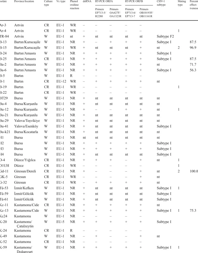
In total, 420 isolates of Cryphonectria parasitica obtained from 12 provinces of Marmara and Black Sea regions of Turkey (Fig. 1) were subjected to various tests for hypovirulence. These isolates were first grown on PDAmb to assess their cultural appear-ance. Seventy-two isolates (16.9 %) showed whitish or cream-coloured culture morphology on this medium and were further characterized (Table1). Among these
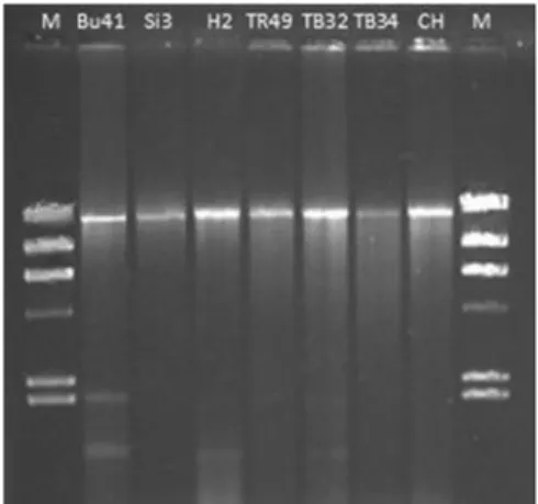
72 isolates, 69 belonged to vc type 1, two to EU-12, and one to EU-5. Out of 45 white isolates tested for phenol oxidase activity, 43 produced no colour reaction when grown on the tannic acid medium. Among the 25 cream-coloured isolates, eight gave no colour reaction and thirteen a weak reaction. Only one white and four cream-coloured isolates showed a strong phenol oxidase activity, which is typical for virulent C. parasitica isolates. A high molecular weight dsRNA was detected in 43 out of 46 white isolates and in six out of 25 cream-coloured isolates. The size of all the dsRNAs was approximately 13 kbp (Fig. 2). Only one cream-coloured isolate yielding dsRNA gave a strong phenol oxidase reaction. The white growing isolate showing a positive reaction for phenol oxidase did not yield dsRNA.
Total RNA was extracted from 57 C. parasitica isolates and used to generate cDNA. The presence of the Cryphonectria hypovirus 1 (CHV-1) was verified in 40 isolates by PCR using specific primer pairs for the virus (Table1, Figs.3and4). The majority of the isolates (n026) amplified a PCR product of the expected size with all four primer pairs. Nine isolates were positive with two to three primer pairs. All iso-lates yielding dsRNA also tested virus-positive by RT-PCR. In addition, virus-specific PCR amplifications were obtained from six more isolates in which dsRNA was not detected (Table1). These results might be due
to the strength of the PCR technique, which can am-plify a small amount of cDNA.
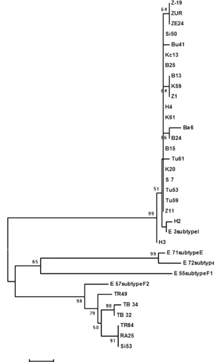
A portion of the hypovirus genome was sequenced from 30 hypoviral dsRNAs. Phylogenetic analysis of these sequences revealed that 24 hypoviruses belonged to the Italian subtype (subtype I) of CHV-1 (Fig.5). Together with the reference sequence of sub-type I, they formed a large cluster, which was separat-ed from the other hypoviruses with high bootstrap support (99 %). Six hypoviruses from Turkey grouped with the reference sequence in the subtype F2 and were assigned to this subtype (Fig.5, Table1). CHV-1 subtype I was dominant in the western part of our study area, while subtype F2 was mainly found in the eastern part of the Black Sea region, namely in the provinces Ordu, Trabzon, Rize and Artvin (Fig. 1). Only one hypovirus in subtype F2 was found west of Ordu, in Sinop, which is located in the centre of the Black Sea region.
Fifty two randomly selected C. parasitica isolates from the Black Sea region were analysed for mating types. Both mating types were found in this region and the ratio of MAT1/MAT2 was 1.79. This ratio was 2.52 for the 25 hypovirulent isolates tested (Table1). The effectiveness of 23 hypovirulent isolates all belonging to vc type EU-1, was tested against an aggressive isolate (K-19) on 3-year-old chestnut sprouts. Twenty of the 23 isolates were infected by a Fig. 1 Provinces in the
Marmara and Black Sea regions of Turkey where Cryphonectria parasitica isolates were collected and occurrence of CHV-1 sub-type I and F2 in the different provinces
Table 1 Characterization of hypovirulent isolates of Cryphonectria parasitica obtained from the Marmara and Black Sea regions of Turkey
Isolate Province/location Culture type
Vc type Phenol oxidase reaction
dsRNA RT-PCR ORFA RT-PCR ORFB CHV-1 subtype Mating type Percent effect-iveness Primers EP713-5/ R2280 Primers OA427F/ OA1323R Primers EP713-6/ EP713-7 Primers OB10193F/ OB11161R
Ar-3 Artvin CR EU-1 WR – – – – –
Ar-4 Artvin CR EU-1 WR – – – – – 1
TR-84 Artvin W EU-1 nt + nt nt nt nt Subtype F2
B-13 Bartın/Kurucaşile W EU-1 NR + – + + + Subtype I 87.5
B-15 Bartın/Kurucaşile W EU-1 WR + nt nt nt + nt 2 96.9
B-24 Bartın/Amasra W EU-1 NR + + + + + Subtype I
B-25 Bartın/Amasra CR EU-1 NR + + + + + Subtype I 87.5
Ba-2 Bartın/Amasra W EU-1 NR + + + + + nt 71.7
Ba-6 Bartın/Amasra W EU-1 NR + + + + + Subtype I 56.3
B-5 Bartın W EU-1 R – – – – –
B-1 Bartın CR EU-12 WR – + + + + nt
B-19 Bartın CR EU-1 WR – – – – – 1
B-22 Bartın CR EU-1 WR – – – – –
BT29 Bursa W EU-1 NR + nt nt nt nt nt
Bu-4 Bursa/Kurşunlu W EU-1 NR + nt nt nt nt nt
Bu-12 Bursa/Kurşunlu W EU-1 NR + – – + + nt 1
Bu-21 Bursa/Kurşunlu W EU-1 NR + nt nt nt nt nt Bu-29 Yalova/Teşvikiye W EU-1 NR + nt nt nt nt nt Bu-41 Yalova/Esenköy W EU-1 NR + nt nt nt nt Subtype I Bu-k21 Bursa/Kocatarla W EU-1 NR + nt nt nt nt nt
H1 Bursa W EU-1 NR nt nt nt nt nt nt
H2 Bursa W EU-1 NR + + + + + Subtype I
H3 Bursa W EU-1 NR + + + + + Subtype I
H4 Bursa W EU-1 NR + nt nt nt nt Subtype I
D-4 Düzce/Yığılca CR EU-1 NR + + + – + nt 1
D1UH Düzce CR EU-1 WR – – – – – 1
Gd-11 Giresun/Dereli CR EU-1 NR + – – – + nt 2 100.0
GK-5 Giresun CR EU-1 WR – – – – + nt
G-32 Giresun CR EU-1 WR – + + + + nt
Tü-53 İzmit/Kefken W EU-1 NR + nt nt nt nt Subtype I 1 Tü-59 İzmit/Gölcük W EU-1 NR + nt nt nt nt Subtype I Tü-61 İzmit/Gölcük W EU-1 NR + nt nt nt nt Subtype I
Kc-11 Kastamonu/Cide CR EU-1 NR + + + + + nt
Kc-13 Kastamonu/Cide W EU-1 NR + + + + + Subtype I 1 75.3
Kc24 Kastamonu W EU-1 NR – – – – – K-20 Kastamonu/ Çatalzeytin W EU-5 NR + + + + + Subtype I K-24 Kastamonu CR EU-1 R – – – – – K-49 Kastamonu W EU-1 NR – + – – + nt K-52 Kastamonu CR EU-1 NR – – – – – K-59 Kastamonu/ Doğanyurt W EU-1 NR + + + + + Subtype I 1
Table 1 (continued)
Isolate Province/location Culture type
Vc type Phenol oxidase reaction
dsRNA RT-PCR ORFA RT-PCR ORFB CHV-1 subtype Mating type Percent effect-iveness Primers EP713-5/ R2280 Primers OA427F/ OA1323R Primers EP713-6/ EP713-7 Primers OB10193F/ OB11161R K-60 Kastamonu/ Doğanyurt W EU-1 NR + + + + + nt K-61 Kastamonu/ Doğanyurt W EU-1 NR + + + + + Subtype I 75.0 K-44 Kastamonu CR EU-1 R – – – – –
TR-49 Ordu W EU-1 nt + nt nt nt nt Subtype F2
Ra-25 Rize/Ardeşen W EU-1 NR + + + + + Subtype F2
Ra-26 Rize/Ardeşen W EU-1 NR + nt nt nt nt nt 100.0
Si-3 Sinop/Erfelek CR EU-1 NR + + + + + nt 62.4
S-7 Sinop/Erfelek W EU-1 NR + + + + + Subtype I 1 52.4
Si-42 Sinop CR EU-1 NR – – + – + nt 1
Si-50 Sinop/Ayancık W EU-1 NR + + + + + Subtype I 1 51.4
Si-53 Sinop/Ayancık W EU-1 NR + + + + + Subtype F2 1 59.0
Si-68 Sinop/Ayancık W EU-1 NR + nt nt nt + nt 1
S-36 Sinop CR EU-1 R – – – – –
S-37 Sinop/Erfelek CR EU-1 R + – + + + Subtype I 45.2
Si-51 Sinop CR EU-1 WR – – – – –
Tb-32 Trabzon/ Beşikdüzü
W EU-1 NR + + + – + Subtype F2 1 52.8
Tb-34 Trabzon/
Beşikdüzü W EU-1 NR + + + – + Subtype F2 1 38.2
T-15 Trabzon CR EU-1 WR – – + – – nt T-18 Trabzon CR EU-1 WR – – – – – 2 Z-1 Zonguldak/ Alaplı W EU-1 NR + + + + + Subtype I 2 87.5 Z-3 Zonguldak/ Alaplı W EU-1 NR + + + + + nt 1 85.9 Z-4 Zonguldak/ Alaplı W EU-1 NR + + + + + nt 1 87.5 Z-5 Zonguldak/ Alaplı W EU-1 NR + + + + + nt Z-11 Zonguldak/
Ereğli W EU-1 NR + + + + + Subtype I 1 83.7
Z-19 Zonguldak/ Uçköy W EU-1 NR + nt nt nt nt Subtype I 86.1 Zç3 Zonguldak W EU-1 NR + – + + + nt Ze-22 Zonguldak/ Soğancıköy CR EU-12 NR – – – – – 2
Ze-5 Zonguldak W EU-1 NR + + + + + Subtype I 2
Ze-24 Zonguldak/ Uçköy
W EU-1 NR + + + + + Subtype I 75.6
Zur Zonguldak/Ereğli W EU-1 NR + + + + + Subtype I 2 54.8
Ze23 Zonguldak CR EU-1 WR – – – – –
ZK9 Zonguldak CR EU-1 WR – – – – –
hypovirus in subtype I and three by a hypovirus in subtype F2. The effectiveness of the hypovirulent isolates ranged from 100 % to 38 % (Table1). Ten hypovirulent
isolates, all with CHV-1 subtype I, prevented can-ker development by more than 80 %. The three isolates with subtype F2 were among the less effective isolates.
Discussion
Identification of hypovirus-infected C. parasitica iso-lates based on dsRNA extractions is a rather laborious and time-consuming process. For this reason we car-ried out hypovirus specific PCR on cDNA generated from total RNA extracts and compared this method with the standard dsRNA extraction. Both methods gave the same results in the vast majority (51 out of 57) of the isolates. Thirty-five isolates were tested double positive and 16 double negative by both dsRNA extractions and PCR. This result suggests that hypovirus-infected isolates can be identified by the PCR based method. This method provides a sensitive and convenient test to screen C. parasitica isolates for the presence of CHV-1. It requires a small amount of mycelia and eliminates the time consuming procedure Fig. 2 Double-stranded RNA bands extracted from
hypoviru-lent isolates of the chestnut blight fungus, Cryphonectria para-sitica in Turkey. Bu41, S13, and H2 represent CHV-1 subtype I and TR49, TB32, and TB34 CHV-1 subtype F2. CH is a hypo-virus isolate from Switzerland in subtype I, which was included as a reference in this agarose gel. M, DNA marker (Lambda DNA digested with HindIII)
Fig. 3 Amplified fragments from total RNA extracted from Cryphonectria parasitica isolates using primer pair EP713-5 and R2280 for a region in ORF A of the hypovirus genome (above) and EP713-6 and EP713-7 for a region in ORF B
(below). The lengths of the amplicons are 1,439 bp for ORF A and 1,702 bp for ORF B. The isolate B-1 is cream coloured, dsRNA negative but PCR positive
of dsRNA isolation. To reduce false-negative results, virus-specific PCRs for different regions of the viral genome can be used.
Phenol oxidase tests were also significantly corre-lated with dsRNA analysis and this method could be applied easily in most laboratories. However, this method does not provide any other useful information about the hypoviruses. Culture morphology gave a broad idea about hypovirulence but in our study it did not always indicate the presence of hypovirulence. The white culture morphology was generally a better indicator for hypovirus infection than the cream-coloured morphology. In fact, only 4.3 % of the white isolates were tested hypovirus-negative compared to 44 % of the cream-coloured isolates. Six out of 25 cream coloured isolates, yielded dsRNA and a positive RT-PCR reaction. Five of the remaining 19 isolates were dsRNA negative but also produced a PCR pos-itive reaction. PCR bands of these isolates were weak compared to the isolates in which dsRNA was detected. These results may suggest that CHV-1 is present in most of the cream-coloured isolates but the virus concentrations are below the detection limits even when using the PCR assays. Alternatively, some
of the cream-coloured isolates might contain a hypo-virus, which cannot be detected by PCR because of low sequence similarity with the known CHV-1 sub-types used for designing the PCR primers.
Phylogenetic analysis grouped the hypoviruses from Turkey into two main clusters together with the reference isolates of the CHV-1 subtype I or F2. This result indicates that two different CHV-1 subtypes occur in the C. parasitica population in Turkey, name-ly the Italian subtype I and the French subtype F2. CHV-1 isolates clustering in subtype I were clearly separated from other subtypes with high bootstrap value supporting the result of Gobbin et al. (2003). Isolates in subtype F2 also formed a distinct cluster supported by high bootstrap value. In the study of Gobbin et al. (2003), grouping of subtype F2 isolates was not consistent in all the phylogenetic analyses. The finding of four additional isolates in subtype F2 in our study might have provided the much clearer clus-tering found in our analysis.
All CHV-1 isolates from the Marmara region and most from the Black Sea region were in subtype I suggesting that this subtype is most widespread in Turkey. Similarly, in Europe CHV-1 subtype I is the Fig. 4 Amplified fragments from total RNA extracted from
Cryphonectria parasitica isolates using primer pair OA-427F/ OA-1323R for a region in ORF A (above) and OB-10193F/OB-11161R and for a region in the helicase domain of ORF B
(below). The lengths of the amplicons are 897 bp for ORF A and 968 bp for ORF B. The isolates Gk-5 and Si-42 are cream coloured, dsRNA negative but PCR positive
dominant subtype across a wide geographic range while other subtypes have restricted distribution areas (Gobbin et al. 2003). Considering that the chestnut blight epi-demic arrived more recently in Turkey, it is most likely that the CHV-1 subtype I found in Turkey originates from Europe. In accordance with this hypothesis, the dominant vc type in Turkey (EU-1) is also frequent in many European areas (e.g. Switzerland, Italy, France,
and eastern Europe) where CHV-1 subtype I occurs (Gobbin et al.2003; Carbone et al.2004; Krstin et al. 2008; Robin et al. 2010). Very likely, subtype I was introduced into Turkey together with its fungal host of vc type EU-1.
The finding of CHV-1 subtype F2 in Turkey is surprising. Until now this subtype was only found in a few C. parasitica isolates in France (Gobbin et al. Fig. 5 Phylogenetic tree of
a partial nucleotide sequence from ORF A of Cryphonec-tria hypovirus 1 (CHV-1) isolates from Black Sea and Marmara Regions of Turkey. Bootstrap consensus tree was constructed using the Neighbour-Joining method. The percentage of replicate trees in which the associated taxa clustered together in the bootstrap test (500 repli-cates) are shown next to the branches. E3, E71, E55, E57, E72 and E71 are refer-ence sequrefer-ences for the dif-ferent CHV-1 subtypes (Gobbin et al.2003) and were taken from Genbank with the accession numbers of AJ577160, AJ577168, AJ577170, AJ577173 and AJ577172, respectively
2003; Robin et al. 2010) and we can only speculate about its origin in the eastern part of Turkey. Possibly, subtype F2 was introduced with C. parasitica-infected plant material from France into this area. However, cultivation of chestnut in orchards and grafting are very rare in eastern Turkey and to our knowledge there is no evidence for any imports of chestnut plants or wood from France. Alternatively, subtype F2 could have migrated from neighbouring Georgia into eastern Turkey. C. parasitica is known to occur in Georgia and other areas in the Caucasus, however, its popula-tion structure and occurrence of hypovirulence have yet to be investigated.
The present study showed that hypovirulence is scattered across a wide area in the Marmara and Black Sea regions of Turkey and it occurs in the most fre-quent vc types of C. parasitica. This situation is to some extent hopeful to start biological control of chestnut blight since hypovirulence is not widespread throughout the country. Our study further indicates that hypovirulent isolates originating from Turkey have the potential to be used as biological control agents. By testing the effectiveness of many hypovir-ulent isolates in situ we could identify several isolates with a promising curative capacity. Each of these iso-lates, which showed medium to high curative potential (>60 % effectiveness) belonged to subtype I. Although only a few isolates in subtype F2 were tested, this finding could indicate that subtype I is more effective as a biological control agent than subtype F2. Our study provides the basis for further hypovirulence testing, which should evaluate both the curative and dissemination potential of selected hypovirulent iso-lates in the field.
Acknowledgments We thank Esther Jung for her help in sequencing of the hypovirus isolates, Ali Uğur Özcan for prep-aration of the map, and Simone Prospero for a critical review of the manuscript. This project was supported by the Ankara University Research Fund and the Swiss National Science Foun-dation (SCOPES project IZ73Z0-127922).
References
Akilli, S., Katircioglu, Y. Z., & Maden, S. (2009). Vegetative compatibility types of Cryphonectria parasitica, causal agent of chestnut blight, in the Black Sea region of Turkey. Forest Pathology, 39, 390–396.
Allemann, C., Hoegger, P., Heiniger, U., & Rigling, D. (1999). Genetic variation of Cryphonectria hypoviruses (CHV1) in
Europe, assessed using restriction fragment length polymor-phism (RFLP) markers. Molecular Ecology, 8, 843–854. Anagnostakis, S. L. (1982). Biological control of chestnut
blight. Science, 215, 466–471.
Anagnostakis, S. L. (1988). Cryphonectria parasitica, cause of chestnut blight. Advances in Plant Pathology, 6, 123–136. Anagnostakis, S. L., Hau, B., & Kranz, J. (1986). Diversity of vegetative compatibility groups of Cryphonectria para-sitica in Connecticut and Europe. Plant Disease, 70, 536– 538.
Bryner, S. F., & Rigling, D. (2011). Temperature-dependent genotype-by-genotype interaction between a pathogenic fungus and its hyperparasitic virus. The American Natural-ist, 177, 65–74.
Carbone, I., Liu, Y. C., Hillman, B. I., & Milgroom, M. G. (2004). Recombination and migration of Cryphonectria hypovirus 1 as inferred from gene genealogies and the coalescent. Genetics, 166, 1611–1629.
Çeliker, N. M., & Onoğur, E. (1998). Determining the hypovir-ulence in the isolates of chestnut blight Cryphonectria parasitica (Murr.) Barr.) in Turkey.‘First record’. Journal of Turkish Phytopathology, 27, 145–146.
Çeliker, N. M., & Onoğur, E. (2001). Evaluation of hypovirulent isolates of Cryphonectria parasitica for the biological con-trol of chestnut blight. Forest Snow and Landscape Re-search, 76, 378–382.
Choi, G. H., & Nuss, D. L. (1992). Hypovirulence of chestnut blight fungus conferred by an infectious viral cDNA. Sci-ence, 257, 800–803.
Cortesi, P., & Milgroom, M. G. (1998). Genetics of vegetative incompatibility in Cryphonectria parasitica. Applied and Environmental Microbiology, 64, 2988–2994.
Cortesi, P., McCulloch, C. E., Song, H. Y., Lin, H. Q., & Milgroom, M. G. (2001). Genetic control of horizontal virus transmission in the chestnut blight fungus, Cryphonectria parasitica. Ge-netics, 159, 107–118.
Delen, N. (1975). Distribution and the biology of chestnut blight (Endothia parasitica) (Murrill) Anderson and Anderson. Journal of Turkish Phytopathology, 4, 93–113.
Erincik, O., Doken, M. T., Acikgoz, S., & Ertan, E. (2008). Characterization of Cryphonectria parasitica isolates col-lected from Aydin province in Turkey. Phytoparasitica, 36, 249–259.
Felsenstein, J. (1985). Confidence limits on phylogenies - an approach using the bootstrap. Evolution, 39, 783–791. Gobbin, D., Hoegger, P. J., Heiniger, U., & Rigling, D. (2003).
Sequence variation and evolution of Cryphonectria hypo-virus 1 (CHV-1) in Europe. Virus Research, 97, 39–46. Grente, M. J. (1965). Les formes hypovirulentes d’Endothia
para-sitica et les espoirs de lutte contre le chancre du châtaignier. Académie d’agriculture de France, 51, 1033–1036. Griffin, G. J., Robbins, N., Hogan, E. P., & Farias-Santopietro,
G. (2004). Nucleotide sequence identification of Crypho-nectria hypovirus 1 infecting CryphoCrypho-nectria parasitica on grafted American chestnut trees 12–18 years after inocula-tion with a hypovirulent strain mixture. Forest Pathology, 34, 33–46.
Gürer, M., Ottaviani, M. P., & Cortesi, P. (2001). Genetic diversity of subpopulations of Cryphonectria parasitica in two chestnut-growing regions In Turkey. Forest Snow and Landscape Research, 76, 383–386.
Gürer, M., Turchetti, T., Biagioni, P., & Maresi, G. (2001). Assessment and characterisation of Turkish hypovirulent isolates of Cryphonectria parasitica (Murr.) Barr. Phyto-pathologia Mediterranea, 40, 265–275.
Heiniger, U., & Rigling, D. (1994). Biological control of chest-nut blight in Europe. Annual Review of Phytopathology, 32, 581–599.
Heiniger, U., & Rigling, D. (2009). Application of the Cryphonec-tria Hypovirus (CHV-1) to control the chestnut blight, expe-rience from Switzerland. Acta Horticulturae, 815, 233–245. Hillman, B. I., & Suzuki, N. (2004). Viruses of the chestnut blight fungus, Cryphonectria parasitica. Advances in Virus Research, 63, 423–472.
Hillman, B. I., Fulbright, D. W., Nuss, D. L., & Van Alfen, N. K. (2000). Hypoviridae. In M. H. V. van Regenmortel, C. M. Fauquet, D. H. L. Bishop, E. B. Carstens, M. K. Esters, et al. (Eds.), Virus taxonomy: Seventh Report of the Interna-tional Committee for the Taxonomy of Viruses (pp. 515– 520). San Diego: Academic.
Krstin, L., Novak-Agbaba, S., Rigling, D., Krajačić, M., & Ćurković Perica, M. (2008). Chestnut blight fungus in Croatia: diversity of vegetative compatibility types, mating types and genetic variability of associated Cryphonectria hypovirus 1. Plant Pathology, 57, 1086–1096.
Krstin, L., Novak-Agbaba, S., Rigling, D., &Ćurković Perica, M. (2011). Diversity of vegetative compatibility types and mating types of Cryphonectria parasitica in Slovenia and occurrence of associated Cryphonectria hypovirus 1. Plant Pathology, 60, 752–761.
MacDonald, W. L., & Fulbright, D. W. (1991). Biological control of chestnut blight - use and limitations of transmis-sible hypovirulence. Plant Disease, 75, 656–661. Marra, R. E., & Milgroom, M. G. (2001). The mating system of
the fungus Cryphonectria parasitica: selfing and self-incompatibility. Heredity, 86, 134–143.
Milgroom, M. G., & Cortesi, P. (2004). Biological control of chestnut blight with hypovirulence: a critical analysis. An-nual Review of Phytopathology, 42, 311–338.
Montenegro, D., Aguin, O., Sainz, M. J., Hermida, M., & Mansilla, J. P. (2008). Diversity of vegetative compatibility types, dis-tribution of mating types and occurrence of hypovirulence of Cryphonectria parasitica in chestnut stands in NW Spain. Forest Ecology and Management, 256, 973–980.
Nuss, D. L. (2005). Hypovirulence: mycoviruses at the fungal-plant interface. Nature Reviews. Microbiology, 3, 632–642. Peever, T. L., Liu, Y. C., Wang, K. R., Hillman, B. I., Foglia, R., & Milgroom, M. G. (1998). Incidence and diversity of double-stranded RNAs occurring in the chestnut blight
fungus, Cryphonectria parasitica, in China and Japan. Phytopathology, 88, 811–817.
Peever, T. L., Liu, Y. C., Cortesi, P., & Milgroom, M. G. (2000). Variation in tolerance and virulence in the chestnut blight fungus-hypovirus interaction. Applied and Environmental Microbiology, 66, 4863–4869.
Prospero, S., Conedera, M., Heiniger, U., & Rigling, D. (2006). Saprophytic activity and sporulation of Cryphonectria par-asitica on dead chestnut wood in forests with naturally established hypovirulence. Phytopathology, 96, 1337– 1344.
Rigling, D., Heiniger, U., & Hohl, H. R. (1989). Reduction of laccase activity in dsRNA-containing hypovirulent strains of Cryphonectria (Endothia) parasitica. Phytopathology, 79, 219–223.
Robin, C., & Heiniger, U. (2001). Chestnut blight in Europe: diversity of Cryphonectria parasitica, hypovirulence and bio-control. Forest Snow and Landscape Research, 76, 361–367. Robin, C., Anziani, C., & Cortesi, P. (2000). Relationship be-tween biological control, incidence of hypovirulence, and diversity of vegetative compatibility types of Cryphonec-tria parasitica in France. Phytopathology, 90, 730–737. Robin, C., Lanz, S., Soutrenon, A., & Rigling, D. (2010).
Dominance of natural over released biological control agents of the chestnut blight fungus Cryphonectria para-sitica in south-eastern France is associated with fitness-related traits. Biological Control, 53, 55–61.
Sotirovski, K., Milgroom, M. G., Rigling, D., & Heiniger, U. (2006). Occurrence of Cryphonectria hypovirus 1 in the chestnut blight fungus in Macedonia. Forest Pathology, 36, 136–143.
Spiegel, S., Scott, S. W., BowmanVance, V., Tam, Y., Galiakparov, N. N., & Rosner, A. (1996). Improved detection of prunus necrotic ringspot virus by the polymerase chain reaction. European Journal of Plant Pathology, 102, 681–685. Tamura, K., Dudley, J., Nei, M., & Kumar, S. (2007). MEGA4:
molecular evolutionary genetics analysis (MEGA) soft-ware version 4.0. Molecular Biology and Evolution, 24, 1596–1599.
Thompson, J. D., Higgins, D. G., & Gibson, T. J. (1994). CLUSTAL W: improving the sensitivity of progressive sequence alignment through sequence weighting, posi-tion-specific gap penalties and weight matrix choice. Nucleic Acids Research, 22, 4673–4680.
Van Alfen, N. K., Jaynes, R. A., Anagnostakis, S. L., & Day, P. R. (1975). Chestnut blight-biological control by transmis-sible hypovirulence in Endothia parasitica. Science, 189, 890–891.


