E L S E V I E R Magnetic Resonance Materials in Physics, Biology and Medicine 6 (1998) 91-93
MAGMA
Magnetic Resonance Materials inPhysics, Biology and Medichqe
Myocardial tagging for the analysis left ventricular function
Eike Nagel
a,b,*
Matthias Stuber e, E. Fleck d, Peter Boesiger °, Otto M. Hess"
a Cardiology', University Hospital, Zurich, Switzerland
b Deutsches Herzzentrum Berlin, Medizinisehe Klinik, Abteilungfiir Kardiologie, Augustenburger Platz 1, 13353 Berlin, Germany c Institute of Biomedical Engineering and Medical Informatics, Universi(y and Federal Institute of Technology, Zurich, Switzerland a Internal Medieine/Cardiology, Charitk, Campus Virchow, Humboldt University and Deutsches Herzzentrum, Berlin, Germany
Keywords: Myocardial tagging; 3D-motion; Heart
1. Introduction
The 3D-motion of the heart is complex and can be described by 3 different motion components, i.e. radial displacement, rotation and translation (Fig. l). Alter- ations in 3D-motion have been reported in LV hyper- trophy, cardiomyopathy and in patients with myocardial ischemia and infarction. Relaxation abnor- malities are one of the most sensitive markers for detection of changes in myocardial energy level (e.g. ischemia) or structure (e.g. hypertrophy). In the follow- ing systolic and diastolic 3D-motion of the left ventricle is discussed in patients after myocardial infarction and patients with aortic stenosis using M R myocardial tagging.
Myocardial tagging is a non invasive technique based on magnetic resonance which allows to label the my- ocardium by spatial modulation of magnetization e.g. with a rectangular grid (Fig. 2). These tags are fixed on the myocardium during systolic contraction and dias- tolic relaxation. From the distortion of the grid and the displacement of the grid-crossing points regional short- ening as well as the rotational motion of the right and left ventricle can be assessed in different projections, e.g. basal, equatorial, and apical short-axis views. An improvement of the original SPAMM technique was achieved by the use of several modifications (CSPAMM, Complementary SPAtial Modulation of
* Corresponding author.
Magnetization). Tag persistence was prolonged to more than 1000 ms by the use of two measurements with complementary tagging signs which were subtracted. Tag contrast was increased by the use of variable flip angles for imaging. Through plane motion was cor- rected for by a slice-following technique. Imaging time was reduced by the combination with an echoplanar imaging technique which allowed to use breath holding to suppress respiratory artifacts.
With these improvements the analysis of one full cardiac cycle with high temporal and spatial resolution within a single breath hold is possible.
2. Image acquisition
CSPAMM was used on a conventional 1.5 T mag- netic resonance system (Gyroscan NT, Philips, Best, The Netherlands) in prone position using a cardiac surface coil. An E C G was recorded and respiratory motion was checked with a strain gauge. After two short scans for the localization of the longitudinal heart axis, 3 short-axis planes (basal, 1 cm below the valvular annulus; equatorial, mid-distance between basal and apical plane; apical, 1 cm within the RV cavity) were imaged and labeled with a rectangular grid (spacing 8 mm). A total of 16 images was acquired for each imaging plane beginning at end-diastole (24 ms after peak R-wave) and ending with the next end-diastole. Temporal resolution was 35 ms and spatial resolution 1352-8661/98/$ - see front matter © 1998 Elsevier Science B.V. All rights reserved.
92 E. Nagel et al./ Magnetic Resonance Materials in Physics, Biology and Medicine 6 (1998) 91-93
3D-motion of the heart tion. A rectangular grid was achieved by
multiplication o f the two images.
Radial Displacement 0 = Center of Gravity
Fig. 1. 3-D cardiac motion.
Fig. 2. Short axLs view. A rectangular tagging grid was placed on the myocardium at end-diastole. Please note the distortion o f the grid at end-systole.
2.3 x 2.3 mm 2 with a slice thickness o f 6 mm at the apex, 7 mm at the equator and 8 mm at the base. Images were acquired during a breath hold o f 16-20 s (TE = 7.2 ms, T R = 800 ms, EPI-factor 3, field of view 300 x 300 ram2). Two sets of images were ac- quired for tagging in the horizontal and vertical direc-
3. Image analysis
The intersection points of the tagging lines were marked and traced semiautomatically in each image using a custom-written evaluation program on a D E C alpha work station. Epi- and endocardial borders were defined manually in the first image and automat- ically determined in all other images using the motion o f the grid crossing points. End-diastole was defined as the first image after the R-wave, end-systole as the image with the smallest cavity volume.
4. Definition of terms Rotation Radial displacement Translation Wringing motion Twisting Untwisting Area change
angular displacement of the grid- crossing points around the center of gravity
displacement o f the grid-crossing points towards the center of gravity longitudinal displacement o f the heart rotation o f the base with opposite ro- tation of the apex
systolic rotation of the heart diastolic rotation of the heart fractional change o f the luminal area Circumferential percent change of the distance be- shortening tween two midmyocardial points
5. 3D-motion of the normal left ventricle
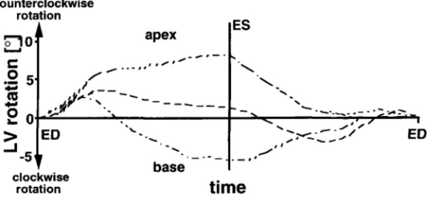
The normal left ventricle performs a systolic clock- wise rotation at the base ( - 3 +_ 2 °) and a counter- clockwise rotation at the apex ( + 1 2 + 4 °) which occurs mainly during isovolumic contraction and re- sults in a systolic 'wringing' motion (Fig. 3). The
counterclockwise rotation ~"~10 ! a p e x s - - - , 0 5' J
*'m
d/,,- ~ ¢ - - - - > ED"-....
j , J ' ' ~ , " 5 " , - - - - base clockwise rotation ES s# _ _ # t t i m eFig. 3. Wringing motion. A clockwise rotation of the cardiac base and a counterclockwise rotation of the apex occur during systolic contraction, resulting in a wringing motion of the left ventricle.
E. Nagel et a l . / Magnetic Resonance Materials' in Physics, Biology and Medicine 6 (1998) 91-93 93 'wringing' motion of the left ventricle allows the heart
to achieve a high intracavitary pressure with minimal radial displacement (analogous to the 'wringing' of a wet towel). An 'untwisting' motion which precedes diastolic filling is observed during isovolumic relax- ation which is directed opposite to the systolic 'wring- ing' motion and results in a counterclockwise rotation at the base and a clockwise rotation at the apex. It is an important determinant of elastic recoil and thus, of diastolic filling and suction. During the systolic ejec- tion and diastolic filling phase hardly any rotation occurs.
7. 3D-motion of the left ventricle in patients with aortic stenosis
In 15 patients with aortic stenosis systolic rotation was reduced at the base ( - 2 _+ 2°; P < 0.05) but in- creased at the apex ( + 15_+6°; P < 0 . 0 5 ) . Diastolic untwisting was delayed and prolonged with a decrease in rotation velocity ( - 7 _+ 1 ° s 1) when compared to controls ( - 11 _+ 2 ° s -
1;
p < 0.001).8. Conclusions
6. 3D-motion of the left ventricle after myocardial in- farction
Regional motion in myocardial infarction depends largely on the loacalization of the infarct and is typi- cally altered with a loss of regional shortening and a prolongation of the 'untwisting' process with a con- secutive reduction in relaxation and diastolic filling. In 14 patients with anterolateral infarcts LV area reduc- tion was reduced in infarct regions (23 _+ 11%) when compared to normal regions (35 ± 10%, P < 0.05). At the same time a reduction of regional circumferential shortening of infarcted (10_+ 5%; P < 0.05 vs remote and controls) and remote regions (15_+6%, P < 0 . 0 5 vs controls) in comparison to controls (21 _+ 5%). In addition a prolongation of the 'untwisting' motion with a reduction in rotation velocity was observed.
Based on our measurements following observations were made:
(1) The left ventricle performs a systolic wringing motion which occurs mainly during isovolumic con- traction.
(2) Diastolic untwisting is found predominantly during isovolumic relaxation and occurs opposite to systolic rotation.
(3) After myocardial infarction regional shortening is reduced in infarcted and remote regions. Predomi- nantly diastolic untwisting is delayed and prolonged. (4) In patients with aortic stenosis apical rotation is enhanced, whereas diastolic untwisting is significantly inhibited, which explains the diastolic dysfunction in these patients.
Myocardial tagging makes an accurate regional wall motion analysis and the assessment of cardiac rota- tion possible and, thus, allow new insight into the mechanical function of the heart.