Remifentanil does not impair left ventricular systolic and
diastolic function in young healthy patients
D. Bolliger
1*, M. D. Seeberger
1, J. Kasper
1, K. Skarvan
1, E. Seeberger
1, G. Lurati Buse
1, P. Buser
2and
M. Filipovic
1,31Department of Anaesthesia and Intensive Care Medicine and2Department of Cardiology, University Hospital Basel, CH-4031 Basel,
Switzerland
3Institute of Anaesthesiology, Cantonal Hospital St. Gallen, Switzerland
* Corresponding author. E-mail: [email protected]
Editor’s key points
† Opioids including remifentanil may directly influence myocardial performance, but in vivo data on the effect of remifentanil on myocardial function in humans are scarce. † In this echocardiographic
study in young healthy subjects, a continuous infusion of remifentanil at a target concentration of 2 ng ml21did not affect systolic and diastolic LV function. † Further studies
investigating higher concentrations of remifentanil and patients with pre-existing cardiac diseases are needed.
Background. Experimental studies and investigations in patients with cardiac diseases suggest that opioids at clinical concentrations have no important direct effect on myocardial relaxation and contractility. In vivo data on the effect of remifentanil on myocardial function in humans are scarce. This study aimed to investigate the effects of remifentanil on left ventricular (LV) function in young healthy humans by transthoracic echocardiography (TTE). We hypothesized that remifentanil does not impair systolic, diastolic LV function, or both.
Methods.Twelve individuals (aged 18 –48 yr) without any history or signs of cardiovascular disease and undergoing minor surgical procedures under general anaesthesia were studied. Echocardiographic examinations were performed in the spontaneously breathing subjects before (baseline) and during administration of remifentanil at a target effect-site concentration of 2 ng ml21 by target-controlled infusion. Analysis of systolic function focused on fractional area change (FAC). Analysis of diastolic function focused on peak early diastolic velocity of the mitral annulus (e′) and on transmitral peak flow velocity (E).
Results.Remifentanil infusion at a target concentration of 2 ng ml21did not affect heart rate or arterial pressure. There was no evidence of systolic or diastolic dysfunction during remifentanil infusion, as the echocardiographic measure of systolic function (FAC) was similar to baseline, and measures of diastolic function remained unchanged (e′) or improved slightly (E).
Conclusion.Continuous infusion of remifentanil in a clinically relevant concentration did not affect systolic and diastolic LV function in young healthy subjects during spontaneous breathing as indicated by TTE.
Keywords: analgesics opioid, remifentanil; heart, myocardial function; monitoring, echocardiography
Accepted for publication: 14 December 2010
Opioids are often used for induction and maintenance of anaesthesia or for sedation in patients at cardiovascular risk on intensive care units, as they are thought to have few haemodynamic side-effects and are claimed to be cardioprotective.1 2 Remifentanil is a newer potent opioid with several distinctive pharmacokinetic properties including short half-life, due to a unique metabolism by plasma and tissue esterase, and potency similar to fentanyl.3 Remifenta-nil has been reported to decrease both heart rate and arterial pressure during general anaesthesia, which may cause severe cardiovascular instability in some cases.4–7 It may affect haemodynamic variables by histamine release or by inhibitory actions on the autonomous and central nervous systems resulting in vasodilation and bradycardia.5–9 In
addition, experimental studies indicate that cardiomyocytes are regulated by opioid receptors (m, d, k). Opioid receptor stimulation causes direct and indirect functional changes in the heart and in myocytes.10–12Therefore, it seems reason-able to speculate that remifentanil influences systolic and diastolic left ventricular (LV) function. In vitro studies on human and animal heart tissue have given conflicting results regarding a direct myocardial effect of different opioids. Fentanyl has been shown to decrease myocardial contractility of isolated rat ventricular myocytes and isolated human heart tissue,13 14 whereas in other in vitro studies, different opioids including fentanyl, sufentanil, and remifen-tanil did not directly impair inotropic and lusitropic properties of both isolated human and perfused rabbit heart tissue.15 16
Differences in species,10 study design, and drug concen-trations may have contributed to the differences between studies.
Although the clinical relevance of experimental studies is acknowledged, it is important that these are supported by direct investigations of the effects of opioids on cardiac func-tion in humans in vivo. The aim of this study was to investi-gate the effects of a clinically relevant concentration of remifentanil on systolic and diastolic LV function in young healthy individuals. We hypothesized that remifentanil at a target effect-site concentration of 2 ng ml2117 would not impair systolic and diastolic LV function as assessed by trans-thoracic echocardiography (TTE).
Methods
Patients
After approval by the local ethics committee (Ethikkommis-sion beider Basel, Basel, Switzerland) and obtaining written informed consent, 12 individuals (Table 1) undergoing minor surgical procedures under general anaesthesia were enrolled. Exclusion criteria were any history or signs of cardiac, pulmonary, or systemic disease, any medication with cardiovascular effects or side-effects, age ,18 or .50 yr, and BMI .30 kg m22. No premedication was given.
After arrival in the preoperative area, i.v. access was established and Ringer’s lactate administered to replace the fluid deficit caused by overnight fasting. The deficit per hour of fasting was calculated as follows: 4 ml kg21h21for the first 10 kg of body weight, 2 ml kg21 h21 for the second 10 kg, and 1 ml kg21h21for every additional kilo-gram. Fifty per cent of the deficit was replaced before the start of the study. To minimize nausea and vomiting, 4 mg of tropisetrone (Navobanw, Novartis Pharma, Basel,
Switzer-land) was given to each patient as soon as i.v. access had been established. Two-lead electrocardiogram with leads II and V5 and pulse oximetry were monitored continuously, and arterial pressure was measured non-invasively every 3 min (PCMS Workstation 90308-15-03, SpaceLab Inc., Redmond, WA, USA). Simultaneously, bispectral index
(BISTM; Aspect 1000TM, Aspect Medical Systems Inc., Natick,
MA, USA; software version 1.01) was monitored continuously. Body temperature was measured continuously and kept above 368C. Hyper- and hypotension, defined as increase and decrease of .30% from baseline value in mean arterial pressure, respectively, were treated with i.v. boluses of gly-ceryl trinitrate (25–50 mg) and phenylephrine (25 –50 mg). Mild tachycardia or bradycardia was not treated, but the study was continued only if the heart rate recovered to values between 50 and 90 beats min21.
The first (baseline) TTE was performed with the patient awake and unpremedicated in a partial left lateral position to optimize imaging quality. This position was maintained until the study was finished. After completion of the baseline TTE, the patient was given oxygen 2 –4 litre min21 by a face mask, and an i.v. infusion of remifentanil (Ultivaw,
GlaxoSmithKline, London, UK) delivered by a target-controlled infusion system (Orchestra& Base Primera, Fresenius Vial, Brezins, France) was started. Target concentration of remifen-tanil was increased stepwise by 0.5 ng ml21. The second TTE was performed as soon as a calculated remifentanil target concentration of 2.0 ng ml2117(corresponding to an infusion rate of 0.08–0.09 mg kg21min21) and stable haemodynamics had been reached. Stable haemodynamics were predefined as ,5% variation of mean arterial pressure and heart rate over three consecutive measurements performed within 6 min. When the second TTE was finished, the study protocol was completed.
Doppler echocardiography
All echocardiograms were obtained with a SonosTM 5500
ultrasonographic system and a 1.8–2.1/3.6 – 4.1 MHz S4 probe (Philips Medical Systems, Best, The Netherlands) according to current guidelines.18 19The echocardiographic data were digitally stored for subsequent off-line analysis. All TTE examinations were performed by the same operator. Standard LV short-axis and two- and four-chamber views were obtained from the parasternal and apical views. For the pulsed-wave Doppler recordings of the mitral inflow, the sample volume was positioned between the tips of the open mitral leaflets using optimal alignment with transmitral blood flow. For recordings of isovolumic relaxation time (IVRT), the beam was slightly moved towards the LV outflow tract to obtain recordings of both LV inflow and LV outflow signals. For recordings of pulsed-wave tissue Doppler imaging, the sample volume was placed at the septal and lateral sides of the mitral annulus, and the acous-tic power and the filter frequencies of the system were set to the lowest possible values. The following variables were measured: end-diastolic and end-systolic areas (EDA and ESA, respectively), peak early and peak late transmitral filling velocities (E and A, respectively), deceleration time (DT), IVRT, early and late diastolic velocities (e′ and a′, respectively), and peak systolic velocity (s′) of the mitral annulus predefined as the average of the septal and lateral mitral annulus measurements obtained by tissue Doppler
Table 1 Patient characteristics. Values are expressed as numbers (%) or median (range). ASA, American Society of Anesthesiology Risk Index; BMI, body mass index
Study patients (n512) ASA class I 12 (100) Women 7 (58) Age (yr) 26 (19 – 45) Weight (kg) 64 (55 – 85) Height (cm) 169 (163 –181) BMI (kg m22) 22 (20 – 26) Haemoglobin (g litre21) 142 (121 –152) Haematocrit (%) 40 (35 – 45)
imaging. The following derived variables were calculated from these data: fractional area change (FAC)¼[(EDA2 ESA)/EDA]×100, ratio E/A, ratio E/e′, and ratio e′/a′. In addition, we determined the myocardial performance index (MPI) as a measure of global cardiac function. This index, defined as the sum of isovolumic contraction and relaxation time divided by the ejection time, was reported by Tei and colleagues20 to be a simple and reproducible indicator of cardiac function and to be independent of heart rate and arterial pressure. LV preload was estimated by EDA and E/e′ ratio (which reflects LV filling pressure); LV afterload was assessed by the end-systolic arterial pressure –area product (SAP×ESA).21
Analysis of systolic function focused on FAC, and analysis of diastolic function focused on e′ and E. FAC correlates well with LV ejection fraction;22E and e′reflect the early dia-stolic filling which depends on the pressure gradient between the atrium and ventricle, LV myocardial relaxation, and early diastolic untwisting.19
All variables were measured at end-expiration over three preferably consecutive cardiac cycles and averaged by one experienced physician-echocardiographer not involved in data acquisition and blinded to all other study data. In a pre-vious study from our group with a similar setting, intra- and inter-observer variability (calculated as the mean absolute difference between two readings divided by their mean and expressed as percentage) were 1.5– 4.2% and 2.6– 6.9%, respectively.23 In the present study, we did not re-evaluate inter- and intra-observer variability.
Statistical analysis
The sample size calculation was based on our previous study24estimating that a size of 12 patients would allow to detect an increase or decrease in e′ or FAC of 20% by the Wilcoxon signed-rank test (a,0.05 and b≥0.8).
Data are presented as median (range) or number (percen-tage) where appropriate. Comparisons for the haemo-dynamic and echocardiographic effects caused by remifentanil were performed by the two-tailed Wilcoxon signed-rank test. A P-value of ,0.05 was considered statisti-cally significant. All statistical analyses were performed using an SPSS for Windows 16.0 computer package (SPSS Inc., Chicago, IL, USA).
Results
The study was safely performed in all patients. No compli-cations occurred during or after the study. No patient showed signs of relevant muscle rigidity or upper airway obstruction during remifentanil infusion. No patient reported nausea. Patients were conscious during the study but became obviously sedated by remifentanil at an end-organ concentration of 2 ng ml21. However, this observation was not reflected by decreased BIS values.
No medication to increase or decrease arterial pressure or heart rate was administered during the study, as arterial
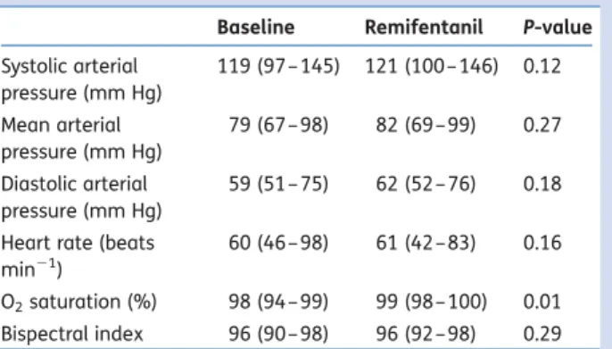
pressure and heart rate did not markedly change during remifentanil infusion (Table2).
Effects of remifentanil on LV systolic function
At baseline, systolic function was normal in all patients: FAC was ≥52%,22 MPI≤0.33,20 and s′≥8.2 cm s21 in each
patient.25 Remifentanil at a target concentration of 2 ng
ml21 did not change the measured echocardiographic
indices of systolic function, that is, FAC, MPI, and s′ were similar to baseline values (Table3).
Effects of remifentanil on LV diastolic function
At baseline, there were no signs of diastolic dysfunction: e′ was ≥12.0 cm s21, E≥64 cm s21, and E/A ratio ≥1.3 in each patient.19 Remifentanil at a target concentration of 2 ng ml21did not induce any echocardiographic signs of dia-stolic dysfunction, i.e. e′ was similar to baseline (P¼0.31), and E and the E/A ratio were slightly increased (P¼0.02 and 0.03, respectively).
Effects of remifentanil on LV preload and afterload
Regarding the echocardiographic indices of LV filling, there was a slight increase in EDA (P¼0.03) but only a trend towards an increased E/e′ ratio (P¼0.06), which better reflects LV filling pressure (Table3).19There was no difference
in the end-systolic arterial pressure –area product before and during remifentanil infusion (P¼0.21).
Discussion
Our study found that remifentanil at a clinically relevant infu-sion rate of 0.08–0.09 mg kg21min21did not impair systolic and diastolic LV function in spontaneously breathing healthy young surgical patients with normal heart function. The echocardiographic indices of systolic function, that is, FAC and s′, were not affected by remifentanil. In contrast, the indices of diastolic function were slightly changed during remifentanil infusion with small but statistically significant increases in E and E/A ratio. However, the lack of any effect
Table 2Physiological variables at baseline and during remifentanil infusion in the 12 study patients. Values are expressed as median (range). P-values were calculated by the Wilcoxon signed-rank test
Baseline Remifentanil P-value Systolic arterial pressure (mm Hg) 119 (97 – 145) 121 (100 – 146) 0.12 Mean arterial pressure (mm Hg) 79 (67 – 98) 82 (69 –99) 0.27 Diastolic arterial pressure (mm Hg) 59 (51 – 75) 62 (52 –76) 0.18
Heart rate (beats min21)
60 (46 – 98) 61 (42 –83) 0.16
O2saturation (%) 98 (94 – 99) 99 (98 –100) 0.01
on e′calls into question the interpretation of these changes as being indicative of improved early diastolic function. Preload was only minimally affected by remifentanil, as indi-cated by slightly increased EDA but insignificantly increased E/e′ratio. Afterload remained similar to baseline during remi-fentanil infusion, as indicated by the unchanged end-systolic pressure2area product. As our study was powered to assess changes in e′and FAC as main diastolic and systolic variables, differences in other echocardiographic indices must be inter-preted cautiously.
Comparison of our findings with previous reports is compli-cated by several facts. First, most previous studies investigated the effects of remifentanil in experimental designs using human and animal heart tissue rather than in vivo.14–16 Sec-ondly, to our knowledge, modern echocardiographic tech-niques have not previously been used to evaluate the effects of opioids on diastolic and systolic cardiac function in adults. An echocardiographic study in children anaesthetized with sevoflurane showed a decreased cardiac output by the addition of remifentanil, mainly as a result of a decrease in heart rate.9 Another echocardiographic study from 1986 using the inferior M-mode technique showed that naloxone, an opioid antagonist, did not affect cardiac dimension and function in healthy adult subjects.26 Thirdly, former in vivo studies investigated patients with cardiac diseases and impaired heart function or critically ill patients.5 7–8 27 28 However, in agreement with our results, remifentanil did not impair systolic and diastolic properties of myocardial tissue and of isolated rabbit hearts in two experimental studies.15
16Also, in support of our findings is the fact that remifentanil
has been successfully used as an anaesthetic agent in high-risk patients undergoing cardiac surgery, implantation of cardiac defibrillators, and Caesarean section in the presence of peripartal cardiomyopathy.7 29 30These investigations and
reports showed that remifentanil provides stable haemo-dynamic conditions even in patients with severely impaired cardiac function.
Conflicting findings have been reported by several studies. A porcine study found that remifentanil directly affects the sinus node,31 thereby inducing bradycardia. Other exper-imental studies found that remifentanil has a vasorelaxant effect on human saphenous veins14 and radial arteries.32 Several human studies on remifentanil found cardiovascular side-effects including severe bradycardia and hypotension due to vasodilation.4–9 In patients with total artificial hearts, remifentanil induced a dose-dependent decrease in systemic arterial pressure and a decreased resistance in arterial vessels but not in veins.8
There are several reasons that may explain the conflicting findings including differences in the patient population,8 11
12 the study protocol,13 14 and the species.10 13 16 For
example, the effects of remifentanil on myocardial function and haemodynamics in healthy individuals with normal heart function, as in our study, may differ from those in criti-cally ill or cardiac patients.7 8 11 12Another reason for differ-ences between the studies is that hypotension and bradycardia seem to be particularly pronounced when a high dose or bolus of remifentanil is administered, or during co-administration of remifentanil together with propofol or other anaesthetics during the induction of anaesthesia.5 9In our study, we did not administer remifentanil at a high dose and administered it as the sole drug. Another reason for con-flicting results is the use of neuromuscular blocking agents and intermittent positive pressure ventilation in several other studies in anaesthetized patients,5 7 8 which both may affect cardiac function by affecting preload and after-load.24 33 Taken together, the results from our study and former in vitro and in vivo studies and also case reports
Table 3 Echocardiographic variables at baseline and during remifentanil infusion in the 12 study patients, and reported normal values of these variables in healthy, awake subjects aged 20 –40 yr.19 20 23 43*Study data are expressed as median (range). P-values were calculated by the Wilcoxon signed-rank test.†Normal values are expressed as mean (SD). N/A, not available
Baseline* Remifentanil* P-value Normal values†
Peak early transmitral filling velocity, E (cm s21) 89 (63 –117) 95 (75 –114) 0.02 76 (15)
Peak late transmitral filling velocity, A (cm s21) 52 (29 –74) 52 (33 –71) 0.53 44 (14)
Ratio E/A 1.6 (1.3– 2.5) 2.0 (1.4 –3.0) 0.03 1.8 (0.6)
Peak early mitral diastolic annular velocity, e′(cm s21) 14.8 (12.0 –20.4) 15.1 (12.7 – 21.4) 0.31 14.1 (2.7)
Peak late mitral diastolic annular velocity, a′(cm s21) 8.1 (5.6– 12.5) 8.6 (4.7 –11.6) 0.53 9.1 (1.7)
Ratio e′/a′ 2.0 (1.1– 3.0) 1.9 (1.2 –3.2) 0.39 1.8 (0.6)
Ratio E/e′ 5.8 (4.0– 7.3) 6.8 (4.6 –7.6) 0.06 5.6 (1.3)
Deceleration time, DT (ms) 189 (153 – 263) 177 (148 –257) 0.17 217 (65)
Isovolumic relaxation time, IVRT (ms) 59 (42 –93) 52 (37 –85) 0.07 91 (17)
Peak systolic mitral annular velocity, s′(cm s21) 10.3 (8.2– 11.7) 10.4 (7.6 –12.2) 0.53 9.4 (1.4)
End-diastolic area, EDA (cm2) 16.6 (12.5 –21.6) 17.2 (13.9 – 23.3) 0.03 18.8 (1.4)
End-systolic area, ESA (cm2) 7.5 (4.4– 10.4) 7.3 (5.1 –10.7) 0.30 8.5 (2.0)
Fractional area change, FAC (%) 56 (52 –65) 58 (52 –63) 0.37 56 (5)
End-systolic arterial pressure – area product, SAP×ESA (mm Hg cm2) 818 (540 – 1298) 836 (631 –1293) 0.21 N/A
suggest that remifentanil at a low-dose infusion rate does not relevantly impair myocardial systolic, diastolic LV function, or both. However, cardiovascular side-effects may become evident with remifentanil bolus administration or infusion at high dose or when co-administrated with propofol or sevoflur-ane.5 9
Besides a pronounced analgesic effect, all opioids also provide dosage-related sedative effects. During remifentanil infusion in this study, patients became sedated but all patients remained conscious and BIS values were unaffected. Previous studies investigating the effect of remifentanil on BIS values have yielded controversial results,6 34 35 but the comparison with our data is complicated by the fact that in those studies remifentanil was administered in critically ill patients27or co-administered with other sedatives.6 34 35As we did not formally assess depth of sedation, conclusions about the effect of the depth of sedation on cardiac perform-ance are not possible from our data.
Our study has several limitations. First, the study protocol did not include a dose –response evaluation. Safety concerns regarding adequate respiratory drive in spontaneously breathing patients27 and regarding haemodynamic stab-ility4–7 kept us from administering high concentrations of remifentanil requiring tracheal intubation and the adminis-tration of other medication that would potentially influence cardiac function. The investigated target concentration of 2 ng ml21 corresponds to remifentanil infusion rates that have been used safely during surgical procedures,36 37 for sedation on the intensive care unit,27 28 or for early post-operative analgesia.38 Therefore, the present study gives clinically important information on the effects of remifentanil on LV function at a lower dose but cannot exclude different effects at a higher dose, or after combined administration of remifentanil together with other analgesic or anaesthetic drugs. In addition, no conclusions must be drawn from our findings with remifentanil to the effects of other opioids.
Secondly, spontaneous breathing during remifentanil infu-sion and oxygen supplementation might result in elevated concentrations of PaCO2 and PaO2. Hypercapnia is known to
decrease systemic vascular resistance and increase stroke volume and cardiac output,39 40 whereas hyperoxaemia may increase systemic vascular resistance.41 Therefore, both increased concentration of PaCO2 and PaO2 may
poten-tially influence LV filling and consequently LV function. However, remifentanil infusion up to 0.1 mg kg21min21in spontaneously breathing critically ill patients did not result in a suppression of respiratory drive or in a relevant increase in PaCO2.
27 The use of an even lower remifentanil
concen-tration in our study suggests unchanged PaCO2
concen-trations in our patients, but we do not have measurements to confirm this. Therefore, we cannot fully exclude an increase in PaCO2in our patients during remifentanil
adminis-tration. Changes in PaCO2and PaO2are unlikely to be
confoun-ders in our study, because previous studies have found that PaCO2 did not relevantly impair diastolic and systolic
func-tion23 40 and that PaO2 may only affect cardiac function at
very high levels.41
Thirdly, we applied a standardized infusion therapy but could not strictly control preload and afterload, both of which may influence echocardiographic indicators of systolic and diastolic function.42 However, stable haemodynamics and unchanged intracardiac pressures, as indicated by the E/e′ ratio and the end-systolic pressure –area product, strongly suggest that there were no relevant changes in preload and afterload that might have confounded our results.
Finally, it must be noted that we did not measure remifen-tanil plasma concentrations. We used a commercially avail-able and widely used calculated pharmacokinetic model taking into account patient’s age, weight, height, and gender.17 A previous study using a similar computerized program showed that the difference between calculated and measured remifentanil plasma concentrations may be substantial at target concentrations ≥5 ng ml21but much smaller at the concentration used in the present study.6 However, we cannot completely exclude potentially substan-tial differences between calculated and existing remifentanil concentrations in our study patients, as we did not measure plasma concentrations.
In conclusion, the present study found that remifentanil at a calculated target effect-site concentration of 2 ng ml21did not impair systolic or diastolic cardiac function in young sur-gical patients free from cardiac disease. These results support previous data suggesting that remifentanil at a low dose is a suitable drug for sedation in patients with normal heart function undergoing monitored anaesthesia care or on intensive care units. Further studies in patients at cardiac risk are needed before such a conclusion is extended to patients with pre-existing cardiac disease.
Acknowledgement
The authors thank Allison Dwileski, BS, Department of Anaes-thesia, University Hospital Basel, Basel, Switzerland, for edi-torial assistance.
Conflict of interest
None declared.
Funding
This study was supported, in part, by grants from the Swiss National Science Foundation, Bern, Switzerland (grant # 3200B0-116229) and from the European Association of Car-diothoracic Anaesthesiologists (2007 Research Grant to D.B.).
References
1 Miller DR, Wellwood M, Teasdale SJ, et al. Effects of anesthetic induction on myocardial function and metabolism: a comparison of fentanyl, sufentanil and alfentanil. Can J Anaesth 1988; 35: 219– 33
2 Sneyd JR. Recent advances in intravenous anaesthesia. Br J Anaesth 2004; 93: 725 –36
3 Michelsen LG, Hug CC Jr. The pharmacokinetics of remifentanil. J Clin Anesth 1996; 8: 679 –82
4 Jhaveri R, Joshi P, Batenhorst R, Baughman V, Glass PS. Dose com-parison of remifentanil and alfentanil for loss of consciousness. Anesthesiology 1997; 87: 253– 9
5 Elliott P, O’Hare R, Bill KM, Phillips AS, Gibson FM, Mirakhur RK. Severe cardiovascular depression with remifentanil. Anesth Analg 2000; 91: 58– 61
6 Hoymork SC, Raeder J, Grimsmo B, Steen PA. Bispectral index, serum drug concentrations and emergence associated with indi-vidually adjusted target-controlled infusions of remifentanil and propofol for laparoscopic surgery. Br J Anaesth 2003; 91: 773– 80 7 Kazmaier S, Hanekop GG, Buhre W, et al. Myocardial conse-quences of remifentanil in patients with coronary artery disease. Br J Anaesth 2000; 84: 578 –83
8 Ouattara A, Boccara G, Kockler U, et al. Remifentanil induces sys-temic arterial vasodilation in humans with a total artificial heart. Anesthesiology 2004; 100: 602– 7
9 Chanavaz C, Tirel O, Wodey E, et al. Haemodynamic effects of remifentanil in children with and without intravenous atropine. An echocardiographic study. Br J Anaesth 2005; 94: 74– 9 10 Barron BA. Opioid peptides and the heart. Cardiovasc Res 1999;
43: 13 –6
11 Bolte C, Newman G, Schultz Jel J. Kappa and delta opioid receptor signaling is augmented in the failing heart. J Mol Cell Cardiol 2009; 47: 493– 503
12 Bolte C, Newman G, Schultz Jel J. Hypertensive state, indepen-dent of hypertrophy, exhibits an attenuated decrease in systolic function on cardiac kappa-opioid receptor stimulation. Am J Physiol Heart Circ Physiol 2009; 296: H967–75
13 Kanaya N, Zakhary DR, Murray PA, Damron DS. Differential effects of fentanyl and morphine on intracellular Ca2+ transients and contraction in rat ventricular myocytes. Anesthesiology 1998; 89: 1532–42
14 Duman A, Saide Sahin A, Esra Atalik K, et al. The in vitro effects of remifentanil and fentanyl on isolated human right atria and saphenous veins. J Cardiothorac Vasc Anesth 2003; 17: 465– 9
15 Hanouz JL, Yvon A, Guesne G, et al. The in vitro effects of remifen-tanil, sufenremifen-tanil, fentanyl, and alfentanil on isolated human right atria. Anesth Analg 2001; 93: 543– 9
16 Lecomte P, Ouattara A, Le Manach Y, Landi M, Coriat P, Riou B. The coronary and myocardial effects of remifentanil and sufentanil in the erythrocyte-perfused isolated rabbit heart. Anesth Analg 2006; 103: 9–14
17 Minto CF, Schnider TW, Egan TD, et al. Influence of age and gender on the pharmacokinetics and pharmacodynamics of remifentanil. I. Model development. Anesthesiology 1997; 86: 10–23
18 Lang RM, Bierig M, Devereux RB, et al. Recommendations for chamber quantification: a report from the American Society of Echocardiography’s Guidelines and Standards Committee and the Chamber Quantification Writing Group, developed in conjunc-tion with the European Associaconjunc-tion of Echocardiography, a branch of the European Society of Cardiology. J Am Soc Echocar-diogr 2005; 18: 1440– 63
19 Nagueh SF, Appleton CP, Gillebert TC, et al. Recommendations for the evaluation of left ventricular diastolic function by echocardio-graphy. Eur J Echocardiogr 2009; 10: 165 –93
20 Tei C, Ling LH, Hodge DO, et al. New index of combined systolic and diastolic myocardial performance: a simple and reproducible measure of cardiac function—a study in normals and dilated car-diomyopathy. J Cardiol 1995; 26: 357 –66
21 Greim CA, Roewer N, Schulte am Esch J. Assessment of changes in left ventricular wall stress from the end-systolic pressure– area product. Br J Anaesth 1995; 75: 583– 7
22 Clements FM, Harpole DH, Quill T, Jones RH, McCann RL. Esti-mation of left ventricular volume and ejection fraction by two-dimensional transoesophageal echocardiography: comparison of short axis imaging and simultaneous radionuclide angiogra-phy. Br J Anaesth 1990; 64: 331– 6
23 Bolliger D, Seeberger MD, Kasper J, et al. Different effects of sevo-flurane, dessevo-flurane, and isoflurane on early and late left ventricu-lar diastolic function in young healthy adults. Br J Anaesth 2010; 104: 547 –54
24 Filipovic M, Wang J, Michaux I, Hunziker P, Skarvan K, Seeberger MD. Effects of halothane, sevoflurane and propofol on left ventricular diastolic function in humans during spon-taneous and mechanical ventilation. Br J Anaesth 2005; 94: 186– 92
25 Ruan Q, Nagueh SF. Effect of age on left ventricular systolic func-tion in humans: a study of systolic isovolumic accelerafunc-tion rate. Exp Physiol 2005; 90: 527– 34
26 Staessen J, Fagard R, Fiocchi R, Lijnen P, Amery A. An echocardio-graphic study on the influence of endogenous opioids on cardiac dimensions and function in normal subjects. J Cardiovasc Ultraso-nography 1986; 5: 71– 7
27 Cavaliere F, Antonelli M, Arcangeli A, et al. A low-dose remifenta-nil infusion is well tolerated for sedation in mechanically venti-lated, critically-ill patients. Can J Anaesth 2002; 49: 1088–94 28 Tipps LB, Coplin WM, Murry KR, Rhoney DH. Safety and feasibility
of continuous infusion of remifentanil in the neurosurgical inten-sive care unit. Neurosurgery 2000; 46: 596 –601
29 Howie MB, Michelsen LG, Hug CC Jr, et al. Comparison of three remifentanil dose-finding regimens for coronary artery surgery. J Cardiothorac Vasc Anesth 2003; 17: 51 –9
30 Lehmann A, Boldt J, Zeitler C, Thaler E, Werling C. Total intrave-nous anesthesia with remifentanil and propofol for implantation of cardioverter-defibrillators in patients with severely reduced left ventricular function. J Cardiothorac Vasc Anesth 1999; 13: 15 –9 31 Zaballos M, Jimeno C, Almendral J, et al. Cardiac
electrophysio-logical effects of remifentanil: study in a closed-chest porcine model. Br J Anaesth 2009; 103: 191 –8
32 Gursoy S, Bagcivan I, Yildirim MK, Berkan O, Kaya T. Vasorelaxant effect of opioid analgesics on the isolated human radial artery. Eur J Anaesthesiol 2006; 23: 496 –500
33 Filipovic M, Michaux I, Wang J, Hunziker P, Skarvan K, Seeberger M. Effects of sevoflurane and propofol on left ventricu-lar diastolic function in patients with pre-existing diastolic dys-function. Br J Anaesth 2007; 98: 12–8
34 Lysakowski C, Dumont L, Pellegrini M, Clergue F, Tassonyi E. Effects of fentanyl, alfentanil, remifentanil and sufentanil on loss of consciousness and bispectral index during propofol induc-tion of anaesthesia. Br J Anaesth 2001; 86: 523– 7
35 Koitabashi T, Johansen JW, Sebel PS. Remifentanil dose/electro-encephalogram bispectral response during combined propofol/ regional anesthesia. Anesth Analg 2002; 94: 1530– 3
36 Gesztesi Z, Mootz BL, White PF. The use of a remifentanil infusion for hemodynamic control during intracranial surgery. Anesth Analg 1999; 89: 1282– 7
37 Albertin A, Casati A, Federica L, et al. The effect-site concentration of remifentanil blunting cardiovascular responses to tracheal intu-bation and skin incision during bispectral index-guided propofol anesthesia. Anesth Analg 2005; 101: 125– 30
38 Schraag S, Kenny GN, Mohl U, Georgieff M. Patient-maintained remifentanil target-controlled infusion for the tran-sition to early postoperative analgesia. Br J Anaesth 1998; 81: 365– 8
39 Carvalho CR, Barbas CS, Medeiros DM, et al. Temporal hemodynamic effects of permissive hypercapnia associated with ideal PEEP in ARDS. Am J Respir Crit Care Med 1997; 156: 1458– 66
40 Foex P, Prys-Roberts C. Effect of CO2on myocardial contractility
and aortic input impedance during anaesthesia. Br J Anaesth 1975; 47: 669– 78
41 Mak S, Azevedo ER, Liu PP, Newton GE. Effect of hyperoxia on left ventricular function and filling pressures in patients with and without congestive heart failure. Chest 2001; 120: 467 –73 42 Pagel PS. Anesthetics and echocardiographic assessment of left
ventricular function: lessons learned from invasive analysis of car-diovascular mechanics. J Am Soc Echocardiogr 2006; 20: 440– 1 43 Dalen H, Thorstensen A, Vatten LJ, Aase SA, Stoylen A. Reference
values and distribution of conventional echocardiographic Doppler measures and longitudinal tissue Doppler velocities in a population free from cardiovascular disease. Circ Cardiovasc Imaging 2010; 3: 614 –22