HAL Id: hal-00419257
https://hal.archives-ouvertes.fr/hal-00419257
Submitted on 23 Sep 2009HAL is a multi-disciplinary open access archive for the deposit and dissemination of sci-entific research documents, whether they are pub-lished or not. The documents may come from teaching and research institutions in France or abroad, or from public or private research centers.
L’archive ouverte pluridisciplinaire HAL, est destinée au dépôt et à la diffusion de documents scientifiques de niveau recherche, publiés ou non, émanant des établissements d’enseignement et de recherche français ou étrangers, des laboratoires publics ou privés.
Power and limitations of electrophoretic separations in
proteomics strategies.
Thierry Rabilloud, Ali R Vaezzadeh, Noelle Potier, Cécile Lelong,
Emmanuelle Leize-Wagner, Mireille Chevallet
To cite this version:
Thierry Rabilloud, Ali R Vaezzadeh, Noelle Potier, Cécile Lelong, Emmanuelle Leize-Wagner, et al.. Power and limitations of electrophoretic separations in proteomics strategies.. Mass Spectrometry Reviews, Wiley, 2009, 28 (5), pp.816-43. �10.1002/mas.20204�. �hal-00419257�
Power and limitations of electrophoretic separations in proteomics
strategies
Thierry. Rabilloud 1,2, Ali R.Vaezzadeh 3 , Noelle Potier 4, Cécile Lelong1,5, Emmanuelle Leize-Wagner 4, Mireille Chevallet 1,2
1: CEA, IRTSV, LBBSI, 38054 GRENOBLE, France.
2: CNRS, UMR 5092, Biochimie et Biophysique des Systèmes Intégrés, Grenoble France 3: Biomedical Proteomics Research Group, Central Clinical Chemistry Laboratory, Geneva University Hospitals, Geneva, Switzerland
4: CNRS, UMR 7177. Institut de Chime de Strasbourg, Strasbourg, France 5: Université Joseph Fourier, Grenoble France
Correspondence :
Thierry Rabilloud, iRTSV/LBBSI, UMR CNRS 5092, CEA-Grenoble, 17 rue des martyrs,
F-38054 GRENOBLE CEDEX 9 Tel (33)-4-38-78-32-12
Fax (33)-4-38-78-44-99
e-mail: Thierry.Rabilloud@ cea.fr
Abstract:
Proteomics can be defined as the large-scale analysis of proteins. Due to the complexity of biological systems, it is required to concatenate various separation techniques prior to mass spectrometry. These techniques, dealing with proteins or peptides, can rely on chromatography or electrophoresis. In this review, the
electrophoretic techniques are under scrutiny. Their principles are recalled, and their applications for peptide and protein separations are presented and critically discussed. In addition, the features that are specific to gel electrophoresis and that interplay with mass spectrometry( i.e., protein detection after electrophoresis, and the process
leading from a gel piece to a solution of peptides) are also discussed.
Keywords: electrophoresis, two-dimensional electrophoresis, isoelectric focusing, immobilized pH gradients, peptides, proteins, proteomics.
Table of contents
I. Introduction
II. The principles at play
III.
How to use electrophoresis in a proteomics strategy
III.A.. Electrophoretic separation of peptides.
III.A.1. Capillary IEF III.A.2. Solution IEF III.A.3. IPG-IEF III.A.4. Off-Gel
III.A.5. A new dimension for data validation
III.B. Electrophoretic separation of proteins.
III.B.1. Electrophoretic separations of native proteins
III.B.2. Electrophoretic separations of denatured proteins-rationale
III.B.3. The implementation of denaturing protein electrophoresis in proteomics III.B.4. The workhorse of protein separation in proteomics: SDS gel electrophoresis III.B.5. Classical high-resolution 2D electrophoresis: the source of proteomics III.B.6. Specialized, IEF-free, 2D electrophoretic systems
IV. From gels to peptides: additional features of electrophoresis in a proteomics
strategy
IV.A. Detection of proteins after gel electrophoresis IV.B. Production of peptides from gel-separated proteins
V. Concluding remarks
I. Introduction
Among the variety of available spectroscopic techniques, the combination of speed, sensitivity, and accuracy makes mass spectrometry an obvious choice in proteomics strategies; i.e., strategies that aim at the wide-scale characterization of proteins and protein variants in biological samples.
However, mass spectrometry alone cannot solve this analytical problem of the wide-scale characterization of proteins, for several major reasons. First of all, biological samples often contain several hundreds to several tens of thousands of different protein forms. The word of “protein forms” encompass, of course, gene products with all of their
post-translational modifications (PTM); e.g., glycosylation, phosphorylation, or protein cleavage. On top of the diversity problem, the dynamic range of protein presence ( i.e., the mass ratio between the rarest protein form and the most abundant ones) is often far beyond the
quantitative dynamic range of a mass spectrometer. For example, this dynamic range covers 4 orders of magnitude in prokaryotic samples, 6 in eukaryotic cells (Lu et al., 2007), and 12 in complex biological fluids such as plasma (Anderson and Anderson, 2002).
Still going on this trend of complexity, the analyte itself (i.e, the protein) is very complex, so that even the most precise mass measurement of a complete, modified protein often does not give an unequivocal answer, in the sense that several modified proteins can fit within the same mass measurement window.
Last but not least, it is quite frequent in biology to have to address quantitation problems. A good example is provided by blood tests, in which normal and pathological values are defined, with the normal values frequently not zero. Thus, a quantitative approach is often needed, and this goal is not always easy to implement for mass spectrometry.
However, the discussion of the quantitation issues in mass spectrometry for proteomics is clearly outside the scope of this review.
Because of the complexity of the analytes, the mass spectrometric measurement is not carried out on intact proteins, but on smaller peptides that are produced from the proteins by a controlled proteolytic process. The multiple measurements made on peptide masses, either on a few peptides by MS/MS or on a combination of peptides that arise from the same protein (e.g., for peptide mass fingerprinting), allow an unequivocal characterization of the proteins of interest and sometimes of some post-translational modifications.
Because of the complexity of biological samples, it is absolutely necessary to separate (and sometimes to quantify) the analytes prior to their measurement with mass spectrometry, so that the complexity of what is introduced into the mass spectrometer is compatible with the performance of the instrument in the chosen measurement mode. This separation can be made on the proteins of the sample or on the peptides that arise from protein digestion (or on both). Among the various separation modes available for proteins and peptides,
chromatography and electrophoresis are almost exclusively used at the current time. Thus the purpose of this review is to provide to the reader a critical review of the input of
electrophoretic techniques in a proteomics strategy.
II. The principles at play
By definition, electrophoresis consists of the separation of analytes (here peptides or proteins) as ions that are driven differently (in order to achieve separation) under an electric field. Two
modes of electrophoresis are mostly used for peptide and protein separation; zone electrophoresis and isoelectric focusing.
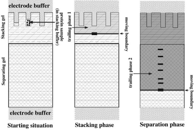
In zone electrophoresis, a speed race is made in the separation medium, usually at constant pH, and the speed of each analyte is dictated by its charge, which drives it under the electric field, and by the friction forces, which slow it down. In order to minimize the impact of diffusion, which always tends to broaden the peaks of analytes, and also to minimize the impact of the volume of the initial sample on the final resolution, a special trick, called discontinuous electrophoresis, is used (Davis, 1964), schematized on figure 1. In this system, a composite separation medium with two phases is used, (figure 1, left panel). The sample is loaded on top of the first phase, called the concentration phase, where an isotachophoresis is carried out. Isotachophoresis (same speed electrophoresis) uses the rules of transport of ions in an electric field to build what is called a moving boundary, in which the ions are ranked in the order of their mobility. Under proper conditions, all analyte ions can be included in this moving boundary, whose dimensions are dictated by the ionic strength of the medium and by the concentration of each ion (for detailed calculations, see Jovin (Jovin, 1973a, Jovin, 1973b). By sweeping this moving boundary through the sample and then through the
concentration phase of the separation medium, all analytes can be concentrated in the moving boundary (figure 1, center panel). Although this produces a spectacular increase in resolution, the concentration zone reflects its name very adequately, because the analytes are extremely concentrated in the moving boundary, with the risk that they exceed their solubility and start to precipitate. But, by definition, no macroscopic separation of the analytes takes place in the concentration phase. Separation takes place in another phase, called the separation phase, in which the speed of the moving boundary is increased to exceed the mobility of the analytes, which are left behind and separated by zone electrophoresis in the trailing phase (figure1, right panel).
Isoelectric focusing exploits special properties of some complex ions, which bear both weak acid and weak bases, which is the case of peptides and proteins via the amino and carboxy termini and by the side chains of some amino acids. Such complex ions have an isoelectric point, also noted pI (i.e. a pH at which the positive and negative charges are equal) so that the resulting electric charge of the analyte is zero. Below the isoelectric point (i.e. at a more-acidic pH) the analyte will be a cation, and above the isoelectric point (i.e. more-basic pH) it will be an anion.
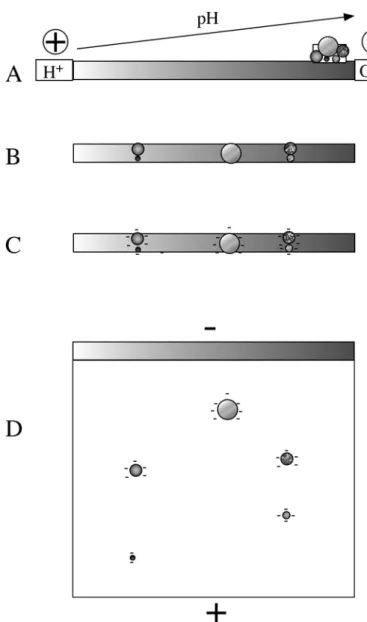
The basis of isoelectric focusing is to use a separation medium in which a smooth pH gradient that encompasses all of the pIs of the analytes of interest (figure 2A). If the acidic end of the gradient is connected to the + electrode and the basic end to the - electrode, then the analytes will be separated according to their pIs, in the sense that each analyte, wherever it is applied in the pH gradient, will migrate to its steady-state position represented by its pI (figure 2B). The method is called isoelectric focusing because each analyte is concentrated at its pI by the counteracting forces induced by diffusion (which tends to broaden the analyte zone) and by the electric field (which tend to drive back the analyte at its exact pI) (see figure 2C). More detailed explanations on isoelectric focusing can be found in dedicated books (Righetti, 1983).
Opposite to the situation in chromatography, the separation in electrophoresis is not strictly dependent on the presence of a solid phase. However, the choice of electrophoresis configurations is indirectly limited by the Joule effect. The heat induced by an electric current provokes important convection flows, which limit in turn the resolution by blurring the separations produced upon electrophoresis. Thus, adequate resolutions are obtained only
when convection flows are limited, and this limitation is achieved mostly by two very different setups.
The most obvious setup, but not the most extensively-used one, is to use a capillary fluid column for separation. Because of the minimal cross-section, minimal electric power is used, and thus minimal heating is produced.
Although such a setup could be, in principle, a real alternative to chromatographic columns for the separation of proteins or peptides, it has never gained widespread use, although there are some publications in the field. This limited use is due to a variety of reasons, among which two prominent ones are worth mentioning.
First, the capillary electrophoresis separation of complete proteins has always been plagued by adsorption on the walls, which has prevented any robustness in separation
Second and more importantly, the loading achievable in capillary electrophoresis is too low to be of practical interest in proteomics studies. Indeed, proteomics studies imply that one can analyze at the same time rather abundant analytes and rather rare ones in the same sample. That requirement implies in turn that one can load rather high amounts of sample. Such loadings are achievable in column chromatography by recirculating the sample in a loading cartridge ,which is pulsed into the capillary column, but this preconcentration process is not so easy to achieve with a capillary electrophoresis setup, despite the use of stacking processes (Locke and Figeys, 2000), solid-phase extraction (Figeys et al., 1998) or isoelectric focusing and its built-in concentrating effect (Simpson and Smith, 2005, Yu et al., 2006, Wang et al., 2007).
All these reasons explain why the second setup for limiting convection ( i.e. gel electrophoresis) has gained the most widespread use. As its name says, it consists of
conducting the electrophoretic separation in a conductive gel; i.e,. a mixed buffer-crosslinked polymer system that has pores of a more or less defined size. The polymer chains that delimit the pores break the convection flows, and ensure adequate resolution. Moreover, they also limit the diffusion, which thereby further enhances resolution. Last but not least, the friction of the analytes on the gel material adds another separation parameter that can be used to enhance the resolution of the complete technique, especially when the size of the analyte is the separating parameter.
In addition to these advantages in separation, working in a gel medium has other important advantages related to the use of solid phase chemistry, but these features are detailed later in this paper (section IV.B).
However, the price to pay for the use of gel media is the loss of automation, but this loss is compensated, at least in part by the fact that gel separations are continuous separations and can be parallel ones, either handling multiple samples on one gel, or providing a
continuous two-dimensional process. This latter aspect is worth discussing in more detail: When a liquid separation is carried out, and when the separated analytes must be further analyzed in another liquid (or gel) separation process, fractions must be collected. When a few analytes are separated, this fraction collection process can be data-dependent, thereby isolating each analyte peak in a limited number of fractions. However, when very complex samples are separated, as is the case in proteomics, data-dependent fraction collection becomes pointless, because it is obvious that any fraction collection scheme will split at least some peaks in two. This peak splitting means in turn that the fraction-collection process that is mandatory in liquid separations introduces artefactual discontinuities in the separations, and splits some analytes into multiple fractions. A good visual illustration of this phenomenon can be found in schemes that couple liquid chromatography to gel
electrophoresis (Szponarski et al., 2004, Szponarski et al., 2007, Schluesener et al., 2007). Conversely, two-dimensional gels, whatever the separation principles used in both
dimensions, represent a happy exception to this rule, because the first-dimension gel, which represents a continuous separation of the analytes, can be loaded as a whole on the top of second dimension gel, to provide a completely continuous two-dimensional separation, therefore without artefactual discontinuity. This continuity in separation, results in higher resolution and higher reproducibility of the two-dimensional gel-based separations.
Of course, there are constraints on the gels that can be used in electrophoretic
separations. Such gels should have pores of an adequate and tunable size, to allow either non-restrictive gels or non-restrictive gels to be used. More importantly, such gels should be devoid of electric charges, which would otherwise cause strong electroendosmosis that complicates or disturb the separation. They should also not adsorb proteins, because adsorption would cause tailing of the separations. In the frame of proteomics studies, these gels should also resist the organic solvents used in the washing, digestion and extraction steps.
This series of constraints have confined the gels used in electrophoresis to crosslinked polyacrylamide. Because of its crosslinked nature, this polymer is stable to many adverse conditions. However, it is not very stable in strongly basic conditions, which lead to the hydrolysis of the amido group. The gel characteristics are also altered by organic solvents (Righetti 1989), which limits the scope of use of this gel. For these reasons, alternate acrylic monomers have been proposed (Artoni et al., 1984, Zewert and Harrington, 1992, Chiari et al., 1994, Simò-Alfonso et al., 1996), but none of them has gained any practical widespread use.
It should be also mentioned that polyacrylamide gels are polymerized in situ, and that process poses additional problems. As a matter of facts, only a fraction of the monomers is polymerized when gelification occurs (Righetti and Caglio, 1993); however the monomer conversion in the polymer continues after gelification to yield much better final incorporation yields (Gelfi and Righetti, 1981a, Gelfi and Righetti, 1981b). However, even with a 90% final conversion rate, the free acrylamide monomer exists in concentrations in the 0.1M range, and this free monomer can react with proteins side chains (Chiari et al., 1992), to lead to complications in the proteomics experiments.
Moreover, acrylamide-based gels can be used to devise immobilized pH gradients (Rosengren et al., 1977) in which the gradient is formed by copolymerization of buffering acrylamide monomers with the standard acrylamide and crosslinker. This way of creating the pH gradient mandatory for isoelectric focusing has revolutioned the scope and use of IEF. Detailed description of the many advantages of immobilized pH gadients is far beyond the scope of this paper, and can be found in monographies (Righetti, 1990).
Although gel-based separations represent by far the most important part of
electrophoretic separations used in proteomics studies, some non-capillary, gel-free systems are sometimes used, most often in a complex separation scheme where they are used in an upstream part, with gel electrophoresis being often the downstream part. When confining the point to proteins and peptide separations, and not to pre-separations of biological samples (e.g. , Zischka et al., 2003), these schemes use almost always isoelectric focusing (Hoffmann et al., 2001, Xie et al., 2005) because this type of separation is less sensitive to convection for several reasons. First of all, IEF operates at very low ionic strength, thereby limiting the Joule effect. Second, IEF is a steady-state process, in which molecules moved away from their steady-state position will be driven back to it by the electric field. Thus, although several devices for free-flow isoelectric focusing of molecules have been described in the literature, those that have been used in proteomics studies belong mainly to the segmented systems, either using ampholytes to build the pH gradients (Egen et al. 1988), (Shang et al., 2003), or
using isolectric membranes (Righetti et al., 1987, Righetti et al., 2005, Han et al., 2008), or a derivative thereof (Ros et al., 2002). Another variant of these IEF-based pre-fractionation techniques is represented by the Sephadex IEF (Radol,a 1973, Radola, 1975, Görg et al., 2002), in which the IEF process is conducted in a layer of granulated beads.
III. How to use electrophoresis in a proteomics strategy
As stated in the introduction, the analytes in a proteomics strategy are by definition proteins, or the peptide that arise from their natural or provoked degradation. Consequently, the application of electrophoresis in proteomics can be split into two subfields, which are, respectively, the separation of proteins and the separation of peptides.
However, a central point crosses both subfields, and is found in all "omics". In contrast to what is done in classical biochemistry, where the separation processes are optimized for each and every protein under study, the large-scale approaches rely on a "one size fits all" dogma. Even if this universality is not possible, at least in the proteomics world, the approach is still a "one size fits many" approach. In other words, the separation techniques that can be successfully used in proteomics must be very generic and wide-scope.
Furthermore, proteomics analysis does not stop at this separation stage. Thus, the separation techniques of interest must be compatible with the downstream analysis, which is now almost always carried out by mass spectrometry. Thus, the resulting sample must be either free of substances that interfere with mass spectrometry (e.g. salts, polymeric reagents, surfactants) or at least cleanable by a method that is compatible with mass spectrometry, generally reverse phase chromatography.
These two constraints, genericity and compatibility with downstream analysis, limit the types of separations that can be used, and electrophoresis is no exception to this rule.
III.A. Electrophoretic separation of peptides.
Although peptides are a much simpler analyte than proteins, their separation by
electrophoresis in proteomics has been described much later than the one of proteins. This delayed used of electrophoresis is due to the fact that the classical electrophoretic techniques, and especially zone electrophoresis, are not well- suited to peptide separation. As a matter of fact, adaptation of zone electrophoresis to peptide separation requires a number of
adjustments (West et al., 1984, Schägger and Von Jagow, 1987), and even though the
resolution is not sufficient to be useful in the 500-3000 Da range, where most of the peptides arising from a complete protein digestion will be. Although the size of the peptides is not a very good separation parameter, parameters that depend importantly on amino-acid
composition are much more useful. Consequently, within the electrophorestic separations, isoelectric focusing is the separation of choice for peptides. However, the generation of the pH gradients needed in IEF has for long relied on the use of carrier ampholytes. These ampholytes molecules are also polycarboxylic polyamino compounds, and thus structurally related to peptides in terms of size and of chemical functions. This structural relation makes the separation between the bulk of carrier ampholytes and the small amounts of peptides difficult, and this contamination of peptides by ampholytes hampers further peptide analysis. This ampolytes-associated problem explains why the separation of peptides in proteomics experiments are most often carried out by immobilized pH gradients, which offer of much cleaner separation in terms of chemical noise.
This example is a typical illustration of the constraints imposed by the downstream analysis. Because the peptides are the real analyte to be studied by mass spectrometry, the separation system must avoid to pollute them too heavily. Otherwise, sample cleaning would be difficult
and/or complicated, with associated losses of some peptides and thus of information. The only exception to this rule is capillary IEF, in which the pH gradient is always generated by ampholytes. However, because of the very low volumes in capillary IEF, the mass contamination by ampholytes is reduced to a more acceptable level.
III.A.1. Capillary IEF
Capillary IEF (CIEF) is a high-resolution separation technique that can be applied for amphoteric compounds, such as proteins or peptides. These analytes are separated according to their pI values, in a pH gradient formed under the influence of an electric field. CIEF was the first IEF technology used for analysis of peptides. Already, in the 1990s there were several groups that used CIEF for peptide fractionation and identification via UV detection or by MS. Mazzeo provided the first report of peptide mapping by CIEF (Mazzeo et al,. 1993). Bovine and chicken cytochrome c were fractionated, and the theoretical and calculated pIs were compared. They found an acceptable correlation between the two values. The major limitation of their method was that, because UV detection at 280 nm was employed, only tryptophan- and tyrosine-containing peptides were detected. Detection at 280nm was selected because the ampholytes used to generate the pH gradient absorb below 280 nm. A year later, Vonguyen et al. published a similar research (Vonguyen et al,. 1994). Kasai’s group demonstrated the utility of synthetic peptides as isoelectric-point markers to accurately determine the pI of some proteins and a peptide by CIEF (Shimura et al., 2000). Other studies reported a multidimensional system to separate tryptic peptides by CIEF coupled to nano-HPLC (Kuroda et al., 2005) or capillary RPLC (Chen et al., 2003). IEF separation of yeast cytosol peptides in fused silica capillaries was performed by Shen et al., and resolved peptides as close as 0.01 pI units (Shen et al,. 2000). Thus, CIEF shows a high resolving power. However, the low loading capacity of this technique and the fact that the system assembly is composed of complicated switching valves capillary traps, makes the routine use of this system difficult, especially for non-expert users. Capillary electrophoresis application to peptides analysis has been extensively reviewed by Kasicka (Kasicka, 2008).
These limitations have driven researchers to use other techniques, which allow for larger sample volumes, but are still gel-free approaches.
III.A.2. Solution IEF
Karger’s group was amongst the first to use solution IEF of peptides, and showed the advantage of fractioning peptides into unique fractions generally not possible by cation-exchange chromatography (Tan et al., 2002). They constructed a miniaturized multi-chamber device with a high sample load of 10 mg of peptides. Preparative Rotofor (Bio-Rad, Hercules, CA, USA) was also used by Groleau and colleagues to fractionate a solution of tryptic hydrolysate of bovine beta-lactoglobulin without ampholytes in order to identify interactions between peptides (Groleau et al., 2002). Xiao et al. utilized the concept of “autofocusing”, which was first presented by Yata and colleagues (Yata et al., 1996) almost a decade before their study (Xiao et al., 2004). They fractionated human serum peptides with a Rotofor in an ampholyte-free method. They identified 844 unique peptides that correspondied to 437 proteins. The authors argued that ampholyte-free IEF is useful to avoid purification steps that result in peptide loss. An and co-workers presented a two-dimensional strategy for peptide separation. This strategy incorporated solution IEF with off-line RPLC (An et al., 2005). Insoluble nuclear proteins isolated from human MCF-7 breast cancer cells were used to evaluate their method. This 2D method was combined with 18O labels for differential
analysis of protein expression between two cell lines. Yates’s group also presented the use ZOOM® IEF fractionator (Zuo and Speicher 2000), which has 7 compartments separated by polyacrylamide discs in fritted polyethylene supports (Cantin et al. 2006). They combined IEF, immobilized metal affinity chromatography (IMAC) and LC-MS to enhance the detection of phosphorylation sites within complex protein samples.
Although solution IEF allows separation of peptides without the use of any detergent and polymeric reagent, free-flow electrophoresis (FFE) has the advantage of narrow-range separations (Moritz et al., 2004). FFE was born as a technique to purify cells and subcellular organelles, which could be recovered highly purified as thin zones, due to their very low diffusion coefficients. FFE would not be ideal for protein prefractionation, though, due to their higher diffusion coefficients, as compared with cells and organelles. However, Kobayashi et al. reported a micro-fabricated FFE device that was useful for continuous separation of proteins (Kobayashi 2004). Baczek also used a home-made solution IEF device to focus standard protein and Sacchromyce cerevisiae-derived peptides (Baczek, 2004; Baczek, 2004). The Baczek study demonstrates one of the disadvantages of liquid-based IEF, which is its low resolving power. In their study, 38% of peptides were found in more than one fraction and 15% were found in more than four fractions. Continuous FFE device was also used to focus cultured human colon cell peptides by Simpson’s group (Moritz et al., 2004) and to analyze a human leukemia cell line by Wang and colleagues (Wang et al., 2004). Griffin’s group also reported the use of FFE to analyze shotgun-produced peptides of the chromatin-enriched fraction of Sacchromyces cerevisiae (Xie et al., 2005). They used peptide pI values as a filtering criterion to help to confirm the identification of some single-hit proteins and semi-tryptic peptides. These identifications were cross-validated with immunoblotting. Aebersold’s group optimized the FFE protocol with a flat pI gradient and with the use of mannitol and urea in the separation media to simplify sample cleanup before downstream LC-MS/MS (Malmstrom et al., 2006). Using samples from Drosophila
melanogaster, they also incorporated the added value of pI in PeptideProphet software
(Keller et al., 2002) to improve data validation. Recently, Griffin’s group used the added pI value of the peptides to enhance identification of PTM (Xie et al., 2007). A peptide mixture from a yeast whole-cell lysate was fractionated with FFE and analyzed by LCMS/MS. They demonstrated the potential of FFE in proteomic studies of protein PTMs. The peptide pI prediction algorithm was adjusted to account for the elimination of charge on lysine residues by acetylation, and these shifted pI values were used to improve the confidence of identifying modified peptides. FFE has a high sample load, short separation time, and no regeneration time between consecutive runs: however, the gradient produced in liquid-based IEF technologies such as FFE is not as reproducible as the one immobilized in the IPG strips.
III.A.3. IPG-IEF
Loo and co-workers developed a bottom-up approach called “virtual 2D” in which, after focusing, IPG strips were washed and sliced without detaching the gel from the plastic backing (Loo et al., 2005). On-gel trypsin digestion was perfomed, and a-cyano-4-hydroxycinnamic acid (CHCA) was applied. Dried, matrix-impregnated strips were mounted on the MALDI sample stage, and proteins were analyzed. Although their approach allowed a direct analysis of the samples from the strip, the diffusion in sliced fractions and the inefficient digestion of the proteins reduced its capacity to provide high resolution. The
contaminants from the strip such as the ampholytes, CHAPS, and urea also disturbed the signal. Virtual 2D inherited the main disadvantage of the 2-DE gels, which is its inability to analyze membrane and hydrophobic proteins.
In 2004, Loo’s group published the first paper on the use of IPG as the first dimension in Shotgun proteomics (Cargile et al,. 2004). In this methodology, proteins were digested with trypsin and purified before being applied to IPG strips by an over-night rehydration method. After focusing, IPG strips were dissected into a number of fractions. Peptides were extracted from the fractions with using a series of washes in acidic solvents. Extracted peptides were separated in second dimension by RPLC. Following LC, peptides were eluted directly onto a MALDI target with a spotting robot or sprayed in an ESI instrument. This step was followed by data-dependent tandem mass spectrometry on a MALDI or an ESI instrument. Spectra were submitted to protein identification and data analysis with powerful bioinformatics tools. Because peptides are separated in the first dimension by their pI and in the second dimension on a RPLC column by their retention time (Rt), these two parameters could be used to coroborate MS identification results. Using IEF-RPLC-MS/MS, Cargile and co-workers could identify more than 6000 peptides from more than 1200 Escherichia coli proteins, from approximately 10µg of starting material. They also showed that this technique improved the sensitivity by 50-100% compared to ion-exchange chromatography (strong cation exchange, SCX). Using pI to filter their data on E.coli proteome analyzed by Shotgun IEF, they could adjust the SEQUEST cross-correlation score (Xcorr) to produce a low peptide false-positive rate of 1% whilst increasing by 23% the number of validated peptides.
Later, Heck’s group compared various time and voltage running conditions for in-gel peptide IEF (Krijgsveld et al., 2006). Optimal focusing conditions were considered to be reflected in a low standard deviation of the average pI and in a narrow interval between the most extreme pIs of the peptides found in one gel slice. They found that focusing occurs even with low voltages. Their data showed that IEF of peptides is a relatively fast process, with focusing times that are ca. 10-times shorter than for proteins. This speed is most likely due to the much smaller peptides that migrate more easily through the gel than the larger proteins. Proper focusing seemed to be achieved more easily in the acidic half compared to the basic half of the strip. However, peptides with a basic pI required longer running times and/or higher focusing voltages than acidic peptides so that the running conditions of an in-gel peptide IEF might directly depend on the chosen pH range of the IPG strip. In contrast to protein IEF, peptide IEF is more tolerant to salt. Although under such conditions high voltages are not reached, efficient focusing is still achieved.
Shotgun IPG-IEF has been recently applied to a broad range of biological applications, and was used to profile different strains of Staphylococcus aureus bacteria (Scherl et al., 2006). Several strains, with different susceptibility levels to vancomycin or teicoplanin, were subjected to whole-genome microarray-based transcription and quantitative proteomic profiling by combining iTRAQ with Shotgun IPG-IEF. Over 5000 unique peptides that represent more than 850 proteins were identified and quantified in a single experiment from the membrane fraction. This number represents more than 30% of the predicted S.
aureus proteome. More than 250 peptides were identified in a single fraction. In addition, a
series of genes and proteins that are possibly involved in the resistance mechanisms were discovered by the analysis of the quantitative data. In this experiment 19% of the identified
proteins were predicted to be integral membrane proteins. Theoretically, 26% of the S. aureus proteins (668 proteins) are predicted to contain one or more Transmembrane (TM) segments. Membrane proteins are inherently difficult to solubilize and maintain in solution during digestion. Nevertheless, Scherl and co-workers showed that, by comparing the theoretical number of proteins with TM segments and proteins identified with GeLC-MS, shotgun IPG-IEF resulted in the highest number of identified membrane proteins. Lengqvist et al. also reported the use of iTRAQ compatibility with the Shotgun IPG-IEF technology by using standard proteins and total cytosolic protein fractions from a human colon cancer cell line (Lengqvist et al,. 2007). They reported that individual peptides with separate iTRAQ labels co-migrate in IEF, because the functional groups in all the tags are the same. They also found that iTRAQ labeling induces a small pI-shift compared to the native peptides that is negligible in the acidic pH range. With shotgun IPG-IEF, Chick et al. compared tryptic digestion of membrane proteins from rat liver, that was carried out in varying concentrations of methanol in 10 mM ammonium bicarbonate: 0%, 40%, and 60% (v/v). A total of 800 proteins identified from 60% methanol increased the protein identifications by 17% and 14% compared to 0% methanol and 40% methanol-assisted digestion (Chick et al., 2008). Sevinsky and co-workers reported a modified protocol for 18O/16O-labeling for shotgun IPG-IEF (Sevinsky et al., 2007). They estimated a different protein expression in human plasma samples from a study on innate and adaptive immunity to infectious disease. By using immobilized trypsin in the initial digestion step, they minimized any post-labeling back-exchange of 18O-labeled peptides into 16O. Bunger et al. employed shotgun IPG-IEF to detect SNPs in breast tumor cells (Bunger et al., 2007). Out of a total of 629 SNPs, 36 were of alternate SNP alleles not found in the reference NCBI or IPI protein databases. Their work highlights that the usefulness of interpreting unassigned spectra as polymorphisms is highly reliant on the ability to detect and filter false positives with pI. Bunger et al. demonstrated another use of pI filtering to identify coding non-synonymous SNPs in un-assigned MS/MS spectra.
A more widely used first step to separate complex mixtures has been SDS-PAGE gel electrophoresis (GeLC-MS). In a pair-wise comparative study on Drosophila nuclear extracts, using the same amount of starting material, Krijgsveld and co-workers compared the two technologies. Shotgun IPG-IEF resulted in a 27% increase in the number of unique peptide identifications with a FT MS instrument. With a LTQ MS, 16% more unique peptides were identified with IPG-IEF. This increased sensitivity of IEF relative to SDS-PAGE could be related to its resolving power (Krijgsveld et al., 2006). On the peptide separation side, SCX has long been the ‘gold standard’ for multidimensional separation coupled to Shotgun approaches (Peng et al. 2003). In 2005, Essader and co-workers published a direct comparison between IEF and SCX as first dimension on shotgun proteomics for the analysis of testis from Rattus norvegicus (Essader et al., 2005). Narrow-range IPG strips were used and identified 17% more peptides and 13% more proteins than an optimized off-line SCX approach. They also showed that the efficiency of the instrument time is much greater for narrow-range IPG than for the equivalent SCX experiment. The major drawback with IPG is that it currently takes more user time to prepare the samples than the equivalent SCX approach. With MS as the rate-limiting step, the method duty cycle is equivalent if multiple samples are analyzed. With IPG, more than one sample can be rehydrated and focused at once, provided that all samples have the same matrix. The Shotgun IPG-IEF method has several merits that make it a promising method for shotgun proteomics. These merits include high capacity and a wide dynamic range in combination with a high-resolution separation. However, the IPG gradients still need some carrier ampholytes to optimize separation, probably to smooth the conductivity differences existing along an immobilized pH gradient
(Righetti, 1983).
III.A.4. Off-Gel
A system called “Off-Gel IEF” has been described by Ros and co-workers (Ros et al., 2002). Just like the multi-compartment separation technique, the off-gel system has been devised to separate proteins and peptides according to their pI, and for their direct recovery in solution without any buffers or ampholytes. The sample is placed in a liquid chamber that is positioned on top of an IPG gel. Theoretical calculations and modeling have shown that the protonation of an ampholyte occurs in the thin layer of solution close to the IPG gel/solution interface. Upon application of a voltage gradient perpendicular to the liquid chamber, the electric field penetrates in the channel and extracts all charged species i.e., with pI value above and below the pH of the IPG gel, thus removing them from the sample cup. After separation, only the globally neutral species (zero net charge) remain in solution. Hörth and colleagues used Agilent’s 3100 OFFGEL to fractionate E.coli peptides (Horth et al., 2006). They identified 3454 peptides and 670 proteins, and found that 74% of the peptides were found in only one fraction. However 1/6 of their fractions contained fewer than 5 peptides. Heller presented a two-stage Off-Gel IEF of proteins and peptides (Heller et al. 2005). Intact human plasma protein isoforms that differed in pI were separated in a first-stage IEF followed by protein digestion and focusing of the derived peptides by the same device. A total of 15 protein and 15 peptide fractions yielded a very large scale (225 fractions) proteome analysis. After immunodepletion, they identified 660 peptides of 81 proteins of a single protein fraction in the pI zone 5.00 to 5.15. In another study, the same group separated peptides derived from a cellular extract of Drosophila Kc167 cells in 15 fractions. They demonstrated the value of pI and Rt to validate peptides that were identified by two search engines (Heller et al., 2005). Off-Gel has a high loading capacity, and has the major advantage of an in solution recovery of the peptides. However, the main disadvantage of the commercial Off-Gel devices is that their number of fractions is predefined, and the user is limited by the predefined shape and size of the wells. Additionally, in comparison to shotgun IPG-IEF, Off-Gel is more time-consuming. The main reason is that, in Off-Gel, the electrical field has vectors in two dimensions whereas in simple IPG-IEF the electrical field projects only in one direction. Consequently, IPG-IEF is a faster procedure than the Off-Gel.
Compared to other separation techniques, IEF has shown a superior resolution, because the focusing effect allows a very narrow peak width. The peak width is inversely related to the voltage, and most of the techniques described above (e.g., capillary IEF, and IPG-IEF) use very high voltages.
In a completely different but also very important trend, IEF relies on a parameter (pI) that can be easily calculated from the amino acid sequence of the peptide and/or protein, and this pI value offers interesting insights to corroborate the results obtained from the mass spectrometry analysis by taking into account the experimental pI of the peptide. This situation differs from what is encountered in chromatography, where retention times (either in ion exchange or in reverse phase) are more difficult to predict, because of the additional parameter represented by the nature of the static phase.
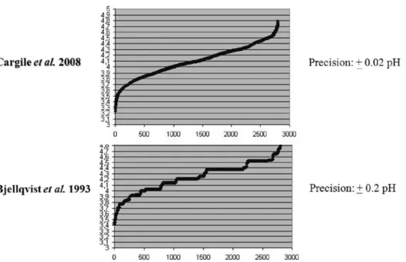
The ability to calculate the pI of proteins within a defined pH gradient was first demonstrated by Bjellqvist and co-workers for 2D gel electrophoresis (Bjellqvist et al., 1993). This approach is based on knowing the pK differences between closely related immobilines by focusing the same sample in overlapping pH gradients. A key component of this experiment is that the ratio of the immobilized groups incorporated into the gel matrix is similar to the ratio found in solution, so that a correct determination of the pK values can be obtained. Using this approach, protein pIs could be predicted within 0.2 pI unit precision. Other approaches have included the use of fluorescent markers in CIEF (Shimura et al. 2000; Shimura et al. 2002) or the use of pI and titration-curve calculations to predict pI shift for PTMs (Gordon et al., 2005).
Shotgun IPG-IEF provides an increased dynamic range and experimental database filtering capabilities for false positive and negative identifications. Most pI calculation algorithms published to date are exclusively based on charged amino acid side chains in a peptide or protein (Bjellqvist et al., 1993; Shimura et al,. 2000). Recently, Cargile and co-workers studied the effect of adjacent amino acid residues on peptide pI (Cargile submitted). They reported a computational method based on a genetic algorithm (Schmitt 2001). This method takes into account three consecutive amino acid residues and their cumulative effect on pI. For pK calculations on the C-terminus, their genetic function took into account any amino acid within three residues of the basic lysine (K) or arginine (R) moiety found in a tryptic peptide. A correction factor for aspartic acid (D) and glutamic acid (E) residues and their interaction with the N- or C-terminus is considered, provided that these amino acids are within four residues of that terminus. Their algorithm was trained on 5000 unique tryptic peptides. This method accurately calculates pI within a range of +/- 0.05 to +/- 0.03 pI units for the acidic part of the gradient. A comparison of the usual algorithm used for proteins and the new algorithm shows a much narrower distribution of peptides across a IPG strip’s gradient as shown in Figure 3.. For the rest of the gradient, Bellqvist’s algorithm was used with corrections for N- and C- terminal residues with charged side chains (Bjellqvist et al., 1993). This algorithm was used for all pI calculations throughout this work.
One of the major issues in MS-based proteomics is the validation of protein and peptide identifications produced by search engines such as Phenyx (Colinge et al. , 2003), Mascot (Perkins et al., 1999) and SEQUEST (Eng et al,. 1994). There are several validation strategies that are based on empirically determined rules such as using a score cutoff (Tabb et
al., 2002) or manual tandem mass spectra validation (Link et al., 1999). These approaches
lack any firm mathematical basis. Because in shotgun IPG-IEF, peptides are separated according to their pI, which is an invariant physicochemical property, this “added value” can be exploited to filter protein/peptide databases. Cargile and co-workers reported the use of pI as a filtering criterion, which resulted in the match of greater than 1300 protein loci identified by a single peptide match (Cargile et al. 2004). They also showed a 23% increase in the number of identified peptides with the pI filtering compared to the SEQUEST’s Xcorr score
alone in an E.coli database. Other groups that used shotgun Off-GEL IEF (Krijgsveld et al., 2006) and shotgun FFE-IEF (Xie et al., 2005) also used pI to recover more false negative peptides while maintaining a low false positive rate, and to confidently identify single hit proteins. Chick et al. evaluated a two-step sequential filtering approach that used false discovery rates (protFDR) at the protein level to assign high-confidence protein identifications (Chick et al. 2008). The distinction between protFDR was that it was calculated at the protein level, where protein scores were ranked and a threshold level was set beyond which assignments were incorrect. However, false positive rates (pepFPR) were calculated at the peptide level, where the following formula applied, 2n(reverse)/n(reverse) + n(forward), and where reverse referred to reverse database assignments and forward refers to
forward database. Their results demonstrated that peptide pI filtering prior to imposing a 1% protFDR increased protein identifications by at least 19% compared to using only a 1% protFDR.
The use of pI in combination with other orthogonal values becomes even more efficient. Cargile et al. showed the utility of using pI in combination with accurate mass (Cargile and Stephenson, 2004). Petritis et al. introduced methods to partially predict the elution time of peptides from reverse-phase columns, and to use this information in the protein identification (Petritis et al., 2003). This information allows some discrimination between peptides to be made, but the ± 10% standard deviation in elution time leaves a large window within which a peptide could theoretically elute. It was shown from offline cation exchange that there is a significant correlation between solution-phase charge and the general elution time of peptides (Peng et al., 2003). Combination of Rt and pI would also be a powerful tool for data validation (Krokhin, 2006). Orthogonal peptide pI filtering is not limited to the identification results. Accurate peptide pI can be applied prior to database searching to constrain the database size and complexity. Sevinsky et al. presented GENQUEST (GENome Queries Using Experimental SpecTra), a method that uses accurate peptide pI to constrain the database size of a six-frame translated human genome, that resulted in accurate and sensitive genome searching comparable to searching the human protein databases (Sevinsky et al., 2008).
III.B. Electrophoretic separation of proteins.
As analytes, proteins are much more complex than peptides, but also much more amenable to separation. Just to keep with generic parameters, proteins differ widely in size, isoelectric point, and hydropathy, which are parameters that can be used for separation. As to
electrophoretic separations, only size and isoelectric point are used.
However, one of the major problems linked to protein separation is their chemical
complexity. Nucleic acids are also long and complex polymers, but their sugar phosphate backbone and the low chemical diversity of the bases gives them a uniform behavior. Conversely, the polypeptide backbone of proteins is proportionally much less important compared to the amino acids side chains, so that the physico-chemical behavior of proteins is extremely complex and strongly depends on the composition and sequence of every protein. In addition, this complexity leads to the 3D folding of proteins, which induces, in turn, a complex surface chemistry, because proteins show surface patches of various features (e.g. cationic, anionic or hydrophobic). Consequently, proteins have a strong tendency to stick to other surfaces, whether these surfaces are those of other cellular molecules or surfaces present in the separation processes.
Thus, when analyzing native proteins, two major problems occur:
i) the equilibrium for each protein between its free form and various complexes and
ii) a tendency to aggregate to other proteins in artefactual complexes and/or precipitates. This aggregation is especially true in the stacking dimension when discontinuous multiphasic gels are used (see section II)
III.B.1. Electrophoretic separations of native proteins
For these reasons, electrophoretic separations of native proteins are very rarely used in proteomics. A happy exception to this rule is represented by the system called Blue-Native PAGE (Schägger and Von Jagow, 1991), where the proteins and protein supercomplexes are
separated by electrophoresis at a neutral pH. To limit aggregation, a charge-shifting agent is added, i.e. a reagent that is able to stick to proteins without unfolding their structure while conferring to them a uniform electric charge. Thus, electrostatic repulsion is maximized between proteins to prevent aggregation. While this system has been designed initially for the analysis of mitochondrial protein complexes (Schägger and Von Jagow, 1991), it has also been applied either to dedicated studies on other types of complexes (e.g., nuclear complexes Nováková et al., 2006) or to even more general large-scale studies (Camacho-Carvajal et al., 2004).
However, despite this trick of the use of a charge-shifting agent, this type of separation is difficult to optimize, because several parameters must be optimized for each system (Schägger and Von Jagow, 1991). Moreover, even with an optimized separation, it is quite frequent to observe some trailing of the bands in BN-PAGE separations, that indicate insufficient protein solubility.
III.B.2. Electrophoretic separations of denatured proteins: rationale
Because of this pivotal problem of protein solubility, the overwhelming majority of electrophoretic separations of proteins is made under denaturing conditions. Stricto sensu, denaturation just means the unfolding of proteins. Unfolding can be brought by physico-chemical perturbations, (e.g. heat or pH extremes), in which case the unfolded proteins aggregate together to form a massive precipitate. However, in some cases, denaturation is brought by conditions that unfold the proteins because they unfavor the internal protein forces in favor of protein-solvent interactions. In this case, proteins are made much less sticky, between themselves and toward surfaces, so that their solubility increases dramatically.
By increasing the solubility (low solubility is the major bottleneck in protein separations), and by decreasing the complexity of the system by dismantling protein complexes, denaturing electrophoresis of proteins is the method of choice for protein separation in proteomics studies.
This kind of solubilizing denaturation can be brought by two types of reagents. The first type is made of chaotropes; i.e., chemicals that disrupt the structure of water. In doing so, these chemicals alter the non-covalent bonds (hydrophobic interactions, hydrogen bonds) that structure proteins and thus unfold them. The chaotropic effect of many chemicals has been investigated (Gordon and Jencks, 1963), and from such studies, three efficient
chaotropes can be used in protein chemistry: urea, thiourea, and guanidine. However, it must be kept in mind that, to be active, chaotropes must be used at multimolar concentrations, thereby precluding the use any ionic chaotrope in electrophoretic separations, and thus the use of guanidine, which is the most potent chaotrope. Because thiourea is not very soluble in water, and furthermore prevents acrylamide polymerization unless special systems are used (Rabilloud, 1998), the chaotrope of choice in electrophoretic separations is urea.
One of the main advantages of neutral chaotropes such as urea is that they do not alter either the molecular weight of the proteins or their isoelectric points, so that they are
compatible with all types of electrophoretic methods.
However, chaotropes are still not the perfect solution. First of all, urea can undergo chemical reactions with protein amino groups, to lead to carbamylation (Cejka et al., 1968). This reaction alters the isoelectric point of the proteins, which is a problem in IEF.
Furthermore, carbamylation (which is artefactual) and acetylation (which is a natural protein modification) induce almost the same mass shift on the peptides and proteins, so that an
artefactuel carbamylation can be misleadingly taken for a bona fide biological acetylation. Second, neutral chaotropes do increase protein solubility, but they just play on an increase in the water solubility of poorly soluble molecules. Thus, they cannot counteract completely protein aggregation, especially at high concentrations. Furthermore, neutral chaotropes have no action on electrostatic interactions. Thus, if such interactions can happen (especially at the low ionic strength conditions that prevail in IEF); then precipitation of proteins of opposite charge or artefacts in migration can occur (Wen et al., 1983).
The second type of solubilizing denaturing agents is composed of ionic detergents; the archetype is SDS (sodium dodecyl sulfate). Their mechanism of action is fairly different from the one of chaotropes. The efficient ionic detergents are made from a long and flexible
hydrocarbon chain that is linked to an ionic, polar head. The detergent molecules will bind through their hydrophobic hydrocarbon tail to the hydrophobic amino acids. This binding favors amino acid-detergent interactions over amino acid-amino acid interactions, thereby promoting denaturation. Moreover, these protein-detergent hydrophobic interactions hold in place many detergent molecules on the proteins, thus anchoring many ionic charges of the same sign. This electrostatic repulsion further promotes denaturation, but also induces a very strong repulsion between protein molecules, and thus an extremely high protein solubility. However, this situation is achieved only when the proper balance between the hydrophilic and hydrophobic properties of the detergent is achieved. If the alkyl chain is too short, and the binding to the proteins is too weak, not enough detergent molecules are bound to promote optimal solubility. If the alkyl chain is too long, only a limited number of sites will exist on the proteins to accomodate such a long alkyl chain. The result is the same; i.e., not enough molecules fixed to ensure optimal solubility under electrophoretic conditions (Lopez et al. 1991). The proper balance differs from one class of detergent to another, but is close to 12 carbons for alkyl sulfate and sulfonates (Reynolds and Tanford, 1970) and of 16 carbons for quaternary ammoniums (MacFarlane, 1983).
Through this mechanism of action, properly chosen ionic detergents can solubilize almost all proteins to very high concentrations. The price to pay for this solubility is the complete disappearance of the protein native electric charge (or pI) that is masked by the charges brought by the detergent. It should also be stressed that the rationale for the efficiency of ionic detergents is hydrophobic interactions, so that concomitant use of
chaotropes and detergents, also found in the literature, is not very helpful. As a matter of fact, urea has been used to remove SDS from proteins (Weber and Kuter, 1971) .
III.B.3. The implementation of denaturing protein electrophoresis in proteomics
The criticism that has been made of capillary electrophoresis in peptide separation holds true for proteins, and to an even greater extent. Because peptides are the final analytes in
proteomics, minimal steps take place between the separation of peptides and their analysis in mass spectrometry. Proteins, conversely, are an indirect analyte for mass spectrometry in most cases, which means, in turn, that several steps must be carried out between protein separation and mass spectrometry. Because each of these steps has a specific, and sometimes low yield, being limited in protein amount, as is the case for capillary electrophoresis,
precludes the use of this technique in proteomics.
Non-capillary free-flow electrophoresis is generally a rather low-resolution technique, and is therefore of low interest in proteomics studies. The only exception to this rule is IEF in free flow (Hoffmann et al., 2001), which shows a much larger capacity than conventional gel-based techniques. However, IEF of proteins has its own problems (detailed later) that does not make it a first choice for proteomics studies.
Thus, the vast majority of protein electrophoretic separations are made in gel media, most often in polyacrylamide gels. Besides the improved resolution, gel media offer the enormous advantage of a continuous separation, as well as additional advantages for downstream processing that will be detailed later (section IV).
III.B.4. The workhorse of protein separation in proteomics: SDS gel electrophoresis In almost every proteomics scheme where a protein electrophoresis separation is carried out, SDS electrophoresis is encountered. This statement shows how widespread SDS
electrophoresis is in proteomics. It can be used as the sole protein separation prior to
digestion and peptide separation (e.g. in Lasonder et al., 2002, Schirle et al., 2003) but it can also be coupled with other protein separations, such as native electrophoresis (Schägger and Von Jagow, 1991), (Hisabori et al., 1991), ion exchange chromatography (Szponarski et al., 2004), denaturing electrophoresis (MacFarlane 1989, Rais et al., 2004), and of course isoelectric focusing, making the well-known high-resolution 2D electrophoresis.
Historically speaking, SDS electrophoresis in its modern form was born in 1970, when Laemmli (Laemmli, 1970) coupled the high resolution of the Tris chloride glycine system of Davis (Davis 1964) to the well-known denaturing power of SDS, which was already used in lower resolution continuous electrophoresis (Shapiro et al., 1967). This method immediately proved extremely versatile and thus became of extremely wide use.
This success can be explained by a combination of factors. The essential factor is the high affinity of SDS for proteins. As stated earlier, SDS is a happy compromise between hydrophobicity and hydrophilicity, and binds extremely well to almost all proteins. In addition to promoting protein denaturation, it confers a very strong negative charge to proteins, which masks their native charge. Consequently, the charge density becomes approximately constant for every protein, so that the separation is driven mainly by the molecular mass. Moreover, because the proteins do repel strongly each other, extremely high solubilities can be achieved. This high solubility allows one to take full advantage of the resolving power of the electrophoretic discontinuous systems, in which the high resolution is coupled with very high protein concentrations in the stacking gel. Although these high concentrations lead to protein precipitation, tailing, and loss of resolution in native systems, such problems are not encountered in the denaturing systems.
In such discontinuous SDS electrophoresis, the migration of the proteins is measured in the gel compared to the migration of the small ions front, visualized with a dye. Because of the extremely wide size distribution of proteins, it is often interesting to optimize this resolution according to the experimental needs. This optmization can be done by two
mechanisms. In the first, and obvious one, the resolution is tuned by changing the pore size of the resolving gel, and thus the acrylamide concentration. When low percentage gels are used, the low molecular weight proteins migration will be almost unhampered by the gel, so that they will run with the small ion front. Meanwhile, high molecular weight proteins can enter more deeply into the gel, and are thus more separated each one from the other. Conversely, if the gel concentration is increased, then low molecular weight proteins will be unstacked from the small ions and separated each one from each other, whereas many high molecular weight proteins will be almost unable to enter the gel and will remain close to the top of the gel, with poor separation. A good compromise is, of course, to make a porosity gradient in the gel with an acrylamide concentration gradient, so that the resolution is optimized for all sizes of proteins. However, this approach is not without drawbacks. For example, low-concentration
gels are very soft, while high-concentration gels are brittle, thereby limiting the scope of this way of optmizing resolution.
The other way to optmize the gel resolution is to play with the buffer system. As shown in buffer calculations (Jovin, 1973a), the speed of the moving ion boundary varies with the composition of the buffer system, in terms of the nature of the ions and in terms of pH. In anionic systems, such as the classical Tris-chloride-glycine system, the speed of the ions increases with increasing pH. Thus, decreasing the pH slows down the ions and makes the low-molecular weight proteins coalesce with the small ion front. However, by stretching the migration time, it also stretches the resolution of higher molecular weight proteins (Johnson, 1982). Conversely, increasing the pH will increase the mobility of the small ions, unstack the low molecular weight proteins, and therefore enable their analysis. However, by shortening the migration time, it will also decrease the resolution of the high molecular weight proteins.
Optimization of the buffer system is therefore another way of tuning the separation, which operates at constant gel concentration. Of course, combining pH changes and gel porosity changes is an optimal way of reaching an adequate resolution in extreme cases, such as giant proteins (Fritz et al., 1989).
However, this play on pH is limited in the case of the classical Tris-chloride-glycine system. in its standard setup, it operates at a gel pH of 8.8, which is already a bit too basic compared to the pK of Tris (8.05). Getting to an even more basic pH to analyze smaller proteins cannot be reached in a Tris system, but can be reached by a more basic system, based on ammediol (Bury, 1981), but at such a basic pH, the acrylamide gel starts to
hydrolyze. In fact, It should be kept in mind that, in anionic systems, the real operating pH of the system is higher than the pH of the starting gel. For example, in the classical Davis system, a pH 8.8 gel operates at pH 9.5, according to Jovin's calculations.
Thus, a better way to analyze low-molecular weight proteins is to use buffer
components with a pK lower than the one of glycine. Initial attempts have been made with MES (Kyte and Rodriguez, 1983), but better results have been obtained with tricine (Schägger and Von Jagow, 1987) or bicine (Wiltfang et al., 1991).
These findings led to a more general trend in the use of gels operating at a more acidic pH, to decrease the problems associated with alkaline hydrolysis of acrylamide. In addition, it was shown that Tricine-based gels show an even better resolution than glycine-based gels (Patton et al. 1991). However, in this case there is the opposite problem with Tris, because optimized resolution for 20-100kDa proteins is reached at pH 7, which again is too far from the buffering range of Tris.
Thus, even more flexibility can be offered by a system that operates close to the pK of Tris; e.g., a taurine-based system (Tastet et al., 2003). In this way, changing the pH of the buffer can be done either on the acidic side or on the basic side, to favor the resolution of high- or low-molecular weight proteins, respectively.
However, despite its outstanding capacities in terms of protein solubilization,
versatility, and flexibility, SDS electrophoresis alone has too low a resolution to be used as the sole separation technique in proteomics. The only exception to this rule lies in interaction proteomics studies, where protein complexes isolated through a tag are first fished out of the cell lysate through a chromatographic process, and then analyzed (Rigaut et al., 1999). Most often, the complexity of the protein complex is low enough to be separable in individual proteins by a single SDS electrophoresis, which can resolve ca. 50 different protein species. It can be argued that, even in the case of low complexity samples, it can happen that two or more proteins have the same apparent molecular weight, which will cause them to comigrate,
even with fine optimization of the separation. Should this comigration happen, some tricks can be used, such as changing the buffer system (Patton et al., 1991), replacing pure SDS by a mixture of alkyl sulfates (Lopez et al., 1991), or even replacing SDS by a mixture of alkyl sulfates and fatty alcohols, because this substitution can lead to the separation of proteins with extremely close molecular weight (Brown, 1988). This separation is due to the fact that these ancillary chemicals will bind to proteins in a structure- or sequence-dependent way, to induce a sequence-dependent electrophoretic mobility
In any event, when the sample to be analyzed becomes more complex, SDS
electrophoresis is no longer sufficient in terms of separation, and must be coupled to other separation techniques. This additional separation can be made either before or after SDS electrophoresis. However, when rationalizing the possible choices, it must be kept in mind that SDS-denatured proteins are soluble in the presence of SDS, but will become easily insoluble when the SDS is removed. The few examples available in the literature demonstrate that interfacing SDS electrophoresis with another downstream protein separation is
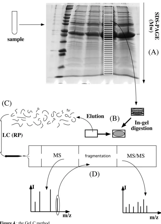
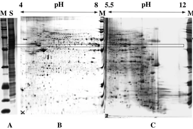
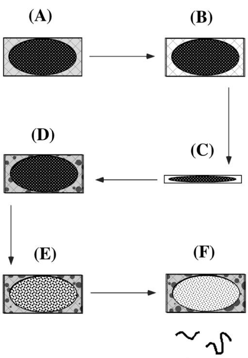
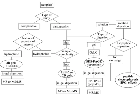
cumbersome (Siemankowski et al., 1978, Tuszynski et al., 1979, Nakamura et al., 1992). Thus, on a practical rule, when a complex separation scheme is needed, SDS electrophoresis is most often the last separation at the protein level. Other protein separations are carried out before SDS electrophoresis. whereas downstream separation is made at the peptide level, as in the GeLC method schematized on figue 4 (Lasonder et al., 2002, Schirle et al., 2003). This GeLC method, i.e., a combination of SDS electrophoresis of proteins, followed by in gel digestion and RP HPLC-MS/MS analysis of the resulting peptides, has shown the ability to deal with many types of samples, such as whole cell lysates (Lasonder et al., 2002) or subcellular fractions (Bell et al., 2001, Andersen et al., 2002). However, the method is still not perfect. As a matter of fact, even with the exquisite sensitivity of modern mass
spectrometry, to be able to positively identify a protein implies to start from a few femtomoles. However, owing to the dynamic range of protein expression in most cells, having a few femtomoles of a minor protein will mean a fairly high total protein load, and consequently high concentrations of the most-abundant proteins (see for example the gel shown on Figure 4). Despite the high loading capacity of SDS electrophoresis, high protein loads often result in some spreading of the major protein peaks, at least just because of their Gaussian shape, not even to speak of tailing induced by overloading. This spreading means, in turn, that these major proteins are identified in many gel fractions, and can divert the MS/MS process and lead to miss the minor proteins. Complicated exclusion lists processes must then be implemented to alleviate this problem.
Thus, a higher resolution approach is to use another protein separation step before SDS electrophoresis.
Owing to the flexibility of SDS electrophoresis, almost any protein separation method, either chromatographic or electrophoretic, can be used prior to SDS electrophoresis. Among this wide variety, the coupling of gel-based electrophoretic separations has been most widely used, due to the fact that it represents a completely continuous bidimensional separation, without the sampling artefacts linked to the necessary sample collection when a liquid-separation is used as the first dimension.
Within this coupling of electrophoretic separations to SDS electrophoresis, the coupling of gel isoelectric focusing to SDS electrophoresis has received the widest audience.
III.B.5. Classical high-resolution 2D electrophoresis: the source of proteomics