HAL Id: inserm-00819393
https://www.hal.inserm.fr/inserm-00819393
Submitted on 1 May 2013
HAL is a multi-disciplinary open access
archive for the deposit and dissemination of
sci-entific research documents, whether they are
pub-lished or not. The documents may come from
teaching and research institutions in France or
abroad, or from public or private research centers.
L’archive ouverte pluridisciplinaire HAL, est
destinée au dépôt et à la diffusion de documents
scientifiques de niveau recherche, publiés ou non,
émanant des établissements d’enseignement et de
recherche français ou étrangers, des laboratoires
publics ou privés.
myogenic differentiation-associated miRNAs and their
target genes.
Petr Dmitriev, Ana Barat, Anna Polesskaya, Mary O’Connell, Thomas
Robert, Philippe Dessen, Thomas Walsh, Vladimir Lazar, Ahmed Turki,
Gilles Carnac, et al.
To cite this version:
Petr Dmitriev, Ana Barat, Anna Polesskaya, Mary O’Connell, Thomas Robert, et al..
Simultane-ous miRNA and mRNA transcriptome profiling of human myoblasts reveals a novel set of myogenic
differentiation-associated miRNAs and their target genes.. BMC Genomics, BioMed Central, 2013,
14 (1), pp.265. �10.1186/1471-2164-14-265�. �inserm-00819393�
M E T H O D O L O G Y A R T I C L E
Open Access
Simultaneous miRNA and mRNA transcriptome
profiling of human myoblasts reveals a novel set
of myogenic differentiation-associated miRNAs
and their target genes
Petr Dmitriev
1,2, Ana Barat
1, Anna Polesskaya
5, Mary J O’Connell
4, Thomas Robert
3, Philippe Dessen
3,
Thomas A Walsh
4, Vladimir Lazar
1, Ahmed Turki
2, Gilles Carnac
2, Dalila Laoudj-Chenivesse
2, Marc Lipinski
1and Yegor S Vassetzky
1*Abstract
Background: miRNA profiling performed in myogenic cells and biopsies from skeletal muscles has previously identified miRNAs involved in myogenesis.
Results: Here, we have performed miRNA transcriptome profiling in human affinity-purified CD56+ myoblasts induced to differentiate in vitro. In total, we have identified 60 miRNAs differentially expressed during myogenic differentiation. Many were not known for being differentially expressed during myogenic differentiation. Of these, 14 (miR-23b, miR-28, miR-98, miR-103, miR-107, miR-193a, miR-210, miR-324-5p, miR-324-3p, miR-331, miR-374, miR-432, miR-502, and miR-660) were upregulated and 6 (miR-31, miR-451, miR-452, miR-565, miR-594 and miR-659) were downregulated. mRNA transcriptome profiling performed in parallel resulted in identification of 6,616 genes differentially expressed during myogenic differentiation.
Conclusions: This simultaneous miRNA/mRNA transcriptome profiling allowed us to predict with high accuracy target genes of myogenesis-related microRNAs and to deduce their functions.
Keywords: microRNA, Skeletal muscle, Myogenesis, Myoblast Background
MicroRNAs (miRNAs) are 20–25 nt-long non-coding RNAs transcribed predominantly by the RNA polymerase II [1,2]. miRNAs play an important role in regulating gene expression at the protein and mRNA levels. They have been previously shown to inhibit translation initiation and elongation and induce deadenylation and degradation of mRNA (for review see [3]). In addition, there are indica-tions that miRNAs may also activate translation [4].
Along with transcription factors, miRNAs contribute to skeletal myogenesis (for review see [5]). Myogenic microRNAs miR-1, miR-133a/b and miR-206 (also called MyomiRs, as suggested by [6]) regulate myogenic
differentiation and proliferation of myogenic cells by targeting important regulators of myogenesis [7,8] (for re-view see [6,9]). MiR-1 and miR-133a are expressed in both skeletal and cardiac muscles [7,10], while miR-133b and miR-206 are expressed solely in skeletal muscles [10].
Most miRNAs expressed in skeletal muscles are found to be upregulated during myogenesis. These include miR-24 [11], miR-26a [12], miR-27b [13], miR-29b/c [14], miR-143 [15], miR-181 [16], miR-208b/499 [17], miR-214 [18], miR-322/424 and miR-503 [19], miR-486 [20], miR-682 [21]. Only few known miRNAs are in-volved in the regulation of myogenic differentiation are downregulated during myogenesis. These include miR-125b [22] and miR-221/222 [23] (for review see [5,24,25]).
Altogether, myogenesis-related miRNAs have been studied during myogenic differentiation of human pri-mary myoblasts [14,15,26,27] and in various model
* Correspondence:vassetzky@igr.fr
1UMR 8126, Univ. Paris-Sud 11, CNRS, Institut de Cancérologie
Gustave-Roussy, 39, rue Camille-Desmoulins, Villejuif 94805, France Full list of author information is available at the end of the article
© 2013 Dmitriev et al.; licensee BioMed Central Ltd. This is an Open Access article distributed under the terms of the Creative Commons Attribution License (http://creativecommons.org/licenses/by/2.0), which permits unrestricted use, distribution, and reproduction in any medium, provided the original work is properly cited.
systems, e.g. the immortalized murine cell line C2C12 [12,16,20,28], murine primary satellite cells and myo-genic progenitor cells [21] as well as quail myoblasts [29]. Several myogenesis-related miRNAs have been dis-covered or confirmed by expression profiling of develop-ing muscular tissues in humans [30], pigs [31] and in the common carp [32] (Table 1).
To perform transcriptome profiling of myogenic cells, it appears interesting to use primary myogenic cells culti-vated in vitro rather than whole muscle tissues. This ap-proach minimizes biases caused by the presence of inflammatory, connective tissue-derived and other non-myogenic cells. Nevertheless, totally avoiding contamin-ation by non-myogenic cells is difficult unless clonal populations of immortalized myogenic cells are used. An alternative to generating immortalized lines of myogenic cells is the utilization of primary cell popula-tion enriched for myogenic precursors. However, in previous miRNA and mRNA expression profiling re-ports, primary skeletal myoblasts cultivated in vitro have not been enriched for myogenic cells prior to analysis.
In the present study, we have thus decided to profile miRNA expression in cultures of CD56+ primary myo-blasts and myotubes isolated from healthy individuals using an affinity purification procedure [35]. A total of 60 miRNAs were found to be differentially expressed during myogenic differentiation induced by serum star-vation of which 20 had not been previously associated with myogenesis. In addition, we have performed micro-array mRNA transcriptome profiling on the same myo-blasts and myotubes samples as used for miRNA expression profiling in order to assign targets for these miRNAs.
Results
Identification of new myogenesis-related MIRNA (MRMIRNA) in human primary myoblasts
To identify new myogenesis-related miRNAs in human cells, CD56+ myogenic precursors were isolated from skeletal muscle biopsies of six healthy individuals (Additional file 1). These were then induced to differ-entiate in vitro using serum starvation and the pro-gression of myogenic differentiation was monitored by following the expression of Myogenin (MYOG) and other myogenic differentiation markers including ID1, TNNT1, TNNT2, MYL4, COL15A1, VIM and CAV1 (Figure 1A). The disappearance of proliferating cells and concomitant emergence of myotubes was moni-tored using Ki67 staining and microscopic observation (Figure 1B and C).
We observed a statistically significant difference of the expression levels of myogenesis-related markers in pro-liferating or differentiated myogenic cells isolated from
different healthy subjects. This difference was considered as a natural heterogeneity of gene expression levels in different subjects that might be explained by their age, sex, physical activity or diet preferences, therefore, all samples analyzed were included in the study.
The expression of 365 different microRNAs was then profiled in human primary myoblasts and myotubes using TaqMan Low Density Array (TLDA) (Additional file 2). In total, 60 miRNAs were found to be differentially expressed during myogenic differentiation with p-values ranging from 3.4x10⁻⁵to 5x10⁻² (Figure 2). These will be referred to as myogenesis-related miRNAs (MR-miRs). Sequences and corresponding TaqMan probe codes for these microRNAs can be found in the Additional file 3.
The majority (43 out of 60) of MR-miRs were found to be upregulated of which 14 had not been previously reported as involved in the regulation of myogenesis ei-ther in humans or in oei-ther species (Table 1). Of the 17 MR-miRs that were found to be downregulated during myogenesis, 6 had not been previously associated with myogenic differentiation.
MRNA transcriptome profiling and functional classification of differentially expressed genes
Until now, mRNA expression profiling of human myoblasts has been performed exclusively on primary cells [36]. How-ever, primary skeletal myoblasts have not been enriched for myogenic cells prior to analysis. To avoid analyzing mixed populations of myoblasts and non-myogenic cells, we have used RNA extracted from CD56+ myoblasts purified from muscular tissues. Then, using Agilent transcriptome 44 K microarrays, we identified 6,616 differentially expressed genes of which 3,445 and 3,245 were down- or up-regulated, respectively (Additional file 4). In most cases, a functional class could be assigned to these transcripts using the DAVID Gene Ontology database (Additional file 5) [37,38].
In agreement with previous reports, the majority of upregulated genes were found to control metabolism, myogenesis, insulin receptor signaling or to positively regu-late transcription (Table 2). Genes involved in the control of cell cycle, DNA damage response, angiogenesis, cell motility and invasion as well as protein modification and NF-kB sig-naling belonged to the group of genes downregulated during myogenic differentiation (Table 2). Genes involved in the regulation of ubiquitination/proteolysis, protein transport/ localization, transcription regulation and apoptosis were found to be either up- or down-regulated (Table 3). The complete list of genes found to be differentially expressed during in vitro myogenic differentiation of human primary myoblasts can be found in the Additional file 4.
Predication of myogenesis-related MIRNA target genes
A common practice to boost target gene predictions ac-curacy is to use several prediction algorithms and then
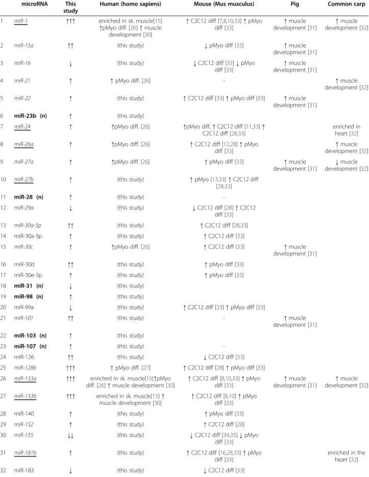
Table 1 microRNAs (MR-miRs) found to be up- and downregulated during myogenic differentiation in this study
microRNA This study
Human (homo sapiens) Mouse (Mus musculus) Pig Common carp 1 miR-1 ↑↑↑ enriched in sk. muscle[15]
↑pMyo diff. [26] ↑ muscle development [30] ↑C2C12 diff [7,8,10,33] ↑ pMyo diff [33] ↑muscle development [31] ↑muscle development [32] 2 miR-15a ↑↑ (this study) ↓pMyo diff [33] ↑muscle
development [31] 3 miR-16 ↓ (this study) ↓C2C12 diff [33] ↓ pMyo
diff [33]
↑muscle development [31]
4 miR-21 ↑ ↑pMyo diff. [26] - ↑muscle development [32] 5 miR-22 ↑ (this study) ↑C2C12 diff [33] ↑ pMyo diff [33] ↑muscle
development [31] 6 miR-23b (n) ↑ (this study)
7 miR-24 ↑ ↑pMyo diff. [26] ↑pMyo diff, ↑ C2C12 diff [11,33] ↑ C2C12 diff [28,33]
enriched in heart [32] 8 miR-26a ↑ ↑pMyo diff. [26] ↑C2C12 diff [12,28] ↑ pMyo
diff [33]
↑muscle development [32] 9 miR-27a ↑ ↑pMyo diff. [26] ↑pMyo diff [33] ↑muscle
development [31]
↓muscle development [32] 10 miR-27b ↑ (this study) ↑pMyo [13,33] ↑ C2C12 diff
[28,33] 11 miR-28 (n) ↑ (this study)
-12 miR-29a ↓ (this study) ↓C2C12 diff [28] ↑ C2C12 diff [33] 13 miR-30a-5p ↑↑ (this study) ↑C2C12 diff [28,33] 14 miR-30a-3p ↑ (this study) ↑C2C12 diff [33]
15 miR-30c ↑ ↑pMyo diff. [26] ↑C2C12 diff [33] ↑muscle development [31] 16 miR-30d ↑↑ (this study) ↑pMyo diff [33]
17 miR-30e-3p ↑ (this study) ↑pMyo diff [33] 18 miR-31 (n) ↓ (this study)
19 miR-98 (n) ↑ (this study)
20 miR-99a ↓ (this study) ↑C2C12 diff [33] ↑ pMyo diff [33]
21 miR-101 ↑↑ (this study) - ↑muscle development [31] 22 miR-103 (n) ↑ (this study)
23 miR-107 (n) ↑ (this study) -24 miR-126 ↑↑ (this study) ↓C2C12 diff [33] 25 miR-128b ↑↑↑ ↑pMyo diff. [27] ↑C2C12 diff [28] ↑ pMyo diff [33] 26 miR-133a ↑↑↑ enriched in sk. muscle[15]↑pMyo
diff. [26] ↑ muscle development [30]
↑C2C12 diff [8,10,33] ↑ pMyo diff [33] ↑muscle development [31] ↑muscle development [32] 27 miR-133b ↑↑↑ enriched in sk. muscle[15] ↑
muscle development [30]
↑C2C12 diff [8,10] ↑ pMyo diff [33] 28 miR-140 ↑ (this study) ↑pMyo diff [33] 29 miR-152 ↑ (this study) ↑C2C12 diff [28] 30 miR-155 ↓↓ (this study) ↓C2C12 diff [34,35] ↓ pMyo
diff [33]
31 miR-181b ↑ (this study) ↑C2C12 diff [16,28,33] ↑ pMyo diff [33]
enriched in the heart [32] 32 miR-183 ↓ (this study) ↓C2C12 diff [33]
to combine their predictions. However, this approach has been proved inefficient [39]. Therefore, we have used a single algorithm, e.g. RNA22 [40] to predict miRNA targets. RNA22 has been chosen for its low rate of false-positive predictions as compared to other algorithms [39]. For each miRNA, the number of target predictions varied between 261 and 3,058.
According to previous reports, the proportion of correct predictions can be as low as 10% [41,42] (for recent review see [43]. To increase the accuracy of bioinformatic
predictions, we used a simultaneous mRNA/miRNA expres-sion profiling approach described in [42,44,45]. Based on the fact that MiRNA regulate gene expression by inhibiting translation or inducing deadenylation of mRNAs followed by their degradation (for review see [46,47]), we reasoned that the expression levels of mRNA and miRNA should be inversely correlated if one regulates the other. Indeed, for upregulated MR-miRNA, the majority of their predicted tar-get genes was found to be underexpressed. Similarly, most downregulated MR-miRNAs had upregulated predicted
Table 1 microRNAs (MR-miRs) found to be up- and downregulated during myogenic differentiation in this study (Continued)
33 miR-192 ↑↑ (this study) ↑pMyo diff [33] 34 miR-193a (n) ↑↑ (this study) -35 miR-204 ↓↓ ↓pMyo diff. [27] -36 miR-206 ↑↑ enriched in sk. muscle[15] ↑
muscle development [30]
↑C2C12 diff [8,10,33] ↑ pMyo diff [33]
↑muscle development 37 miR-210 (n) ↑ (this study)
38 miR-221 ↓ ↑pMyo diff. [26]↓ pMyo diff. [27] ↓quail myoblasts diff, ↓ C2C12 diff [29] 39 miR-222 ↓ ↑pMyo diff. [26] ↓ pMyo diff. [27] ↓quail myoblasts diff, ↓ C2C12
diff [29] ↓ C2C12 diff [28]
↓muscle development [32] 40 miR-320 ↓ (this study) ↑pMyo diff [33]
41 miR-324-3p (n) ↑↑ (this study) 42 miR-324-5p (n) ↑ (this study)
43 miR-331 (n) ↑ (this study)
-44 miR-339 ↓ (this study) ↑C2C12 diff [33] ↑ pMyo diff [33] 45 miR-361 ↑ (this study) ↑pMyo diff [33] 46 miR-362 ↑↑ (this study) ↑C2C12 diff [28] 47 miR-374 (n) ↑ (this study)
48 miR-432 (n) ↑ (this study) 49 miR-451 (n) ↓↓↓ (this study) 50 miR-452 (n) ↓↓ (this study)
51 miR-500 ↑↑ (this study) ↑C2C12 diff [28,33] ↑ pMyo diff [33] 52 miR-501 ↑↑↑ (this study) ↑C2C12 diff [28,33] 53 miR-502 (n) ↑↑ (this study)
-54 miR-503 ↑ (this study) ↑C2C12 diff [19,28,33] ↑ pMyo diff [33]
55 miR-532 ↑↑ (this study) ↑C2C12 diff [28,33] ↑ pMyo diff [33] 56 miR-550 ↓ ↓pMyo diff. [27] -57 miR-565 (n) ↓ (this study) -58 miR-594 (n) ↓ (this study)
59 miR-659 (n) ↓↓↓ (this study) -60 miR-660 (n) ↑↑↑ (this study)
-In bold are human-specific MR-miRs (hMR-miRs, 16 in total); Italicized are microRNAs shown to be differentially expressed during myogenesis in non- human cells; microRNAs with a known function in myogenesis are underscored. (n): microRNAs that we have shown to be differentially expressed during myogenesis for the first time in our study. The number of arrows corresponds to the level of up- or downregulation of microRNAs. One arrow: fold change 1 to 3 fold, two arrows: fold change 3 to 7 times, three arrows: fold change 7 to infinity.
targets. Indeed, for every microRNA tested, the distribution of Pearson correlation coefficients of its predicted target genes was shifted towards negative values. This was the case neither for randomly selected genes, nor when the sample correspondence was permuted (Figure 3A and the Additional file 6) which was used to establish a signifi-cance threshold of Pearson correlation coefficients. Target genes with correlation coefficients below the threshold had less than 5% chances to be random. We call these genes “supported targets” to underscore the fact that the prediction was supported by mRNA tran-scriptome profiling (Additional file 7). On average, 11% of microRNA target genes predicted by the RNA22 algorithm were “supported” by mRNA tran-scriptome profiling (Figure 3B).
We then compared the accuracy of predictions made by RNA22 and TargetScan, another popular miRNA tar-get prediction algorithm. As an indication of accuracy, we have used a percentage of predicted targets that were supported by transcriptome data. For 11 of 17 target genes, RNA22 demonstrated higher percentage of pre-dictions supported by transcriptome data, for 2 more microRNAs the difference in the accuracy of predictions was insignificant (Figure 3C and Additional file 8).
QRT-PCR validation of supported MIRNA target genes
To demonstrate the relevance of our miRNA target pre-dictions, we altered the expression of several microRNAs and measured the expression of their predicted target genes, supported by the transcriptome data. To illustrate the impact of miRNA repression on the expression of their target genes, we have chosen to inhibit the expres-sion of four well-characterized myogenic microRNAs miR-1, -133a, -133b and −206 in human immortalized myoblasts using LNA (Locked Nucleic Acids) antisense microRNAs (anti-miRs). Human immortalized myoblasts were differentiated in vitro and then transfected separ-ately with anti-miRs targeting 1, 133a, miR-133b and miR-206. miR-1 and miR-206 have the same
seed sequence and, therefore, target very similar lists of genes. The same is true for miR-133a and miR-133b. We have then randomly selected 12 and 10 genes predicted to be targeted by miR-1/206 and miR-133a/b respect-ively and supported by our transcriptome data and tested their expression using qRT-PCR in the cells transfected with corresponding anti-miRs. Repressing miR-1/206 resulted in the upregulation of six of its tar-gets: AP3D1 (adaptor-related proteins complex 3, delta 1 subunit), COL3A1 (collagen, type III, alpha 1), HDAC1 (histone deacetylase 1), PDCD4 (programmed cell death 4), PRKAB2 (protein kinase, AMP-activated beta 2 non-catalytic subunit) and ZNF365 (zinc finger protein 365). These target genes were considered as novel “qRT-PCR val-idated” target genes of miR-1 and −206 (Figures 4A). Six other target genes supported by our transcriptome data in-cluding ATP2B1, C11orf45, FOXP, G2E3, MBOAT7 and TUBB could not be validated via qRT-PCR experimentally, as we did not observe significant changes of their expres-sion following anti-miR transfection (data not shown).
In the case of miR-133a and -133b, immortalized myotube transfection anti-miRs resulted in the upregulation of six of ten randomly selected target genes, supported by our tran-scriptome data. These novel qRT-PCR validated targets of miR-133a and -133b include ASAP2 (ArfGAP with SH3 do-main, ankyrin repeat and PH domain 2), ARFGAP1 (ADP-ribosylation factor GTPase activating protein 1), ATP1A2 (ATPase, Na+/K+ transporting, alpha 2 polypeptide), C7orf51 (chromosome 7 open reading frame 51), RBM15B (RNA binding motif protein 15B) and TMCC2 (transmem-brane and coiled-coil domain family 2) (Figures 4B). Four other target genes supported by our transcriptome data in-cluding LMCD1, NAT9, PRPF38A and SYNGR2 could not be validated via qRT-PCR, as we did not observed significant changes of their expression following anti-miR transfection (data not shown).
To illustrate the impact of miRNA overexpression on the expression of their target genes, we have selected two microRNAs, miR-30 and −128, that were previously
(See figure on previous page.)
Figure 1 Expression of myogenic differentiation markers and miRNA profiling. A. Expression of muscle differentiation markers MYOG (myogenin, myogenic factor 4, p-value=0.0016), TNNT1 (Troponin T type 1, skeletal slow, p-value=0.00064), MYL4 (myosine light chain 4, p-value=0.0016), COL15A1 (collagen, type XV, alpha 1, p-value=0.013), TNNT2 (troponin T type 2 (cardiac), p-value=4.8x10^-6), VIM (vimentin, p-value=0.00034), ID1 (inhibitor of DNA binding 1, p-value=1.9x10^-5) and CAV1 (caveolin 1, p-value=0.00015) in proliferating myoblasts (labeled as P) and
differentiated myotubes (labeled as D) was measured using qRT-PCR, normalization was performed using ΔΔCt method using GAPDH as a control gene and proliferating sample #1 as a reference sample (expression level 1). The average of three independent experiments is shown. Numbers from 1 to 6 indicate the sample number (for full description refer to Table S1). Insets indicate the average of 6 samples of proliferating myoblasts (P) and differentiated myotubes (D). Asterisk corresponds to p-values<0.05. Error bars correspond to standard deviation (SD) in the case of individual samples and standard error of the mean (SEM) in the case of average expression levels (inset). B. Results of immunofluorescence microscopy analysis of cells stained with anti-Ki67 (red), and anti-Desmin (green) antibodies and DAPI nuclear staining (blue) showing normal cellular localization of these proteins in proliferating myoblasts and differentiated myotubes, 20x magnification. Bar=10 μm. C. The number of Ki67+ cells is significantly reduced during myogenic differentiation in vitro. Results of quantification of the DES+ and Ki67+ cells representing 300 individual cells. Statistically significant difference between cell cultures of proliferating myoblasts and differentiated myotubes is indicated by an asterisk (t-test p-value<0.05).
shown to be upregulated during myogenic differentiation [26,27], but their function in myogenesis is not well-established. Two plasmids coding for the corresponding pre-miRNAs or scrambled sequence were transfected separately into human rhabdomyosarcoma cells, and the expression of their target genes was then tested via qRT-PCR. Ectopic overexpression of miR-128 led to the downregulation of three of its four target genes, randomly selected among the genes supported by our transcriptome data. These novel qRT-PCR validated miR-128 target genes include EDARADD (EDAR-associated death domain), CAV1 (Caveolin 1) and DTYMK (deoxythymidylate kin-ase) (Figures 4C). One transcriptome-supported miR-128 target gene, RBM15B, could not be validated using qRT-PCR, as we did not observed significant changes in its ex-pression level following miRNA precursor tranfection (data not shown).
Similarly, overexpressing miR-30 led to the downregulation of three of its five randomly selected transcriptome-supported targets. These qRT-PCR validated miR-30 tar-gets include PLXNB (plexin B2), C11orf45 (chromosome 11 open reading frame 45) and ATP2B1 (ATPase, Ca++ transporting, plasma membrane 1) (Figures 4D). Two remaining miR-30 target genes, PPIA and CASD1 could not be validated via qRT-PCR, as we did not observed sig-nificant changes in its expression level following miRNA precursor transfection (data not shown).
Overall, at least 50% of the supported target genes tested in this study appeared to be regulated by their corresponding microRNA either directly or indirectly which is significantly better than the result of ordinary bioinformatic prediction with only 10% of correctly pre-dicted target genes.
Functional analysis of MR-MIRNA target genes supported by transcrptome data
After functional classification of genes differentially expressed during myogenic differentiation in vitro, we thought to determine the proportion of microRNA-controlled genes within each functional class. Almost 30% of all protein coding genes contain miRNA seeds in their 30UTRs and are, therefore, potential miRNA
tar-gets [48]. Indeed, we have found that a significant propor-tion of genes differentially expressed during myogenic
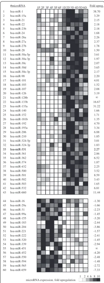
#microRNA 1P 2P 3P 4P 5P 6P 1D2D 3D 4D 5D 6D Fold upreg. 1 hsa-miR-1 38.70 2 hsa-miR-15a 3.29 3 hsa-miR-21 2.15 4 hsa-miR-22 2.18 5 hsa-miR-23b 2.18 6 hsa-miR-24 1.68 7 hsa-miR-26a 2.26 8 hsa-miR-27a 2.37 9 hsa-miR-27b 1.78 10 hsa-miR-28 1.50 11 hsa-miR-30a-5p 3.61 12 hsa-miR-30a-3p 1.97 13 hsa-miR-30c 2.07 14 hsa-miR-30d 3.65 15 hsa-miR-30e-3p 2.17 16 hsa-miR-98 2.20 17 hsa-miR-101 4.01 18 hsa-miR-103 1.52 19 hsa-miR-107 2.08 20 hsa-miR-126 3.19 21 hsa-miR-128b 22 hsa-miR-133b 16.67 23 hsa-miR-133a 31.22 24 hsa-miR-140 1.66 25 hsa-miR-152 1.71 26 hsa-miR-181b 1.35 27 hsa-miR-192 4.70 28 hsa-miR-193a 3.34 29 hsa-miR-206 6.88 30 hsa-miR-210 1.85 31 hsa-miR-324-5p 3.01 32 hsa-miR-324-3p 1.83 33 hsa-miR-331 2.27 33 hsa-miR-331 2.27 34 hsa-miR-361 2.86 35 hsa-miR-362 6.52 36 hsa-miR-374 1.87 37 hsa-miR-432 2.25 38 hsa-miR-500 3.52 39 hsa-miR-501 8.59 40 hsa-miR-502 3.43 41 hsa-miR-503 1.65 42 hsa-miR-532 6.81 43 hsa-miR-660 11.44 44 hsa-miR-16 -1.30 45 hsa-miR-29a -1.66 46 hsa-miR-31 -2.23 47 hsa-miR-99a -1.45 48 hsa-miR-155 -3.20 49 hsa-miR-183 -2.44 50 hsa-miR-204 -3.86 51 hsa-miR-221 -1.57 52 hsa-miR-222 -2.41 53 hsa-miR-320 -1.45 54 hsa-miR-339 -2.94 55 hsa-miR-451 56 hsa-miR-452 -4.20 57 hsa-miR-550 -2.48 58 hsa-miR-594 -1.48 59 hsa-miR-565 -2.72 60 hsa-miR-659 -7.33 1 2 4 6 8 10 microRNA expression. fold upregulation
Figure 2 miRNA profiling in proliferating myoblasts (left) and differentiated myotubes (right). Out of 365 miRNAs tested, this table shows 60 miRNAs that were differentially expressed in myotubes as compared to myoblasts. Gray levels indicates the expression level of microRNA in each individual sample tested. Labels 1P to 6P correspond to samples of proliferating myoblasts described in Table S1, labels 1D to 6D correspond to the samples of differentiated myotubes described in Additional file 1: Table S1.
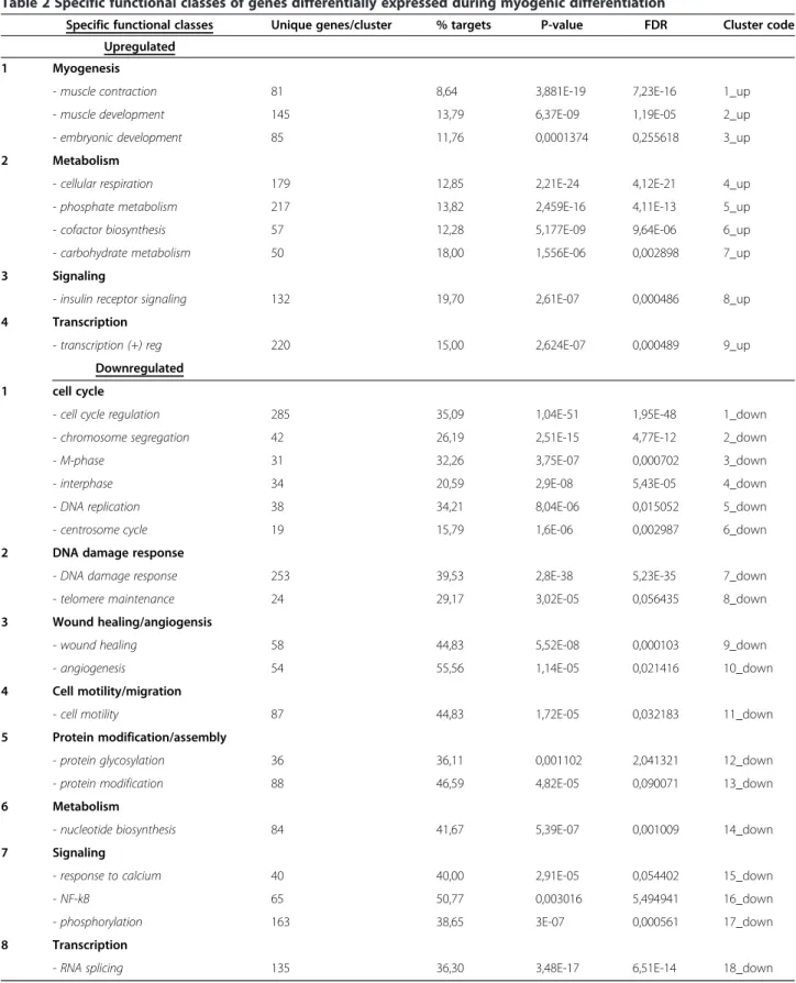
Table 2 Specific functional classes of genes differentially expressed during myogenic differentiation
Specific functional classes Unique genes/cluster % targets P-value FDR Cluster code Upregulated
1 Myogenesis
- muscle contraction 81 8,64 3,881E-19 7,23E-16 1_up - muscle development 145 13,79 6,37E-09 1,19E-05 2_up - embryonic development 85 11,76 0,0001374 0,255618 3_up 2 Metabolism
- cellular respiration 179 12,85 2,21E-24 4,12E-21 4_up - phosphate metabolism 217 13,82 2,459E-16 4,11E-13 5_up - cofactor biosynthesis 57 12,28 5,177E-09 9,64E-06 6_up - carbohydrate metabolism 50 18,00 1,556E-06 0,002898 7_up 3 Signaling
- insulin receptor signaling 132 19,70 2,61E-07 0,000486 8_up 4 Transcription
- transcription (+) reg 220 15,00 2,624E-07 0,000489 9_up Downregulated
1 cell cycle
- cell cycle regulation 285 35,09 1,04E-51 1,95E-48 1_down - chromosome segregation 42 26,19 2,51E-15 4,77E-12 2_down - M-phase 31 32,26 3,75E-07 0,000702 3_down - interphase 34 20,59 2,9E-08 5,43E-05 4_down - DNA replication 38 34,21 8,04E-06 0,015052 5_down - centrosome cycle 19 15,79 1,6E-06 0,002987 6_down 2 DNA damage response
- DNA damage response 253 39,53 2,8E-38 5,23E-35 7_down - telomere maintenance 24 29,17 3,02E-05 0,056435 8_down 3 Wound healing/angiogensis
- wound healing 58 44,83 5,52E-08 0,000103 9_down - angiogenesis 54 55,56 1,14E-05 0,021416 10_down 4 Cell motility/migration
- cell motility 87 44,83 1,72E-05 0,032183 11_down 5 Protein modification/assembly
- protein glycosylation 36 36,11 0,001102 2,041321 12_down - protein modification 88 46,59 4,82E-05 0,090071 13_down 6 Metabolism
- nucleotide biosynthesis 84 41,67 5,39E-07 0,001009 14_down 7 Signaling
- response to calcium 40 40,00 2,91E-05 0,054402 15_down - NF-kB 65 50,77 0,003016 5,494941 16_down - phosphorylation 163 38,65 3E-07 0,000561 17_down 8 Transcription
- RNA splicing 135 36,30 3,48E-17 6,51E-14 18_down
differentiation appeared to be controlled by microRNAs. However, the proportion of genes targeted by microRNAs was not the same within different functional classes. For example, 50% to 55% of genes related to angiogenesis and NF-kB signaling were potentially targeted by miRNAs, while in the case of myogenesis, potential miRNA targets represented only 8.6% of genes (Table 2). Interestingly, more miRNA targets were found among downregulated than upregulated genes. For example, among apoptosis-related genes, 17.6% of upregulated and 38.4% of downregulated genes were found to be targeted by miRNAs (Table 3). This observation prompted us to search for known functions of each supported target gene of every MR-miRNA described in this study using DAVID functional annotation tool [37,38].
miR-1 and miR-206
It has been previously demonstrated that miR-1 and −206 target genes linked to the regulation of chromatin modifi-cations [8], transcription [20,49] and cell cycle [50]. Func-tional analysis of predicted miR-1 target genes supported
by transcriptome data indicated that these microRNAs might also target genes involved in the regulation of apop-tosis, DNA damage response, cell motility and protein modification, cell signaling and kinase activity [33]. Our own functional analysis of predicted target genes of miR-1 and −206 supported by transcriptome analysis confirmed that miR-1 and miR-206 might target genes involved in in these biological processes. In addition, miRNA-1 and −206 might target genes implicated in cytoskeleton organization, ubiquitination and nucleotide biosynthesis (Figure 5, Additional file 9).
miR-21
It has been demonstrated that miR-21 targets genes in-volved in in apoptosis [51,52] and regulation of kinase ac-tivity [53-56] and regulation of gene expression [57]. Here we confirm that miR-21 might target genes involved in these biological processes and might also target genes in-volved in transport, cell cycle regulation, chromatin assem-bly, protein modification, NF-kB signaling, protein complex assembly and DNA damage (Figure 5, Additional file 9).
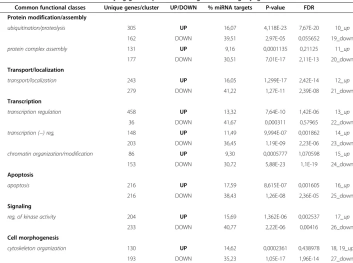
Table 3 Common functional classes unifying genes up- and downregulated during myogenic differentiation
Common functional classes Unique genes/cluster UP/DOWN % miRNA targets P-value FDR 1 Protein modification/assembly
ubiquitination/proteolysis 305 UP 16,07 4,118E-23 7,67E-20 10_up 162 DOWN 39,51 2,97E-05 0,055652 19_down protein complex assembly 131 UP 9,16 0,0001135 0,21125 11_up
177 DOWN 30,51 7,01E-17 2,11E-13 20_down 2 Transport/localization
transport/localization 243 UP 16,05 1,299E-17 2,42E-14 12_up 279 DOWN 41,22 1,27E-11 2,39E-08 21_down 3 Transcription
transcription regulation 458 UP 13,32 7,64E-10 1,42E-06 13_up 36 DOWN 41,67 0,000311 0,57965 22_down transcription (−) reg, 148 UP 11,49 9,994E-07 0,001862 14_up
203 DOWN 36,45 1,19E-09 2,23E-06 23_down chromatin organization/modification 86 UP 9,30 0,0005777 1,070598 15_up
153 DOWN 30,72 5,88E-23 1,1E-19 24_down 4 Apoptosis
apoptosis 216 UP 17,59 8,615E-07 0,001605 16_up 216 DOWN 38,43 1,26E-08 2,36E-05 25_down 5 Signaling
reg, of kinase activity 204 UP 15,69 1,362E-06 0,002537 17_up 233 DOWN 40,77 2,22E-06 0,00416 26_down 6 Cell morphogenesis
cytoskeleton organization 130 UP 14,62 0,0002361 0,438978 18, 19_up 193 DOWN 35,23 1,05E-17 1,96E-14 27_down
miR-24
It has been demonstrated that miR-24 is involved in DNA damage response [58,59], regulation of cell cycle progres-sion [59,60], regulation of kinase activity and protein phosphorylation [61], transcription [62] and apoptosis [63]. We confirm that miR-24 might control genes impli-cated in these biological processes. In addition, miR-24 might control genes implicated in the regulation of trans-port , NF-kB signaling, protein ubiquitination and com-plex assembly, RNA splicing and chromatin modification (Figure 5, Additional file 9).
miR-26a
It has been previously demonstrated that miR-26a target genes are implicated in cell cycle control [64], apoptosis [65], regulation of kinase activity and protein phosphor-ylation [66], as well as chromatin modification [12]. Here we confirm that miR-26a might target genes involved in the regulation of cell cycle, apoptosis, kinase activity and protein phosphorylation, but not in chromatin modifica-tion. In addition, miR-26a might target genes involved in the regulation of transport, DNA damage response, pro-tein modification and propro-tein complex assembly, as well as nucleotide biosynthesis and RNA splicing (Figure 5, Additional file 9).
miR-27a
miR-27b is involved in the regulation of transcription by inhibiting the expression of various transcription factors [13,67-70]. Here we confirm the role of miR-27b as an im-portant transcriptional regulator, in addition, our predic-tions suggest that miR-27a might be involved in the regulation of intracellular transport, apoptosis, kinase ac-tivity, protein complex assembly, cytoskeleton organization and other biological functions including DNA damage re-sponse, cell cycle, motility, nucleotide biosynthesis and protein phosphorylation (Figure 5, Additional file 9).
miR-30c
miR-30 was shown to induce apoptosis [71] and regulate cell motility by influencing extracellular the matrix re-modelling process [72-74]. This microRNA is also known to regulate protein modification e.g. sumoylation [75] and transcription [72,73,76,77]. We here confirm that miR-30c
might target genes participate in the regulation of apop-tosis and cell motility regulation but not transcription control. Instead, our predictions suggest that miR-30 might target genes involved in the regulation of protein phosphorylation and kinase activity, cell cycle control, intracellular transport, cytoskeleton organization, protein ubiquitination, DNA damage response and nucleotide bio-synthesis (Figure 5, Additional file 9).
miR-128b
It has been previously demonstrated that miR-128b tar-gets genes involved in the regulation of cell cycle [78,79], transcription [78,79] and apoptosis [79,80]. In addition, transcriptome analysis of cell transfected with miR-128 re-vealed an alteration of the expression of genes implicated in cytoskeleton organization, kinase activity and protein phosphorylation [81]. Here we confirm the target genes of miR-128 might be involved in these biological functions. In addition, our predictions suggest that miR-128 might target genes in DNA damage response, transport, protein modification and ubiquitination and cell motility (Figure 5, Additional file 9).
miR-133a and miR-133b
Previously, these microRNAs have been shown to control genes involved in the regulation of transcription [8,82], cytoskeleton structure [83-87], apoptosis [88] and mRNA splicing [89]. Here we confirm that miR-133a/b might tar-get genes involved in these biological functions. In addition, miR-133a/b might target genes involved in intracellular transport, cell cycle regulation, DNA damage response, protein phosphorylation and ubiquitination (Figure 5, Additional file 9).
miR-204
It has been previously shown that miR-204 is involved in the regulation of transcription [90-92], controls cell migra-tion cytoskeleton organizamigra-tion [92-94]. miR-204 has been also shown to be involved in regulation of apoptosis by targeting multiple genes [95] and in the regulation of cell signaling [94, 96]. Here we confirm that miR-204 might target genes involved in transcription regulation, cytoskel-eton organization and apoptosis but not cell signaling and cell migration. Furthermore, our predictions suggest that
(See figure on previous page.)
Figure 3 Bioinformatic predictions of microRNA target genes. A. Density plot of Pearson correlation coefficients between expression of miRNA and their target genes (continuous line) predicted by RNA22 algorithm. Number of genes is expressed as% of total predictions indicated within each plot. The distribution of Pearson correlations is clearly shifted towards negative values; the density plot of Pearson correlation coefficient after permutation of the list of microRNAs (dashed line). In this case the distribution is centered around zero. Several examples are shown, for other microRNAs see the Additional file 6. B. Diagram showing the proportion of bioinformatic predictions made by RNA22 algorithm (taken for 100%) that were supported by transcriptome profiling (black). Light grey shows unsupported predictions. The graphs corresponding to TargetScan predictions are presented in the Additional file 8. C. Comparison of the accuracy of target gene predictions by RNA22 and TargetScan algorithms.% of target genes predicted by both algorithms and supported by transcriptome data is shown.
this microRNA might target genes involved in the control of ubiquitination, cellular respiration and intracellular transport (Figure 5, Additional file 9).
miR-221 and miR-222
Previously, these microRNAs have been show to target genes involved in the process of protein degradation [97], apoptosis [98], kinase activity [99,100], cell cycle progression [29] and cell motility [101]. We here con-firm that miR-221 and −222 might target genes involved in protein degradation (via ubiquitination), apoptosis, and kinase activity regulation, but not cell cycle progres-sion or cell motility. In addition, miR-221 and −222 might also target genes that play a role in the regulation of transcription, insulin signaling, intracellular transport and cellular respiration (Figure 5, Additional file 9).
miR-550
No target genes and functions of this microRNA were not previously described. Our predictions suggest that miR-550 might target genes involved in the regulation of transcription, protein ubiquitination, phosphate metab-olism, apoptosis, insulin signaling, muscle development, intracellular transport and cellular morphology (Figure 5, Additional file 9).
The full list of supported target genes of MR-miRs with and without assigned functions can be found in the Additional file 7.
Discussion and conclusion
MicroRNAs, particularly miR-1, -206 and -133a/b, play crucial roles in myogenesis [6]. These are called MyomiRs to underscore their importance in myogenesis (for review see [24]). Previously, high-throughput profil-ing of miRNA expression performed in human myo-blasts have resulted in identification of miRNAs differentially expressed during myogenic differentiation [26,27]. Besides that, a larger number of miRNAs have been found to be differentially expressed during myo-genic differentiation of mouse C2C12 myoblasts [19,28], mouse myogenic progenitors [18,21] or at various stages of development of skeletal muscles in pig [31] or in the common carp [32].
Here, we have compared miRNA expression profiles in human proliferating primary CD56+ myoblasts and differen-tiated myotubes. Sixty differentially expressed miRNAs were identified (MR-miRs). Twenty of these had not been previ-ously implicated in myogenesis. Conversely, several miRNAs previously reported as related to myogenic differentiation were not identified in the present study. These included miR-214 [18], hsa-miR-424 (identical to mmu-miR-322) [19], miR-29b/c [14], miR-143, miR-208a [15], miR-208b, miR-499 [17], miR-125b [102] (Additional file 10). One miRNA, miR-682, previously shown to be upregulated in mouse myogenic precursors [21] could not be tested here. The difficulties to confirm previously reported data for sev-eral miRNAs associated with myogenesis in model organ-isms could be due to species-specific characteristics in the myogenesis program or could originate from differences in experimental conditions. Indeed, high-throughput miRNA expression profiling has been previously performed using mixed cell populations isolated from skeletal myoblasts [26,27]. The resulting presence of non-myogenic cells, in-cluding fibroblasts and inflammatory cells, could have al-tered the resulting miRNA expression profiles. It thus appears that the existing repertoire of myogenic miRNAs is still incomplete or contaminated with miRNAs unrelated to myogenesis. For example, we have demonstrated that miR-221 and miR-222 were downregulated during myogenic dif-ferentiation of human myoblasts while others reported their upregulation [26]. Our results are in agreement with previ-ous reports demonstrating that these microRNAs are downregulated during myogenesis in mice [28,29].
In addition to microRNA expression profiling, we have profiled mRNA expression in myogenic cells in the course of myogenic differentiation. Transcriptome ana-lysis of myogenesis has been previously reported by others. In mice, several models have been used for tran-scriptome profiling. These include C2C12 immortalized cell line considered as the “golden standard” of in vitro myogenic differentiation [103-107], primary mouse myo-blasts [108] and an in vivo model of muscle regeneration following injury [109]. In humans, the only published mRNA expression profiling has been performed on pri-mary cells grown in vitro [36]. The transcriptome profil-ing of human myoblasts in the course of myogenic
(See figure on previous page.)
Figure 4 Confirmation of bioinformatic predictions of miR targets. Human immortalized myoblasts (iMyo) cultivated in growth medium (P) or differentiation medium for 3 days (D) were transfected with either control LNA (anti-miR-C) or LNA against miR-1 and miR-206, miR-133a and miR-133b (A). Human rhabdomyosarcoma cells (RD) were transfected with plasmids coding for miR-128 and miR-30 precursors or scrambled sequence (scr) (B). Then the expression of corresponding microRNA and their randomly selected target genes was tested using qRT-PCR, normalization was performed using ΔΔCt method using GAPDH as a control gene and proliferating sample #1 as a reference sample (expression level 1). Transcriptome-supported target genes of miR-1/206 (C) and miR-133a/b (D) were downregulated during normal myogenic differentiation but not when it was accompanied by the transfection with corresponding anti-miRs. Transcriptome-supported target genes of miR-128 and miR-30 were downregulated when human rhabdomyosarcoma cells were transfected with lentiviral constructs overexpressing miR-128 (E) and miR-30 (F). The average of three independent experiments is shown. (*) indicates p-value <0.05; (G): Diagram showing the proportion of supported predictions that were qRT-PCR validated in this study. Black: validated targets, gray: non-validated targets.
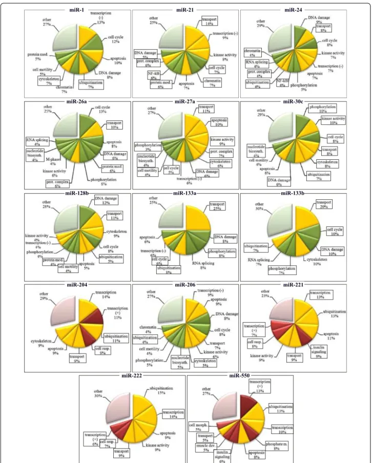
Figure 5 Predicted functions of MR-miRs. Green: functions downregulated during myogenic differentiation, Red: functions upregulated during myogenic differentiation. Yellow: functions that are both up- and downregulated during myogenic differentiation. Framed are the functions that have not been previously ascribed to a given microRNA.
differentiation in vitro described here, has two important advantages over the previously published study. Firstly, we used CD56+ myogenic cells that allowed us to ex-clude from the analysis genes unrelated to myogenesis. Secondly, our study is the first example of a simultan-eous miRNA/mRNA expression profiling of myogenic cells.
Simultaneous miRNA/mRNA was used here to identify novel target genes of myogenesis-related microRNAs. This approach has been proven efficient for increasing the pre-cision of bioinformatics predictions for miRNA targets [42], and it has been applied previously to identify miRNA target genes in neural tissue [45] and cancer cell lines [44,110], but not in myogenic cells or muscle tissue.
Several methods are used to validate the target genes of miRNA. These include qRT-PCR, luciferase assays and western blot (for review see [111] and [112]). Lucif-erase assay based on a reporter plasmid containing miRNA recognition sites in the 30UTR of the luciferase
gene is the method used to demonstrate the direct in-hibition of mRNA expression by miRNA. However this method does not provide information as to whether the miRNA-dependent regulation occurs at the level of tran-script stability or at the level of translation, nor the dir-ect miRNA:mRNA interaction demonstrated by this method does guarantee the physiological significance of the observed miRNA effect. Here, to validate the pre-dicted target genes, we have used qRT-PCR validation approach. This method is more physiological than lucif-erase assay as it measures the expression of target gene from their natural genomic context, although it can not discriminate between direct and indirect effects of miRNA expression. Hereby, we have randomly selected 32 genes targeted by 6 different microRNAs including miR-1, -206, -133a, -133b, -128 and −30 and tested their expression using pRT-PCR in the cells where corre-sponding microRNA was ectopically overexpressed or inhibited. We have demonstrated the miRNA-dependent inhibition for 18 of these target genes. We called these genes “qRT-PCR validated” to formally distinguish them from simply “validated”, as the latter term is usually re-served for miRNA target genes validated using luciferase reporters.
On average, we estimate that the lists of predicted tar-get genes supported by transcriptome data contain at least 50% of genes that will change their expression, according to our predictions, in response to the alter-ations of microRNA expression.
Using the lists of target genes supported by transcrip-tome data we have deduced functional impact of MR-miRs using DAVID functional annotation tool [37,38]. Our predictions generally corresponded to already known functions of MR-miRs demonstrated by others, but also allowed us to make suggestions about novel
functions of MR-miRs. It has to be noted, that these pre-dictions have to be handled with caution for two reasons: (i) according to our estimation, among target genes sup-ported by transcriptome analysis, up to 50% might not react to miRNA alteration, as predicted; (ii) the functions of MR-miR have been deduced using bioinformatic pre-diction algorithms that can not substitute for a true ex-perimental validation of microRNA functions.
To summarize, we have discovered 20 human miRNAs not previously known to be differentially expressed during myogenic differentiation and confirmed 40 myogenesis-related microRNAs. We then used an approach that in-cluded a simultaneous miRNA/mRNA expression profiling to predict targets genes of these microRNAs with more than 50% accuracy, although whether these are direct or indirect targets of miRNA remains unknown. Finally, we deduced novel functions of these microRNA from the known functions of their target genes. Further studies are necessary to confirm our predictions of functional contri-bution of newly discovered miRNAs to the process of myo-genic differentiation.
Methods
Cell culture conditions and transfection
Primary human myoblasts were isolated from skeletal muscles of healthy subjects as described in [35], for de-tails see Additional file 4: Table S4), purified with an immuno-magnetic sorting system (Miltenyi Biotec, USA) using an anti-CD56/NCAM antibody according to the manufacturer’s instructions. CD56-positive myoblasts were seeded in collagen-coated Petri dishes (P1) and cul-tured in DMEM, 10% FCS, 1% Ultroser G, at 37°C with 5% CO2.All experiments were carried out between P1
and P5 to avoid cell senescence. Myoblast purity was de-termined by staining for Desmin. Purified myoblasts were plated in collagen-coated Petri dishes and cultured in growth medium containing DMEM supplemented with 20% fetal bovine serum at 37°C in humidified at-mosphere with 5% CO2. Myogenic differentiation of
confluent cells was induced after 5 days by changing to DMEM containing 2% FBS (differentiation medium). Cells were kept in differentiation medium for 3 days. Myogenic fusion index at day three was 37-70%. Human immortalized myoblasts (iMyo) (kind gift of Dr. V. Mouly) were grown and differentiated as described in [113]. RD and TE671 (a kind gift of Dr. S. Leibowitz) were grown as described [114]. phrGFP-1 vector was from Stratagen, pcDNA3.2/V5 hsa-mir-128 (#26308) [115] and pCMV-miR30 (#20875) [116] vectors were pro-vided by Addgene. Transient transfection of RD cells was performed in 6-well plate format using Lipofectamine 2000 according to manufacturer’s instructions with minor modification: 600.000 cells were added to the transfection mixture prepared directly in the cell culture plate. To
transfect human immortallized myoblasts, anti-sense LNA inhibitors of miR-1, -206, 133a and b, and the irrelevant control were purchased from Exiqon (Denmark) and were mixed with LipoRNAiMAX (Invitrogen) in 6-well plates at 100nM final concentration, using 500 000 cells per well.
Reverse transcription and real-time PCR
For microRNA expression analysis 400 ng of total RNA purified via Trizol (Invitrogen) was converted into cDNA using 8 independent pools of primers (#4384791, Ap-plied Biosystems, AB) and TaqMan microRNA Reverse transcription kit (#4366596, AB). cDNA was quantified using via qPCR using TaqMan 2x Universal PCR Master Mix, No AmpErase UNG (#4324018, AB) and human microRNA panel version 1.0 TLDA (TaqMan Low Density Array, AB), data were acquired on AB7900HT Real-Time PCR machine. Sequences of microRNAs and corresponding codes of TaqMan assays can be found in Additional file 2. For mRNA expression analysis total RNA was isolated from 2x10⁶proliferating myoblasts, or differentiated myotubes using Trizol (Invitrogen) and re-verse transcribed using the High Capacity cDNA Arch-ive kit (Applied Biosystems, AB) according to the manufacturer protocol. cDNA was mixed with 2x Taqman PCR mix (AB) and amplified customized TLDA (TaqMan Low Density Array) using Abiprism 7900HT apparatus (AB). In case of miRNA and mRNA the ex-pression level was calculated from Ct values using ΔΔCt method using GAPDH as a control [117]. The calcula-tion of SEM in case of average of multiple samples has been done as described in [118].
Transcriptome profiling and data processing
Human primary myoblasts were sacrificed directly on plates at 30% confluency using Trizol, RNA was prepared using organic extraction and ethanol precipitation as de-scribed [119] followed by silica column cleanup on silica columns (Nucleospin RNA Extraction kit, Macherey Nagel). RNA extracted from individual myoblast lines was Cy5-labeled, mixed with with a pool of RNA samples la-beled with Cy3 and hybridized to Gene Expression microarrays (4x44k #G4112F Agilent) and scanned as instructed by the manufacturer. Scanned images were then analysed using the Feature Extraction Software (Agilent), the subsequent gene expression data treatment was performed using R and Bioconductor. Spots with in-tensity lower than 50 or lower than background in more than 50% of biological replicates have been removed from further analysis. The background correction and intensity normalization procedures were applied for the re-maining ~30 000 probes using the Bioconductor package vsn [120]. A background offset and a scaling factor for each array and dye channel were calculated using the least squares regression procedure, then the generalized
log-transformation was applied. The ordinary least squares re-gression approach is based on the assumption that “most genes are not differentially expressed”. However, in the case of myogenic differentiation, where many genes are differentially expressed, this assumption does not hold. Therefore, to apply the above mentioned approach to myogenic differentiation, the vsn least squares model has first been applied to a subset of features, and then ex-tended for the whole set of features. To select the sub-set of features, a pool of samples prepared from five different proliferating myoblast lines before and after myogenic differentiation were hybridized to two add-itional microarrays. Then 14.358 features that did not exceed the cutoff value of 1.23 fold change between the pools have been selected. To determine the differentially expressed genes, a t-test analysis was conducted using the limma package from Bioconductor [120]. Using this pack-age, a linear model is fitted to the expression data for each gene. An empirical Bayes moderation of the standard errors were performed. This method borrows informa-tion across genes in order to arrive at more stable esti-mates of each individual gene’s variance, even for experiments with small number of arrays. The microarray data related to this paper have been submitted to the Array Express data repository at the European Bioinformatics In-stitute (http://www.ebi.ac.uk/arrayexpress) under the ac-cession number E-MTAB-1577 (release date 2014-3-22).
miRNA targets predictions
RNA22 [40] and TargetScan algorithms [121] have been used to predict miRNA target genes in this study. miRNA targets identified by RNA22 have been downloaded from the RNA22 website http://cbcsrv.watson.ibm.com/rna22. html. Predictions were processed in the form of 30UTR
binding sites occurrence matrices, containing the number of 30UTR binding sites for each combination of miRNA
(columns) and mRNA (rows).
Pearson correlation between mRNA and miRNA
expression profiles and permutation tests for significance
All the following has been performed using the R soft-ware. Pearson correlation values were computed using expression levels of each single transcript available and for each miRNA, found to be differentially expressed during myogenic differentiation in this study. Pearson correlation values were retained for further analysis, if they were corresponding to miRNA and mRNA pares having at least one occurrence in at least one of the two 30UTR binding sites occurrence matrices. To distinguish
between biologically significant and random correlations, we performed a permutation test. We performed nine different permutations by calculating correlation values using a miRNA table with permuted sample designa-tions. Then, the distribution of the real and permuted
data were graphically plotted. For each miRNA, the 5% percentile of the Pearson correlation obtained with the permuted data was used as a threshold (th) for selecting biologically significant target genes. For each differen-tially expressed miRNA, the Pearson correlation value is compared to th and if smaller, the gene is retained as a biologically significant target gene regulated by the miRNA, with a 5% risk of error.
Additional files
Additional file 1: Table S1. Samples of myogenic cells used in the study were isolated from six healthy subjects.
Additional file 2: Table S2. Results of miRNA expression analysis using TaqMan low density arrays.
Additional file 3: Table S3. Sequences of mature microRNAs and corresponding TaqMan probes used in this study.
Additional file 4: Table S4. mRNAs differentially expressed during myogenic differentiation of human primary myoblasts. (XLSX 761 kb) Additional file 5: Table S5. Functional classification of genes differentially expressed during myogenic differentiation. (XLSX 904 kb) Additional file 6: Figure S1. A. Density plot of Pearson correlation coefficients between expression of miRNA and their target genes (continuous line) predicted by RNA22 algorithm and the the density plot of Pearson correlation coefficient after permutation of the list of microRNAs (dashed line). B. Schematic representation of ectopic overexpression/knockdown of miRNA in the cells that served to confirm their targets genes supported by transcriptome data.
Additional file 7: Table S6. Target genes of microRNAs differentially expressed during myogenic differentiation, supported by transcriptome analysis of mRNA expression. (XLSX 223 kb)
Additional file 8: Figure S2A. Density plot of Pearson correlation coefficients between expression of miRNA and their target genes (blue) predicted by TargetScan algorithm and the density plot of Pearson correlation coefficient after permutation of the list of microRNAs (red). B. Diagram showing the proportion of bioinformatic predictions made by TargetScan algorithm (taken for 100%) that were supported by transcriptome profiling (black). Blue bars show unsupported predictions. Additional file 9: Table S8. Functions of miRNA target genes supported by transcriptome data. Functional classification has been performed using DAVID functional annotation tool.
Additional file 10: Table S7. microRNAs that were previously shown to be differentially expressed during myogenesis but could not be confirmed in the present study.
Competing interests
The authors declare that they have no competing interests. Authors’ contributions
YSV and PDm designed the experiments, PDm, AP, TR, TAW, VL, AT, GC and DL-C performed the experiments, PDm, AB, MO’C and PDe conducted bioinformatic analysis of the data, YSV, PDm and ML wrote the manuscript. All authors read and approved the final manuscript.
Acknowledgements
This research was supported by grants from the Association Française contre les Myopathies (AFM) to YSV and DL PD is co-funded by the University Montpellier I and the association “Amis FSH”.
Author details
1
UMR 8126, Univ. Paris-Sud 11, CNRS, Institut de Cancérologie
Gustave-Roussy, 39, rue Camille-Desmoulins, Villejuif 94805, France.2INSERM
U-1046, 371 Avenue du Doyen Gaston Giraud, Montpellier F-34295, France.
3Institut de Cancérologie Gustave-Roussy, Villejuif F-94805, France.
4Bioinformatics and Molecular Evolution Group, School of Biotechnology,
Dublin City University, Glasnevin, Dublin 9, Ireland.5CNRS FRE 3377, CEA
Saclay, Gif-sur-Yvette F-91191, France.
Received: 19 November 2012 Accepted: 26 March 2013 Published: 18 April 2013
References
1. Kim VN, Nam JW: Genomics of microRNA. Trends Genet 2006, 22(3):165–173. 2. Olena AF, Patton JG: Genomic organization of microRNAs. J Cell Physiol
2010, 222(3):540–545.
3. Valencia-Sanchez MA, Liu J, Hannon GJ, Parker R: Control of translation and mRNA degradation by miRNAs and siRNAs. Genes Dev 2006, 20(5):515–524. 4. Vasudevan S, Tong Y, Steitz JA: Switching from repression to activation:
microRNAs can up-regulate translation. Science (New York, NY 2007, 318(5858):1931–1934.
5. Callis TE, Deng Z, Chen JF, Wang DZ: Muscling through the microRNA world. Experimental biology and medicine (Maywood, NJ 2008, 233(2):131–138. 6. McCarthy JJ: MicroRNA-206: the skeletal muscle-specific myomiR.
Biochim Biophys Acta 2008, 1779(11):682–691.
7. Zhao Y, Samal E, Srivastava D: Serum response factor regulates a muscle-specific microRNA that targets Hand2 during cardiogenesis. Nature 2005, 436(7048):214–220.
8. Chen JF, Mandel EM, Thomson JM, Wu Q, Callis TE, Hammond SM, Conlon FL, Wang DZ: The role of microRNA-1 and microRNA-133 in skeletal muscle proliferation and differentiation. Nat Genet 2006, 38(2):228–233. 9. Townley-Tilson WH, Callis TE, Wang D: MicroRNAs 1, 133, and 206: critical
factors of skeletal and cardiac muscle development, function, and disease. Int J Biochem Cell Biol 2009, 42(8):1252–1255.
10. Rao PK, Kumar RM, Farkhondeh M, Baskerville S, Lodish HF: Myogenic factors that regulate expression of muscle-specific microRNAs. Proc Natl Acad Sci USA 2006, 103(23):8721–8726.
11. Sun Q, Zhang Y, Yang G, Chen X, Zhang Y, Cao G, Wang J, Sun Y, Zhang P, Fan M, et al: Transforming growth factor-beta-regulated miR-24 promotes skeletal muscle differentiation. Nucleic Acids Res 2008, 36(8):2690–2699.
12. Wong CF, Tellam RL: MicroRNA-26a targets the histone methyltransferase Enhancer of Zeste homolog 2 during myogenesis. J Biol Chem 2008, 283(15):9836–9843.
13. Crist CG, Montarras D, Pallafacchina G, Rocancourt D, Cumano A, Conway SJ, Buckingham M: Muscle stem cell behavior is modified by microRNA-27 regulation of Pax3 expression. Proc Natl Acad Sci USA 2009, 106(32):13383–13387. 14. Wang H, Garzon R, Sun H, Ladner KJ, Singh R, Dahlman J, Cheng A, Hall BM,
Qualman SJ, Chandler DS, et al: NF-kappaB-YY1-miR-29 regulatory circuitry in skeletal myogenesis and rhabdomyosarcoma. Cancer Cell 2008, 14(5):369–381. 15. Sempere LF, Freemantle S, Pitha-Rowe I, Moss E, Dmitrovsky E, Ambros V:
Expression profiling of mammalian microRNAs uncovers a subset of brain-expressed microRNAs with possible roles in murine and human neuronal differentiation. Genome Biol 2004, 5(3):R13.
16. Naguibneva I, Ameyar-Zazoua M, Polesskaya A, Ait-Si-Ali S, Groisman R, Souidi M, Cuvellier S, Harel-Bellan A: The microRNA miR-181 targets the homeobox protein Hox-A11 during mammalian myoblast differentiation. Nat Cell Biol 2006, 8(3):278–284.
17. van Rooij E, Quiat D, Johnson BA, Sutherland LB, Qi X, Richardson JA, Kelm RJ Jr, Olson EN: A family of microRNAs encoded by myosin genes governs myosin expression and muscle performance. Dev Cell 2009, 17(5):662–673. 18. Juan AH, Kumar RM, Marx JG, Young RA, Sartorelli V: Mir-214-dependent
regulation of the polycomb protein Ezh2 in skeletal muscle and embryonic stem cells. Mol Cell 2009, 36(1):61–74.
19. Sarkar S, Dey BK, Dutta A: MiR-322/424 and −503 are induced during muscle differentiation and promote cell cycle quiescence and differentiation by down-regulation of Cdc25A. Mol Biol Cell 2010, 21(13):2138–2149. 20. Dey BK, Gagan J, Dutta A: miR-206 and −486 induce myoblast
differentiation by downregulating Pax7. Mol Cell Biol 2011, 31(1):203–214. 21. Chen Y, Gelfond J, McManus LM, Shireman PK: Temporal MicroRNA
Expression during in vitro Myogenic Progenitor Cell Proliferation and Differentiation: Regulation of Proliferation by miR-682. Physiol Genomics 2011, 43:621–630.
22. Ge Y, Sun Y, Chen J: IGF-II is regulated by microRNA-125b in skeletal myogenesis. J Cell Biol 2011, 192(1):69–81.
23. le Sage C, Nagel R, Egan DA, Schrier M, Mesman E, Mangiola A, Anile C, Maira G, Mercatelli N, Ciafre SA, et al: Regulation of the p27(Kip1) tumor
suppressor by miR-221 and miR-222 promotes cancer cell proliferation. EMBO J 2007, 26(15):3699–3708.
24. Ge Y, Chen J: MicroRNAs in skeletal myogenesis. Cell cycle (Georgetown, Tex 2011, 10(3):441–448.
25. Guller I, Russell AP: MicroRNAs in skeletal muscle: their role and regulation in development, disease and function. J Physiol 2010, 588 (Pt 21):4075–4087.
26. Ciarapica R, Russo G, Verginelli F, Raimondi L, Donfrancesco A, Rota R, Giordano A: Deregulated expression of miR-26a and Ezh2 in rhabdomyosarcoma. Cell cycle (Georgetown, Tex 2009, 8(1):172–175. 27. Granjon A, Gustin MP, Rieusset J, Lefai E, Meugnier E, Guller I, Cerutti C,
Paultre C, Disse E, Rabasa-Lhoret R, et al: The microRNA signature in response to insulin reveals its implication in the transcriptional action of insulin in human skeletal muscle and the role of a sterol regulatory element-binding protein-1c/myocyte enhancer factor 2C pathway. Diabetes 2009, 58(11):2555–2564.
28. Sun Y, Ge Y, Drnevich J, Zhao Y, Band M, Chen J: Mammalian target of rapamycin regulates miRNA-1 and follistatin in skeletal myogenesis. J Cell Biol 2010, 189(7):1157–1169.
29. Cardinali B, Castellani L, Fasanaro P, Basso A, Alema S, Martelli F, Falcone G: Microrna-221 and microrna-222 modulate differentiation and maturation of skeletal muscle cells. PLoS One 2009, 4(10):e7607.
30. Koutsoulidou A, Mastroyiannopoulos NP, Furling D, Uney JB, Phylactou LA: Expression of miR-1, miR-133a, miR-133b and miR-206 increases during development of human skeletal muscle. BMC Dev Biol 2011, 11:34. 31. Zhou B, Liu HL, Shi FX, Wang JY: MicroRNA expression profiles of porcine
skeletal muscle. Anim Genet 2010, 41(5):499–508.
32. Yan X, Ding L, Li Y, Zhang X, Liang Y, Sun X, Teng CB: Identification and profiling of microRNAs from skeletal muscle of the common carp. PLoS One 2012, 7(1):e30925.
33. Marzi MJ, Puggioni EM, Dall’Olio V, Bucci G, Bernard L, Bianchi F, Crescenzi M, Di Fiore PP, Nicassio F: Differentiation-associated microRNAs antagonize the Rb-E2F pathway to restrict proliferation. J Cell Biol 2012, 199(1):77–95.
34. Seok HY, Tatsuguchi M, Callis TE, He A, Pu WT, Wang DZ: miR-155 inhibits expression of the MEF2A protein to repress skeletal muscle
differentiation. J Biol Chem 2011, 286(41):35339–35346.
35. Barro M, Carnac G, Flavier S, Mercier J, Vassetzky Y, Laoudj-Chenivesse D: Myoblasts from affected and non-affected FSHD muscles exhibit morphological differentiation defects. J Cell Mol Med 2010, 14(1–2):275–289. 36. Sterrenburg E, Turk R, Hoen PA T, Van Deutekom JC, Boer JM, Van Ommen GJ,
Den Dunnen JT: Large-scale gene expression analysis of human skeletal myoblast differentiation. Neuromuscul Disord 2004, 14(8–9):507–518. 37. da Huang W, Sherman BT, Lempicki RA: Systematic and integrative
analysis of large gene lists using DAVID bioinformatics resources. Nat Protoc 2009, 4(1):44–57.
38. da Huang W, Sherman BT, Lempicki RA: Bioinformatics enrichment tools: paths toward the comprehensive functional analysis of large gene lists. Nucleic Acids Res 2009, 37(1):1–13.
39. Ritchie W, Flamant S, Rasko JE: Predicting microRNA targets and functions: traps for the unwary. Nat Methods 2009, 6(6):397–398.
40. Miranda KC, Huynh T, Tay Y, Ang YS, Tam WL, Thomson AM, Lim B, Rigoutsos I: A pattern-based method for the identification of MicroRNA binding sites and their corresponding heteroduplexes. Cell 2006, 126(6):1203–1217. 41. Alexiou P, Maragkakis M, Papadopoulos GL, Reczko M, Hatzigeorgiou
AG: Lost in translation: an assessment and perspective for computational microRNA target identification. Bioinformatics 2009, 25(23):3049–3055.
42. Huang JC, Babak T, Corson TW, Chua G, Khan S, Gallie BL, Hughes TR, Blencowe BJ, Frey BJ, Morris QD: Using expression profiling data to identify human microRNA targets. Nat Methods 2007, 4(12):1045–1049. 43. Huang Y, Zou Q, Song H, Song F, Wang L, Zhang G, Shen X: A study of
miRNAs targets prediction and experimental validation. Protein Cell 2010, 1(11):979–986.
44. Wang YP, Li KB: Correlation of expression profiles between microRNAs and mRNA targets using NCI-60 data. BMC Genomics 2009, 10:218. 45. Nunez-Iglesias J, Liu CC, Morgan TE, Finch CE, Zhou XJ: Joint genome-wide
profiling of miRNA and mRNA expression in Alzheimer’s disease cortex reveals altered miRNA regulation. PLoS One 2010, 5(2):e8898.
46. Sayed D, Abdellatif M: MicroRNAs in development and disease. Physiol Rev 2011, 91(3):827–887.
47. Bartel DP: MicroRNAs: target recognition and regulatory functions. Cell 2009, 136(2):215–233.
48. Lewis BP, Burge CB, Bartel DP: Conserved seed pairing, often flanked by adenosines, indicates that thousands of human genes are microRNA targets. Cell 2005, 120(1):15–20.
49. Chen JF, Tao Y, Li J, Deng Z, Yan Z, Xiao X, Wang DZ: microRNA-1 and microRNA-206 regulate skeletal muscle satellite cell proliferation and differentiation by repressing Pax7. J Cell Biol 2010, 190(5):867–879. 50. Kim HK, Lee YS, Sivaprasad U, Malhotra A, Dutta A: Muscle-specific
microRNA miR-206 promotes muscle differentiation. J Cell Biol 2006, 174(5):677–687.
51. Frankel LB, Christoffersen NR, Jacobsen A, Lindow M, Krogh A, Lund AH: Programmed cell death 4 (PDCD4) is an important functional target of the microRNA miR-21 in breast cancer cells. J Biol Chem 2008, 283(2):1026–1033. 52. Yao Q, Xu H, Zhang QQ, Zhou H, Qu LH: MicroRNA-21 promotes cell
proliferation and down-regulates the expression of programmed cell death 4 (PDCD4) in HeLa cervical carcinoma cells. Biochem Biophys Res Commun 2009, 388(3):539–542.
53. Kim YJ, Hwang SJ, Bae YC, Jung JS: MiR-21 regulates adipogenic differentiation through the modulation of TGF-beta signaling in mesenchymal stem cells derived from human adipose tissue. Stem cells (Dayton, Ohio) 2009, 27(12):3093–3102.
54. Roy S, Khanna S, Hussain SR, Biswas S, Azad A, Rink C, Gnyawali S, Shilo S, Nuovo GJ, Sen CK: MicroRNA expression in response to murine myocardial infarction: miR-21 regulates fibroblast metalloprotease-2 via phosphatase and tensin homologue. Cardiovasc Res 2009, 82(1):21–29.
55. Talotta F, Cimmino A, Matarazzo MR, Casalino L, De Vita G, D’Esposito M, Di Lauro R, Verde P: An autoregulatory loop mediated by miR-21 and PDCD4 controls the AP-1 activity in RAS transformation. Oncogene 2009, 28(1):73–84. 56. Hatley ME, Patrick DM, Garcia MR, Richardson JA, Bassel-Duby R, van Rooij E,
Olson EN: Modulation of K-Ras-dependent lung tumorigenesis by MicroRNA-21. Cancer Cell 2010, 18(3):282–293.
57. Iliopoulos D, Jaeger SA, Hirsch HA, Bulyk ML, Struhl K: STAT3 activation of miR-21 and miR-181b-1 via PTEN and CYLD are part of the epigenetic switch linking inflammation to cancer. Mol Cell 2010, 39(4):493–506. 58. Srivastava N, Manvati S, Srivastava A, Pal R, Kalaiarasan P, Chattopadhyay S,
Gochhait S, Dua R, Bamezai RN: miR-24-2 controls H2AFX expression regardless of gene copy number alteration and induces apoptosis by targeting antiapoptotic gene BCL-2: a potential for therapeutic intervention. Breast Cancer Res 2011, 13(2):R39.
59. Lal A, Navarro F, Maher CA, Maliszewski LE, Yan N, O’Day E, Chowdhury D, Dykxhoorn DM, Tsai P, Hofmann O, et al: miR-24 Inhibits cell proliferation by targeting E2F2, MYC, and other cell-cycle genes via binding to “seedless” 30UTR microRNA recognition elements. Mol Cell 2009, 35(5):610–625. 60. To KH, Pajovic S, Gallie BL, Theriault BL: Regulation of p14ARF expression
by miR-24: a potential mechanism compromising the p53 response during retinoblastoma development. BMC Cancer 2012, 12:69. 61. Dogar AM, Towbin H, Hall J: Suppression of latent transforming growth
factor (TGF)-beta1 restores growth inhibitory TGF-beta signaling through microRNAs. J Biol Chem 2011, 286(18):16447–16458.
62. Melkman-Zehavi T, Oren R, Kredo-Russo S, Shapira T, Mandelbaum AD, Rivkin N, Nir T, Lennox KA, Behlke MA, Dor Y, et al: miRNAs control insulin content in pancreatic beta-cells via downregulation of transcriptional repressors. EMBO J 2011, 30(5):835–845.
63. Singh R, Saini N: Downregulation of BCL2 by miRNAs augments drug induced apoptosis: Combined computational and experimental approach. J Cell Sci 2012.
64. Singh R, Saini N: Downregulation of BCL2 by miRNAs augments drug induced apoptosis: Combined computational and experimental approach. J Cell Sci 2012, 125(6):1568–1578.
65. Zhu Y, Lu Y, Zhang Q, Liu JJ, Li TJ, Yang JR, Zeng C, Zhuang SM: MicroRNA-26a/b and their host genes cooperate to inhibit the G1/S transition by activating the pRb protein. Nucleic Acids Res 2011, 40(10):4615–4625. 66. Huse JT, Brennan C, Hambardzumyan D, Wee B, Pena J, Rouhanifard SH,
Sohn-Lee C, le Sage C, Agami R, Tuschl T, et al: The PTEN-regulating microRNA miR-26a is amplified in high-grade glioma and facilitates gliomagenesis in vivo. Genes Dev 2009, 23(11):1327–1337.
67. Chinchilla A, Lozano E, Daimi H, Esteban FJ, Crist C, Aranega AE, Franco D: MicroRNA profiling during mouse ventricular maturation: a role for miR-27 modulating Mef2c expression. Cardiovasc Res 2011, 89(1):98–108.