Université de Montréal
Molecular mode of action of Cry6Aa1, a new insecticidal
Bacillus thuringiensis toxin
par
Eva Fortea Verdejo
Département de physiologie moléculaire et intégrative Faculté de médecine
Mémoire présenté en vue de l’obtention du grade de Maîtrise
en physiologie moléculaire, cellulaire et intégrative option physiologie et biophysique moléculaires
Membres du jurée Lucie Parent, Réjean Couture et Jean-Louis Schwartz
Août 2016
ABSTRACT
Cry6Aa1 is a new toxin produced by Bacillus thuringiensis (Bt), which displays insecticidal activity against the Western corn rootworm (WCRW). The present work demonstrates that Cry6Aa1 is a pore-forming toxin (PFT) in planar lipid bilayers (PLBs). Contrary to other Bt toxins tested so far, pore formation by Cry6Aa1 does not require protease pretreatment and takes place at doses that are two to three orders of magnitude lower than those required for other Bt toxins under similar conditions.
Pore formation by Cry6Aa1 is pH-dependent; the conductances of the pores range between 31 and 689 pS under symmetrical 150 mM KCl conditions; they are cationic and display a complex kinetic behaviour. The treatment of the toxin with midgut juice (Cry6Aa1 WCR1) does not change the biophysical properties of the pores. However, the treatment with trypsin (Cry6Aa1 TT) affects their conductance and selectivity at pH 5.5 (the WCRW gut physiological pH). The incorporation in PLBs of native membrane material from WCRW midgut affects the behaviour of the Cry6Aa1 pores. The molecular determinants of the mode of action of this new PFT appear therefore to differ from those reported before for other Bt toxins.
The three-dimensional (3-D) atomic structure of Cry6Aa1 has just been elucidated. It shows that the toxin assumes an α-helix-rich configuration, which is quite similar to that of the ClyA PFT produced by E. coli. Based on the data available for ClyA, we have studied how different changes in the N- and C-terminal regions of Cry6Aa1 affect its pore formation ability in PLBs.
Key words: Planar lipid bilayer, bacterial toxin, Bacillus thuringiensis, pore-forming toxin, Western corn rootworm, Cry6Aa1, δ-endotoxin.
RÉSUMÉ
Cry6Aa1, une nouvelle toxine produite par Bacillus thuringiensis (Bt), agit comme insecticide sur la chrysomèle du maïs (WCRW). Dans cette étude, on démontre que Cry6Aa1 est une toxine formeuse de pores (TFP) en bicouches lipidiques planes (BLP). Contrairement aux autres toxines de Bt étudiées jusqu’à présent, la formation de pores par Cry6Aa1 ne requiert pas de prétraitement par protéases et se produit à des doses de toxine deux à trois ordres de grandeur plus faibles que celles nécessaires pour les autres toxines de Bt dans les mêmes conditions.
La formation de pores par la forme non traitée de Cry6Aa1 dépend du pH; les pores obtenus ont des conductances comprises entre 31 et 689 pS en conditions symétriques de 150 mM de KCl; ils sont cationiques avec un comportement cinétique complexe. Les propriétés biophysiques des pores ne changent pas lorsque la toxine est traitée avec le suc du mésenthéron de l’insecte (Cry6Aa1 WCR1). Par contre, un traitement à la trypsine (Cry6Aa1 TT) modifie la conductance et la sélectivité des pores à pH 5,5 (le pH physiologique de l’intestin de WCRW). La reconstitution en BLP de fraction de membrane native du mésenthéron de WCRW affecte les propriétés des pores formés par Cy6Aa1. Les déterminants moléculaires du mode d’action de cette nouvelle toxine formeuse de pores semblent donc différer de ceux décrits précédemment pour d’autres toxines de
Bt.
La structure atomique tridimensionnelle de Cry6Aa1 vient tout juste d’être élucidée. Elle montre que la toxine adopte une conformation riche en hélices α qui ressemble fortement à celle de la TFP ClyA produite par E. coli. En se fondant sur les données disponibles pour ClyA, on a étudié l’effet de divers changements dans les régions N et C terminales de Cry6Aa1 sur sa capacité de former des pores en BLP.
Mots-clés: Bicouche lipidique plane, toxine bactérienne, Bacillus thuringiensis, toxine formeuse de pore, Western corn rootworm, Cry6Aa1, δ-endotoxine
CONTENTS
ABSTRACT ... iii
RÉSUMÉ ... iv
CONTENTS ... v
LIST OF FIGURES ... vii
LIST OF TABLES ... ix ABBREVIATION TABLE ... x ACKNOWLEDGEMENTS ... xii CHAPTER 1. INTRODUCTION ... 1 1.1. Bacillus thuringiensis (Bt) ... 1 1.1.1. Nomenclature ... 1
1.1.2. Organisms affected by Bt toxins ... 2
1.2. Mode of action of Bt toxins ... 3
1.2.1. Ingestion of Bt toxins ... 4
1.2.2. Solubilization of Bt toxins ... 4
1.2.3. Activation of Bt toxins ... 4
1.2.4. Crossing the peritrophic membrane ... 5
1.2.5. Molecular recognition of Bt toxins ... 6
1.2.6. Pore formation ... 9
1.2.7. Bt toxin effect on the membrane potential of lepidopteran insect midgut cells ... 10
1.2.8. Intracellular effects of Bt toxins ... 11
1.2.9. Proposed models of Bt mode of action ... 12
1.3. Bt toxins structures and function ... 13
1.3.1. Cry toxins ... 13
1.3.2. Other toxins ... 19
1.4. Diabrotica virgifera virgifera (Western corn rootworm, WCRW) ... 21
1.4.1. Geographic distribution ... 22
1.4.2. Biological cycle and ecology ... 22
1.4.3. Physiology relevant to Bt studies ... 23
1.4.4. Bt toxins currently used to control WCRWs ... 24
1.5. Cry6Aa1 toxin ... 25
1.5.1. Target organisms ... 25
1.5.2. Structure ... 26
1.5.3. Similarity of Cry6A to other toxins ... 27
1.7. Objectives and hypotheses ... 32
1.7.1. Hypotheses ... 33
1.7.2. Objectives ... 33
CHAPTER 2. MATERIALS AND METHODS ... 34
2.1. Cry6Aa preparation ... 34
2.1.1. Toxins used ... 34
2.1.2. Solubilisation ... 34
2.1.3. Activation ... 35
2.2. Planar lipid bilayers (PLBs) ... 36
2.2.1. Experimental setup ... 36
2.2.2. Channel current recording ... 40
2.3. Midgut brush border membrane reconstitution into PLBs ... 40
2.3.1. Preparation of BBMs from WCRW dissected midguts ... 40
2.3.2. BBM-enriched liposome preparation ... 41
2.3.3. BBM fusion into PLBs ... 42
2.4. Analysis of pore formation in PLBs ... 42
2.4.1. Current-voltage relationships (IV curves) ... 42
2.4.2. Ionic selectivity ... 43
2.5. Data presentation ... 44
CHAPTER 3. MANUSCRIPT ... 45
CHAPTER 4. ADDITIONAL RESULTS AND DISCUSSION ... 77
4.1. Similarity between Cry6Aa1 and ClyA: primary and secondary structures ... 77
4.2. Testing structure-function relationships in Cry6Aa1 ... 79
4.3. Biophysical properties of the pores formed in PLBs ... 79
4.4. Structure-function relationships ... 84
4.4.1. The head subdomain: interaction with the membrane ... 84
4.4.2. The tail subdomain: selectivity and pore-formation efficiency ... 85
4.4.3. Role of the disulphide bridges ... 87
CHAPTER 5. DISCUSSION, FUTURE RESEARCH AND CONCLUSION ... 88
LIST OF FIGURES
Fig. 1. Spores and parasporal crystals of Bacillus thuringiensis from strain H29.3. ... 1
Fig. 2. Schematic representation of the « classical » mode of action. ... 3
Fig. 3. Pore formation demonstration of Cry1Aa, Cry1B and Cry1C in PLBs. ... 10
Fig. 4. 3-D ribbon representation of (A) Cry3A activated toxin (1DLC) and (B) Cry1Aa activated toxin (1CIY). (C) Superimposed Cry1Aa (yellow) and Cry3A (red). ... 14
Fig. 5. 3-D ribbon representation of Cry1Ac protoxin (4W8J) ... 15
Fig. 6. Model for domain I disassembly. ... 17
Fig. 7. 3-D ribbon structure of (A) Cry34Ab1 (4JOX), (B) Cry35Ab1 (4JP0) and (C) BinB (3WA1). ... 18
Fig. 8. 3-D ribbon structure of (A) Cry46 (Parasporin 2) (2ZTB) (B) Cry51Aa1 protoxin (4PKM) and (C) Proaerolysin (1PRE) ... 19
Fig. 9. 3-D ribbon representation of (A) Cyt2Ba (2RCI), (B) Cyt1Aa (3RON) and (C) Vovlatoxin (1PP0). ... 20
Fig. 10. 3-D ribbon representation of Vip2 (1QS1) ... 21
Fig. 11. Life cycle of Diabrotica virgifera virgifera ... 23
Fig. 12. 3-D ribbon representation of Cry6Aa1 trypsin-treated (5J65) ... 26
Fig. 13. Conformational change of ClyA monomer into a protomer ... 29
Fig. 14. Structural similarity between A) Hbl, B) the homology model of NheB, C) the homology model of NheC and D) ClyA ... 31
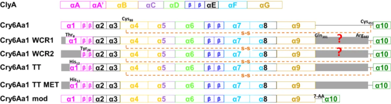
Fig. 15. Diagram of the secondary structures of ClyA and Cry6Aa1 TT, and the predicted secondary structures of the other forms of Cry6Aa1. ... 34
Fig. 16. Simplified schematic representation of the experimental setup for PLB experiments .... 37
Fig. 17. Schematic drawing of the different parts of the disposable holder ... 37
Fig. 18. Chemical structure of the lipids used in PLBs ... 38
Fig. 19. Simplified description of the lipid mixture preparation protocol. ... 38
Fig. 20. Simplified schematic protocol used to make agar bridges. ... 39
Fig. 21. Simplified representation of the protocol used for testing Bt toxins in PLBs. ... 40
Fig. 22. Simplified schematic protocol of enriched liposome preparation ... 42
Fig. 23. Demonstration of the analysis of one type of pore conductance by doing an IV curve .. 43
Fig. 24. Relation of Cry6Aa1 TT secondary structure to its amino acid sequence ... 77
Fig. 25. Relation of ClyA secondary structure to its amino acid sequence ... 77
Fig. 26. Sequence alignment of Cry6Aa and ClyA using ClustalW ... 78
Fig. 27. Representative IV curves for one experiment for each form of Cry6Aa1 ... 82
Fig. 28. Cumulative frequency distribution of pore conductances for the different forms of Cry6Aa1 ... 83
Fig. 29. β tongues (blue) and α1 and αA (magenta) of Cry6Aa1 and ClyA, respectively ... 85
LIST OF TABLES
Table I. Three-domain Cry toxins that have been crystalized so far ... 14 Table II. Composition of the buffer solution used to solubilise Cry6Aa1. ... 35 Table III. Biophysical properties of the different forms of Cry6Aa1. ... 81
ABBREVIATION TABLE
3-D Three-dimensional
ABC ATP-binding cassette
ALP Alkaline phosphatase
APN Aminopeptidase N
BBM Brush border membrane
BBMF Brush border membrane fragment
BBMV Brush border membrane vesicle
Bt Bacillus thuringiensis
Bt-R1 Bt cadherin receptor
CAPS 3-(Cyclohexylamino)-1-propanesulfonic acid
CHES 3-[(3-Cholamidopropyl) dimethylammonio]-1-propanesulfonate.
ClyA Hemolysin E
Cry Crystal protein
Cyt Cytolytic protein
DPhPC Diphytanoylphosphatidylcholine
EGTA Ethylene glycol-bis(β-aminoethyl ether)-N,N,N',N'-tetraacetic acid
HEPES 4-(2-hydroxyethyl)-piperazine-1-ethanesulfonic acid
HBL Hemolysin BL
JNK c-Jun N-terminal kinase
kDa kilodalton
MAPK Mitogen-activated protein kinase
MES 2-(N-morpholino)ethanesulfonic acid
MET b- mercaptoethanol
Mtx3 Mosquitocidal toxin
NCRW Northern corn rootworm
PC L-alpha-phosphatidylcholine
POPC 1-palmitoyl-2-oleoyl-sn-glycero-3-phosphocholine
POPE 1-palmitoyl-2-oleoyl-sn-glycero-3-phosphoethanolamine
PLB Planar lipid bilayer
Sip Secreted insecticidal protein
TEP
TPCK N-tosyl-L-phenylalanine chloromethyl ketone
Tris Tris(hydroxymethyl)aminomethane
Vip Vegetative insecticidal protein
ACKNOWLEDGEMENTS
I would like to thank everyone that has contributed to this thesis and to my Master’s degree during the past two years.
First of all, I would like to thank Dr. Jean-Louis Schwartz for giving me the opportunity to work in this incredible project. For guiding me during the whole project and introducing me to the field of biophysics.
I would also like to thank James-Christopher Bernard and Maxime Schmidt for being so supportive inside and outside the laboratory. For the great afternoons having a beer and for being such amazing friends. I would also like to thank Lena Potvin for her tough love and for teaching me how to properly do bilayers. Thank you Vincent Lemieux for starting this project and introducing me into the lipid bilayer technique. I would also like to thank Dr. Vincent Vachon for his patience and for his help during the writing process. I would also like to thank all the internship students, who have always been great supporters and have always made me feel at home in Montreal and in the laboratory. Daline Tho, Maxime Schmidt Jr., Emilie Huynh and especially to
Thomas Cauchi, whose help with the abstract in French has been very much appreciated.
Moreover, I would like to thank Dr. Lucie Parent and Dr. Rikard Blunck for accepting to be in my Master’s committee and giving me advice during our meeting. Also, I would like to thank Dr. Réjean Couture for accepting to judge my thesis. Thank you also to all the other members of the GEPROM and the Départament de physiologie moléculaire et integrative for supporting me and always helping me in every way they could. Especially, thanks to Michel Brunette for all the technical support that has made it possible to complete my experiments. Also, thank you to all the members of Dow AgroDciences for being so open to our ideas and for providing us with all of the toxins. It has been a very enriching and interesting process to have the opportunity to collaborate.
I would also like to thank NSERC for funding the project and Université de Montréal and Départament de physiologie moléculaire et integrative for the financial support through the recruitment scholarship, the scholarship of exemption of international tuition fees and the end of master’s scholarship.
CHAPTER 1. INTRODUCTION
1.1. Bacillus thuringiensis (Bt)Bacillus thuringiensis (Bt) is a rod-shaped, Gram-positive bacterium that forms spores
when facing difficult conditions. This bacterium is a ubiquitous soil microorganism. It can be found in Asia, Europe, Africa, America and Australia (1). Strains have been isolated from insects, stored product dust, water and leaves (2).
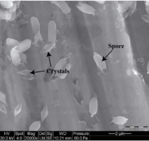
The most noticeable characteristic of this microorganism, is that it is pathogenic to invertebrates, mostly insects, by the production of proteins that are toxic to specific species of several orders (3,4). These toxins are produced either as parasporal crystals during sporulation (Fig. 1) or during the vegetative growth of the bacterium (5).
Fig. 1. Spores and parasporal crystals of Bacillus thuringiensis from strain H29.3 (5).
Until now more than 700 crystal toxins that are produced by different strains of Bt have been discovered (Crickmore, N. et al. "Bacillus thuringiensis toxin nomenclature"
http://www.btnomenclature.info, June 2016). The genes that code for these proteins are usually located on large plasmids and, for most of them, between transposable elements (6).
1.1.1. Nomenclature
Since the identification and cloning of the first gene that codes for an insecticidal crystal protein, Cry1 (2), the number of novel insecticidal proteins has increased every year, which has produced a need for a coherent nomenclature system. In the first classification system that was
created, the names of Cry toxins and their genes included a primary rank distinction with a Roman numeral, which depended on the insecticidal activity of each protein: CryI for Lepidoptera, CryII for Lepidoptera and Diptera, CryIII for Coleoptera and CryIV for Diptera (7). However, this system presented important drawbacks for the fast classification of these toxins once discovered, the main problem being the fact that, to classify the protein, it had to be bioassayed against different insects to find its target, which was not always possible. This why a new nomenclature system was proposed where, instead of concentrating on the toxicity of the proteins, the classification is based on amino acid sequence (8).
In this system, all of the toxins are given a four-rank name depending on their degree of pairwise amino acid identity with previously discovered and classified toxins. As a result, the toxin denomination and classification is independent of the three-dimensional (3-D) protein structure, host range and even mode of action. Arabic numbers are used for the first and fourth ranks and uppercase and lowercase letters are assigned for second and third ranks, respectively. Proteins that share less than 45% amino acid identity are assigned a different primary rank, proteins sharing less than 78% amino acid identity are assigned a different secondary rank, proteins sharing less than 95% amino acid identity are assigned a different tertiary rank and a quaternary rank is assigned for already established toxins that differ only in a few amino acids because of mutational changes or an imprecision in sequencing (8). For a protein to be included in this nomenclature, it has to be a Bt parasporal inclusion that shows toxicity against an organism or share more than 95% amino acid identity with an already established Bt toxin (Crickmore, N. et al. "Bacillus thuringiensis toxin nomenclature" http://www.btnomenclature.info, June 2016).
1.1.2. Organisms affected by Bt toxins
Many Cry proteins have properties that are useful for the control of pests in agriculture to limit the use of hazardous chemical insecticides. However, this is not the only target for Bt toxins. Bt proteins target a wide range of organisms that are mainly insects, which extend their use in health protection (medical entomology, disease vector control), although some of them are active against other invertebrates and even mammalian cancer cells. More specifically, these toxins have well documented toxicity against Lepidoptera, Coleoptera, Diptera, Hemiptera,
nematodes, snails and human cancer cells of various origins (2,5). Some Cry toxins might possess novel toxin properties that have not been discovered yet, since there are some proteins whose toxicity has not been reported (5).
Some proteins such as Cry11A and Cry1Ab have shown toxicity against several anaerobic bacteria and archaea, such as Clostridium butyricum, Clostridium acetobutylicum and
Methanosarcina barkeri. Furthermore, some protein fragments, like a 49-kDa fragment of
Cry3Aa, have been shown to have a lytic effect on some bacteria (9). An interesting fact about the mode of action of these Cry toxins on prokaryotes is that they are far less specific than when they target insects.
1.2. Mode of action of Bt toxins
From a biological point of view, it has always been thought that the main purpose of Bt toxin action was to destroy the epithelial barrier of insects so that the spores would germinate and colonize the hemolymph of the insect, a perfect environment in which the bacteria would live (10). The mode of action by which this would happen has been studied in different organisms but is still far from being fully elucidated.
During the last 30-40 years, most of the research has concentrated on studying the “classical mode of action” (Fig. 2), which states that the toxin is ingested, solubilised, activated, crosses the peritrophic membrane, binds to one or more membrane proteins and forms a pore that leads to osmotic lysis of the cell. In recent years, two more models of Bt mode of action have been proposed, which are poorly supported by experimental data so far (11).
1.2.1. Ingestion of Bt toxins
The first step of the mode of action is for the crystal to enter the digestive system of the target insect. This step could explain why some sucking insects and other invertebrates such as spiders and mites are not sensitive to Cry proteins.
1.2.2. Solubilization of Bt toxins
After ingestion, the crystal is solubilised inside the midgut of the insect larvae. The difference in the solubility of the inclusion bodies of different Bt toxins is mostly determined by the cysteine composition of their protoxins. Although some crystals lack disulphide bridges (12), most of the cysteine residues present in the crystal protein form interchain disulphide bonds. These cysteine residues are highly conserved among the different Bt toxins, and are found in the C-terminal half of the toxin. The specific orientation of the thiol groups also plays a role in crystal formation and ultimately in insecticidal activity (13,14). Moreover, solubility of the crystals is also affected by temperature. Crystals that have been frozen (-80 °C) or subjected to very high temperatures were found to be less soluble (15).
Generally, the toxins that target Lepidoptera are solubilised at alkaline pH (9.5 – 11), which is coherent since the midgut of lepidopteran larvae is a highly alkaline environment. The solubilisation step is more difficult to understand for toxins that target coleopteran insects: they are also optimally solubilised at alkaline pH, whereas the midgut of coleopteran larvae has an acidic environment. For example, Cry3Aa remains mostly in its crystal form (95%) between pH 5 and pH 9.5, while the midgut pH of coleopteran larvae is around 6 (16,17) . Therefore, although low solubility does not seem to prevent toxicity in Coleoptera, its role is still not fully understood.
1.2.3. Activation of Bt toxins
After solubilisation, the crystal proteins undergo the next step in the mode of action, activation. During this step the proteins are cleaved by insect midgut proteases in various ways, which are determined by the insect species. Depending on how this activation happens, the toxins will show specificity to different species of insects. For example, Cry2 activated by Pieris
brassicae midgut juice was only toxic to lepidopteran cells, while the same toxin was activated
The activation step has been mostly studied in the years of Bt research that dealt with Cry1, Cry2 and Cry3. Cry1 protoxins are cleaved at the N-terminal and C-terminal, losing 70-kDa. Cry2 protoxins are only cleaved at the C-terminal, losing around 5-70-kDa. Cry3 is cleaved at the N-terminal, losing between 6 and 18-kDa, depending on the activation process (19,20).
Activation has been demonstrated to be an important step for toxicity. Moreover, it has been shown that in vitro activation of some Cry toxins could render them toxic towards orders of insects against which they are not naturally active (21).
An alteration of protein activation by midgut proteases has been found to be involved in insect resistance to Bt toxins (22). Bt resistant insects have been shown to have either proteases with a higher proteolytic activity or a higher concentration of proteases in the gut that resulted in the complete hydrolysis of the toxin core, which therefore resulted in resistance to the toxin. This was also supported by some experiments in planar lipid bilayers (PLBs) where, depending on the enzyme used to activate the protein, the pore formation ability changed, suggesting that proteases directly influence pore formation (23).
1.2.4. Crossing the peritrophic membrane
Once the toxin has been solubilised and activated, and before it reaches to the brush border membrane (BBM) of the insect midgut, where it will interact with different membrane proteins that act as a receptors, it has to go through the peritrophic membrane.
The peritrophic membrane is a layer that surrounds the food bolus in the midgut of various organisms, such as insects. It is mainly composed of chitin, which forms interconnected chains arranged in different layers. There are also around 35-40% of associated proteins, some of them proteases that could cleave the toxins under specific conditions. This composition varies depending on the insect. The main function of this membrane is to protect the midgut epithelium from food particles and microorganisms (24,25).
Some cry toxins are thought to bind to the peritrophic through sugar groups (25). For example, the truncated Cry1Ie, that shows strong insecticidal activity against the Asian corn borer, binds to the peritrophic membrane in a specific way and its domain III is involved in this process (26).
1.2.5. Molecular recognition of Bt toxins
The binding of Cry toxins to insect midgut epithelial membrane molecules, which constitutes the next step in their mode of action after crossing the peritrophic membrane, is one of the most important factors in specificity. Direct correlation between species toxicity and specific binding in brush border membrane vesicles (BBMVs) has been demonstrated in some cases. For example, in Manduca sexta and P. brassicae BBMVs, where Cry1b would only bind to M. sexta BBMV while Cry1Ab and Cry1Ba bound specifically to P. brassicae BBMVs (27). Binding of Cry toxins to their molecular recognisers has been proposed to be a two-step kinetic model, one step where binding can be reversible and the toxin can dissociate from the receptor irreversible and another step where the toxin binding to the receptor is irreversible. It has been shown that the rate constant for irreversible binding, which is thought to reflect the insertion step of the toxin into the membrane, is better correlated with toxicity than the overall binding. This was proved by showing a direct relationship of Cry1A toxins binding to Lymantria dispar BBMVs (28).
Many Cry toxin receptors have been reported so far, although not all of them have been studied in the same depth. The best characterized receptors are aminopeptidase N (APN), cadherin-like molecules, glycolipids and alkaline phosphatases (ALPs). APN, cadherins and ALPs have been studied mainly in Lepidoptera, while glycolipids have been studied as receptors of toxins that target nematodes (29).
1.2.5.1. Aminopeptidase N (APN) receptors
The APN protein family is a class of enzymes whose function is to cleave neutral amino acids from the N-terminus of polypeptides. In 1994 a 120-kDa glycoprotein that showed high similarity with members of the APN family was purified from the midgut membrane of M. sexta larvae (30). That same year it was also demonstrated that the mixture of APN from M. sexta enhanced Cry1Ac pore formation in lipid vesicles. Actually, these proteins were shown to increase the toxin binding by 35% (31). This interaction was further demonstrated by using PLBs to which the M. sexta receptor complexes had been fused. This caused Cry1Aa, Cry1Ac and Cry1Ca to form pores at concentrations that were much lower than when the receptor was not present in the bilayers (32).
So far, several different Cry toxins have been shown to bind APN molecules. Their different patterns of binding complicates further understanding of the whole mechanism (29). It has also been attempted to express APN in cell lines to study how the binding of Cry toxins affects cell toxicity. However, most of the efforts have been unsuccessful (29). On the other hand, expressing M. sexta APN in Drosophila, was successful and demonstrated that Cry1Ac was 100% toxic to transformed flies at a concentration of 50 ug/ml, in comparison to the control larvae which were unaffected by toxin concentrations up to 1 mg/ml (33).
1.2.5.2. Cadherin receptors
The cadherin superfamily of proteins serves diverse functions such as cell adhesion, migration, cytoskeletal organization and morphogenesis. The most important characteristic of this family of proteins is the presence of repeating calcium-binding domains. These proteins are also glycosylated and anchored to the membrane by a single transmembrane domain, although there are also some GPI-anchored variants. Normally, cadherins are primarily located within adherent junctions and are involved in cell-to-cell adhesion. However, the cadherins that have been identified to be involved in Cry recognition are found in the apical membrane of midgut columnar cells and their physiological role is unknown. In this case glycosylation does not seem to be essential for toxin binding (29). The first identified and purified cadherin that serves as a
Bt toxin receptor is a 210-kDa protein found in BBMVs of M. sexta. It specifically recognizes
and binds Cry1Ab with high affinity (34).
Contrary to APN receptors, cadherins have been successfully expressed in cell lines, such as Sf21 cells. Cells that express the Bt-R1 (a Bt cadherin receptor) have acquired sensitivity
to Cry1Aa, Cry1Ab and Cry1Ac. This toxicity is similar to the sensitivity of M. sexta to these three toxins (35). Cadherin-like receptors have also been expressed in COS7 and HEK293 cell lines. The cells bound and became susceptible to Cry1Aa, Cry1Ab and Cry1Ac (36). Cadherins have also been demonstrated to bind dipteran targeting toxins such as Cry4Ba (37).
1.2.5.3. Alkaline phosphatase (ALP) receptors
Alkaline phosphatases (ALP) have also been identified as Cry toxin receptors, but the knowledge about ALP-Cry toxin interaction is considerably smaller than for other receptors. ALP is a molecule that removes phosphate groups from proteins and nucleotides. In 1994, it
was suggested that ALP might act as a Cry1Ac receptor in M. sexta. It was also noted that the ALP expression levels were reduced in some Bt resistant strains of Heliothis virescens, suggesting that it played a role in the toxin’s mode of action (31). However, the role of ALP in mediating Cry1Ac susceptibility has yet to be established. ALP has been shown to be an important receptor for Cry11Aa in A. aegypti (38). It has been demonstrated that A. aegypti larvae can increase their resistance to Cry11Aa 124-fold by modifying the expression of receptor proteins such as ALPs and APNs (39).
1.2.5.4. Glycolipid receptors
The interaction between glycosphingolipids and Cry toxins was first proposed in 1986 (40). It was also shown that Cry1A bind to different parts of glycolipids depending on activation (41). It was not until 2005 that the presence of an invertebrate-type glycolipid receptor for Cry toxins was found in Caenorhabditis elegans (42). This was discovered when mutants that failed to produce ceramide-based glycolipids that bind Cry5Ba were found, which made them resistant to the toxin. Some preliminary results also suggested that glycolipids were not only responsible for binding Cry5Ba toxins in C. elegans, but that they were also found in other order of insects such as Lepidoptera. Glycolipids extracted from the midgut of M. sexta were shown to bind Cry1Aa, Cry1Ab and Cry1Ac (42). Yet, the importance of this interaction and its role in toxin susceptibility remains to be explored.
1.2.5.5. ATP-binding cassette (ABC) transporter receptors
ATP-binding cassette (ABC) transporters are membrane proteins that are widespread in prokaryotes and eukaryotes. They are responsible for transporting molecules through cell membranes. Linkage mapping analysis showed that resistance to Cry1Ab in some strains of H.
virescens was linked to several mutations in an ATP-binding cassette transporter gene, which
had never before been considered to play a role in the mode of action of Cry toxins (43). This was later confirmed by a study showing that the same mutation in ABCC2 from Cry1Ab-resistant insects resulted in a much reduced susceptibility to Cry1Ac and Cry1Ab in Sf9 cells that were expressing the truncated ABCC2 (44).
1.2.6. Pore formation
After the molecular recognition step, the toxins are thought to insert into the membrane and form pores, which eventually leads to osmotic lysis of the cell. This step of the mode of action has been extensively studied using different biophysical techniques such as light scattering assays, fluorescence assays, PLBs and cellular electrophysiology.
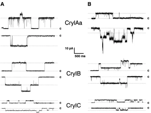
Pore formation has been shown in receptor-free PLBs (Fig. 3A), the first time using toxins Cry1Ac and Cry3A, which target lepidopteran and coleopteran insects respectively. These early experiments showed that the toxins were able to form pores at very alkaline pH with a strong cationic selectivity. It was also shown that the pores had several conductance levels. In the case of Cry1Ac main conductances of the pores are 450 pS and Cry3A forms pores of around 500 pS, both in alkaline conditions (45-48). Selectivity experiments performed later have proven that the general cationic selectivity of Bt toxins is not as strong as it was thought in the first place (49,50).
Pore formation by the binary toxin Cry34Ab1/Cry35Ab1 has also been investigated (51). This toxin was not a very efficient pore former under alkaline conditions, which may be expected since the midgut conditions of the coleopteran target insect are acidic (17). While the two components were able to form pores separately in PLBs, optimal pore formation was attained when they were used together (51).
Pore formation in the presence of receptors has also been studied (Fig. 3B). Initially, this was done by doing light scattering assays with BBMVs isolated for M. sexta (52). It was demonstrated that activated Cry1Ac produced a change in the solute permeability of the BBMVs, which could be explained by pore formation. Pore formation by Cry1Ac occurred at physiologically relevant toxin concentrations and it was shown that these pores are also not very selective.
The effect of the receptor on pore formation was also demonstrated by fusing BBMVs or purified receptors into PLBs (32,53). It was shown that the integration of purified APN molecules, which are receptors for Cry1Ac, into PLBs increased the conductance of the Cry1Ac pore, decreased the concentration needed for pore formation by 250-fold and induced current rectification. Yet for Cry1Aa, the concentration of toxin in receptor rich bilayers was only reduced 100-fold, conductance was also enlarged and there was no rectification, which could mean that Cry1Ac and Cry1Aa did not interact in the same way with their receptor (54). Pore
formation by Cry1Ca and its receptor modulation also been studied in Sf9 cells by using patch clamp (55). The pore formation mechanism in Sf9 cells was proven to be very similar than that in PLBs. The only difference being in the selectivity, which afterwards was shown to be due to the difference in pH conditions (50) .
Finally, the size of the pores of Cry1Ac has been estimated by using two different techniques. Initially, this was done by using an osmotic swelling assay on M. sexta BBMVs using molecules of different sizes, such as polyethylene glycols (56). Later, it was estimated by using PLBs and polyethylene glycols (57). The pores made by Cry1C had a uniform radius around 1-1.3nm in both cases. The results in the latter study show that the multiple conductance levels observed experimentally may be due to clusters composed of different numbers of similar pores that are cooperatively gated.
Fig. 3. Pore formation demonstration of Cry1Aa, Cry1B and Cry1C in PLBs. In the absence of receptors (A) and in the presence of M. sexta APN receptor (B). The currents correspond to +40mV (upper traces) and -40mV (lower traces) (32).
1.2.7. Bt toxin effect on the membrane potential of lepidopteran insect midgut cells In lepidopteran midgut cells, the electrochemical K+ gradient across the apical
membrane, serves as the main driving force for the absorption of solutes, such as amino acids. A vacuolar-type proton ATPase, coupled with an electrogenic K+/H+ exchanger, generates this
gradient (58-60). When a Bt toxin forms pores in the apical membrane of these cells, this gradient is abolished and the cells lose their capacity to transport the solutes and allow the equilibration of pH between the highly alkaline content of the midgut lumen and the cells’ cytoplasm. To study how Bt toxins directly affect the potential of the midgut cells, the glass microelectrode technique was used on freshly isolated midguts of lepidopteran larvae (61). The potential across the membrane midgut cells of Lymantria dipar and Bombyx mori, which was around -80 mV in the absence of toxin, decreased rapidly in the presence of different activated Cry toxins. Cry1Aa, Cry1Ab, Cry1Ac, Cry1C, Cry1E and Cry1F all caused a fast depolarization of the apical membrane of the cells from the midgut of L. dispar. However, toxins such as Cry1Aa and Cry1C had no effect on the potential of the basolateral membrane of these cells. Also, Cry1B and Cry3A did not have any effect on the potential of these cells, which is expected since they also show no toxicity in vivo against this insect. Yet, the proteins’ toxicity in vivo and in vitro did not fully correlate in L. dispar since for example Cry1Ac was not very active in the bioassays but triggered the same depolarization as Cry1Aa, which had a very high toxicity bioassays. For B. mori Cry1Aa caused a fast depolarization of the apical membrane of the midguts but Cry1Ab and Cry1Ac were inactive, a result which correlates with in vivo toxicities.
1.2.8. Intracellular effects of Bt toxins
The effect of Bt toxins in Sf9 cells has been not only studied at the pore-formation level, but also at the level of intracellular events. When the cells were exposed to Cry1C, there was a very fast rise in the intracellular calcium followed by the activation of anion selective channels. This was the first demonstration that intracellular signals occurred in response to Bt toxin exposure (55).
Lately, researchers have started to be interested in putative defense mechanisms in cells under attack by pore-forming toxins (PFTs). Such mechanisms have been studied using Cry5B, a Bt toxin active against C. elegans and streptolysin O, which permeabilizes mammalian cells (62). One hundred and six genes have been identified though RNA interference screening, which play some kind of cellular protection role against a PFTs. Two previously identified mitogen-activated protein kinase (MAPK) pathways, the p38 pathway and the c-Jun N-terminal kinase (JNK) pathway, are crucial components of the defense system. It was demonstrated that the JNK MAPK pathway is the key central regulator of PFT induced transcriptional and functional
responses, while the p38 is not. The transcription factor activator protein 1 (AP-1) is the first cellular component involved in this defense mechanism. This research area on cell protection against PFTs may provide a solid starting point to further understand the intracellular effect of
Bt toxins.
1.2.9. Proposed models of Bt mode of action
Since the early 2000s, two different models of Bt toxin mechanisms of action have been proposed. One is the sequential binding model that attempts to describe a number of intermediate steps leading to pore formation and osmotic-colloidal lysis of target cells (63,64). The other model is the signalling pathway model, which states that pore formation is not important. Cell death is triggered by the signalling pathways activated by Bt toxins in target cells (65,66).
1.2.9.1. The sequential binding model
The sequential binding model postulates that following activation of Bt toxin by midgut proteases, the toxin first binds to a cadherin molecule with low affinity but high capacity. This binding causes a conformational change of the toxin that eases the proteolytic cleavage of its first α-helix (63). Once this helix is removed, the toxin oligomerizes and forms different pre-pore structures, depending on the activation step (64), that bind to APN receptors with very high affinity. This allows the insertion of the pore into the membrane, which results in the osmotic unbalancing and death of the cell (63). Unfortunately, the data in which this model is based have not been independently obtained in other laboratories and may have been overinterpreted (11).
1.2.9.2. The signalling pathway model
The signalling pathway model states that pore formation, which has been demonstrated in a number of laboratories worldwide for decades, does not kill the cells. More specifically, in this model Cry1Ab binds to Bt-R1, which activates Mg2+- dependent pathways never reported
elsewhere (65). The binding also stimulates the production of alpha G proteins (GαS) and cAMP, which activates a protein kinase A leading to necrotic cell death (66). This model suffers from severe experimental flaws that lead to rushed conclusions. It cannot reasonably be supported (11).
1.3. Bt toxins structures and function
Bt produces a wide range of toxins such as δ-endotoxins, enterotoxins, β-endotoxins,
haemolysins, Vegetative insecticidal proteins (Vip) and Secreted insecticidal proteins (Sip). The
Bt toxins that have been studied the most, are the δ-endotoxins, Cry (crystal) toxins and Cyt
(Cytolytic) toxins.
1.3.1. Cry toxins
So far, the most studied Cry toxins display a 3-D structure of three domains. However, in the past decade, due to the fast rise in insect resistance to commercialized toxins, efforts have been made to discover toxins that may possess a different molecular structure and possibly a different mode of action (67).
1.3.1.1. Cry toxins with 3-domain structures
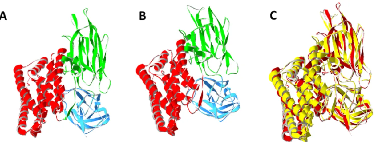
The first atomic structure of a Bt toxin was that of the activated coleopteran Cry3Aa (Fig. 4A) at a resolution of 2.5Å (68). A few years later, the 3-D structure at 2.25Å resolution of the activated lepidopteran-targeting Cry1Aa toxin (Fig. 4B) was described (48). Both structures showed a very similar arrangement of their three different domains (Fig. 4C).
Domain I of Cry3Aa starts at Thr61 up to Leu290 and is a 7-helix bundle, with a central helix surrounded by 6 helices (68). On the other hand, in Cry1Aa, domain I is composed by 8 α-helices, where α2 corresponds to α2a and α2b. In the 3-D structure, α5 is surrounded by the others (48). This α-helix bundle shows structural similarity to other PFTs such as colicin A. Due to this similarity, it was suggested and later demonstrated that Domain I was responsible for pore formation in the membrane (47,48,69).
Domain II is a β-prism that contains 3 antiparallel β-sheets packed around a hydrophobic core. It has a triangular cross-section and goes from Tyr291 to Phe500 in Cry3Aa (68). Cry1Aa is very similar. However, apart from the β-sheets there are two short α-helices in the structure (48).
Domain III, which is the C-terminal part of the protein, extends from Phe501 to Asn644 in Cry3Aa. It is very similar in Cry1Aa, with a jelly-roll topology with the strands forming a β-sandwich (48,68).
Fig. 4. 3-D ribbon representation of (A) Cry3A activated toxin (1DLC) and (B) Cry1Aa activated toxin (1CIY). Domain I is colored in red, Domain II is colored in green and Domain III is colored in Blue. (C) Superimposed Cry1Aa (yellow) and
Cry3A (red).
The structures of other 3-domain Cry toxins have been determined later. One of them, at a 2.2Å resolution, is that of the Cry2Aa protoxin, the first available structure of a toxin that is specific to both Diptera and Lepidoptera. The overall topology of Cry2Aa is similar to that of Cry3Aa and Cry1Aa (70), despite little sequence identity between Cry2Aa and the two other toxins (20% or less) (71). In recent years more structures of 3-domain toxins have been elucidated (Table I).
Table I. Three-domain Cry toxins that have been crystalized so far (Modified from (72)).
Toxin Target Insects Activation Resolution PDB ID References
Cry1Aa Lepidoptera Trypsin
65-kDa 2.25Å 1CIY (48)
Cry1Ac Lepidoptera Trypsin
65-kDa 2.35Å 4ARX (73)
Cry1Ac Lepidoptera Protoxin
130-kDa 3.2Å 4W8J (74)
Cry2Aa Diptera /
Lepidoptera
Protoxin
62-kDa 2.2Å 1I5P (70)
Cry3Aa Coleoptera Papain
67-kDa 2.5Å 1DLC (68)
Cry3Bb Coleoptera Chymotrypsin
Cry4Aa Diptera Trypsin
65-kDa 2.8Å 2C9K (76)
Cry4Ba Diptera Chymotrypsin
68-kDa 1.75Å 1W99 (77)
Cry8Ea Coleoptera Chymotrypsin
66.2-kDa 2.2Å 3EB7 (78)
Cry5B Nematodes Elastase
66.14-kDa 2.3Å 4D8M (79)
Until 2014, all of the available Bt toxin crystal structures, except for that of Cry2Aa, were those of the activated form of the toxins. However, the full length structure of Cry1Ac protoxin was recently solved (74). The structure of the full-length Cry1Ac protoxin was solved a 3.2-3.5Å resolution (Fig. 5). It is made of 7 domains, the already known three domains, and four more. Two of these are mainly α-helices and the other two display β-sheet arrangements. Interestingly, the part of the structure that corresponds to the activated Cry1Ac and of Cry1Ac protoxin are very similar (74).
Fig. 5. 3-D ribbon representation of Cry1Ac protoxin (4W8J). Domain I is colored in red, Domain II is colored in green, Domain III is colored in blue, Domain IV is colored in purple, Domain V is colored in orange, Domain VI is colored in brown
and Domain VII is colored in yellow.
1.3.1.1.1. Structure-function relationships of 3-domain toxins
Once the structure of the first 3-domain Bt toxin was elucidated, a considerable amount of effort was put into understanding its structure-function relationships.
Domain I was proposed to be the pore-forming domain (48,68). It was demonstrated that the first domain of these Bt toxins was sufficient for pore formation in PLBs and Rb+ efflux
in lipid vesicles (47,80-82). Interestingly, the first domain alone of the toxin did not have insecticidal activity towards H. virescens (80). Other studies on the mosquitocidal toxin Cry4B and its α1-α5 fragment demonstrated that they formed slightly cation selective pores in PLBs, supporting the initial experiments showing that the first domain of Cry3Aa formed a functional pore that had similar properties to those of the full toxin (47,83). It was also shown that the conductance and gating depended on the pH conditions used for the processing of the protoxin, which provided a link between pore formation and the earlier steps of solubilisation and activation. The umbrella model for toxin insertion was proposed based on the Cry3Aa structure and on its similarity to colicin A (84). In this model α4 and α5 of domain I would be the ones penetrating the membrane. This hypothesis was further studied by using different techniques such as the biotinylation of several cysteine residues in α4, α5 and surroundings that affected the toxin’s pore formation (85) and disulphide bridge engineering(85,86). It was demonstrated that the mutation of amino acid residues of α4 of domain I had a large impact on the pore’s biophysical characteristics, which not only supported the umbrella model but showed that α4 lines the lumen of the pore (87-89). Several studies done also concentrate on α3, using circular dichroism, protease sensitivity, electrophoretic mobility analysis and point mutations. It was shown that several mutations in α3 affected pore formation and oligomerization (85,90). Finally, Domain I interhelical loops appear to be important for conformational changes that lead to pore insertion, and unfolding of the protein around a hinge region between domain I and II is thought to play an important role (85,86,89,91).
Domain II was proposed to be the binding domain due to the identification of specificity-determining regions (92). For instance, the specificity of Cry2A for Lepidoptera and Diptera as opposed to the Lepidoptera specificity of Cry2B was due to residues 307-382, which are located in domain II (71). Further studies of Cry2Aa, and correlation with chimeric scanning data of Cry2Aa and Cry2Ab, indicated that the putative receptor binding epitope lied on the core of the β-sheets of the domain (70,71).
Domain III, which was thought to be mainly involved in structural stability of Bt toxins, also played a role in binding and pore formation. Several mutations in a conserved alternating arginine region had a direct effect in pore formation and pore conductance (93,94).
Finally, taking into account the structural and functional features of 3-domain toxins mentioned before, a model of Domain I disassembly was proposed (78). In this model, after the binding of the Cry toxin to its receptors, there is a conformational change in the β-sheets of domain II. This change mainly influences the interface between domain I and domain II, which makes α7, which has a kink, move towards the center of the helix bundle in domain I. This forces helices α2b and α3, which are acting as a lid in domain I, to swing away from the central helix α5, a displacement that is facilitated by proline 105 of α3 that is highly conserved among the 3-domain Cry toxins. Finally, after the lid is open, α4 and α5 are exposed and can penetrate the membrane.
Fig. 6. Model for domain I disassembly (78).
1.3.1.2. Cry toxins with non 3-domain structures
Other Bt atomic structures that do not display three domains such as binary toxins, like Cry34Ab1/Cry35Ab1, and aerolysin-like toxins, like parasporin 2, have also been elucidated.
1.3.1.2.1. Binary toxin: Cry34Ab1/Cry35Ab1
Cry34Ab1 and Cry35Ab1 is a binary toxin produced by Bt. The structures of Cry34Ab1 and Cry35Ab1 have been elucidated at resolutions of 2.15Å and 1.8Å respectively (95). Cry34Ab1 (Fig. 7A) is a 14-kDa protein. Its structure is similar to that of actinoporins, PFTs produced by sea anemones, or hemolysins, which are membrane interacting proteins. The
protein folds in a β-sandwich conformation with two β-sheets packed against each other. The entire protein has a relatively flat layer-like conformation with only two slightly twisted β-sheets. Cry35Ab1 (Fig. 7B), on the other hand, is a 44-kDa protein that has structural features of the Toxin_10 family, which includes insecticidal proteins such as the binary toxin BinA and BinB (Fig. 7C) from Lysinibacillus sphaericus. It has an elongated rectangular shape and is composed of two domains. The N-terminal domain has a β-trefoil fold with a very hydrophobic core, while the C-terminal domain contains 6 helices and three antiparallel β-sheets. An interesting characteristic about this toxin is that it contains two cysteine residues that are too far apart to form a disulphide bridge, but the replacement of one of these cysteines reduces the toxin’s activity.
Although both Cry34Ab1 and Cry35Ab1 were able to form pores in PLBs at pH 5.5 separately, maximum pore formation efficiency was achieved when both toxins were combined (51).
Fig. 7. 3-D ribbon structure of (A) Cry34Ab1 (4JOX), (B) Cry35Ab1 (4JP0) and (C) BinB (3WA1).
1.3.1.2.2. Aerolysin-like toxins: Parasporin-2 (PS2) and Cry51Aa1
In the last decade new Bt toxins have been discovered (96,97) that share structural properties with aerolysin (98), a protein produced by Aeromonas hydrophila that belongs to the family of β-PFTs, even though they share very little sequence similarity.
Parasporin-2 (PS2), also classified as Cry46 (97), is a Bt toxin active against cancer cells. Upon activation by proteases, the 37-kDa protoxin is transformed into a 30-kDa toxin. The structure of this toxin was elucidated at 2.38Å resolution (Fig. 8A) (99). It has an unusually elongated structure that is mostly dominated by β-strands, which twist and run along the long axis of the molecule. The structure is divided in 3-domains. Domain I comprises a small β-sheet
and short α-helices and is thought to be the part of the molecule that binds to the cancer cells. The other two domains are thought to be involved in protein oligomerization and pore formation. One of the most interesting features of the toxin, can be found in domain II, where there is a putative pore-forming β-hairpin that is characteristic of the aerolysin-type toxins. Furthermore, at the surface of domain II, there is a large number of exposed side chains of serine and threonine residues that might orient the molecule on the cell membrane when domain I binds to the target. This protein is a PFT as demonstrated in PLBs and by patch clamp experiments on HepG2 cells (unpublished data, Schwartz’ laboratory).
The complete structure of the Cry51Aa1 protoxin has been elucidated by X-ray crystallography at 1.65Å resolution (Fig. 8B) (100). This is the first coleopteran active Bt toxin that has a high structural similarity to the aerolysin-type PFTs. This protein is structurally very similar to PS2. Almost a quarter of the residues are serines or threonines scattered throughout the protein. They may be involved in allowing the molecule to move freely and closely to the membrane when the N-terminal domain binds to the receptor, in guiding the amphipathic loop towards the membrane and in promoting oligomerization.
These proteins have very little sequence similarity with most of the Cry toxins with an already elucidated structure. It also has a very limited sequence similarity to the family of aerolysin-like PFTs too even if the structure (Fig. 8C) is similar.
Fig. 8. 3-D ribbon structure of (A) Cry46 (Parasporin 2) (2ZTB) (B) Cry51Aa1 protoxin (4PKM) and (C) Proaerolysin (1PRE). The β-hairpin is colored in red.
1.3.2. Other toxins
In addition to Cry toxins, Bt produces Cyt, Vip and Sip toxins, which have been shown to have very different 3-D structures.
1.3.2.1. Cyt
Cyt toxins are produced as parasporal crystals, at the same time as Cry toxins by Bt subsp. israelensis (101) mainly, but have cytolytic activity and their molecular mode of action is poorly understood (102). These toxins have specific insecticidal activity towards dipteran insects, such as mosquitoes and black flies (103). They are also toxic against mammalian cell lines in vitro (104,105). Until now, three different Cyt toxins have been crystalized and their structure is very different from that of any other Bt toxin, which might be related to their different mode of action.
Cyt2Aa and Cyt2Ba (Fig. 9A) structures have been resolved at 2.6Å and 1.8Å resolution, respectively (106,107). They are composed of a single αβ-domain that comprises a β-sheet between two outer layers of α-helix hairpins. In the protoxin form, Cyt2Aa is a nonhemolytic dimer that is linked by the N-terminal β-strands. Activation cleaves the N-terminal and C-terminal domain segments, which leads to dimer dissociation (106).
The activated monomeric form of Cyt1Aa, which is the most toxic protein in the Cyt family, was crystalized and solved at 2.2Å resolution (Fig. 9B) (108). Cyt1Aa adopts a similar structure as Cyt2Aa. It has been hypothesized that Cyt toxins could undergo a conformational change that would be necessary for membrane insertion. The α-helical layers would swing away, exposing the hydrophobic β-sheets that would then insert into the membrane. This hypothesis has been supported by the identification of a lipid binding pocket between the β-sheet and the helical layer of Cyt1Aa, which explains the binding of this Cyt toxin to specific unsaturated membrane phospholipids. It should be noted that the Cyt structure resembles that of volvatoxin (Fig. 9C), a pore-forming cardiotoxic protein (109).
1.3.2.2. Vip
Vip toxins (vegetative insecticidal proteins) are produced by Bt and Bacillus cereus during their vegetative growth phase (For review see (110)). Vip1 and Vip2 is a binary toxin that targets coleopteran insects. At least one Vip toxin, Vip1, has been shown to be a PFT (111), while Vip2 is NAD-dependent enzyme that interferes with polymerization of actin (112).
Vip2Aa has been crystalized and its structure has been elucidated at 1.5Å resolution (Fig. 10) (113). It shows a high structural homology in the C-terminal and N-terminal αβ-domains with the family of ADP ribosylating toxins, which regulate post translational modification of proteins. He structure of Vip3 has not been elucidated yet, however it has been shown that it is a PFT (114).
Fig. 10. 3-D ribbon representation of Vip2 (1QS1)
1.3.2.3. Sip
Sip toxins are insecticidal proteins secreted by Bt during growth. It is active against coleopteran larvae. So far, only one Sip toxin has been identified, Sip1Aa1, a 367 amino acid, 41-kDa protein (115). It shows a significant similarity to Mtx3, a mosquitocidal toxin from L.
sphaericus. Its mode of action remains unknown, although based on its homology to Mtx3 it
has been hypothesised that Sip1Aa might be a pore former. The structure of this toxin has not been elucidated yet.
1.4. Diabrotica virgifera virgifera (Western corn rootworm, WCRW)
Diabrotica virgifera virgifera LeConte (Western corn rootworm, WCRW) is one of the
maize producing states of the north-central USA (116). In North America only, the cost of insect control and losses to WCRWs account for more than 1000 million dollars every year. In Europe, 472 million euros a year have been estimated only in crop losses due to this pest, since it was first introduced in the 1990s (117). A closely related species, the Northern corn rootworm (NCRW, Diabrotica barberi), is becoming a major pest in Northern United States and Canada (Centre for agriculture and biosciences international, http://www.cabi.org/, August 2016).
1.4.1. Geographic distribution
The WCRW originates from America, more specifically from Central America (118). It uses different means of dispersal: while larvae can only move relatively small distances, adults can fly to different maize fields and are able to migrate short and long distances. Adults may be carried by wind during cold fronts or thunderstorms (116). Several studies have determined that in-field dispersal of male WCRWs respond to pheromone release by reproductive females and by available food (119). WCRWs have been detected in all North America and Eastern Europe, while NCRWs have only been detected in North America.
1.4.2. Biological cycle and ecology
The biological cycle of WCRWs consists in four different stages (Fig. 11): egg, larvae, pupae and adult. Flattened oval eggs, around 500 µm long, are laid just before the winter and require a cold-induced diapause period before hatching. These are usually concentrated in the top 5 to 20 cm of soil. Three larval instars develop in corn roots. The larvae are slender and white to pale yellow. The third instar larvae can be up to 10 mm long. The pupae are white, turning brownish just before adult emergence. They are found in the soil near the roots of corn plants. Finally the adults emerge in the summer and prevail in the maize field until the autumn. These adults are elongated and of a greenish-yellowish colour (118). WCRW insects are univoltine, there is one generation per year (120).
Fig. 11. Life cycle of Diabrotica virgifera virgifera (permission asked, www.pioneer.com, August 2016).
1.4.3. Physiology relevant to Bt studies
The physiology of WCRWs has not been as well investigated as that of lepidopteran or other coleopteran insects, one of the reasons being the very small size of the larvae that are difficult to study with available tools and methods.
1.4.3.1. Midgut pH and proteases
The general midgut pH has been measured in homogenized midgut juice of third instar WCRW larvae. It was found to be acidic around 5.75 units of pH by using a pH microelectrode (17). However, it cannot be excluded that pH is compartmentalized and the presence of more alkaline microenvironments in the larval gut.
A large number of proteases has been detected in third instar larvae of WCRWs. The main serine endopeptidases are trypsin, chymotrypsin, elastase, cathepsin G, plasmin and thrombin. Cysteine endopeptidases, which are often combined with aspartic endopeptidases, are thought to be the main proteases in the midgut of WCRWs due to its low pH. The main proteases of this category that have been identified are cathepsin L, papain, cathepsin B and cathepsin H. There is also an exopeptidase aminopeptidase, which is surprising considering that this protease activity is optimum at pH 9 (17).
1.4.3.2. Receptors
Finding Bt toxin receptors in WCRWs has proven to be a very difficult task. Using an expressed sequence tag, a coleopteran cadherin was identified as a putative Bt receptor. This
cadherin contains several regions of high identity to lepidopteran cadherin proteins (121), suggesting a common molecular basis for susceptibility to Cry toxins in lepidopteran and coleopteran species (122).
So far, no other receptor in coleopteran species has been identified. However, competitive binding studies in WCRW BBMs have demonstrated that Cry34Ab1/35Ab1 does not share binding sites with other coleopteran-active Bt proteins such as Cry3Aa, Cry6Aa and Cry8Ba (123).
1.4.4. Bt toxins currently used to control WCRWs
Bt toxins have been used for WCRW pest control for a number of years with different
strategies and mitigated success. In European countries pheromone traps are used to detect WCRW infestation, followed by insecticide treatment (124). In the US, where this pest is a main problem, other methods are also used such as crop rotation (125), which consists in planting different kinds of plants that are not affected by WCRWs so that WCRWs will not be able to feed and will not complete its biological cycle. Biological control has also proven to be a somehow efficient method to fight WCRW pests by using a dipteran parasite like Celatoria
compressa or entomophatogenic nematodes such as Heterorhabditis bacteriophora (126).
Chemical control still represents the most economically feasible approach, using chlorinated hydrocarbons, carbamates or organophosphates (127).
Bt toxins have also proven to be a very efficient tool to control WCRWs. Commercial Bt plants contain Cry3Bb1, mCryA and Cry34Ab1/35Ab1 toxins. They are either being
commercialized as traits with one or two Bt toxin genes registered in the US (120). However, insect resistance to Bt continues to increase. There is, therefore, a strong need to find new Bt toxins against WCRWs and to use different strategies such as setting refuges, host plants that do not produce Bt toxins, or developing transgenic plants that express more than one Bt toxin in order to delay resistance (120).
1.4.4.1. Cry3Bb1
Cry3Bb1 is currently used as a bioinsecticide against WCRWs. However, this protein is not very toxic to WCRWs. WCRWs became rapidly resistant to this toxin in the laboratory,
greenhouses and fields (120). Evidence of resistant populations of WCRW to Cry3Bb1 was reported in Iowa fields in 2009 (128). Experimental evidence has demonstrated that WCRW’s rapid evolution of resistance to Cry3Bb1 happened when adequate refuges were not provided.
1.4.4.2. mCry3A
Cry3A is the first Bt toxin that showed activity against a coleopteran insect species,
Leptinotarsa decemlineata (129), but it has no effect on WCRW pests. However, it was
demonstrated that the addition of a chymotrypsin/cathepsin H protease recognition site in an exposed region of domain I, in the loop between α2 and α3 of Cry3A, results in toxicity of the modified protein (mCry3A) against neonate WCRW larvae. It is thought that the introduction of this site enhanced the activation and binding of the toxin to the BBM of midgut cells (130). Nevertheless, after four generations of greenhouse selection, the survival of the selected WCRW strain was similar on mCry3A Bt corn and non-Bt corn (120).
1.4.4.3. Cry34Ab1/Cry35Ab1
Cry 34Ab1/35Ab1 is one of the newest Bt toxins currently commercialized for WCRW pest management. However, after 8 generations of selection, the survival rate of WCRWs increased up to 58.5 fold (120). This suggests that there is a very high risk of resistance of WCRWs to Bt corn producing this binary toxin. This risk is thought to be similar to the one for Cry3Bb1 corn.
1.5. Cry6Aa1 toxin
There are two different proteins that belong to the Cry6 family, Cry6A and Cry6B. They are both active against coleopteran pests (131,132). Furthermore, Cry6A is also active against several nematode species (133). The two toxins share 50% identity. Cry6B lacks 88 amino acids found in the C-terminus of Cry6A (133).
1.5.1. Target organisms
Cry6Aa has been shown to be toxic to one coleopteran species, Diabrotica virgifera
Distolabrellus veechi (131,133). The interaction of Cry6A with C. elegans has been
investigated. This toxin suppresses growth, decreases brood size, reduces feeding and alters the locomotion ability of this nematode (134). Besides, all C. elegans bre mutants, selected for resistance to Cry5B, remain susceptible to Cry6Aa (135). Until now, it has only been possible to identify one coleopteran species that is susceptible to Cry6B, Hypera postica (132).
1.5.2. Structure
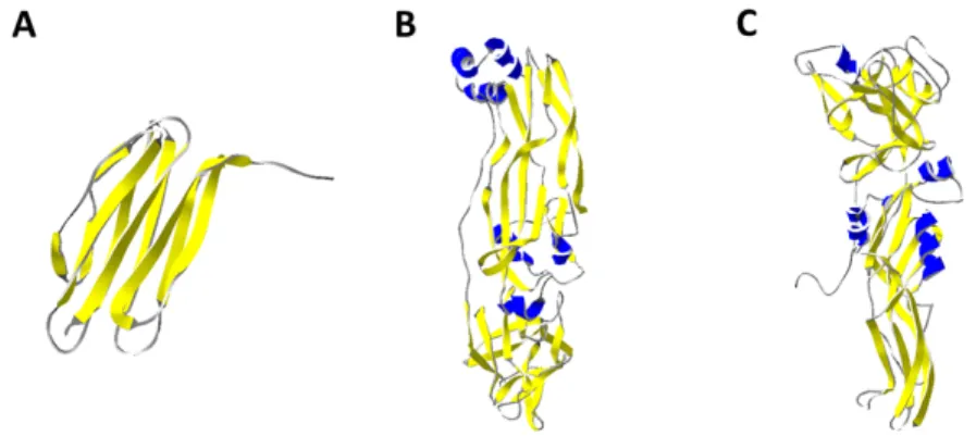
The crystal structure of Cry6Aa, a 475 amino acid protein, has been elucidated at 2.70 Å resolution (136) for the native form and by two different research groups at 2.0 Å and 1.90Å resolution for the trypsin-treated form (136,137). Nematode bioassays showed that the minimal toxic fragment of Cry6Aa was located between Arg11 and Asn389, indicating a molecular weight of 43 kDa, whereas the fragment starting at His12 was much less active than the ones that started at Arg11 (133).
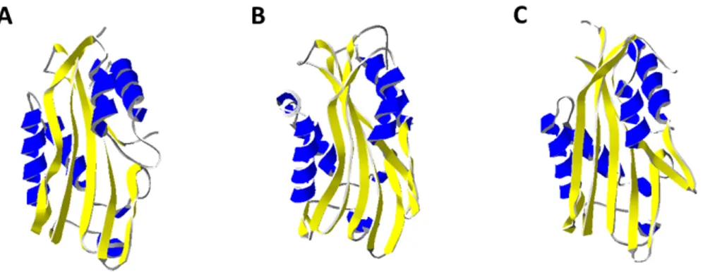
Fig. 12. 3-D ribbon representation of Cry6Aa1 trypsin-treated (5J65). α1 is colored in magenta, the β-tongue is colored in yellow, α9 is colored in blue and α10 is colored in green.
1.5.2.1. Structure of the trypsin-treated Cry6Aa
The trypsin-treated Cry6Aa1 is composed of two polypeptide chains, one that goes from His12 to Ser390 and the other that starts at Ser445 and ends at Trp472. These two polypeptides are linked by a disulphide bridge between residues Cys88 and Cys451. The trypsin-treated
Cry6Aa1 (Fig. 12A) is a helix bundle made out of 10 helices and two β-sheets, where the last helix is attached to the rest of the protein by a disulphide bridge. The head subdomain is folded across the helices at one end and is formed by the residues closer to the C-terminal of the protein. One of the most interesting characteristics of the head domain is that it contains a very hydrophobic β-tongue that confers on this protein a structural similarity to other proteins such as hemolysin E. The tail subdomain is made of residues close to the N-terminal of the toxin (136). The main difference between both elucidated structures of Cry6Aa is that the one solved at 1.9Å resolution ends at α9 (137), while the other one shows α10 attached by a disulphide bridge (136).
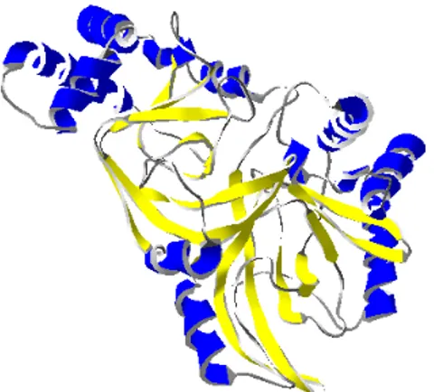
1.5.2.2. Structure of the native Cry6Aa
The full length structure of the native Cry6Aa was also elucidated, and shown to be very similar to that of the trypsin-treated Cry6Aa (Fig. 12B) (136). Because there were some segments of the protein that were too flexible to be identified by X-ray analysis, Cry6Aa1 treated with trypsin was used to model these missing parts. The native Cry6Aa, a 55-kDa protein, extends from Met1 to Asn475. The structure contains 70% α-helices and 1.7% β-sheets, consistent with circular dichroism data.
The native Cry6Aa1 has a total of five cysteine residues at positions 88, 162, 402, 404 and 451. Cys88 and Cys451 are forming a disulphide bridge, which after treatment with trypsin keeps the two fragments of the toxin together. The disulphide bridge between Cys402 and Cys404 is lost during trypsin treatment and Cys162, which is buried in the core of the protein, is not forming any disulphide bond in either form of the toxin. The pairing of the cysteines and the buried one also indicate that intermolecular disulphide bonds do not stabilize Cry6Aa crystals.
1.5.3. Similarity of Cry6A to other toxins
The Cry6Aa1 structures are strikingly similar to those of previously described bacterial toxins, such as HblB of Bacillus cereus, NheA of B. cereus and hemolysin E of E. coli. The hemolysin E structure-function relationships have been studied in depth.