https://doi.org/10.1080/01694243.2017.1321178
Correlation between cell surface physicochemical properties
of bacterial strains and their chromium removal potential
Meryem Asria , Alae Elabeda,c, Nabil Tirrya , Aziza Kouchoua,Saad Ibnsouda Koraichia,b, Naïma El Ghachtoulia and Soumya Elabeda,b
alaboratoire de Biotechnologie Microbienne, faculté des sciences et Techniques, université sidi Mohamed Ben abdellah, fès, Morocco; bcentre universitaire régional d’interface, université sidi Mohamed Ben abdellah, fès, Morocco; claboratoire de génie chimique, cnrs – université de Toulouse (inPT), Toulouse, france
ABSTRACT
Physicochemical characterization of microbes has gained recently a great interest by scientific community. It is proved of extreme importance in several fields of science and technology applications such as bioremediation. In this work, we investigated the establishment of a possible correlation between chromium removal capacity of seven bacterial strains isolated from contaminated sites with industrial wastes including tanning processing and their cell surface physicochemical properties. Thus, hydrophobicity and donor/acceptor electrons character were obtained using contact angle measurements. Statistical analysis showed a high significant positive correlation between hexavalent chromium (Cr(VI)) removal by the strains and their acceptor electron character γ+(r = 0.90). While significant negative correlation between the Cr(VI) removal potential and the ΔGiwi value (r = −0.844) and also with their donor electron character γ− (r = −0.746) were observed. These results may contribute to determine a selectrion criteria of bacteria that can be operated in bioremediation applications.
Introduction
Hexavalent chromium (Cr(VI)) is a highly dangerous heavy metal that has been classified as one of the top 16th toxic polluants [1]. The extensive use of Cr(VI) by a large number of chromium related industries such as steel industries, metal finishing and tannery leads to the production of massive amounts of chromium containing effluents [2]. These effluents directly discharged into municipal sewers and environment without adequate treatment may cause a serious threat to human beings, animals and plants [3,4]. It is recommended that the permissible limit concentration of Cr(VI) in aqueous media should be 0.1 mg L−1
[5]. Therefore, it is essential to intensify reaserches for the removal of chromium from industrial effluents and aqueous environmental systems.
KEYWORDS
Bacterial strains; cr(Vi) removal; cell surface physico-chemical characteristics; correlation; bioremediation
Microbial biosorption is a low cost, eco-friendly and effective method, that receives considerable attention for the elimination of Cr(VI) [6,7]. It seems to be the most promis-ing alternative to convential methods includpromis-ing adsorption, electro-chemical precipitation, reverse osmosis, etc. [8].
Enourmous investigations aimed to understand biosorption mechanisms and factors influ-encing biosorption potential. In fact, Göksungur et al. (2003) reported that the biosorption of heavy metal ions by microorganisms is influenced by several parameters. These factors include the specific surface properties of the microorganism (biosorbent), physicochemical parameters of the solution such as temperature, pH, initial metal ion concentration, the amount of biomass and the existence of other ions [1,9]. Unlike the other parameters extensively studied [6,10], the effect of microbial surface properties has not been rigorously investigated yet.
Indeed, the relation between cell surface physicochemical properties of bacterial strains and their chromium biosorption potential was not specifically identified. Furthermore, a scrutiny of literature indicates a paucity in depth investigations on important physicochem-ical parameters such as the cell surface acid-basic component.
Thus, in this work, we investigated for the first time bacterial cells surface characteristics (hydrophobicity, acid-basic component), using contact angle measurement (CAM), in rela-tion with their chromium removal capacity. This work would contribute to understand the mechanisms of the chromium biosorption and to establish a possible correlation between the adsorption potential and the surface cell properties and to determine the most suitable cell surface characteristics for chromium adsorption.
Materials and methods
Bacterial isolation, identification and growth conditions
Seven bacterial strains were isolated from soil, sediment and wastewater samples contaminated with chemical industrial wastes including tanning process in Fez, Morocco. The pH of sampling sites presented values of 7.5–8.2.
The isolation of the bacterial strains was carried out by enrichment culture technique in Luria broth (LB) medium (peptone 10 g L−1, NaCl 10 g L−1 and yeast extract 5 g L−1 in 1000 mL
distilled water, pH 7.0) which was amended with 100–300 mg L−1 of a sterile solution of Cr(VI)
as K2Cr2O4 and 2 g of soil or 1 mL of wastewater. The medium was incubated at 30 °C, under aerobic conditions, on a shaker incubator at 150 rpm. After 6 days, enriched bacterial strains were isolated by plating on LB agar plate amended with 100–300 mg L−1 of Cr(VI). The bacterial
strains that showed high tolerance were purified and stored at −4 °C for further uses.
The isolates were streaked on LB agar plates in order to get a single pure culture and were used for molecular identification based on the rDNA 16S sequence analysis. Polymerase chain reaction amplification was performed by using FD1 (5’AGAGTTTGATCCTGGCTCAG3’) and RS16 (5’TACGGCTACCTTGTTACGACTT3’) primers. The DNA sequencing was performed by using ABI 3130 (Applied Biosystems, France) according to the manufacturer’s instructions and the GenBank BLAST (Basic Local Alignment Search) tools served for sequence analysis. Biosorption experiments
The bacterial growth was obtained at 30 °C, using LB medium at pH 8 where 0.1 M NaOH solutions were used for pH adjustment if needed, on a rotary shaker at 150 rpm.
After 24 h of incubation, bacterial cells were harvested by centrifugation at 7000 g for 10 min, at 4 °C.
In order to study the chromium biosorption potential of bacterial strains, batch experiments were realized. It was carried out by suspending a loopful of biomass in LB medium prepared in sterile distilled water, containing an initial concentration of metal ions of 100 mg L−1 of Cr(VI) as K
2Cr2O7. The cell concentration was adjusted to
an optical density of 0.7–0.8 at 660 nm (A0) (approximately 108 CFU mL−1 cell density).
Experiments were conducted at 30 °C under agitation system with speed of 150 rpm. Aliquots of 1 mL were taken every 24 h, centrifuged at 6000 g for 10 min at 4 °C and the residual Cr(VI) concentration was determined using diphenylcarbazide method [11]. The assay of Cr(VI) concentration in a medium was carried out using hexavalent chro-mium specific colorimetric reagent S-diphenyl carbazide (DPC). The reaction mixture was set up in 10 mL volumetric flask as follows: 200 μL of supernatant followed by the addition of 330 μL of 6 M H2SO4 and 400 μL of DPC (0.25% (w/v) prepared in acetone), the final volume was made to 10 mL using distilled water. The concentrations of Cr(VI) in the aqueous solutions were measured immediately at a wavelength of 540 nm using a UV–vis spectrophotometer.
The biosorption capacity was calculated as follows: Qt = V(Ci − Ct)/mB [12]. Where Qt is the amount of metal adsorbed per unit mass of biosorbent at a given time (mg g−1 or
mmol g−1), V is the volume of solution (L), C
i and Ct are respectively the initial
concentra-tions and the residual concentraconcentra-tions of metal ions at a given time (mg L−1), m
B is the dry
weight of the biosorbent applied (g). The difference between the initial and the residual concentrations of the metal ion in the solution at the sampling time allows the determina-tion of biosorpdetermina-tion capacity.
The removal percentage was calculated as: Removal (%) = ((Ci − Ct)/Ci) × 100.
Biosorption experiments were realized in triplicate to assess reproducibility. To eliminate the abiotic reduction of Cr(VI), an abiotic control set was prepared throughout the course of the study without bacterial cell.
Contact angle measurements (CAM) Bacterial lawn preparation
Allong with biosorption experiments, CAM were conducted. These measurments were assessed on lawns of organisms deposited on membrane filters [13]. In order to pre-pare bacterial lawns, a loopful of bacterial strains was inoculated in LB medium and incubated at 30 °C for 24 h under a shaker system where the log phase is attained. The exponential phase was determined following the evolution of OD600 with time where OD600 is the optical density of the culture neasured at 600 nm. Bacterial cells were then obtained by centrifugation at 8000 g for 15 min, at 4 °C. The cell pellet was washed twice using sterile KNO3 (0.1 M) and resuspended in the same buffer. The cell concentration was adjusted to an optical density of 0.7 - 0.8 at 660 nm (A0) (approx-imately 108 CFU.mL−1 cell density) in 0.1 M KNO
3. The microbial cell suspensions
were then deposited on a cellulose acetate membrane filter (0.45 μm) using negative pressure. Cells lawns were placed to air dry for 30–60 min to be stabilizated [13,14]. The bacterial lawns for the CAM were realized in triplicate with separately cultured bacterial strains.
Contact angles measurements and calculation of the physicochemical characteristics
The measurement of bacterial cell surface hydrophobicity was carried out using a goniom-eter (GBX Instruments, France) by the sessile drop method [14]. By using three different liquids with known surface parameters values 𝛾lLW, 𝛾
+ l , and 𝛾
−
l, two polar liquids (water and
formamide) and one non-polar liquid (diiodomethane) (Table 1), the unknown surface tension components of microbial surface (the Lifshitz–van der Waals component (γLW),
the electron donor or Lewis base (γ–) and the electron acceptor or Lewis acid (γ+)) was
calculated using the young equation (1). An average of the contact angles and the standard deviation were calculated and reported ((S) and (L) denote solid surface and liquid phases respectively):
The Lewis acid-base component was obtained by:
The cell surface hydrophobicity was also estimated using Van Oss approach [15,16], where the degree of hydrophobicity of a given surface (i) is expressed as the free energy of interac-tion between two entities of that material immersed in water (w). The materiel is hydropho-bic when ΔGiwi is negative and conversely when ΔGiwi is positive the material is considered as being hydrophilic. ΔGiwi is calculated according to the following equation:
where 𝛾iLW is Lifshitz-van der Waals component, 𝛾 LW
w is Lifshitz-van der Waals component
of water, 𝛾i+ electron acceptor of a given material (i), 𝛾i− electron donor of a given material (i), 𝛾w+ and 𝛾
−
i are respectively electron acceptor and electron donor of water.
Statistical analysis
The results were subjected to statistical calculations for means comparison using XLSTAT software. One-way analysis of variance (ANOVA) was used to test the significance of the results followed by post hoc tests (TUKEY HSD) [17].
(1) 𝛾L(cos 𝜃 + 1) = (𝛾LW S 𝛾 LW L ) 1∕2+ (𝛾+ S𝛾 − L) 1∕2+ (𝛾− S𝛾 + L) 1∕2 𝛾SAB= 2(𝛾S−𝛾 + S) 1∕2 ΔGiwi = −2𝛾iw = −2[((𝛾LW i ) 1∕2− (𝛾LW w ) 1∕2) + 2((𝛾+ i 𝛾 − i ) 1∕2+ (𝛾+ w𝛾 − w) 1∕2− (𝛾+ i 𝛾 − w) 1∕2− (𝛾+ w𝛾 − i ) 1∕2)]
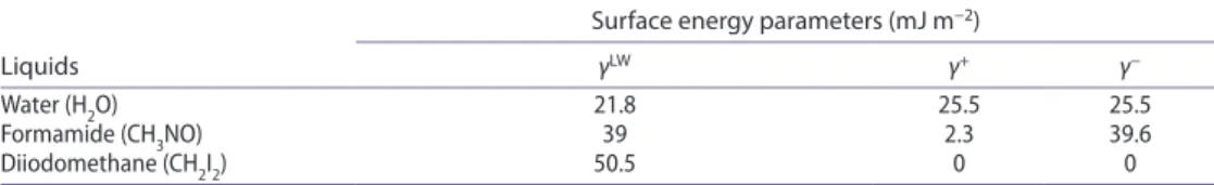
Table 1. energy characteristics (lifshitz–van der Waals (γlW), electron-donor (γ−) and electron-acceptor
(γ+) parameters (mJ m−2)) of pure liquids used to measure contact angles. Liquids
Surface energy parameters (mJ m−2)
γLW γ+ γ−
Water (h2o) 21.8 25.5 25.5
formamide (ch3no) 39 2.3 39.6
Results and discussions
Biosorption of Cr(VI) by bacterial cells
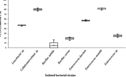
Under identical conditions, the seven bacterial strains showed, after 48 h incubation, dif-ferent percentages of Cr(VI) removal, ranging from 5.32 to 99.87%, (Figure 1). Statistical analysis showed that Cr(VI) removal potential of tested bacterial stains are distribued within four independant groups with significant difference (p = 0.05). Strains Cellulosimicrobium sp. and Enterococcus mundtii offered an excellent potential of Cr(VI) removal, with a removal percentage of 79.87 and 81.39% respectively, grouped within the same group (a). Leucobacter sp. and Enterococcus faecium presented a good Cr(VI) removal potential with a percentage of 46.71 and 56.85% respectively (group (b)). Enterococcus sp. and Bacillus cereus presented a lower chromium removal capacity (24.99 and 19.12%, respectively) (group (c)). Bacillus subtilis presented the weakest chromium removal potential with a percentage of 5.32% (group (d)).
Biosorption is an energy independent binding of metal ions to the cell wall of micro-organisms [7,18]. Thus, we hypothesized that the cell surface physicochemical properties of the seven isolated bacterial strains would be correlated with their different chromium uptake potentials. Indeed, Several studies aiming to determinate the factors affecting the bioremediation mechanisms revealed that hydrophobicity and surface properties play a key role [19], mainly in the adsorption of charged molecules such as heavy metals.
Physicochemical characteristics of bacterial cell surfaces
The physicochemical characterization of microbial cell surface properties can be realized by several techniques, such as microbial adhesion to solvent (MATH) [20] and CAMs [14].
Figure 1. chromium removal percentage by isolated bacterial strains in lB medium, after 48 h incubation at 30 °c, 150 rpm.
In this work, contact angle technic by the sessile drop served as a prompt and a simple way, to obtain precise and valuable surface energy values. It yields precise estimations of cell surface hydrophobicity and electron donor/acceptor character [13,21].
According to Vogler (1998), a given surface is considered as hydrophobic when the value of θw exceeds 65° and it is characterized as hydropholic when θw is less than 65° [22]. Moreover, according to Van Oss approach (1988, 1996), a positive value of the free energy surface (ΔGiwi) indicates that the surface is hydrophilic and a negative value means that it is hydrophobic [15,16].
The results of hydrophobicity obtained are expressed qualitatively (θw) and quantitatively (in terms of ΔGiwi) (Table 2). The seven isolated bacterial strains had positive values of the free energy of interaction ΔGiwi and thus were classified as hydrophilic.
Statical analysis showed that cell surface hydrophobicity of the bacterial stains tested are grouped within five independant groups with significante difference. Thus, group (a) includes ΔGiwi values of 42.17 mJ m−2 (B. subtilis) and 38.79 mJ m−2 (B. cereus). Group (b)
is formed by ΔGiwi values of 38.79 mJ m−2 (B. cereus) and 36.07 mJ m−2 (Enterococcus sp.).
Enterococcus sp. and Leucobacter sp. exibits a lower hydrophobic character with ΔGiwi val-ues 36.07 and 34.93 within group (c). Group (d) includes Leucobacter sp. and Enterococcus faecium with ΔGiwi values of 34.93 and 32.21 respectively and group (e) regroups the lowest ΔGiwi values 26.48 (Cellulosimicrobium sp.) and 26.13 (E. mundtii).
These findings agree with the previous results of Elabed et al. (2011), which reported that different bacterial species exihibit different ΔGiwi values [14]. Many studies have aimed to understand the cell surface properties. A linear correlation between surface properties and the chemical composition of cell surface has been found [21]. It is known that the most hydrophilic microbial surfaces tends to have lower protein/carbohydrate ratios [14].
The results concerning electron donor/acceptor character are presented in Table 2. All iso-lated strains are predominantly electron donors with values of γ− ranging from 49.73 ± 0.364
to 59.69 ± 1.396 mJ m−2 and presents a weak electron acceptor character with values of γ+
0.13 ± 0.053 to 0.69 ± 0.025 mJ m−2.
Table 2. contact angle values (θw, θf, θd), lifshitz–van der Waals (γlW), electron-donor (γ−) and
electron-acceptor (γ+) parameters and surface energies (ΔGiwi) of bacterial cells.
note: letters indicates independent groups with significant difference (p = 0.05) according to anoVa test. Bacterial
strains
Contact angles (°) Surface tension: components and parameters (mJ m−2) energiesSurface
θw θF θD γLW γ+ γ− ΔGiwi (mJ m−2) Leucobac-ter sp. 16.10 ± 0.95 19.80 ± 1.28 28.13 ± 0.72 44.88 ± 0.28 0.30 ± 0.06 (c) 55.49 ± 1.24 (b,c) 34.93 ± 2.00 (c,d) Cellulosimi-crobium sp. 20.90 ± 0.26 15.13 ± 0.40 27.03 ± 0.50 45.3 ± 0.19 0.52 ± 0.01 (b) 49.73 ± 0.36 (d) 26.48 ± 0.65 (e) Bacillus
subtilis 14.23 ± 0.55 24.73 ± 1.83 29.23 ± 0.50 44.44 ± 0.20 0.13 ± 0.05 (d) 59.69 ± 1.39 (a) 42.17 ± 2.56 (a) Bacillus cereus 18.93 ± 0.59 25.83 ± 2.56 30.77 ± 1.12 43.81 ± 0.47 0.18 ± 0.09
(c) 57.02 ± 1.83 (b) 38.79 ± 3.44 (a,b) Enterococcus
faecium 12.87 ± 0.61 13.13 ± 1.10 27.73 ± 0.21 45.04 ± 0.08 0.49 ± 0.03 (b) 54.53 ± 0.29 (c) 32.21 ± 0.02 (d) Enterococcus
mundtii 17.20 ± 0.60 8.10 ± 1.22 27.47 ± 0.15 45.14 ± 0.06 0.69 ± 0.03 (a) 50.43 ± 0.49 (d) 26.13 ± 0.73 (e)
The Statistical analysis showed that the electron acceptor character of tested bac-terial stains are grouped within four independant groups with significante difference (p = 0.05). Thus, group (a) included E. mundtii (γ+ = 0.025 mJ m−2), group (b) formed by
Cellulosimicrobium sp. and E. faecium with γ+ values of 0.52 and 0.49 mJ m−2, group (c)
with γ+ values of 0.18 mJ m−2 (B. cereus) and 0.19 (Enterococcus sp.), group (d) regroups B.
subtilis with γ+ value of 0.13 mJ m−2.
Our findings are in agreements with a previous study reported by Van der Mei et al. (1998) showing that microbial cell surfaces are electron-donating, while seldom electron-accepting cell surfaces can be found [13]. The surface of microorganisms is essentially composed of polysac-charides, proteins and lipids. This composition offers a dominant electron donor character due to the presence of many negatively charged functional groups such as carboxylate, hydroxyl, thiol, sulphonate, phosphate, amino and imidazole groups [19,23]. Their presence is involved in polluted aqueous media bioremediation and capacity of binding metal ions [23].
Relation between cell surface physicochemical properties of bacterial strains and their chromium removal potential
Voleskey et al. (1995) had reported that the microbial cell walls are mainly responsible for the metal biosorption behavior. In fact, the first phase of heavy metal biosorption is attributed to surface adsorption, essentially based on anion exchange with the participation of the functional groups on the cell surface [24]. In addition, it was reported that the adsorption onto the biomass surface is the main mechanism of heavy metal uptake [9].
In order to get an insight into the biosorption process, we proposed to examinate the possible correlation between bacterial physicochemical properties and their chromium removal capacity. For this purpose, CAM were employed to determinate both of CSH and microbial acid-base components of surface free energy. Cell wall properties are a factor that can significantly influence Cr(VI) biosorption efficiency, since it was reported that chemical affinity is the most important driving force that favours ions adsorption to the biosorbent surface [5]. Furthermore, the content of complexing functional groups in microorganisms cellular wall may influence their surface properties and hence their biosorption capacity [7].
It is noteworthy that Cr(VI) exists in aqueous media in different anionic forms. It may exist predominately as chromate (CrO2–4), dichromate (Cr
2O2–7) or hydrogen chromate
(HCrO−4) according to aquatic chemistry of chromium [18] depending on environmental
pH as follows the Cr(VI) speciation diagram [25].
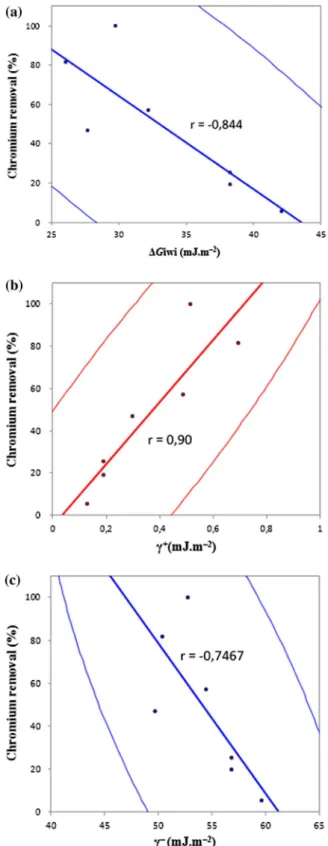
Our results show a negative significant correlation obtained between bacterial CSH and their chromium removal potential (r = −0.844) (Figure 2). Thus, the more hydrophobic bacterial strains have a better chromium removal potential and have a better biosorption capacity of chromium anions. It is generally accepted that the increase of cell surface hydro-phobicity is a defense system against stress conditions and several studies showed a good correlation between microorganisms hydrophobicty and their bioremediation potential [20]. Płaza et al. (2005) reported that microorganisms with higher CSH have a better poten-tial in petroleum bioremediation [26]. It was also reported that Serratia spp. with higher hydrophobicity could adhere to betacypermethrin, absorb and degrade it more easily [20]. Obuekwe et al. (2009) demonstrated that cell surface hydrophobicity of microbial strains was an important factor in biodegradation of crude oil and the most hydrophobic variants were the best degraders [27].
Figure 2. correlation between chromium removal percentage by isolated bacterial strains and their surface physicochemical properties: (a) cell surface hydrophobicity (csh), (b) acceptor electron character γ+ and
It is noticeable that these previous investigations relating cell surface properties to micro-bial bioremediation capacities mainly focused on cell surface hydrophobicity character (CSH). These works especially dealed with the interaction of microorganisms with organic substrates, where the hydrophobic strains were considered as more efficient strains in envi-ronmental applications [27–30]. Our results also suggest that hydrophobic microbial strains may have a great bioremediation capacity in heavy metal removal. However, CSH should not be the only selection criteria for the isolation of performant bacterial strains especially in the case of metal ions bioremediation.
Van Oss (1996) reported that the energy of acid-base interactions can be twice greater than that due to hydrophobic interactions [16], but their role in the phenom-enon of bioremediation is rarely studied. For this reason, we suggested to study the role of acid-base properties of the cell surface in the phenomenon of bioremediation of heavy metal ions.
The results show a negative correlation between the bacterial chromium removal poten-tial and the donor electron character γ− (r = −0.746) (Figure 2). Bacterial strains with high
donor electron character present weak chromium uptake potential. Thus, the higher is the surface electron donor character, the greater the approach of negatively charged metal anions will be inhibited. This result is consistent with the Gupta et al. (2009) work, which reported a relation between functional groups and chromate ions biosorption. This work showed that the more the functional groups in algal cell surface become negatively charged at high pH values, the more they tend to repulse the negatively charged ions chromate and thus affects the adsorption to the algal wall [1].
Our results showed also a high positive correlation between bacterial electron acceptor character γ+ and their chromium removal potential (r = 0.90) (Figure 2). These findings
suggest that surface bacterial donor/acceptor character can significantly influence their chro-mium biosorption capacity. The acid component of cell surface is related to their capacity of Cr(VI) adsorption, an increase in the acid component promotes the adsorption process of chromium anions.
Conclusion
Under their own optimum removal conditions, the chromium removal capacity of iso-lated bacterial strains presents a significant positive correlation with their electron acceptor character, while it shows a negative significative correlation with ΔGiwi value and electron donor character. To our knowledge, this is the first report about the correlation between cell surface hydrophobicity, acid-basic component and chromium removal using CAM. This paper confirms the importance of physicohemical characteristics of microbial strains as selection criteria of bacteria for their use in bioremediation projects.
Acknowledgments
The authors thankfully acknowledge the financial and scientific support of Microbial Biotechnology Laboratory, Faculty of Sciences and Technology, SMBA University, Fez, Morocco.
Disclosure statement
ORCID
Meryem Asri http://orcid.org/0000-0002-0886-9582
Nabil Tirry http://orcid.org/0000-0002-6127-2857
References
[1] Gupta VK, Rastogi A. Biosorption of hexavalent chromium by raw and acid-treated green alga Oegoronium hatei from aqueous solution. J Hazard Mater. 2009;163:396–402.
[2] Leyva-Ramos R, Jacobo-Azuara A, Diaz-Flores PE, et al. Adsorption of chromium(VI) from an aqueous solution on a surfactant-modified zeolite. Colloids Surf A. 2008;330:35–41.
[3] Batayneh AT. Toxic (aluminum, beryllium, boron, chromium and zinc) in groundwater: health risk assessment. Int J Environ Sci Technol. 2011;9:153–162.
[4] Dhankhar R, Hooda A. Fungal biosorption – an alternative to meet the challenges of heavy metal pollution in aqueous solutions. Environ Technol. 2011;32:467–491.
[5] Suh YJ, Chae JW, Jang HD, et al. Role of chemical hardness in the adsorption of hexavalent chromium species onto metal oxide nanoparticles. Chem Eng J. 2015;273:401–405.
[6] Tahri Joutey N, Bahafid W, Sayel H, et al. Remediation of hexavalent chromium by consortia of indigenous bacteria from tannery waste-contaminated biotopes in Fez. Morocco. 2011;6:37–41. [7] Bahafid W, Joutey N, Sayel H, et al. Chromium adsorption by three yeast strains isolated from
sediments in Morocco. Geomicrobiol J. 2013;30:422–429.
[8] Pandey PK, Sharma SK, Sambi SS. Kinetics and equilibrium study of chromium adsorption on zeoliteNaX. Int J Environ Sci Technol. 2010;7:395–404.
[9] Gӧksungur Y, Üren S, Güvenç U. Biosorption of copper ions by caustic treated waste baker’s yeast biomass. Turkish J Biol. 2003;27:23–29.
[10] Bahafid W, Sayel H, Tahri Joutey N, et al. Removal mechanism of hexavalent chromiul by a novel strain of Pichia anomala isolated from industrial effluents of Fez (Morocco). J Environ Sci Eng. 2011;5:980–991.
[11] Pattanapipitpaisal P, Brown NL, Macaskie L. Chromate reduction and 16S rRNA identification of bacteria isolated from a Cr(VI)-contaminated site. Appl Microbial Biotechnol. 2001;57:257–261. [12] Dang VBH, Doan HD, Dang-Vu T, et al. Equilibrium and kinetics of biosorption of cadmium(II)
and copper(II) ions by wheat straw. Biores Technol. 2009;100:211–219.
[13] van der Mei HC, Bos R, Busscher HJ. A reference guide to microbial cell surface hydrophobicity based on contact angles. Colloids Surf B. 1998;11:213–221.
[14] Elabed S, Mostakim M, Berguadi F, et al. Study of microbial adhesion on some wood species: Theoritical prediction. Microbiology. 2011;80:43–49.
[15] Van Oss CJ, Chaudhury MK, Good RJ. Interfacial Lifshitz-van der Waals and polar interactions in macroscopic systems. Chem Rev. 1988;88:927–941.
[16] Van Oss CJ. Interfacial forces in aqueous media. New York (NY): Dekker; 1996.
[17] Avellan A, Levard C, Auffan M, et al. Influence of structural defects of Ge-imogolie nanotubes on their toxicity towards Pseudomonas brassicacearum. Environ Sci Nano. 2016;3:839–846. [18] Gupta VK, Shrivastava AK, Jain N. Biosorption of chromium (VI) from aqueous solutions by
green algea Spirogyra specices. Water Res. 2001;35:4079–4085.
[19] Liu ZF, Zeng GM, Zhong H, et al. Effect of saponins on cell surface properties of Penicillium simplicissimum: Perfomance on adsorption of cadmium (II). Colloids Surf B. 2011;86:364–369. [20] Zhang C, Jia L, Wang S, et al. Biodegradation of beta-cypermethrin by two Serratia spp. with
different cell surface hydrophybicity. Biores Technol. 2010;101:3423–3429.
[21] Hamadi F, Latrache H, Zahir H, et al. The relation between Escherichia coli surface functional groups’composition and their physicochemical properties. Brazilian J Microbiol. 2008;39:10–15. [22] Vogler EA. Structure and reactivity of water at biomaterial surfaces. Adv Coll Interface Sci.
1998;74:69–117.
[23] Gong R, Ding Y, Liu H, et al. Lead biosorption and desorption by intact and pretreadted Spirulina maxima biomass. Chemosphere. 2005;58:125–130.
[25] Li Y, Gao B, Wu T, et al. Hexavalent chromium removal from aqueous solution by adsorption on aluminum magnesium mixed hydroxide. Water Res. 2009;43:3067–3075.
[26] Płaza GA, Ulfig K, Brigmon RL. Surface active properties of bacterial strains isolated from petroleum hydrocarbon-bioremediated soil. Polish J Microbiol. 2005;54:161–167.
[27] Obuekwe CO, Al-Jadi ZK, Al-Saleh ES. Hydrocarbon degradation in relation to cell-surface hydrophobicity among bacterial hydrocarbon degraders from petroleum-contaminated Kuwait desert environment. Int Biodeterior Biodegrad. 2009;63:273–279.
[28] Pijanowska A, Kaczorek E, Chrzanowski Ł, et al. Cell hydrophobicity of Pseudomonas spp. and Bacillus spp. bacteria and hydrocarbon biodegradation in the presence of Quillaya saponin. World J Microbiol Biotechnol. 2007;23:677–682.
[29] Kaczorek E, Urbanowicz M, Olszanowski A. The influence of surfactants on cell surface properties of Aeromonas hydrophila during diesel oil biodegradation. Colloids Surf B. 2010;81:363–368. [30] Al-Tahhan RA, Sandrin TR, Bodour AA, et al. Rhamnolipid-induced removal of
lipopolysaccharide from Pseudomonas aeruginosa: Effect on cell surface properties and interaction with hydrophobic substrates. Appl Environ Microbiol. 2000;66:3262–3268.